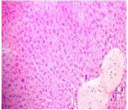
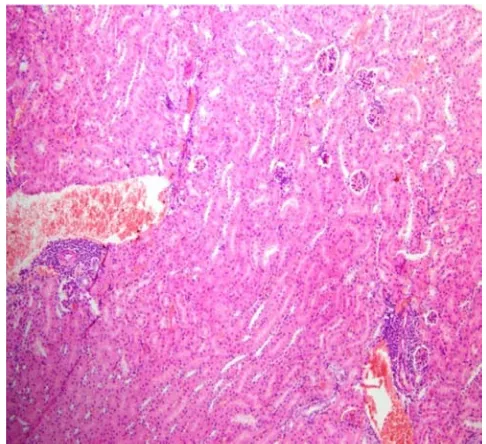
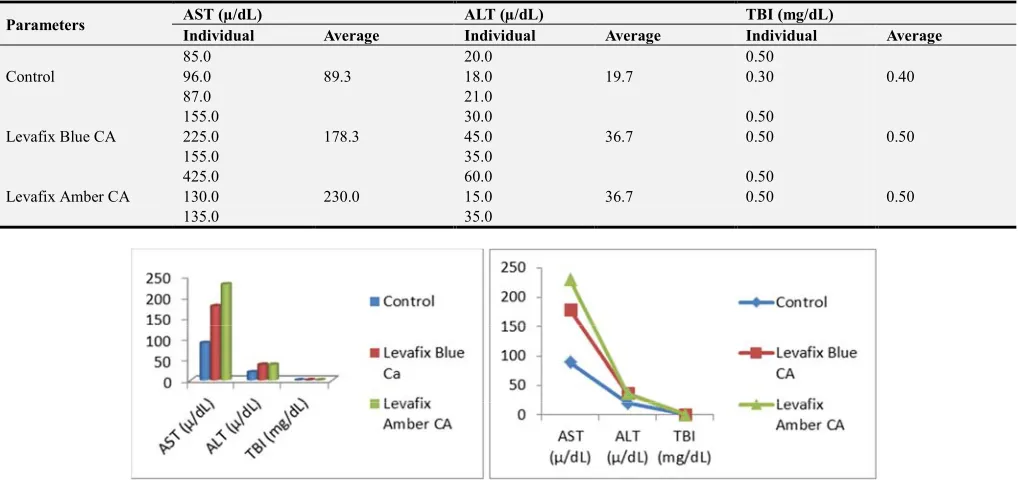
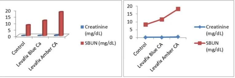
Toxic Effects of Levafix Blue CA and Levafix Amber CA Reactive Dyes on Liver and Kidney in Mice
Full text
Figure
Related documents
In many instances, surgery is not required as the condition may be mild so the patient does not know they have a condition or it simply does not affect their lives; in many
The objective of this paper is to estimate the quarterly net fi xed capital stock in the Czech Republic at constant prices in industry classifi cation (CZ-NACE rev. 2) compatible
If the first place winner has won other certified contests and is in attendance at the National Oldtime Fiddler's Contest they are still the winner, and this does not create
APO03.02 Define reference architecture APO03.03 Select opportunities and solutions APO03.04 Define architecture implementation APO03.05 Provide enterprise architecture
[black line] force coefficient (5.4.1), derived from one spline fit to all the position data (same as figure 6-6b); [discontinuous purple line] composite force coefficient
We studied tree weta ( Hemideina thoracica ) in an urban kahikatea ( Dacrycarpus dacrydioides ) forest remnant in Hamilton City: we estimated relative abundance of tree weta,..
Our field study confirmed that silicon addition increased the chlorophyll fluorescence and some photosynthetic parameters as well as antioxidant enzymes and grain yield in
