University of Pennsylvania
ScholarlyCommons
Publicly Accessible Penn Dissertations
2018
Structure, Function & Dynamics At The
Membrane
Evan O'brien
University of Pennsylvania, [email protected]
Follow this and additional works at:https://repository.upenn.edu/edissertations Part of theBiochemistry Commons, and theBiophysics Commons
This paper is posted at ScholarlyCommons.https://repository.upenn.edu/edissertations/3165
Recommended Citation
O'brien, Evan, "Structure, Function & Dynamics At The Membrane" (2018).Publicly Accessible Penn Dissertations. 3165.
Structure, Function & Dynamics At The Membrane
Abstract
The biological membrane is necessary for maintaining cellular identity, yet must also allow for interaction with the extracellular environment in order to respond to stimuli. Proteins that are directly embedded in the membrane or that interact more peripherally are responsible for these extracellular signaling events, which lie at the heart of cell communication. The first major goal of this work was to interrogate the peripheral
interaction of cytochrome c and the mitochondrial lipid cardiolipin at atomic resolution using solution nuclear magnetic resonance (NMR) techniques; this interaction is key to promoting apoptosis. After demonstrating that the protein was correctly folded in the reverse micelle solution used as a membrane mimetic, cardiolipin was introduced to confirm two previously predicted sites of interaction as well as to identify and propose a novel third site. Next, NMR-derived methyl side chain order parameters have been shown to be important in the thermodynamics of intermolecular interactions. Molecular simulation has become routine in investigations of protein dynamics with atomic-level information, yet their accuracy in replicating experimental dynamics measurements is unknown. Using a variety of standard “force-fields”, it becomes apparent that both common implementations perform comparably, yet outside of the model ubiquitin system, much progress remains in this area. Simulations were then used to interrogate the role of backbone motions in protein thermodynamics. Finally, though we now know much about the role of methyl dynamics in protein conformational entropy, this view has been attained solely with soluble protein systems; the dynamic behavior of membrane proteins remains to be elucidated. Utilizing a newly designed labeling technique for producing deuterated, appropriately methyl-labeled samples, we collected the first quantitative side chain dynamics experiments on several large, integral membrane protein systems. These experiments revealed that membrane proteins apparently contain massive wells of residual conformational entropy, manifested in the extremely dynamic average behavior of the side chain methyl groups. This extraordinary average behavior is the result of the emergence of a previously unobserved “hyper-dynamic” band of methyl groups that explore extensive amounts of rotameric space. In contrast, a series of structural waters and buried polar residues are very rigid by simulation and appear necessary for maintaining a single tertiary structure.
Degree Type Dissertation
Degree Name
Doctor of Philosophy (PhD)
Graduate Group
Biochemistry & Molecular Biophysics
First Advisor Joshua Wand
Keywords
STRUCTURE, FUNCTION & DYNAMICS AT THE
MEMBRANE
Evan S. O’Brien
A DISSERTATION
in
Biochemistry and Molecular Biophysics
Presented to the Faculties of the University of Pennsylvania
in
Partial Fulfillment of the Requirements for the
Degree of Doctor of Philosophy
2018
Supervisor of Dissertation
A. Joshua Wand, Ph.D., Benjamin Rush Professor of Biochemistry & Biophysics
Graduate Group Chairperson
Kim A. Sharp, Ph.D., Associate Professor of Biochemistry & Biophysics
Dissertation Committee
Ronen Marmorstein, Ph.D., George W. Raiziss Professor of Biochemistry & Biophysics (Committee Chair)
Kim A. Sharp, Ph.D., Associate Professor of Biochemistry & Biophysics Ravi Radhakrishnan, Ph.D., Professor of Bioengineering
STRUCTURE, FUNCTION & DYNAMICS AT THE MEMBRANE
COPYRIGHT
2018
Evan Stephen O’Brien
This work is licensed under the Creative Commons Attribution-NonCommercial-ShareAlike 3.0 License.
Acknowledgements
First, I would like to thank Prof. Josh Wand for all of his help and support throughout my thesis. His mentorship combined the best aspects of freedom to really follow my interests and encouragement to continue when things seemed the most difficult. Josh’s insights and guidance, as well as his ability to push the boundaries, allowed for some science that I don’t this would be possible anywhere else, and for this I am truly grateful for everything. I would also like to thank Prof. Kim Sharp for his extensive support along the way, in everything from our rotation project turned collaboration(s) to impromptu office biophysics lessons.
Thank you to the other members of my thesis committee, Prof. Ronen Marmorstein and Prof. Ravi Radhakrishnan, for their continued encouragement.
My thesis work certainly relied extensively upon friends and colleagues in the Wand lab. First, I can’t thank Prof. Nathaniel Nucci enough for the many roles he played throughout my time here; from mentor to friend to officiant, he was a continual source of support for me for the last 6 years. I likely learned as much from mentoring Danny Lin as he did from me, and I am grateful to him for all of his help and hard work in getting my thesis project to actually work. My contemporaries and friends in the Wand lab (Dr. Brian Fuglestad, Dr. Bryan Marques, and Christine Jorge) were continual sounding boards along the way that provided much insight (and beer) along the way. I would like to also acknowledge the pivotal role that Dr. Matt Stetz played in making sure that the lab had all of the big-protein NMR experiments I needed to make this project work. I would also like to thank past (Dr. Vignesh Kasinath, Dr. Kyle Harpole, Dr. Jackwee Lim, Dr. Shannen Cravens) and future (Nikki Kerstetter, Sravya Kotaru, Dr. Alfredo Caro) members of the lab for helpful discussions and insights throughout.
I am particularly thankful for the help and insights on NMR techniques from Dr. Kathy Valentine and the assistance and support of Dr. Li Liang in the wet lab.
ABSTRACT
STRUCTURE, FUNCTION & DYNAMICS AT THE MEMBRANE
Evan S. O’Brien
A. Joshua Wand, Ph.D.
CONTENTS
Acknowledgements . . . iii
Abstract . . . iv
List of Tables . . . .vii
List of Figures . . . viii
1. Introduction. . . 1
1.1.Proteins. . . 1
1.1.1. Interplay of structure, dynamics, and thermodynamics . . . 3
1.1.2. Allostery . . . 7
1.2. Membrane proteins . . . 8
1.2.1. Classes . . . .10
1.2.2. Biochemical interrogations . . . .13
1.2.3. G-protein coupled receptors . . . 16
1.3.Nuclear magnetic resonance (NMR) techniques . . . 20
1.3.1. General description . . . 20
1.3.2. Protein NMR . . . 23
1.3.3. Protein structure determination by NMR . . . 28
1.3.4. Protein dynamics by NMR . . . 34
1.4.Objectives of dissertation . . . 40
2. Exploration of the cytochrome c/cardiolipin binding event in reverse micelles 41 2.1.Introduction . . . 41
2.2. Methods . . . .
44
2.2.1. Protein purification & reverse micelle encapsulation . . . 44
2.2.2. NMR spectroscopy . . . 45
2.2.3. Structure refinement . . . 47
2.3.Cyt c structure in the reverse micelle . . . 52
2.4. Interaction with CL observed in the RM. . . 61
3.1.Replication of experimental dynamics through simulation . . . 73
3.1.1. Introduction . . . 73
3.1.2. Simulation conditions and analysis . . . 76
3.1.3. Results . . . 78
3.1.4. Discussion & Suggestions . . . 83
3.2.Calculation of residual protein backbone conformational entropy . . . 84
3.2.1. Introduction . . . 84
3.2.2. Results . . . 85
3.2.3. Conclusions & Discussion . . . 89
4. Membrane protein dynamics by NMR . . . 91
4.1.Improving yields of 2H, methyl labeled proteins by growth in 1H2O . . . 91
4.1.1. Introduction . . . 91
4.1.2. Materials & methods . . . 93
4.1.3. Strategy . . . .102
4.1.4. Uniform labeling strategies . . . .103
4.1.5. Methyl labeling . . . 108
4.1.6. Discussion . . . 112
4.2.The unique dynamic nature of membrane proteins . . . 116
4.2.1. Introduction . . . 116
4.2.2. Materials & methods . . . 123
4.2.3. Structural scaffolds of pSRII and OmpW . . . 138
4.2.4. The nature of side-chain dynamics in membrane proteins . . . 145
4.2.5. OmpW and pSRII retain rigid polar cores . . . 154
4.3. Conclusions & future implications . . . 157
5. Conclusions . . . 161
5.1.Summary . . . 161
5.2. Future Directions . . . 165
A Appendix Chapter 2 . . . 168
B Appendix Chapter 3 . . . 197
C Appendix Chapter 4 . . . 204
LIST OF TABLES
Chapter 2
2.1 NMR and refinement statistics for oxidized, RM-encapsulated cytochrome c . . . . 55
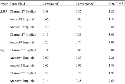
Chapter 3 3.1 Proteins and force field parameters used in force field testing . . . 76
3.2 Relationship between simulation convergence and stability . . . 80
Chapter 4 4.1 Composition of growth medium and variable components . . . 95
4.2 pSRII fitted relaxation parameters and determined order parameters . . . 130
4.3 OmpW fitted relaxation parameters and determined order parameters . . . 134
4.4 Bayesian banding statistics of pSRII in micelles . . . 136
4.5 Example soluble protein average methyl O2axis values . . . 149
Appendix A A.1 Assigned chemical shifts for RM-incorporated, oxidized cytochrome c . . . 168
Appendix B B.1 Experimental and simulated O2axis values for α3D . . . 197
B.2 Experimental and simulated O2axis values for ADBP . . . 198
B.3 Experimental and simulated O2axis values for CaM-sMLCK . . . 199
B.4 Experimental and simulated O2axis values for HEWL . . . 201
B.5 Experimental and simulated O2axis values for ubiquitin . . . 202
Appendix C C.1 Simulated methyl order parameter values for pSRII . . . 205
C.2 Simulated polar group order parameters in pSRII . . . 207
C.3 Simulated methyl order parameter values for OmpW . . . 209
LIST OF FIGURES
Chapter 1
1.1 Proteins are complex macromolecules . . . 2
1.2 The lipid bilayer and its inhabitants . . . 11
1.3 GPCR topology . . . 17
1.4 Utilizing hydrogens in proteins for structure determination . . . 29
1.5 Example autocorrelation functions of different motional modes . . . 38
1.6 Protein backbone dynamics are dominated by secondary structural elements . . . . 40
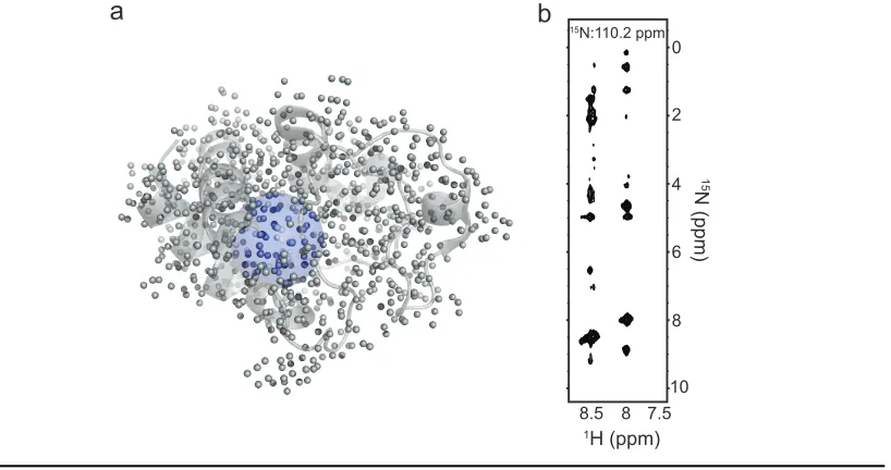
Chapter 2 2.1 RM-incorporated cyt c retains structural fidelity . . . 43
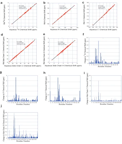
2.2 Comparison of aqueous cyt c and RM-encapsulated cyt c . . . 46
2.3 The structure of oxidized cytochrome c encapsulated in reverse micelles . . . 53
2.4 Structural waters of ferricytochrome c . . . .56
2.5 Cytochrome c undergoes structural changes upon change in redox state . . . 59
2.6 Identification of cardiolipin interaction sites on ferricytochrome c . . . 61
2.7 Both redox states of cyt c were tested independently for interactions with CL . . . . 64
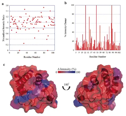
2.8 Many peaks change intensity during the course of CL addition . . . 66
2.9 Energetics of cyt c confinement . . . 70
Chapter 3 3.1 Correspondence between experimental O2axis parameters and those derived from molecular dynamics simulations using various force fields . . . 78
3.2 Experimental vs. simulated methyl order parameters in villin . . . .81
3.3 Accuracy of simulated entropy in a variety of protein systems . . . 82
3.4 Different relationships between backbone and side chain order parameters . . . 86
3.5 Histogram of simulated backbone amide order parameters . . . 87
3.6 Relationship between individual methyl and backbone order parameters . . . 88
3.7 Simulated entropy vs. backbone order parameter . . . 89
Chapter 4 4.1 Idealized growth curve . . . 98
4.2 Percent isotopic labeling vs. amount of labeled amino acids . . . 103
4.3 15N-TROSY spectra for pSRII, AKR1c3, and LacI . . . 104
4.6 ILVM labeled pSRII . . . 112
4.7 Example backbone and methyl spectra of pSRII and OmpW . . . 139
4.8 Global structural folds of pSRII and OmpW . . . 140
4.9 Structural fidelity of pSRII dynamics samples . . . 141
4.10 The helical and sheet backbones of pSRII and OmpW are largely dynamically silent . . . 142
4.11 Distribution of effective tumbling times for pSRII and OmpW . . . 143
4.12 Example cross-correlated relaxation build up curves for pSRII . . . 144
4.13 Single-quantum experiments alone cannot replicate build-up curves . . . 146
4.14 The distribution of fast side chain dynamics in membrane proteins is distinct from their soluble counterparts . . . 148
4.15 Comparison with O2axis values derived from MD simulations of pSRII . . . 150
4.16 Spatial distribution of fast side chain motion in pSRII . . . 151
4.17 Burial depth dependence of methyl-bearing side chain motion . . . 152
4.18 Protein-water contacts . . . 154
Chapter 1
Introduction
1.1
Proteins
Proteins are responsible for nearly all of the complex chemistry accomplished in
living organisms; they are capable of this versatility by way of their massive potential for
variety. They display extensive variation in character (the chemical diversity of the 20
common building blocks), sequence (the ordering of the building blocks), structure (the
averaged 3D spatial relationship of the protein sequence), and dynamics (the motions of
each building block in the final structural ensemble) (Fig. 1.1). This versatility and
adaptability (via evolution) has allowed proteins to function in cells as chemical catalysts,
functional motors, scaffolds, intercellular transport, and intra/intercellular
communication. Different evolved protein sequences code for different protein structures;
in essence, the final structure and dynamics, indeed the full energy landscape, are
“encoded” in the protein sequence.1 As every protein begins as a “string” of unfolded
amino acids in solution, they have adapted relatively fast and efficient mechanisms of
reaching their final “folded” state(s). Proteins accomplish this feat through a series of
elements,2 though given the complex nature of protein energetics, each intermediate is
necessarily composed of large numbers of excursions outside of local topologies. Each
intermediate structure can then serve as a catalyst for efficient incorporation of other
structural elements until the final fold is achieved.
Once proteins have reached their final “native state”, they can begin their
functioning lives. In addition to all of the above complexities of proteins, one of the most
important for the development of higher organisms is increasingly intricate regulation of
their functions. Regulation of function by means of intercellular signaling is particularly
important from a biological stand point as a means of quick and efficient response to
external stimuli; this property makes signaling proteins especially susceptible to mutation
in cancers as well as useful targets for pharmaceuticals. Proteins can have their functions
leucine (L) lysine (K)
+
H2O
LK+ISTYQRLAAS...
Building Blocks Sequence
Structure Dynamics
modulated by either covalent or non-covalent interactions with other chemical groups.
Covalent modifications to amino acid side chains, such as phosphorylation and
acetylation, can have dramatic impacts on protein structure, dynamics, and function. Just
as important are the non-covalent interactions that proteins make with everything from
water to salts to ligands, substrates, and other protein interaction partners. These
interactions with partner molecules can have drastic impacts on protein function,
sometimes imparting large-scale rearrangements at the opposite ends (> 40 Å away). This
phenomenon of allostery is fundamental to complex signaling events and will be
explored further in the introduction, and is at the heart of most of the work presented
herein. Governing all changes in protein structure, dynamics and subsequent functional
changes are the fundamental laws of statistical thermodynamics.
1.1.1
Interplay of structure, dynamics, and thermodynamics
X-ray crystallographic studies of proteins and protein complexes have largely
enforced a structural viewpoint of protein function. Such studies have been instrumental
in elucidating the global folds of proteins, determining intra- and inter-residue interaction
propensities, and conducting atomic-resolved interrogations of protein interactions.
However, crystal structures only give one static piece of the puzzle, ideally the most
energetically favorable one. It is now known, largely through NMR relaxation based
experiments, that proteins undergo extensive exploration of conformational space beyond
the single structures observed in crystals. In order to more fully explore and sample how
proteins move in solution, so-called “molecular dynamics” (MD) simulations have been
taken a relatively simple approach centered on calculating the net force on each atom in a
protein system at discrete time steps and applying Newton’s laws to subsequently move
atomic coordinates accordingly for the next time step. These forces are calculated using
empirically optimized coefficients for equilibrium bond lengths, torsion angles, dihedral
angles, and improper angles using familiar spring-like potentials, in addition to
longer-range non-bonded interactions determined largely by charge-charge interactions. Several
different “force-fields” exist for the purpose of observing protein motions in solution.
While it is clear that much can be learned from Newtonian physical models of protein
dynamics, it remains to be determined exactly how accurate such simulations are at
recapitulating realistic protein fluctuations. Force fields are continually being modified to
better recapitulate various experimental observables, though the limitations of Newtonian
models may lead to fundamental limitations in accuracy due to the abundance of quantum
mechanical interactions that are insufficiently modeled (e.g. hydrogen bonding).3
At a more macroscopic level, protein motions can be thought of instead as an
ensemble of different structures, the populations of which are determined by the energy
differences between them, and the rates at which they interconvert determined by the
relevant energy barrier between them. Thus, many relevant features of protein energetics
and function are best thought of in the context of the Gibbs free energy (G):
G U PV TS= + − (1.1)
where U is the total internal system energy, S is the entropy, and P, V, ant T are pressure,
volume, and temperature, respectively. Using the definition of the system enthalpy (H),
as the internal energy plus the amount of “pressure-volume work” (PV) needed for the
system, we can redefine the Gibbs free energy with the classic equation
G=H TS− (1.3)
Entropy can then be defined as a function of the total number and population of states
from Boltzmann as follows
ln
B i i
S k= ∑p p (1.4)
where kB is Boltzmann’s constant and pi is the probability of any given state within the
system. Experimentally, it is often easiest to calculate changes in thermodynamic
parameters upon some change in state rather than absolute values. For instance, changes
upon protein folding or unfolding, introduction of binding partner, or experimental
conditions are common perturbations used to better understand protein function and
thermodynamics. Thus, the more useful definition of the change in Gibbs free energy
(ΔG) is widely applicable in protein thermodynamics:
G H T S
Δ =Δ − Δ (1.5)
where ΔH is the change in enthalpy and ΔS is the change in entropy. Using its inherent
relationship with the equilibrium constant between any two states of a system, Keq, we
can then use changes in Gibbs free energy to directly calculate the relative populations of
those states by
ln eq
G RT K
Δ =− (1.6)
where R is the gas constant. Rearranging gives the exponential relationship between the
equilibrium constant and the change in Gibbs free energy
/
G RT eq
K e−Δ
For two states of a protein, for instance the fully folded state A and a partially unfolded
state B, separated by a Gibbs energy difference ΔGAB, the equilibrium constant can be
defined as pB/pA where pB and pA are the populations of state B and A, respectively.
Accordingly, the populations of the two states are related to the energy difference as
/ AB
A G RT
B p e p −Δ = (1.8)
Similarly, for the interaction of a protein P with its ligand L, where the unbound protein
(P) has some energy difference from the bound protein (PL) of ΔGP-PL, the population of
the free (pP) protein, free ligand (pL) and bound complex (pPL), the equilibrium
dissociation constant kD can be defined as
L P D PL p p k p ⋅ = (1.9)
Similar to the standard relationship between free energy and equilibrium constants
defined previously (Eq. 1.6), we can define a similar expression here for kD:
D lnk G RT
Δ = (1.10)
Finally, in addition to the more common thermodynamic terms discussed already (G, H,
S, U), intrinsic heat capacity (Cp) is often a very useful parameter particularly in its
various relationships with other state variables (subscript p indicates that pressure is
assumed to be constant for these definitions). Most intuitively, heat capacity can be
interpreted as how much the enthalpy (heat) of a system changes with changes in
Or, conversely, how much heat must be put into a system to change its temperature. For
instance, water has a very high heat capacity, as a large amount of energy much be put
into the system to bring it to high temperatures, unlike oils and alcohols, which typically
require far less heat to change their temperature. In addition to its relationship with
enthalpy, heat capacity can be described in terms of the temperature dependence of
entropy as follows,
p
p S
C T
T ∂
⎛ ⎞
= ⎜ ⎟
∂
⎝ ⎠ (1.12)
1.1.2
Allostery
Proteins are not simply static objects, randomly diffusing throughout cells
performing a single dedicated function. Rather, the many proteins have the ability to
respond to some sort of signal and modulate their biological activity accordingly. Kinases
commonly await an external phosphorylation event before their own activity as a
phosphoryl-transferase is revealed. Transcription factor proteins bind to specific genes of
interest only upon some external signal tells them to do so. Proteins embedded in the cell
membrane are extraordinarily important for sensing extracellular signaling molecules and
transmitting that signal to the cell interior (cytosol). The binding of some regulatory
molecule at a distal site on a protein often promotes changes in activity at a separate
active site, a phenomenon called allostery. Many interrogations of allosteric mechanisms
often center around questions of whether binding of allosteric regulators results in
conformational changes transmitted to the active site (termed induced fit) or whether such
conformational selection); such questions are likely to largely be determined by simple
interaction kinetics.4 Internal protein motions are responsible for both mechanisms of
allosteric regulation. Regardless, binding of modulators at specific allosteric sites on
proteins result in structural and/or dynamic changes at the active site that cause specific
changes in function. Classically, changes in function are thought to arise by changes in
structure, as originally shown for hemoglobin structural changes upon oxygen binding.5
However, modern views of allostery are driven by the knowledge that structural and
dynamic changes are often tied together to determine changes in function, and it has long
been speculated that changes in dynamics can be solely responsible for allosteric
modulation.6 Changes in global or local structure and/or global or local dynamics are of
course determined by the thermodynamic relationships described previously for proteins,
descriptions of which can be used to quantitatively describe and predict allostery.7
Membrane proteins are extremely important allosteric systems in that they are essential
for cell signaling responses, and, accordingly, must pass signals over great distances; they
will be described further below.
1.2 Membrane Proteins
Cell membranes provide a necessary boundary, separating “cell” from “not cell”.
Membranes are principally composed of phospholipids, which contain an extended
polycarbon lipid chain attached to a polar, charged head group (Fig. 1.2a). Lipid bilayers
are then composed of a tail-to-tail arrangement of phospholipids with the charged head
the membrane (Fig. 1.2c). Different types of phospholipids have very different physical
properties, which can be important for promoting complex membrane behavior. Head
groups can be neutral, positive, or negatively charged, and even contain sugar molecules.
Lipid tails can range in length, saturation, and number of chains (Fig. 1.2a). Different
types of lipids can promote different physical properties in membranes themselves, as
well as promote differential interactions with other membrane components, such as
membrane proteins.
Typical globular soluble proteins largely elect to bury nonpolar (aromatic and
aliphatic) side chains within the core of the protein; release of water from the burial of
these types of residues is a major energetic driving force for protein folding.8 This leaves
largely charged and polar amino acids exposed to water on the surface of the protein.
Proteins that are embedded into the cell membrane have a very different arrangement of
amino acid composition. So-called “transmembrane” regions of proteins must expose
nonpolar amino acid side chains on their surface to avoid unfavorable charge-lipid tail
interactions. Polar and charged residues are then typically exposed at the extracellular
and cytosolic faces of the protein, and in some cases they are found buried in the protein
core. Proteins that have such transmembrane regions make up ~20-30% of protein coding
regions in most organisms,9 yet they only occupy a small fraction of structures deposited
in the Protein Data Bank (PDB).10 Also, due to their unique location as cellular
gatekeepers, they are overrepresented as drug targets, with more than 50% of modern
drugs targeting membrane proteins.11 Clearly, much remains to be learned about this
1.2.1
Classes
Proteins can interact with cell membranes in a variety of manners (Fig. 1.2c).
Some are peripheral membrane proteins, which are generally soluble proteins that
contain a region that interacts with a particular phospholipid, effectively increasing their
propensity to be found in close proximity to the membrane. Cytochrome c (cyt c), for
instance, spends much of its time interacting with the membrane, either with its two
membrane protein binding partners or with negatively-charged phospholipids,
particularly cardiolipin (CL), even though it is highly soluble in aqueous solution (Fig.
1.2c). Conversely, otherwise soluble proteins can have lipids covalently attached to
particular side chains, allowing for direct “tethering” to the lipid bilayer (Fig. 1.2c). The
myristoylated protein recoverin responds to increased intracellular Ca2+ by revealing to
solution its normally buried lipid tag, dramatically increasing its propensity to engage the
cell membrane.12 Some membrane-associated proteins are tethered through hydrophobic
interactions with one side of the membrane, either through loops or amphiphilic helices.
The above are examples of proteins that have a propensity to interact with the membrane,
but not a necessity.
Integral membrane proteins contain at least part of their amino acid sequence that
is sequestered within the membrane itself. This can be through a single transmembrane α
-helix connecting two otherwise soluble domains (e.g. receptor tyrosine kinases), multiple
transmembrane helices coming together to form a tertiary integral membrane protein (e.g.
GPCRs and ion channels), or large hydrophobic β-strand assemblies referred to as β
Figure 1.2. The lipid bilayer and its inhabitants. a) Lipids have a large amount of variety, in both their head group (small or large, positive or negative or neutral, chemical composition) and lipid tail (length, number of chains, saturated or unsaturated). b) Detergent micelles have emerged as a tool for studying membrane proteins; they have a comparable degree of complexity in head group and lipid tail. Micelles are roughly spherical particles that expose polar head groups to solution and bury their lipid tail groups. c) Alternatively, lipid bilayers are composed of the longer-chain lipid molecules and form large extended sheets with two layers, each exposing their polar head group to different sides of the bilayer and burying the lipid tails. Membrane proteins can embed in the membrane in a variety of manners. From left to right, 1) peripheral MPs associate with lipid head groups, 2) tethered MPs have a covalently attached lipid anchor that inserts into one leaflet of the bilayer, 3) single-pass MPs have a single transmembrane helix that spans both leaflets and have variable soluble domains on either side, 4) integral MPs embed themselves across the whole lipid bilayer and can potentially interact with molecules on both the interior and exterior of the cell, and some
dimyristoyl phosphocholine
dioleoyl phosphoserine
phosphatidyl inositol
head group poly-carbon tail
lipid tag TM helix soluble domain
integral membrane protein
water phospholipid a
c
b
diheptanoyl phosphocholine
variety of mechanisms of achieving functional outcomes. They can be regulated by an
assortment of physiological signals such as light, force, pH, ions, membrane components,
and small molecules. Certain classes of channel proteins for instance only open and allow
for flow of ions upon activation by ligand binding (e.g. glutamate binding to ionotropic
glutamate receptors), and are otherwise closed.13 Similarly, specific ion channels
modulate their structure and activity in response to changes in the voltage across the
membrane (voltage-gated ion channels), allowing for rapid responses to changes in ion
concentration in neuronal signaling.14 Channel and transport proteins of all kinds are
responsible for obtaining nutrients for cells (e.g. glucose transporters) that would
otherwise be impermeable to the membrane (Fig. 1.2c). Flip- and floppases are catalysts
for the transport of a lipid molecule from one membrane leaflet to the other in order to
change membrane properties.15 Changes in the ternary structure of membrane proteins
can also have important physiological consequences; ligand binding by extracellular
domains of receptor tyrosine kinases causes a dimerization event and activation of the
intracellular kinase domains, promoting cell signaling events.16 Glucose is converted to
cellular energy in the form of adenosine triphosphate (ATP) by a large series of integral
membrane protein complexes called the electron transport chain; some members of this
chain are only stable in the presence of the mitochondrial lipid cardiolipin, and the whole
process is facilitated by the membrane-associated cyt c protein described previously.17
Finally, GPCRs (discussed in further detail below) are essential in responding to all kinds
of cell signals, making them perhaps the most sought after drug targets in the human
crucial functions for cell maintenance and communication, experimental difficulties have
led to large gaps in our knowledge of how those functions are carried out.
1.2.2
Biochemical interrogations
There are many reasons for the relatively few structural and biochemical
investigations of membrane proteins. In order to obtain purified, stable membrane protein
samples for any such studies, many additional steps need to be optimized relative to
soluble protein purifications. First, membrane proteins must be properly extracted from
the membrane of the expression organism, or they must be able to fully and correctly
refold after purification in an unfolded state. This extraction step largely involves the use
of fairly harsh detergent molecules, and often must be optimized for each specific protein
target. To maintain stability in solution techniques, an appropriate membrane mimetic
must be used. Commonly, detergent micelles are used as a relatively simple method to
keep membrane proteins soluble in aqueous solution (Fig. 1.2b). Micelles are composed
of short-chain detergent molecules with the carbon chains towards the interior of the
roughly spherical micelle particles, with their charged or polar head groups exposed to
aqueous solution (Fig. 1.2b). The hydrophobic portions of transmembrane domains are
then able to selectively interact with lipid tails. Many different properties of micelles can
have drastic effects on their ability to stabilize membrane proteins; detergent tail length
or character, head group charge or chemistry, and how these combine to modify bulk
properties of the resulting micelles (e.g. the critical micelle concentration [c.m.c.], and
micelle size) can all impact the behavior of the final sample. Micelles can be an easy
validated by structural and/or biochemical activity assays relative to activity or structure
in the biological membrane in order to be sure the resulting samples properly recapitulate
biologically relevant behaviors.
Due to the potentially non-native effects induced by micelles, it is sometimes
preferable to incorporate the membrane protein system into a true lipid bilayer. Until
recently, the most common method of generating relatively small bilayers in solution was
to use bicelles. Bicelles are composed of small “patches” of lipid bilayer, solubilized in
solution by short-chain detergents surrounding the edges of the bilayers. Bicelles are
potentially advantageous as they can be tuned to the needed purpose – bilayer properties
can be directly selected for by changing the lipid(s) used, and size of the resulting
particles can be easily modulated by changing the ratio of short-chain detergents to
long-chain lipids. Older methods of generating bicelles often resulted in unfolded protein
samples due to the harshness of the methodology, but newer processes are fast, easy, and
relatively gentle on the target membrane protein.18 Similar to bicelles, nanodiscs are a
more recent lipid system that results in small to medium sized, monodisperse, soluble
particles.19-21 Discs are also composed of patches of lipid bilayer, but rather than being
solubilized by short-chain detergents, they are solubilized in solution by amphiphilic
helix proteins derived from apolipoprotein-A1, with their hydrophilic residues towards
solution and hydrophobic residues interacting with lipid molecules. Also like their bicelle
counterparts, different sized nanodisc particles can be produced by using different protein
constructs for the solubilizing component; deleting select helices in the disc sequence
Reverse micelle particles have been used as a mechanism to directly improve the
molecular reorientation properties of proteins in solution for nuclear magnetic resonance
(NMR) spectroscopy (discussed further below).22-23 Primarily used for soluble protein
systems, reverse micelles incorporate short-chain detergent molecules into a bulk alkane
phase. Into this, concentrated protein in aqueous solution is introduced, causing the polar
head groups of the detergents to interact with aqueous phase with detergent tails exposed
to bulk alkane. Membrane anchored proteins such as myristoylated HIV matrix protein or
recoverin expose their lipid anchors to the lipid tail/bulk alkane phase to maintain
stability and solubility of the resulting particles.24 Integral membrane proteins can also be
incorporated into reverse micelles, resulting into so-called “shower-cap” particles,
demonstrated for the ion channel KcsA.25-26 Reverse micelles can also be used to study
lipid-protein interactions for peripherally membrane interacting soluble proteins, as
discussed further in Chapter 2.
When deciding what membrane mimetic is appropriate for studies of membrane
protein systems, all of the above benefits and detriments must be taken into account.
When using techniques such as NMR, the overall size of the resulting particle must
always be taken into account. Chemical compatibility with the protein target is also of
interest – specific lipid head groups can modify the activity of proteins. Often, extensive
trials must be conducted with different detergents, different bicelle or nanodisc lipid
mixtures, and different final protein concentrations. When size is not an issue, simple
large lipid vesicles can be used for studies such as biological assays (in particular cases).
final solubilizing conditions, while other less stable systems will require much more
extensive searches for proper conditions, some of which may require a bilayer with select
lipids for native function.27
1.2.3
G-protein coupled receptors
As discussed previously, membrane proteins respond to an impressively wide
array of extracellular signaling events. Nowhere is this diversity more apparent than in
the large class of receptors known as G-protein coupled receptors (GPCRs). GPCRs are
composed of 7 transmembrane (TM) helices separated by loops of various size and
composition, and extracellular N-terminal and intracellular C-terminal domains (Fig.
1.3). There are approximately 800 GPCRs coded in the human genome, making them the
largest class of membrane protein receptors.28 Additionally, 30-40% of modern drugs are
targeted against GPCRs, demonstrating that they are highly sought after candidates for
pharmaceutical intervention.29 This large family of proteins has been clustered into
different classes. Class A is the largest with ~650 members which share sequence
homology with rhodopsin-like GPCRs. Slightly more than half of these receptors are
involved in olfactory response (vision, taste, smell), while the rest respond to endogenous
small molecule ligands; ~150 of Class A receptors have unknown function or ligands,
making them orphan receptors. There are 15 Class B receptors that respond to peptide
ligands and have a large extracellular domain involved in peptide recognition.30 The 22
Class C receptors respond to glutamate and pheromone ligands.31 Receptors in different
classes generally have no sequence homology to each other, but all retain the 7TM helix
GPCRs respond to many different extracellular stimuli. Rhodopsin-type receptors
have a covalently attached retinal cofactor that acts as its ligand. Upon absorption of the
appropriate wavelength photon, the retinal switches cis/trans configuration, causing
changes in the protein structure that are propagated to the intracellular face, activating its
bound G-protein partner. Non-covalent, diffusible ligands activate most GPCRs. These
molecules range from ions to amino acids to growth hormone peptides to whole proteins.
GPCRs are key regulatory proteins for immune response and inflammation; chemokine
receptors respond to immune system ligands and histamine receptors mediate
inflammation response pathways.32 They play a major role in cognition by responding to
many neurotransmitters including serotonin, dopamine, glutamate, and γ-aminobutyric
acid (GABA). Numerous other physiological functions are modulated by GPCR signaling
pathways.
GPCRs are so-named due to their ability to signal through intracellular
G-proteins. While there are a large number of GPCRs that respond to a wide array of
signals, the partner G-proteins are much less diverse. There are only 4 main classes of
so-called heterotrimeric G-proteins, the main coupling partners for GPCRs; the diversity of
extracellular signaling is unified into a consistent intracellular signaling response.33
Heterotrimeric G-proteins are composed of 3 subunits, called α, β and γ; Gα subunits
retain the nucleotide binding site and the intrinsic GTPase activity. Upon activation by
extracellular ligand, the 6th transmembrane helix (TM6) swings ~10-20 Å outwards
exposing the G-protein binding interface.33-36 The activated GPCR then facilitates the
exchange of GDP for GTP in the nucleotide bound G-protein complex.33 GTP-bound Gα
then dissociates from the complex (leaving Gβγ) and promotes a wide array of
intracellular signaling events, including activating ion channels, inhibiting or activating
adenylyl cyclase (inhibiting or activating cyclic AMP formation), and activation of
phosphodiesterase (inhibiting cyclic AMP formation).33 The free G
βγ complex can cause
its own downstream signaling events.33 GTP-bound G-protein promotes its respective
signaling response until the intrinsic GTPase property of the G-protein cleaves off the
terminal phosphate, leaving inactive, GDP-bound G-protein. This process is also
facilitated by so-called “regulators of G-protein signaling” RGS proteins.37
In addition to signaling through G-proteins, GPCRs have the ability to signal
signaling through arrestins is facilitated by binding to the receptor core as well as a
previously phosphorylated C-terminal tail, both of which can activate the arrestin.39
Arrestin interaction with GPCRs both inhibits signaling through G-proteins directly by
steric occlusion and promotes G-protein independent signaling pathways and facilitates
GPCR internalization.38 Some ligands can signal exclusively through the G-protein
pathway (G-protein biased), some signal through arrestins (β-arrestin biased), and some
signal through both to an equal extent (non-biased ligands). Expressly signaling through
one pathway can have beneficial therapeutic effects, as side effects are commonly
associated with one or the other; biased ligands are highly sought after pharmaceutical
modulators.40
Not only can GPCRs signal through both G-protein and arrestins, different ligands
can activate either pathway to varying extents (different efficacy), while some only serve
to inhibit signaling by other ligands (neutral antagonists). Further, GPCRs in the absence
of ligand promote some degree of coupling to G-proteins, called basal signaling;
so-called inverse agonists can attenuate basal signaling. Such complex signaling properties
imply a complex energy landscape for human signaling GPCRs – in fact, NMR and MD
studies of GPCRs have shown that in the absence of ligand, at least 4 distinct
slowly-exchanging states of the protein exist.35,41 Selection of a “fully active” ensemble of states
only occurs upon ligand and G-protein binding, suggesting “weakly coupled” allostery
between the ligand-binding and G-protein binding interfaces.35,41 Further exploration of
such energy landscapes is needed to fully understand the complex signaling behavior of
While diffusible ligand GPCRs have extraordinarily complex behavior, covalently
bound ligand GPCRs act as simple, binary switches. In the absence of light, they have
minimal basal signaling, and upon photoactivation, they specifically and quickly engage
G-proteins.42 These 7TM helix bundles exist across broad swaths of life in all 3 major
domains of life and facilitate a wide array of responses to light. Human rhodopsins
respond to absorption of light by activating intracellular G-protein partners (they are thus
GPCRs),42 while bacteriorhodopsin acts as a light-driven proton pump for generating
energy,43 and sensory rhodopsins from archaea act to induce motor responses towards or
away from different wavelengths of light.44 Much can potentially be gleaned about the
function of these complex proteins by studying model rhodopsin systems from single-cell
organisms.
1.3 Nuclear magnetic resonance (NMR) techniques
NMR, similar to other spectroscopic techniques, inherently involves using
radiation to probe differences in energy between atomic states. While ultraviolet and
visible spectroscopies deal with different energy electronic states and infrared
spectroscopy probes bond vibration energy levels, NMR spectroscopy uses
radiofrequency radiation to interrogate differences between nuclear spin state energies.
1.3.1
General Description
Atomic nuclei can have different values of spin angular momentum, depending on
the makeup of protons and neutrons within the given nucleus. Nuclear spin can be
momentum. Spin was originally conceived of as a product of individual subatomic
particles physically “spinning” about an axis (as a spinning charged particle creates a
magnetic field); while this is a useful analogy for understanding properties of nuclear (or
electronic) spin, it is an inherently quantum mechanical property not meant to be
physically interpreted in this manner. The magnetic moment µ of a “spin-active” nucleus
is defined as
I
µ=γ (1.13)
Where y is the gyromagnetic ratio of the atom and I is the directional quantum number
associated with the given spin (m) times the reduced Plank’s constant (ℏ)
I =mh (1.14)
Any spin ½ nucleus (the value most commonly associated with NMR spectroscopy, such
as 1H, 13C, or 15N) can take the value of either spin-up (+½) or spin-down (-½). The
energy of each individual spin state can be calculated as
0
E=−µB (1.15)
Where B0 is the strength of the static external magnetic field. Therefore, when a nucleus
is not exposed to a large magnetic field, the two energy levels are redundant with no
difference observed between them. Correspondingly, the larger the external magnetic
field that is “felt” by the nucleus, the larger the energy difference between spin states.
Similar to previous descriptions of state populations in the presence of differences in
energy, the population of each spin state (p+1/2 or p-1/2) can be described as an exponential
/ / 1/2
1/2
E kT h kT p
e e
p
ν
−Δ −
+
−
= =
(1.16)
As differences between nuclear spin states are very small, the population of each
sub-state is nearly equivalent, even at large static magnetic field strengths. Thus, relatively
weak radio frequency radiation must be used to stimulate transitions between nuclear spin
states. Due to the very small population differences observed, NMR is also an inherently
insensitive spectroscopy, requiring relatively large amounts of target nucleus to stimulate
and detect these transitions.
The intrinsic frequency (Larmor frequency, ω) at which a given nucleus precesses
about the static magnetic field (assumed to be in the z-direction) is a function of the
gyromagnetic ratio of the nucleus and the magnetic field itself.
0 B
ω=−γ (1.17)
Each different type of nucleus has a different intrinsic Larmor frequency associated with
it. Of course, different atoms can be involved in different sorts of bonded and nonbonded
interactions depending on the molecular context, each of which has an impact on the
observed precession frequency, known as the chemical shift, due to shielding effects.
Shielding effects from local electron clouds can change the field strength observed by the
nucleus of interest. In complex molecular environments, such as in proteins, the local
electronegative environments experienced by individual nuclei can vary quite
dramatically.
There are two main types of interactions between NMR-active nuclei that can be
through-space interactions (nuclear Overhauser effect [NOE]). Two adjoining, covalently bonded
atoms share electrons due to the wave function overlap involved in covalent bond
formation. Due to the electronic wave function overlap, there is also a significant overlap
in the corresponding nuclear wave functions. The extent of this nuclear overlap is
determined by the angle between them as well as the number of bonds separating the
NMR-active nuclei. In the context of the energetics of spin states, such “coupled”
systems are then a function of the spin states of both nuclei, the presence of the coupling
serving to modulate the energy difference between states. Many common NMR
experiments take advantage of the relatively strong coupling between covalently joined
1H-13C, 1H-15N, or 13C-13C nuclei. Atoms do not have to be covalently bonded to each
other to experience such coupling of nuclear wave functions. If they are sufficiently close
in space, their spins can interact via dipole-dipole effects. Due to the weak nature of
nuclear spin coupling, this effect drops off fairly dramatically as a function of the
distance between atom i and atom j (rij) as
6 1
ij I
r ≈
(1.18)
Where I is a measure of the intensity of the observed interaction. Due to the steep r6
dependence on distance, dipole-dipole interactions further than ~5-6 Å are typically not
observable via the NOE effect. Both interactions are important in the study of complex
macromolecules, where inter-atomic distances and scalar (J) couplings can be useful
probes of global structure.45
NMR spectroscopy has enjoyed widespread use in the analysis of small molecules
for organic chemistry purposes; however, most advancement in NMR machinery and
techniques over the past several decades have been driven by the explosion of interest in
using NMR for studying protein structure and dynamics.45 Proteins contain large numbers
(~thousands) of inherent NMR-active hydrogen atoms engaged in a variety of molecular
interactions that can all potentially be used as probes of local structure & dynamics (Fig.
1.4a). However, with these large numbers, even small proteins begin to run into severe
“overlap” of protons in simple 1-dimensional experiments. This limits practical aspects of
NMR experiments. One of the first major advancements in protein NMR utilized
bacterial overexpression of protein targets in order to achieve full isotopic labeling of
heavy heteroatoms in proteins (predominantly 13C & 15N) in combination with
experiments that can efficiently take advantage of heteronuclear J-coupling effects for
covalently-bonded 13C-H or 15N-H pairs.46 The ability to “separate out” bonded pairs of
atoms into two dimensions allowed for atomic-resolved interrogations of proteins.
Even when peaks arising from appropriately isotopically labeled atoms (via their
bonded interactions) in proteins are fully separable and clearly identifiable, we do not
know which atoms within the protein they correspond to. There are several methods for
identifying which peaks arise from which atoms in a protein structure, termed protein
resonance assignment methods. Prior to the implementation of heteronuclear J-coupling
methods, protons could be assigned through using NOESY experiments in concert with
knowledge about average chemical shifts within a given residue type.46 Predictably, these
NMR-active nuclei can significantly interfere with interpretation (i.e. protons very far in
sequence can just as easily yield NOEs to the proton of interest if they are close in
tertiary structure).
Heteronuclear J-coupling experiments allowed for much more consistent, reliably
interpretable data for full resonance assignment of proteins.47 The basic premise of
current resonance assignment experiments is to begin with the backbone amide
resonances, which are commonly spectrally distinct and easy to label. For each backbone
amide peak, one then collects a series of experiments aimed at determining the chemical
shifts the of 13Cα, 13Cβ, and 13CO (carbonyl carbon) atoms within that particular residue
(i), as well as the 13Cα and 13Cβ atoms of the previous residue (i-1).48 These experiments
are named in the manner of the J-coupling connectivity utilized, for example, the
HN(CO)CA experiment correlates a given amide group with the previous residues 13Cα
by going through the carbonyl carbon.49 In this manner, it is relatively facile to go
through the resulting matrix of chemical shifts and “connect” adjacent residues in
sequence, again taking advantage of unique residue-specific chemical shifts (i.e. alanine
and threonine Cβ atoms). Once the backbone (amide nitrogen and proton, 13Cα, 13Cβ, and
13CO) has been assigned, side chain carbons and protons can be assigned by various
forms of a TOCSY (TOtal Correlated SpectroscopY) experiment designed to connect
backbone amide resonances with all of the attached side chain carbons (and protons).50-51
For large proteins, protein methyl groups are easier assigned by specifically isotopically
labeling methyl groups of interest (see above) with 13CH3 groups and using single 13C-13C
backbone assignment experiments.52 Once all atoms within a protein are assigned, they
can then be interrogated to determine average protein structures and distributions about
those averages (dynamics).
Large protein systems (>30 kD) present a number of challenges for experimental
NMR. First, the sheer number of NMR-active nuclei increases continually with size,
increasing the potential for overlap of peaks even when spread out into two (or three)
dimensions. Next (and more importantly), the larger the protein system becomes, the
slower the overall global tumbling time (τc) of the particle. As the tumbling becomes
slower, T2 relaxation (discussed further below) becomes very fast, leading to very large
linewidths of the observed peaks (as expected due to the inverse relationship between T2
and linewidth).53 As peaks get wider and more numerous, NMR spectra become very
difficult to interpret and all of the J-coupled experiments described above become nearly
impossible. Several methods have been introduced over the years in an effort to combat
tumbling-related line-broadening, the most common of which has been extensive protein
deuteration in conjunction with the TROSY (Transverse Relaxation Optimized
SpectroscopY) effect.54
T2 relaxation times are impacted by so-called “dipole-dipole” effects: interactions
of the nuclei of interest with nearby NMR-active dipoles. One of the major advancements
in achieving high signal-to-noise experiments for large protein systems has been to
simply eliminate as many sources of external dipole relaxation as possible through
extensive protein deuteration. Because deuterons (m=1) interact with protons via the
significantly diminished.54 Protein deuteration is typically accomplished by expression of
protein in bacteria adapted to growth on a bulk D2O medium, with incorporation of
deuterons (rather than protons) into as many external sources as possible, such as the use
of deuterated glucose. While these methods are now the standard source of protein
deuteration, several issues arise. Expression of proteins in D2O medium is highly
variable, but most commonly results in fairly significant decreases in final protein yield
due to poor bacterial growth and inconsistent behavior in overexpressed deuterated
protein.55 Also, bulk D2O results in full deuteration of every hydrogen atom in the
system; nearly all NMR experiments rely on at least select retention of protons, typically
the backbone amide hydrogen. This requires deuterated proteins to be partially unfolded
in order to “back-exchange” for protons in bulk H2O medium. Many proteins are
incapable of being fully refolded properly, even in partially denaturing conditions,
resulting again in decreases in final protein yield. Membrane proteins are even more
difficult to exchange due to deep burial within membrane mimetic and general lack of
reversibility in folding (with exceptions). However, well-behaved, highly expressing
large proteins can typically be fully deuterated and back-exchanged as needed for
high-resolution NMR experiments.
T2 relaxation times are also impacted by chemical shift anisotropy (CSA) in
addition to the previously discussed dipole-dipole effect. J-coupled systems interrogated
by NMR, typically the 15N-1H system, show up as quadruplets in the absence of so-called
decoupling schemes.54 Importantly, each component of the quadruplet is impacted by
destructive interference. The TROSY effect relies on “selecting” the component of the
multiplet with the narrowest inherent linewidth, eliminating the broadest components.
When used in conjunction with protein deuteration, the TROSY effect has been used
extensively to interrogate proteins up to several hundred kDa (including the megadalton
proteasome complex).56
1.3.3
Protein structure determination by NMR
With the wealth of NMR-active atoms within proteins, it is no surprise that a
variety of measurements have been designed to probe average structural features of
proteins. First and foremost is the NOE. Every NMR structural interrogation of protein
structure begins with a number of NOESY experiments to build a series of distance
restraints for as many atoms within the protein as possible. These are typically collected
for the backbone amide protons and side chain (largely methyl) protons. For each of these
individual protons (or groups of protons), “strips” of peaks with every atom within 5-6 Å
of that proton are collected (Fig. 1.4b). The intensity of those NOE “cross-peak” is then
used to translate it into an approximate distance range. No true high-resolution NMR
structures are possible without a large number of proton-proton distances. Amide
backbone protons typically yield information on its local secondary structure; backbone
protons involved in helical structure will have characteristic NOE cross-peaks with HN
and Hα atoms within close proximity, while backbone protons involved in sheet structure
will show characteristic inter-strand (long-range) cross-peaks largely with Hα atoms in
the adjoining strand.45 Occasionally, longer-range side chain or backbone protons will be
defining local secondary structure, side-chain NOEs are necessary for defining accurate
tertiary structure interactions. Long-range (meaning > 5 residues apart in sequence)
distances between methyl groups is neighboring helices, strands, or loops assist in
“bringing together” secondary structural elements into an accurate global fold. The more
long-range interactions in the background of well-defined structural elements, the better
the resolution of the final structure.
In addition to NOEs, raw chemical shifts of backbone atoms (HN, N, CO, Cα, Hα,
[Cβ, Hβ]) have a clear relationship with the dihedral angles of the backbone. Thus,
simply by assigning the protein, one has access to secondary structure restraints by
analyzing assignments with programs like TALOS,57 which output ranges of predicted
backbone angles and associated errors. Though less common in modern structures,
J-10 8 6 4 2 0
8.5 8 7.5 15N:110.2 ppm
1H (ppm)
15
N (ppm)
a b
coupling values between backbone atoms also have a clearly defined relationship with
backbone angles, and can be a useful secondary structure restraint.58
Most NMR phenomena (NOEs, J-coupling & chemical shifts) are local in nature,
meaning they are responsive to local backbone angles and local distances (< 6 Å). It is
therefore useful to as many global restraints as possible; several experimental approaches
yield such values. First, residual dipolar couplings (RDCs) have emerged as a powerful
measure of the relative orientation of bond vectors within a tertiary structure.59-60 While
most solution NMR measurements are collected under the assumption of random
isotropic motion in solution, agents can be introduced to NMR samples to induce partial
alignment of protein particles with the main magnetic field. This alignment of proteins
with the magnetic field causes changes in the intrinsic J-coupling value for bond vectors
within the protein, largely measured for the 15N-HN vector (typically ~94 Hz coupling
constant). The change in coupling constant in response to alignment is measured for each
bond vector within the protein and interpreted in the context of an overall alignment
tensor, yielding an orientation of each bond vector relative to this global tensor.60 RDCs
can then be valuable restraints on global structure. While NOEs are limited to short
distances, the interaction between nuclear spins and unpaired electrons is much longer
range and can be used to obtain approximate distances between atoms on a protein and a
covalently attached free radical spin label (typically a nitroxide such as MTSL).61 Such
paramagnetic labels enhance relaxation of all nuclear spins in the region in a distance
dependent manner through a decrease in observed T2, thus these restraints are termed
2 2 2
3 4
1 c c
H c K
r τ τ
ω τ
⎡ ⎛ ⎞⎤
=⎢ ⎜ + ⎟⎥
Γ ⎝ + ⎠
⎣ ⎦ (1.19)
where Γ2 is the transverse PRE relaxation rate, τc is the global macromolecular tumbling
time, ωH is the Larmor frequency (for the proton in this case), and K is a constant
composed of
2 2 2 1
( 1) 15
K = S S+ γ g β
(1.20)
And γ is the proton gyromagnetic ratio, g is the electronic g-factor, and β corresponds to
the Bohr magneton. Atoms closer to the spin label have a greatly enhanced T2 and thus
are significantly line-broadened, while those further away (>25 Å) are unaffected. Peak
intensities in the oxidized and reduced forms of the spin label can then be used as a proxy
for the distance of any given atom from the average coordinate of the spin label.61
Comparably, when the magnetic moment of this unpaired electron has a structural
dependence on the protein (i.e. in the case of a structural paramagnetic metal such as
Fe3+) then there is an observed change in the averaged chemical shift of atoms within the
protein. This change in chemical shift is due to direct dipolar coupling between the
nucleus and the unpaired electron.62 Because this pseudocontact shift (PCS) decays with
r3 (rather than r6 for NOEs) its effects are much more widespread throughout proteins,
potentially giving distance information past 10 Å. In addition to the distance dependence
of the PCS, the given changes in chemical shift are also dependent on the angular
dependence of the nucleus-electron vector with the overall magnetic susceptibility tensor