Role of the donor nuclei in cloning efficiency:
can the ooplasm reprogram any nucleus?
YOKO KATO* and YUKIO TSUNODA
Laboratory of Animal Reproduction, College of Agriculture, Kinki University, Nara, Japan
ABSTRACT Cloning efficiency has not been dramatically improved after the first success of somatic cell nuclear transfer (SCNT) in sheep in 1997. The reasons for the low efficiency of SCNT embryos must be attributed to the insufficient reprogramming of the donor nucleus in ooplasm. It has been clarified that the methylation and acetylation status are disordered in SCNT embryos and the gene expression pattern is different and widely varied in SCNT embryos, compared with fertilized embryos. In this paper, we focused on the role of the donor nuclei in cloning efficiency, and discuss whether ooplasm can reprogram any nucleus.
KEY WORDS: cloning, donor nucleus, nuclear transfer, efficiency, reprogram
Introduction
Despite several attempts to improve the cloning efficiency of somatic cell nuclear transfer (SCNT) since the first successful
production of a sheep in 1997 (Wilmut et al., 1997), dramatic
improvements have not yet been realized. Reprogramming the meiotic deacetylation process by inhibiting deacetylation with the potent and specific histone deacetylase inhibitor trichostatin A (TSA) has been effective for in vitro and in vivo cloning efficiency (Rybouchkin et al., 2006; Tsuji et al., 2009; Kishigami et al., 2006), but has not induced sufficiently dramatic improvement. Although the low efficiency of SCNT embryos appears to be due to insufficient reprogramming of the donor nucleus in the ooplasm, this has not been clarified and overcoming this insufficient repro-gramming presents several challenges. The nuclear reprogram-ming process is likely to be very complex; upon nuclear transfer, donor chromatin is exposed to the ooplasm (the first and most important reprogramming step), and after artificial activation of SCNT oocytes, reconstituted oocytes begin preimplantation de-velopment (the second reprogramming process), including zy-gotic genome activation, compaction, and the first differentiation into ICM and TE cell lineages. Postimplantation development is the last reprogramming step and is even more complex.
In the first reprogramming process, donor information should be reprogrammed from the somatic type to the embryonic type. Reprogramming factors in the ooplasm have also been exam-ined, and several factors that promote cloning efficiency have been identified (Miyamoto et al., 2007, 2009; Jullien et al., 2010). In the second reprogramming process, SCNT embryos begin to
BIOLOGY
www.intjdevbiol.com*Address correspondence to: Yoko Kato. Laboratory of Animal Reproduction, College of Agriculture, Kinki University, 3327-204, Nakamachi, Nara 631-8505, Japan. e-mail: [email protected]
Final author corrected PDF published online: 17 February 2011.
ISSN: Online 1696-3547, Print 0214-6282
© 2011 UBC Press Printed in Spain
Abbreviations used in this paper: SCNT, somatic cell nuclear transfer.
cleave and develop to the blastocyst stage with a time schedule similar to that of fertilized embryos. Although the gene expression pattern of SCNT preimplantation embryos is largely different and varies widely compared with fertilized embryos (Li et al., 2006a,b, 2008), it is not known whether the different gene expression patterns are incompatible with successful SCNT cloning. The last reprogramming process involves very complex fero-maternal communication, an in vivo process that remains unclear. Admin-istration of human chorionic gonadotrophin (hCG) to control the physiology of recipient females was recently reported to improve cloning efficiency (Tsuji et al., 2010).
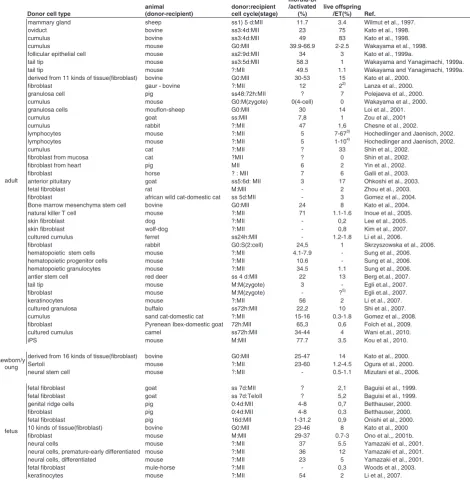
Several studies have focused on determining which donor cell type or donor cell status is best for successful cloning (Table 1). The state of the donor cell is one of the most important factors for cloning efficiency. In the present study, we discuss mainly the role and effect of the donor nucleus type in cloning efficiency.
Cell cycle combination
promot-ing factor (MPF) activity is decreased durpromot-ing S-phase.
In contrast, when somatic cells are used as donors for nuclear transfer, the cell cycle combination might be important for devel-opment. The G0-phase was thought to be the most adequate cell cycle for nuclear reprogramming of the donor nucleus (Campbell,
et al., 2006; Wilmut et al., 1997), but it was later clarified that the G1-phase and M-phase are also reprogrammed in MII-phase oocytes and can develop to full-term (Table 1). Even if cells are not
induced to the G0-phase by serum starvation or contact inhibition, up to approximately 60% cells of cultured somatic cells are in the G1 phase. When donor cells at the G0 and G1-phase are used, emission of the first polar body in reconstituted oocytes must be suppressed by cytochalasin B or a similar chemical to maintain the diploid status of the donor cell in the ooplasm, as in blastomere transfer. When M-phase cells are used as donor cells, as reported in mouse (Ono, 2001a,b) and rat (Zhou, 2003), emission of the
TABLE 1
THE EFFECTS OF DONOR CELL TYPE AND CELL CYCLE COMBINATION BETWEEN DONORS AND RECIPIENTS, ON DEVELOPMENTAL ABILITY OF NT EMBRYOS RECEIVING SOMATIC,
EMBRYONIC, ES, EG AND PGC CELLS
Donor cell type
animal (donor-recipient)
donor:recipient cell cycle(stage)
morula-Bl /activated
(%)
live offspring /ET(%) Ref.
mammary gland sheep ss1) 5 d:MII 11.7 3.4 Wilmut et al., 1997.
oviduct bovine ss3:4d:MII 23 75 Kato et al., 1998.
cumulus bovine ss3:4d:MII 49 83 Kato et al., 1998.
cumulus mouse G0:MII 39.9-66.9 2-2.5 Wakayama et al., 1998.
follicular epithelial cell mouse ss2:9d:MII 34 3 Kato et al., 1999a.
tail tip mouse ss3:5d:MII 58.3 1 Wakayama and Yanagimachi, 1999a.
tail tip mouse ?:MII 49.5 1.1 Wakayama and Yanagimachi, 1999a.
derived from 11 kinds of tissue(fibroblast) bovine G0:MII 30-53 15 Kato et al., 2000.
fibroblast gaur - bovine ?:MII 12 22) Lanza et al., 2000.
granulosa cell pig ss48:72h:MII ? 7 Polejaeva et al., 2000.
cumulus mouse G0:M(zygote) 0(4-cell) 0 Wakayama et al., 2000.
granulosa cells mouflon-sheep G0:MII 30 14 Loi et al., 2001.
cumulus goat ss:MII 7,8 1 Zou et al., 2001
cumulus rabbit ?:MII 47 1,6 Chesne et al., 2002.
lymphocytes mouse ?:MII 5 7-673) Hochedlinger and Jaenisch, 2002.
lymphocytes mouse ?:MII 5 1-104) Hochedlinger and Jaenisch, 2002.
cumulus cat ?:MII ? 33 Shin et al., 2002.
fibroblast from mucosa cat ?MII ? 0 Shin et al., 2002.
fibroblast from heart pig MII 6 2 Yin et al., 2002.
fibroblast horse ? : MII 7 6 Galli et al., 2003.
anterior pituitary goat ss5:6d: MII 3 17 Ohkoshi et al., 2003.
fetal fibroblast rat M:MII - 2 Zhou et al., 2003.
fibroblast african wild cat-domestic cat ss 5d:MII - 3 Gomez et al., 2004. Bone marrow mesenchyma stem cell bovine G0:MII 24 8 Kato et al., 2004. natural killer T cell mouse ?:MII 71 1.1-1.6 Inoue et al., 2005.
skin fibroblast dog ?:MII - 0,2 Lee et al., 2005.
skin fibroblast wolf-dog ?:MII - 0,8 Kim et al., 2007.
cultured cumulus ferret ss24h:MII - 1.2-1.8 Li et al., 2006.
fibroblast rabbit G0:S(2:cell) 24,5 1 Skrzyszowska et al., 2006.
hematopoietic stem cells mouse ?:MII 4.1-7.9 - Sung et al., 2006.
hematopoietic progenitor cells mouse ?:MII 10.6 - Sung et al., 2006.
hematopoietic granulocytes mouse ?:MII 34.5 1.1 Sung et al., 2006.
antler stem cell red deer ss 4 d:MII 22 13 Berg et.al., 2007.
tail tip mouse M:M(zygote) 3 - Egli et.al., 2007.
fibroblast mouse M:M(zygote) - ?5) Egli et.al., 2007.
keratinocytes mouse ?:MII 56 2 Li et al., 2007.
cultured granulosa buffalo ss72h:MII 22,2 10 Shi et al., 2007. cumulus sand cat-domestic cat ?:MII 15-16 0.3-1.8 Gomez et al., 2008. fibroblast Pyrenean Ibex-domestic goat 72h:MII 65,3 0,6 Folch et al., 2009.
cultured cumulus camel ss72h:MII 34-44 4 Wani et.al., 2010.
adult
iPS mouse M:MII 77.7 3.5 Kou et al., 2010.
derived from 16 kinds of tissue(fibroblast) bovine G0:MII 25-47 14 Kato et al., 2000.
Sertoli mouse ?:MII 23-60 1.2-4.5 Ogura et al., 2000.
newborn/y oung
neural stem cell mouse ?:MII - 0.5-1.1 Mizutani et al., 2006.
fetal fibroblast goat ss 7d:MII ? 2,1 Baguisi et al., 1999.
fetal fibroblast goat ss 7d:TeloII ? 5,2 Baguisi et al., 1999.
genital ridge cells pig 0:4d:MII 4-8 0,7 Betthauser, 2000.
fibroblast pig 0:4d:MII 4-8 0,3 Betthauser, 2000.
fetal fibroblast pig 16d:MII 1-31.2 0,9 Onishi et al., 2000.
10 kinds of tissue(fibroblast) bovine G0:MII 23-46 8 Kato et al., 2000
fibroblast mouse M:MII 29-37 0.7-3 Ono et al.,, 2001b.
neural cells mouse ?:MII 37 5.5 Yamazaki et al., 2001.
neural cells, premature-early differentiated mouse ?:MII 36 12 Yamazaki et al., 2001.
neural cells, differentiated mouse ?:MII 23 5 Yamazaki et al., 2001.
fetal fibroblast mule-horse ?:MII - 0,3 Woods et al., 2003.
Somatic cells
fetus
inner cell mass (ICM) cells mouse G16):MII 23-64 8 Tsunoda and Kato, 1998. mural trophectoderm (TE) cells mouse G16):MI 32-62 11 Tsunoda and Kato, 1998. embryonic disc sheep ss 5 d:MII 6.7-21 11 Campbell et al., 1996.
1) serum starved; d: days, h: hours, 2) later-term abortion at day 202, 3) ntES cells were injected into 2n host blastocysts, 4) ntES-cells were injected into 4n blastocysts, 5) 5 chimeras after injection of ntES-cells into blastocysts. 6) ICM and TE cell were isolated from blastocysts previously treated with nocodazole followed by achidicolin. 7) serial nuclear transferred at 1-cell stage, 8) ntES-cells were injected into 4n bl;astocysts, 10) day 10.5 chimera after serial nuclear trasnfer at the 2-cell stage, 11) day10.5 fetuses.
second polar body after artificial activation is essential to maintain the diploid status in the ooplasm. This differs from blastomere transfer, because somatic cells at the S-phase do not develop in activated ooplasm. Also, when G0/G1-phase and M-phase donor cells are transferred to activated ooplasm, the in vitro develop-mental ability of reconstituted oocytes is significantly decreased (Tani et al., 2003).
The Table summarizes the effect of the cell cycle of the donor and recipient and of the donor cell types on the development of manipulated embryos. The first report of each animal species and the specific cell cycle combination of the donor and recipient was selected from the vast literature. As shown in Table, a G0/G1 donor with MII recipient ooplasm was used in almost all success-ful reports.
There have been some attempts to use fertilized oocytes such as zygotes and 2-cell embryos as SCNT recipients, but the success has been limited. Zygotes at the S-phase seem to support the development of SCNT up to the 4-cell stage and then
development stops (Wakayama et al., 2000). Zygotes at the
M-phase seem to support the development of SCNT to the blasto-cyst stage (Egli et al., 2007), but if embryonic stem (ES) cells are used, zygotes at the M-phase can develop to full term after embryo transfer to foster mothers (Egli et al., 2007).
When primordial germ cells at the arrested stage obtained from day 15.5 fetuses or at the mitotic phase obtained from day 11.5 fetuses were fused with enucleated zygotes at the M-phase (Kato and Tsunoda, 1995a) and/or enucleated blastomeres of one of the 2-cell embryos at the late S-phase (Kato and Tsunoda, 1992), reconstituted zygotes or chimeric 2-cell embryos(Kato and Tsunoda, 1992) developed to the blastocyst stage. Moreover, in rabbit, chimeric embryos, i.e., adult fibroblast cells fused with one enucleated blastomere of a 2-cell embryo, develop to chimeric offspring (Skrzyszowska et al., 2006).
Based on these studies, MII oocytes might possess some
reprogramming factors in the ooplasm (Miyamoto et al., 2009),
but gradually lose these reprogramming factors in the cytoplasm after fertilization or artificial activation (Tani et al., 2003). We examined the effect of ooplasm aging on the developmental potential of SCNT embryos in bovine (Tani et al., 2003) and in mouse (Liu et al., 2007). When bovine cumulus cells at the
G0-phase and M-G0-phase are fused every hour after activation for 6 h, the potential to develop into blastocysts after SCNT gradually decreased with time after activation. Ooplasm 2 h and more after activation did not support the development of G0-phase cumulus nuclei to blastocysts. When M-phase cumulus cells were fused, the ability to develop into blastocysts dramatically decreased beginning at 6h postactivation. These findings clearly demon-strate that some reprogramming factors present in the ooplasm decrease after artificial activation. We isolated some reprogram-ming factors in the ooplasm before activation, and identified one
of the candidate reprogramming factors as TCTP (Tani et al.,
2007), which expressed in ooplasm, but is no longer present in activated ooplasm as the same type. Although, oocytes injected with TCTP peptide after SCNT produce much healthier offspring in bovine than in non-injected oocytes, (Tani et al., 2007), the role played by TCTP in the whole reprogramming process remains unclear.
Donor cell type
After the first report in which mammary gland cells were used as the donor cells, cumulus cells have been the most popular donor cell type in SCNT because of the experimental conve-nience (Table 1). Many attempts have been made to determine the most efficient donor cell types, especially for bovine and mouse. To examine which cell types are the most successful for SCNT in bovine, we compared the development potential of 39 cell types from adults, newborns, and fetuses of both sexes, but there was no big difference (Kato et al., 2000). When tissue or biopsy samples were used for the cell culture, fibroblast cells were the most common cell type to easily increase. Cells that were not identified as a specific cell before nuclear transfer were catego-rized as fibroblast cells. As shown in the table, although the developmental potential was higher in bovine compared with mice, the overall conclusion is that cloning efficiency is similar among somatic cell types. Although some types of stem cell, such
as bone marrow mesenchymal stem cells (Kato et al., 2004),
efficiency of SCNT must not depend on the cell type, but that ooplasm alone is also not sufficient for complete reprogramming of the somatic nucleus. To improve the efficiency of SCNT cloning, several possibilities are considered.
When male primordial germ cells on day 15.5 arrested at the G0-phase of the cell cycle were used as donor cells, nuclear-transferred oocytes developed to blastocysts, but after their transfer to recipient mice, they stopped developing at around day 10.5 due to the lack of a proper imprint (Tsunoda and Kato, 1995;
Kato and Tsunoda, 1999b; Kato et al., 1999b). Later, Miki et
al.,2005) demonstrated that PGCs on day 10.5 developed to term
after nuclear transfer.
Based on these results, epigenetic modification of DNA such as methylation in imprinted genes might not be reprogrammed by the ooplasmic factor(s) alone. If donor cell chromatin undergoes epigenetic modification in genes important for development, such as imprinting before nuclear transfer, nuclear-transferred oocytes must be very difficult to completely reprogram. It might be sug-gested that imprinting status is more important for the success of cloning than the origin of the donor cells. It is possible that successful cloning requires the use of donor cells with an ad-equate methylation pattern that are then reprogrammed in the ooplasm and develop to term.
Cytoplasmic factors
In conducting nuclear transfer, cytoplasmic factors of donor cells often contaminate the recipient ooplasm. Somatic cytoplas-mic factors affect the developmental potential of not only nuclear-transferred embryos but also parthenogenetic oocytes (Takeda et al., 2005, 2010) and fertilized oocytes (Thuan et al., 2006). Thuan
et al. (2006) also demonstrated that the injection of cumulus cytoplasm into oocytes before fertilization induced a decrease in preimplantation development and impaired full-term develop-ment. When isolated mitochondria from ear epithelial cells in the G0-phase were injected into mouse oocytes, the developmental potential of parthenogenones into blastocysts was significantly decreased (Takeda et al., 2010), suggesting that mitochondrial heteroplasmy or foreign mitochondria introduction affects the developmental potential of parthenogenetic oocytes.
In SCNT, donor mitochondria comprise only about 1%, which is difficult to detect. In some cases, however, the amount of mitochondria from donor somatic cells increased and survived to become clones (Takeda et al., 2003). It has remained unclear how mitochondria heteroplasmy affects SCNT development.
In nuclear transfer, serial nuclear transfer at the late pro-nucleus (Ono et al., 2001b) or 2-cell stages in the mouse (Kato et al., 1999a), during which zygotic genome activation has occurred in the mouse, improves the development of SCNT embryos. The serial nuclear transfer has mainly two effective meanings; dilution of the donor cytoplasmic factors incorporated in the ooplasm, and fertilized cytoplasm after zygotic genome activation must be much better than parthenogenetic ooplasm (Kato et al., 1999a).
Direct reprogramming of somatic cells into pluripotency
The term “reprogramming” may be used to refer to either the induction of totipotency(Tsunoda and Kato, 2002), leading to successful cloning or the induction of pluripotent capabilities.
Tada et al.,2001) demonstrated that somatic cells such as lym-phocytes fused with ES cells become pluripotent. Activation of the Oct4 gene and reactivation of the X chromosome in lymphocyte nuclei in hybrid cells results in chimeric fetuses. These acquired characteristics in the lymphocytes are very similar to that of ES cells. Tada et al. (1997, 2001) also demonstrated that embryonic germ cells have more developmental potential than ES cells; the methylation status of embryonic germ cells is downregulated compared to that of ES cells and therefore the imprint of ES cells is erased after fusion in the hybrid cells. Although, it is not clear which factors contribute to controlling the methylation pattern and to erasing the imprint from ES cells, factor(s) including imprint status, in EG-cells and PGCs seemed to be dominant compared with that from ooplasm and ES-cells.
Somatic cells were recently directly reprogrammed (Mitalipov and Wolf, 2009) to induce pluripotency in cells in vitro, termed induced pluripotent stem (iPS) cells (Takahashi and Yamanaka, 2006) and MUSE cells (Kuroda et al., 2010). When iPS cells were used as donor cells, the cloning efficiency was slightly improved
to a level between that of somatic cells and ES cells (Kou et
al.,2010). MUSE cells can be isolated from cultured skin fibro-blasts and bone marrow stromal cells, or directly from bone marrow aspirates. MUSE cells can be produced without gene transfer and are capable of self-renewal and expression of a set of genes associated with pluripotency. Moreover, they are nontumorigenic stem cells with the ability to generate multiple cell types of the three germ layers. At present, MUSE cells show low proliferation activity, but will be useful as candidate SCNT donors, especially in domestic animals, because the produced cloned animals are not transgenic.
One possibility is that clones are produced via nuclear transfer of ES cells (ntES-cells). Although ES cells have varied gene expression patterns (Furusawa et al., 2006), the cloning effi-ciency of ES cells is higher (12.3%-33%) than that of somatic cells (1.1%-3.4%, Mizutani et al., 2008; Amano et al., 2001), and may be useful for improving SCNT efficiency. But, three steps are needed for using ntES cells as donors for the second NT; first, NT with somatic cells, then ES cell establishment from SCNT blasto-cysts, and last, the second NT.
Because the direct reprogramming of somatic cells induces pluripotency, such as in ES cells or somatic cells in vitro, direct reprogrammed somatic cells might be interesting SCNT donor candidates.
Pre- and post-treatment of SCNT embryos
Treatment of reconstituted oocytes with TSA, which inhibits the activity of classical histone deacetylases, improves the potential of young to develop into mice. TSA treatment might also stimulate DNA demethylation (Cervoni & Szyf, 2001; Geiman & Robertson,
2002; Kishigami et al., 2006), leading to improved
reprogram-ming.
potential of SCNT embryos. 5-aza-dC might be too toxic for cells and long-term culture is not adequate for cells and embryos (Tsuji
et al.2009).
The morphology of SCNT blastocysts is not a criteria of embryo quality, because even SCNT embryos with a visually-perfect morphology under a microscope develop at a very low rate to full-term. The developmental ability of SCNT embryos and ES cell nuclear-transferred embryos gradually decreases in vivo (Amano
et al., 2001; Yabuuchi et al., 2001) Many attempts to improve the potential of SCNT have focused mainly on the preimplantation stages, and not focused on the postimplantation stages. Ewes carrying SCNT clone pregnancies have significantly lower serum progesterone levels than ewes carrying control pregnancies, suggesting that a low serum P4 level is one reason for the low potential of SCNT embryos to reach full term (Alexander et al., 2008). Enhancement of the recipient by daily injection of hCG from day 3.5 to day 6.5 of pregnancy after embryo transfer of SCNT significantly increases the implantation and fetal develop-ment rates compared to controls (Tsuji et al., 2010). The potential of SCNT embryos to develop to full term, however, was not greater than that of controls, even if hCG administration was continued to day 11.5 or day 17.5 and progesterone was admin-istered from day 7.5 to day 17.5 after hCG injection. These
findings demonstrated that injection of hCG to recipients protects the in vivo development of SCNT embryos until day 10.5, but other treatment is necessary to support the progression of the embryos to full-term development.
Selection of SCNT embryos before transfer
Although the morphologic appearance of SCNT embryos does not differ from that of in vitro-fertilized embryos, the potential to develop to term dramatically differs between SCNT and IVF embryos (Li et al., 2005, 2006a, b, 2008; Kato et al., 2007.). Markers that will be useful for predicting the potential of NT embryos to develop into young are needed. We examined the relation between the morphology of embryos with gene expres-sion of development-related genes, such as Oct 4, Nanog, Stat3, FGF4, Stella, and Sox2 (Li et al., 2006a,b). In that study, six kinds of blastocysts were produced; in vivo fertilized/in vivo-developed,
in vivo fertilized/in vitro-developed, pronuclear exchanged, morula blastomere NT, ES-NT, and cumulus cell NT. Based on the small
variations in the gene expression levels among the in vivo
-developed blastocysts, and the significant differences in gene
expression between in vivo-developed (high developmental
po-tential), and ES cell and cumulus cell NT blastocysts (low devel-opmental potential), the downregulation of Sox2 and Oct4 genes is considered to be a candidate marker for the low potential of NT embryos to develop into young. A method of preselecting donor cells before SCNT is needed (Furusawa et al., 2006)
Interspecies nuclear transfer
The use of SCNT methods has been extended to a wide variety of fields. Interspecies nuclear transfer of endangered species is
a new purpose for SCNT (Beyhan et al., 2007). Many attempts
have been taken to produce animals by interspecies nuclear transfer, developmental potential of interspecies nuclear-trans-ferred embryos was very low, and full-term development was very
limited (Beyhan et al., 2007). The interactions between the donor nucleus and recipient ooplasm should be matched for develop-ment. Mitochondrial interaction, transcription, and translation, which occur in reconstituted ooplasm, must be overcome. As shown in the table, several experiments using interspecies SCNT have succeeded.
Conclusion
Despite continuing problems with SCNT, it is also clear that SCNT can be used to produce healthy cloned animals. After the first successful SCNT more than 10 years ago, researchers have tried several methods to improve the efficiency of SCNT. Never-theless, the efficiency remains low. It is still unclear if the success-ful cloning was derived from the elite donor cells were happened to be selected as nuclear donor, or some kinds of donor nucleus happened to be reprogrammed in ooplasm. Ooplasm reprogram-ming should be further investigated as a factor in the direct reprogramming of somatic cells to become pluripotent. If ooplasm which can reprogram any donor nucleus, it will be much useful for extended to a wide variety of fields more than expection, although it is still in the beginning.
Acknowledgements
This work was supported by the Ministry of Education, Science, and Culture (21028022).
References
ALEXANDER, B., COPPOLA, G., MASTROMONACO, G.F., John, E.St., Reyes, E.R., Betts, D.H., King, W.A. (2008). Early pregnancy diagnosis by serum progesterone and ultrasound in sheep carrying somatic cell nuclear
transfer-derived pregnancies. Reprod Domest Anim 43: 207–211.
AMANO,T., TANI,T., KATO,Y., TSUNODA,Y. (2001). Mouse cloned from
embry-onic stem(ES) cells synchronizing in metaphase with nocodazole. J Exp Zool
289: 139-145.
BAGUISI, A., BEHBOODI, E., MELICAN, D.T., POLLOCK, J.S., DESTREMPES, M.M., CAMMUSO,C., WILLIAMS, J.L., NIMS, S.D., PORTER, C.A., MIDURA, P., PALACIOS, M.J., AYRES,S.L., DENNISTON, R.S., HAYES,M.L., ZIOMEK, C.A., MEADE,H.M., GODKE, R.A., GAVIN,W.G., OVERSTROM, E.W.,
ECHELARD, Y. (1999). Production of goats by somatic cell nuclear transfer. Nat
Biotechnol 17: 456–461.
BERG,D.K., Li,C., ASHER, G., WELLS, D.N., OBACK, B. (2007). Red deer cloned
from antler stem cells and their differentiated progeny. Biol Reprod 77: 384–394.
BETTHAUSER, J., FORSBERG, E., AUGENSTEIN, M., CHILDS L., EILERTSEN, K., ENOS, J., FORSYTHE,T., GOLUEKE, P., JURGELLA, G., KOPPANG, R., LESMEISTER, T., MALLON, K., MELL,G., MISICA, P., PACE, M., PFISTER-GENSKOW, M., STRELCHENKO, N., VOELKER,G, WATT,S.,
THOMPSON,S.,BISHOP,M. (2000). Production of cloned pigs from in vitro
systems. Nat Biotechnol 18: 1055-1059.
BEYHAN,Z., IAGER,A.E., CIBELLI,J.B. (2007). Interspecies nuclear transfer:
im-plications for embryonic stem cell biology. Cell Stem Cell 1: 502-512.
CAMPBELL,K.H., McWHIR,J., RITCHIE, W.A., WILMUT, I. (1996). Sheep cloned
by nuclear transfer from a cultured cell line. Nature 380: 64-66.
CERVONI, N., SZYF, M. (2001). Demethylase activity is directed by histone
acetylation. J Biol Chem 276: 40778–40787.
CHANG, G., MIAO, Y.L., ZHANG, Y., Liu,S., KOU, Z., DING, J., CHEN, D.Y., SUN, Q.Y., Gao, S. (2010). Linking incomplete reprogramming to the improved pluripotency of murine embryonal carcinoma cell-derived pluripotent stem cells.
PLoS One. 5:e10320.
CHESNE, P., ADENOT, P.G., VIGLIETTA, C., BARATTE, M., BOULANGER, L., RENARD, J.P. (2002). Cloned rabbits produced by nuclear transfer from adult
EGLI, D., ROSAINS, J., BIRKHOFF, G., EGGAN, K. (2007). Developmental
reprogramming after chromosome transfer into mitotic mouse zygotes. Nature
447: 679-685.
ENRIGHT, B.P., KUBOTA,C., YANG,X., TIAN, X.C. (2003). Epigenetic character-istics and development of embryos cloned from donor cells treated by trichostatin
A or 5-aza-2’ -deoxycytidine. Biol Reprod 69: 896–901.
ENRIGHT, B.P., SUNG,L.Y., CHANG,C.C., YANG,X., TIAN,X.C. (2005). Methyla-tion and acetylaMethyla-tion characteristics of cloned bovine embryos from donor cells
treated with 5- aza-2 2 -deoxycytidine. Biol Reprod. 72: 944–948.
FOLCH, J., COCERO, M.J., CHESNE, P., ALABART, J.L., DOMINGUEZ, V., COGNIE, Y., ROCHE, A., FERNANDEZ-ARIAS,A., MARTI, J.I., SANCHEZ,P., ECHEGOYEN, E., BECKERS,J.F.,BONASTRE, A.S., VIGNON, X. (2009). First birth of an animal from an extinct subspecies (Capra pyrenaica pyrenaica) by
cloning. Theriogenology 71: 1026-1034.
FURUSAWA, T., IKEDA,M., INOUE,F., OHKOSHI, K., HAMANO, T., TOKUNAGA, T. (2006). Gene Expression Profiling of Mouse Embryonic Stem Cell
Subpopu-lations. Biol Reprod 75: 555–561.
GALLI, C., LAGUTINA, I., CROTTI, G., COLLENONI, S., TURINI, P., PONDERATO, N., DUCHI, R., LAZZARI, G.,(2003). Pregnancy: a cloned horse born to its dam
twin. Nature 424: 635.
GEIMAN,T.M.,ROBERTSON, K.D. (2002). Chromatin remodeling, histone
modifications and DNA methylation- how does it all fit together? J Cell Biochem
87: 117–125.
GOMEZ, M.C., POPE,C.E., GIRALDO,A., LYONS,L.A., HARRIS,R.F., KING, A.L., COLE, A., GODKE,R.A., DRESSER, B.L. (2004). Birth of African Wildcat cloned
kittens born from domestic cats. Cloning Stem Cells 6: 247–258.
GOMEZ, M.C., POPE,C.E., KUTNER,R.H., RICKS,D.M., LYONS, L.A., RUHE, M., DUMAS, C., LYONS,J., LOPEZ, M., DRESSER, B.L., REISER, J. (2008). Nuclear transfer of sand cat cells into enucleated domestic cat oocytes is
affected by cryopreservation of donor cells. Cloning Stem Cells. 10: 469-483.
HOCHEDLINGER,K., JAENISCH,R. (2002). Monoclonal mice generated by nuclear
transfer from mature B and T donor cells. Nature 415: 1035-1038.
INOUE,K., WAKAO,H., OGUNOKI,N., MIKI,H., SEINO,K., NAMBU-WAKAO,R., NODA,S., MIYOSHI, H.,KOSEKI,H.,TANIGUCHI,M., OGURA,A. (2005).
Gen-eration of cloned mice by direct nuclear transfer from natural killer T cells. Curr
Biol 15: 1114–1118.
JONES, K.L., HILL, J., SHIN, T.Y., LUI, L.,WESTHUSIN, M. (2001). DNA
hypomethylation of karyoplasts for bovine nuclear transfer. Mol Reprod Dev 60:
208–213.
JULLIEN,J., ASTRAND,C., HALLEY-STOTT,R.P., GARRETT,N., GURDON,JB. (2010). Characterization of somatic cell nuclear reprogramming by ooytes in
which a linker histone is required for pluripotency gene reactivation. Pceed Natl
Acad Sci 107: 5483-5488.
KATO,Y., TSUNODA,Y. (1992). Nuclear transplantation of mouse fetal germ cells
into enucleated two-cell embryos. Theriogenology 37: 769-778.
KATO,Y., TSUNODA,Y. (1995a). Nuclear transfer of inner cell mass cells and fetal germ cells at a different cell cycle into enucleated zygotes at M-phase in the
mouse. J Reprod Dev 41: 345-351.
KATO,Y., TSUNODA,Y. (1995b). Germ cell nuclei of male fetal mice can support development of chimeras to midgestation following serial transplantation.
Development 121: 779-783.
KATO,Y., TANI,T., SOTOMARU,Y., KUROKAWA,K.,KATO,J, DOGUCHI,H.,YASUE,H., TSUNODA,Y. (1998). Eight calves cloned from
so-matic cells of a single adult. Science 282: 2095–2098.
KATO,Y., YABUUCHI,A., MOTOSUGI, N., KATO,J., TSUNODA,Y. (1999a). Devel-opmental potential of mouse follicular epithelial cells and cumulus cells after
nuclear transfer. Biol Reprod 61: 1110-114.
KATO,Y., RIDEOUT III,W.M., HILTON,K., BARTON,S.C., TSUNODA,Y., SURANI,M.A. (1999). Developmental potential of mouse primordial germ cells.
Development 126: 1823-1832.
KATO,Y., TANI, T., TSUNODA,Y. (2000). Cloning of calves from various somatic
cell types of male and female adult, newborn and fetal cows. J Reprod Fertil 120:
231-237.
KATO, Y., IMABAYACHI,H., MORI,T., TANI,T., TANIGUCHI, M., HIGASHI,M., MATSUMOTO,M., UMEZAWA,A., TSUNODA,Y. (2004). Nuclear transfer of adult bone marrow mesenchymal stem cells: developmental totipotency of
tissue-specific stem cells from an adult mammal. Biol Reprod. 70: 415-418.
KATO,Y., LI,X.P., AMARNATH, D., USHIZAWA,K., HASHIZUME,K., TOKUNAGA,T., TANIGUCHI,M., TSUNODA,Y. (2007). Comparative gene expression analysis of bovine nuclear-transferred embryos with different developmental potential by cDNA microarray and real-time PCR to determine genes that might reflect calf
normality. Cloning and Stem Cells 9: 495-511.
KIM,M.K., JANG,G., OH,H.J., YUDA,F., KIM,H.J., HWANG, W.S., HOSSEIN, M.S., KIM, J.J., SHIN, N.S., KANG, S.K., LEE, B.C. (2007). Endangered wolves
cloned from adult somatic cells. Cloning Stem Cells 9: 130–137.
KISHIGAMI,S., THUAN,N.V., HIKICHI,T., OHTA,H., WAKAYAMA,S.,MIZUTANI,E., WAKAYAMA,T. (2006). Epigenetic abnormalities of the mouse paternal zygotic
genome associated with microinsemination of round spermatids. Dev Biol 289:
195–205.
KOU,Z., KANG, L., YUAN,Y., TAO,Y., ZHANG,Y., WU,T., HE,J., WANG, J., LIU, Z.,
GAO, S. (2010). Mice Cloned from Induced Pluripotent Stem Cells (iPSC). Biol
Reprod 83: 238-243 (doi: 10.1095/ biolreprod.110.084731).
KURODA,Y., KITADA,M., WAKAO,S., NISHIKAWA,K., TANIMURA,K., MAKINOSHIMA, H., GODA,M., AKASHI,H., INUTSUKA,A., NIWA,A., SHIGEMOTO,T., NABESHIMA,Y., NAKAHARA,T.,NABESHIMA,Y., FUJIYOSHI,Y., DEZAWA,M. (2010). Unique multipotent cells in adult human
mesenchymal cell populations. Proc Natl Acad Sci USA. 107: 8639-8643.
LANZA,R.P., CIBELLI,J.B., DIAZ,F., MORAES,C.T., FARIN,P.W., FARIN,C.E., HAMMER,C.J., WEST, M.D., DAMIANI,P. (2000). Cloning of an endangered
species (Bos gaurus) using interspecies nuclear transfer. Cloning 2: 79–90.
LEE,B.C., KIM, M.K., JANG, G., OH, H.J., YUDA, F., KIM,H.J., HOSSEIN, M.S., KIM,J.J., KANG, S.K., SCHATTEN, G., HWANG, W.S. (2005). Dogs cloned
from adult somatic cells. Nature 436: 641.
LI, J., GRECO, V., GUASCH,G., FUCHS, E., MOMBAERTS, P. (2007). Mice cloned
from skin cells. Proc Natl Acad Sci USA 104: 2738–2743.
LI,X.P., KATO,Y., TSUNODA, Y. (2005). Comparative analysis of development-related gene expression in mouse preimplantation embryos with different
developmental potential. Mol Reprod Dev 72: 152-160.
LI,X.P., AMARNATH, D., KATO.Y., TSUNODA,Y. (2006a). Analysis of develop-ment-related gene expression in cloned bovine blastocysts with different
developmental potential. Cloning Stem Cells 8: 41–50.
LI,X.P., KATO,Y., TSUNODA, Y. (2006b). Comparative Studies on the mRNA Expression of Development-Related Genes in an Individual Mouse Blastocyst
with Different Developmental Potential. Cloning and Stem Cells 8: 214-224.
LI,X.P., KATO,Y., TSUJI,Y., TSUNODA, Y. (2008). The effect of trichostatin A on mRNA expression of chromatin structure-, DNA methylation-, and
develop-ment-related genes in cloned mouse blastocysts. Cloning and stem Cells 10:
133-142.
LI,Z., SUN, X., CHEN, J., LIU, X., WISELY, S.M., ZHOU, Q., RENARD, J.P., LENO,G.H., ENGELHARDT, J.F. (2006). Cloned ferrets produced by somatic
cell nuclear transfer. Dev Biol 293: 439–448.
LIU,G, KATO,Y., TSUNODA,Y. (2007). Aging of recipient oocytes reduces the
development of cloned embryos receiving cumulus cells. J Reprod Develop 53:
785-790.
LOI,P., PTAK,G., BARBONI,B., FULKA,J.JR., CAPPAI,P., CLINTON,M. (2001). Genetic rescue of an endangered mammal by cross-species nuclear transfer
using post-mortem somatic cells. Nat Biotechnol 19: 962–964.
MIKI,H., INOUE,K., KOHDA,T., HONDA,A., OGONUKI,N., YUZURIHA,M., MISE,N., MATSUI,Y., BABA,T., ABE,K., ISHINO,F., OGURA,A. (2005). Birth of mice
produced by germ cell nuclear transfer. Genesis 41: 81–86.
MITALIPOV,S.,WOLF,D. (2009). Totipotency, pluripotency and nuclear
reprogramming.Adv Biochem Eng Biotechnol 114: 185-199.
MIYAMOTO,K., FURUSAWA,T., OHNUKI,M., GOEL,S., TOKUNAGA, T., MINAMI,N., YAMADA,M., OHSUMI,K., IMAI,H. (2007). Reprogramming Events
of Mammalian Somatic Cells Induced by Xenopus laevis Egg Extracts. Mol
Reprod Dev 74: 1268–1277.
MIYAMOTO,K., TSUKIYAMA,T., YANG,Y., LI,NINGi, MINAMI,N., YAMADA,M., IMAI,H. (2009). Cell-Free Extracts from Mammalian Oocytes Partially Induce
Nuclear Reprogramming in Somatic. Biol Reprod 80: 935-943.
857.
MIZUTANI,E., ONO,T.,LI,C., MAKI-SUETSUGU,R., WAKAYAMA,T. (2008) Propa-gation of senescent mice using nuclear transfer embryonic stem cell lines.
Genesis. 46: 478-483.
OGURA,A,. INOUE, K., TAKANO, K., WAKAYAMA,T., YANAGIMACHI,R. (2000).
Birth of mice after nuclear transfer by electrofusion using tail tip cells. Mol
Reprod Dev 57: 55–59.
OHKOSHI,K., TAKAHASHI,S., KOYAMA,S., AKAGI,S., ADACHI,N., FURUSAWA,T., FUJIMOTO,J.,TAKEDA,K.,KUBO,M., IZAIKE,Y.,
TOKUNAGA,T(2003). In vitro oocyte culture and somatic cell nuclear transfer
used to produce a live-born cloned goat. Cloning Stem Cells. 5: 109-115.
ONISHI,A., IWAMOTO,M., AKITA,T., MIKAWA,S., TAKEDA,K., AWATA,T., HANADA,H., PERRY,A.C. (2000). Pig cloning by microinjection of fetal
fibro-blast nuclei. Science 289: 1188–1190.
ONO,Y., SHIMOZAWA,N., MUGURUMA,K., KIMOTO,S, HIOKI,K., TACHIBANA,M, SHINKAI,Y., ITO,M., KONO,T. (2001a). Production of cloned mice from
embry-onic stem cells arrested at metaphase. Reproduction 122: 731-736.
ONO,Y., SHIMOZAWA,N., ITO,M., KONO,T. (2001b). Cloned mice from fetal
fibroblast cells arrested at metaphase by a serial nuclear transfer. Biol Reprod
64: 44-50.
POLEJAEVA,I.A., CHEN,S.H., VAUGHT,T.D., PAGEMR.L., MULLINS,J., BALL,S., DAI,Y., BOONE,J., WALKER,S., AYARES,D.L., COLMAN,A., CAMPBELL,K.H. (2000). Cloned pigs produced by nuclear transfer from adult somatic cells.
Nature 407: 86–90.
RYBOUCHKIN,A., KATO,Y., TSUNODA,Y. (2006). Role of histone acetylation in
reprogramming of somatic nuclei following nuclear transfer. Biol Reprod 74:
1083-1089.
SHI,D., LU,F., WEI,Y., CUI,K., YANG,S., WEI, J., LIU,Q. (2007). Buffalos (Bubalus
bubalis) cloned by nuclear transfer of somatic cells. Biol Reprod 77: 285-291.
SHI,W., ZAKHARTCHENKO,V., WOLF,E. (2003). Epigenetic reprogramming in
mammalian nuclear transfer. Differentiation 71: 91–113.
SHIN,T., KRAEMER,D., PRYOR,J., LIU,L., RUGILA,J., HOWE,L., BUCK,S., MURPHY,K., LYONS,L., WESTHUSIN,M. (2002). A cat cloned by nuclear
transplantation. Nature 415: 859.
SKRZYSZOWSKA,M., SMORAG,Z., SBOMSKI,R., KATSKA-KSIAZKIEWICZ,L., KALAK,R., MICHALAK,E., WIELGUS,K., LEHMANN,J., LIPINSKI,D., SZALATA,M., PBAWSKI,A., SAMIEC,M., JURA,J., GAJDA,B., RYNSKA,B., PIENKOWSKI,M. (2006). Generation of transgenic rabbits by the novel
tech-nique of chimeric somatic cell cloning. Biol Reprod. 74: 1114-1120.
SUNG,L.Y., GAO,S., SHEN,H., YU, H., SONG, Y., SMITH, S.L., Chang,C.C., INOUE,K., KUO,L., LIAN,J., LI,A.,, TIAN,X.C., TUCK,D.P., WEISSMAN,S.M., YANG,X., CHENG,T. (2006). Differentiated cells are more efficient than adult
stem cells for cloning by somatic cell nuclear transfer. Nat Genet 38: 1323-1328.
TAKAHASHI,K., YAMANAKA,S. (2006). Induction of pluripotent stem cells from
mouse embryonic and adult fibroblast cultures by defined factors. Cell 126:
663-676.
TAKEDA,K., AKAGI,S., KANAYAMA,K., KOJIMA,T., TAKAHASHI,S., IMAI,H., YAMANAKA,M., OHISHI,A., HANADA,H. (2003). Proliferation of donor mito-chondrial DNA in nuclear transfer calves (Bos taurus) derived from cumulus
cells. Mol Reprod Dev 64: 429-437.
TAKEDA,K., TASAI,M., IWAMOTO,M., ONISHI,A., TAGAMI,T., NIRASAWA,K., HANADA,H., PINKERT,C.A. (2005). Microinjection of cytoplasm or mitochon-dria derived from somatic cells affects parthenogenetic development of murine
oocytes. Biol Reprod 72: 1397-1404.
TAKEDA,K., TASAI,M., AKAGI,S., MATSUKAWA,K., TAKAHASHI,S., IWAMOTO,M., SRIRATTANA,K., ONISHI,A., TAGAMI,T., NIRASAWA,K., HANADA,H., PINKERT,C.A. (2010). Microinjection of serum-starved mitochon-dria derived from somatic cells affects parthenogenetic development of bovine
and murine oocytes. Mitochondrion 10: 137-142.
TADA,M., TADA,T., LEFEBVRE,L., BARTON,S.C., SURANI,M.A. (1997). Embry-onic germ cells induce epigenetic reprogramming of somatic nucleus in hybrid
cells. EMBO J. 16: 6510-6520.
TADA,M., TAKAHAMA,Y., ABE,K., NAKATSUJI,N., TADA,T. (2001). Nuclear
re-programming of somatic cells by in vitro hybridization with ES cells. Curr Biol 11:
1553-1558.
TANI,T., KATO,Y., TSUNODA,Y. (2003). Reprogramming of bovine somatic cell nuclei is not directly regulated by maturation promoting factor or
mitogen-activated protein kinase activity. Biol. Reprod. 69: 1890-1894.
TANI,T., SHIMADA,H., KATO,Y., TSUNODA,Y. (2007). Bovine oocytes with the potential to reprogram somatic cell nuclei have a unique 23-kDa protein,
phosphorylated transcriptionally controlled tumor protei (TCTP). Cloning and
Stem Cells 9: 267-280.
THUAN, N.V., WAKAYAMA,S., KISHIGAMI,S, OHTA,H., HIKICHI,T., MIZUTANI,E., BUI,H.T., WAKAYAMA,T. (2006). Injection of Somatic Cell Cytoplasm into Oocytes Before Intracytoplasmic Sperm Injection Impairs Full-Term
Develop-ment and Increases Placental Weight in Mice Biol Reprod 74:865–873.
TSUJI,Y., KATO,Y., TSUNODA,Y. (2009). The developmental potential of mouse somatic cell nuclear-transferred oocytes treated with trichostatin A and
5-aza-2'-deoxycytidine. Zygote 17: 109-115.
TSUJI,Y., KATO,Y., TSUNODA,Y. (2010). Effect of Human Chorionic Gonadotro-pin and Progesterone Administration on the Developmental Potential of Mouse
Somatic Cell Nuclear-Transferred Oocytes. Cell Reprog 12: 183-189.
TSUNODA,Y., KATO,Y. (1995). Development of enucleated mouse oocytes receiv-ing fetal male germ cells treated with PDGF and FGF after activation with
electrical stimulation. J Reprod Dev 41: 71-75.
TSUNODA,Y., KATO,Y. (1998). Not only inner cell mass cell nuclei but also trophectoderm nuclei of mouse blastocysts have a developmental totipotency.
J Repord Fertil 113: 181-184.
TSUNODA,Y., KATO,Y. (2002). Recent progress and problems in animal cloning.
Differentiation 69: 158-161.
VIGNON,X., ZHOU,Q., RENARD,J.P. (2002). Chromatin as a regulative architec-ture of the early developmental functions of mammalian embryos after
fertiliza-tion or nuclear transfer. Cloning Stem Cells 4: 363–377.
WAKAYAMA,T., PERRY,A.C., ZUCCOTTI,M., JOHNSON,K.R., YANAGIMACHI,R. (1998). Full-term development of mice from enucleated oocytes injected with
cumulus cell nuclei. Nature 394: 369–374.
WAKAYAMA,T. and YANAGIMACHI,R.(1999a). Cloning of male mice from adult
tail-tip cells. Nat Genet 22: 127-128.
WAKAYAMA,T., RODRIGUEZ,I., PERRY,A.C., YANAGIMACHI,R., and
MOMBAERTS, P. (1999b). Mice cloned from embryonic stem cells. Proc Natl
Acad Sci USA 96: 14984–14989.
WAKAYAMA,T., TATENO,H., MOMBAERTS,P., YANAGIMACHI,R. (2000). Nuclear
transfer into mouse zygotes. Nat Genet 24: 108-109.
WANI,N.A., WERNERY,U., HASSAN,F.A., WERNERY,R., SKIDMORE,J.A. (2010).
Production of the first cloned camel by somatic cell nuclear transfer. Biol Reprod
82: 373-379.
WILMUT, I., SCHNIEKE,A.E., McWHIR,J., KIND,A.J., CAMPBELL,K.H. (1997).
Viable offspring derived from fetal and adult mammalian cells. Nature 385: 810–
813.
WOODS,G.L., WHITE,K.L., VANDERWALL,D.K., LI,G.P., ASTON,K.I., BUNCH,T.D., MEERDO,L.N., PATE,B.J. (2003). A mule cloned from fetal cells
by nuclear transfer. Science 301: 1063.
YABUUCHIM,A., TANI,T., KATO,Y., TSUNODA,Y. (2001). Nuclear transfer of mouse follicular epithelial cells pretreated with spermine, protamine, or putrecine.
J Exp Zool 289: 208–212.
YAMAZAKI,Y., MAKINO,H., HAMAGUCHI-HAMADA,K., HAMADA,S., SUGINO,H., KAWASE,E., MIYATA,T., OGAWA,M., YANAGIMACHI,R., YAGI,T. (2001). Assessment of the developmental totipotency of neural cells in the cerebral
cortex of mouse embryo by nuclear transfer. Proc Natl Acad Sci USA 98: 14022–
14026.
YIN,XI,JUN, TANI,T., KATO,Y., TSUNODA,Y. (2002). Production of cloned pigs from adult somatic cells by chemically assisted removal of maternal
chromo-somes. Biol Reprod 67: 442-446.
ZHOU,Q., RENARD,J.P., LE,FRIEX,G., BROCHARD,V., BEAUJEAN, N., CHERIFI,Y., FRAICHARD,A., COZZI, J. (2003). Generation of fertile cloned
rats by regulating oocyte activation. Science 302: 1179.
ZOU,X., CHEN,Y., WANG,Y., LUO, J., ZHANG, Q., ZHANG, X., YANG,Y., JU, H., SHEN,Y., LAO,W., XU,S., DU,M. (2001). Production of cloned goats from enucleated oocytes injected with cumulus cell nuclei or fused with cumulus
Further Related Reading, published previously in the
Int. J. Dev. Biol.
See our recent Special Issue Placenta edited by Joan S. Hunt and Kent L. Thornburg at: http://www.ijdb.ehu.es/web/contents.php?vol=54&issue=2-3
Enhancing somatic nuclear reprogramming by Oct4 gain-of-function in cloned mouse embryos
Martin J. Pfeiffer, Sebastian T. Balbach, Telma C. Esteves, Nicola Crosetto and Michele Boiani Int. J. Dev. Biol. (doi: 10.1387/ijdb.103197mp)
Uteroplacental vascular development and placental function: an update
Lawrence P. Reynolds, Pawel P. Borowicz, Joel S. Caton, Kimberly A. Vonnahme, Justin S. Luther, David S. Buchanan, Shireen A. Hafez, Anna T. Grazul-Bilska and Dale A. Redmer
Int. J. Dev. Biol. (2010) 54: 355-365
Factors engaged in reactivation of DNA replication in the nuclei of growing mouse oocytes introduced into the cytoplasm of parthenogenetic one-cell embryos
Ewa Borsuk and Renata Czolowska Int. J. Dev. Biol. (2010) 54: 21-31
Dynamic alterations of linker histone variants during development
James S. Godde and Kiyoe Ura Int. J. Dev. Biol. (2009) 53: 215-224
Interplay between DNA methylation, histone modification and chromatin remodeling in stem cells and during development
Kohta Ikegami, Jun Ohgane, Satoshi Tanaka, Shintaro Yagi, and Kunio Shiota Int. J. Dev. Biol. (2009) 53: 203-214
The 2-cell block occurring during development of outbred mouse embryos is rescued by cytoplasmic factors present in inbred metaphase II oocytes
Mario Zanoni, Silvia Garagna, Carlo A. Redi and Maurizio Zuccotti Int. J. Dev. Biol. (2009) 53: 129-134
Accessory nuclei in insect oogenesis: in search of the function of enigmatic organelles
Mariusz K. Jaglarz, Malgorzata Kloc and Szczepan M. Bilinski Int. J. Dev. Biol. (2008) 52: 179-185
Cytoskeletal mechanisms of ooplasmic segregation in annelid eggs.
T Shimizu
Int. J. Dev. Biol. (1999) 43: 11-18
Effects of some cytoskeleton inhibitors on ooplasmic segregation in the Nereis virens egg.
A K Dondua, R P Kostyuchenko and Z E Fedorova Int. J. Dev. Biol. (1997) 41: 853-858
Study of yolk precursor transport in the avian ovary with the use of horseradish peroxidase.
K D’Herde and L Vakaet
Int. J. Dev. Biol. (1992) 36: 435-438