1 College of Korean Medicine, Kyung Hee University, 24 Kyungheedae-ro, Dongdaemun-gu, Seoul 02447, Republic of Korea
2 Natural Products Research Institute, Korean Institute of Science and Technology, 679 Saimdang-ro, Gangneung, Gangwon-do 25451, Republic of Korea
3 Department of Botany and Microbiology, College of Science, King Saud University; Riyadh -11451, Kingdom of Saudi Arabia,
4 School of Biomedical Sciences, Curtin Health Innovation Research Institute, Curtin University, Perth WA 6009, Australia
5 Department of Pharmacology, Yong Loo Lin School of Medicine, National University of Singapore, Singapore 117600
* Correspondence: [email protected]; Tel.: 82-2-961-2316; [email protected]; Tel.: (65) 65163267; Fax: (65) 68737690
Abstract: 2,5-Dihydroxyacetophenone (DHAP) is an active compound obtained from Radix Rehmanniae Preparata, which is widely used as a herbal medicine in many Asian countries. DHAP has been found to possess anti-inflammatory, anti-anxiety, and neuroprotective qualities. For the present study, we evaluated the anti-cancer effects of DHAP on multiple myeloma cells. It was discovered that DHAP downregulated the expression of oncogenic gene products like Bcl-xl, Bcl-2, Mcl-1, Survivin, Cyclin D1, IAP-1, Cyclin E, COX-2, and MMP-9, and upregulated the expression of Bax and p21 proteins, consistent with the induction of G2/M phase cell cycle arrest and apoptosis in U266 cells. DHAP inhibited cell proliferation and induced apoptosis, as characterized by the cleavage of PARP and the activation of caspase-3, caspase-8, and caspase-9. Mitogen-activated protein kinase (MAPK) pathways have been linked to the modulation of the angiogenesis, proliferation, metastasis, and invasion of tumors. We therefore attempted to determine the effect of DHAP on MAPK signaling pathways, and discovered that DHAP treatment induced a sustained activation of JNK, ERK1/2, and p38 MAPKs. DHAP also potentiated the pro-apoptotic and anti-proliferative effects of bortezomib in U266 cells. Our results suggest that DHAP can be an effective therapeutic agent to target multiple myeloma.
Keywords: 2,5-Dihydroxyacetophenone; multiple myeloma; MAPK; apoptosis
1. Introduction
Radix Rehmanniae is obtained from the root of the herbaceous plant Rehmannia glutinosa Libosch. It is a traditional Chinese medicinal herb and has been found to have biological properties like anti-inflammatory and wound-healing effects and the ability to attenuate diabetic nephropathy [1-3]. 2,5-Dihydroxyacetophenone (DHAP) is a one of the bioactive compounds isolated from Radix Rehmanniae Preparata, the steamed root of Radix Rehmanniae, and has also been reportedto have anti-inflammatory properties [4], through the modulation of NF-κB pathway-mediated anti-inflammatory responses in the activated macrophages. Dysregulated inflammatory responses are important in a number of chronic diseases, including cancer [5-8], and we therefore postulate that DHAP may also exhibit promising anti-cancer properties.
threonine protein kinases that are vital to the regulation of certain cellular properties, e.g. differentiation, cell growth and proliferation, and apoptosis. MAPKs consist of stress-activated JNK and p38, and growth factor-regulated ERK1/2 [11, 21].
For this study, we attempted to determine if DHAP’s anti-cancer properties stem from the triggering of apoptosis in the multiple myeloma cell lines. We discovered that DHAP effectively inhibited multiple myeloma cell growth, induced apoptosis, and upregulated MAPK signaling pathways in vitro. In addition, DHAP exhibited synergistic effects when combined with proteasome inhibitor, bortezomib. Our results therefore demonstrate the potential effectiveness of DHAP for the treatment of multiple myeloma.
2. Results
For this study, we investigated the anti-tumor activity of DHAP in multiple myeloma cell U266 cells. The basic structure of DHAP is displayed in Fig. 1A.
(a)
(c)
(d)
(e)
(g)
Figure 1. Effect of DHAP on anti-apoptotic and pro-apoptotic gene expressions in U266 cells. (A) The
chemical structure of 2,5-Dihydroxyacetophenone (DHAP). U266 cells were treated with 100 μM of DHAP for the time periods stated. (B) Bcl-xl, Bcl-2, Survivin, Mcl-1, and IAP1 protein levels were determined via Western blot analysis. (C) The gene expression levels of Bcl-xl, Bcl-2, and Survivin were determined by RT-PCR. (D) Cyclin D1, Cyclin E, COX-2, and MMP-9 protein levels were determined via Western blot analysis. (E)Cyclin D1, COX-2 and MMP-9 gene expression levels were determined by RT-PCR. (F) The protein levels of Bax and p21 were determined via Western blot analysis. (G) The gene expression levels of Bax and p21 were determined by RT-PCR. The immunoblot was stripped and reprobed for β-actin to ensure equal protein loading. GAPDH mRNA expression was used as an internal control for normalization purposes. The results shown are representative of the three independent experiments.
2.1. DHAP modulates the expression of certain proteins connected to apoptosis, metastasis, and proliferation.
The cell survival proteins Bcl-2, Bcl-xl, Mcl-1, Survivin, and IAP1 have been linked to resistance to apoptosis [22, 23], and so we investigated the effect of DHAP on the constitutive expression of these proteins in U266 cells. It was noted that DHAP inhibited the expression of anti-apoptotic gene products in a time-dependent fashion (Fig. 1B and C). In addition, DHAP down-regulated the expression of cell cycle protein (Cyclin D1 and Cyclin E) and proteins relevant to metastasis (COX-2 and MMP-9) (Fig. 1D and E). DHAP also induced the expression of pro-apoptotic proteins Bax and p21 in a time-dependent manner (Fig. 1F and G). These results indicate that DHAP can trigger apoptosis by down-regulating proliferative, anti-apoptotic, and metastatic proteins and upregulating pro-apoptotic proteins in tumor cells.
2.2. DHAP inhibits cell proliferation and induces apoptosis in U266 cells.
(a)
(b)
(c)
(e)
(f)
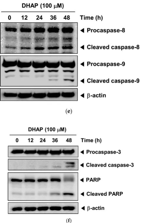
Figure 2. Effect of DHAP on apoptosis and proliferation of U266 cells. The cells were treated with 100
μM of DHAP for 24 h and 48 h. (A) Cellular DNA staining incorporating PI and flow cytometric analysis was performed to ascertain the cell cycle distribution. (B) The cells were incubated with an FITC-conjugated Annexin V, then examined for an early apoptotic effect with flow cytometry. (C) The cells were fixed and incubated with a TUNEL reaction solution, then examined for DNA fragmentation with flow cytometry. (D) U266 cells were treated with 50 and 100 μM of DHAP, then subjected to an MTT assay after 12, 24, 36, and 48 h, to enable cell proliferation to be examined. (E) U266 cells were treated with 100 μM of DHAP for the time periods stated; whole-cell extracts were then prepared and examined via Western blot analysis for caspase-8 and caspase-9. (F) U266 cells were treated with 100 μM of DHAP for the time periods stated; whole-cell extracts were then prepared and analyzed via Western blot analysis for caspase-3 and PARP. To confirm equal protein loading, the immunoblot was stripped and reprobed for β-actin.
2.3. DHAP activates MAPK signaling pathways.
(a)
(b)
(d)
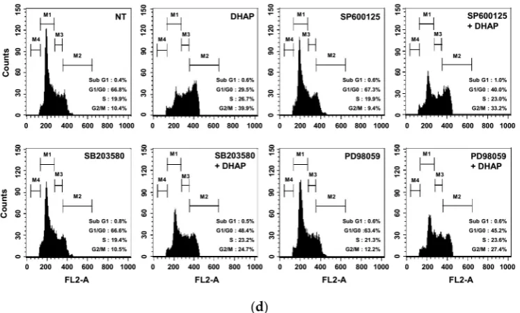
Figure 3. Effect of DHAP on MAPK activation in U266 cells. (A) The cells were treated with 100 μM
of DHAP for the time periods stated; whole-cell extracts were then prepared and analyzed via Western blot analysis for p-p38 (Thr180/Tyr182), p-JNK (Thr183/Tyr185), and p-ERK1/2 (Thr202/Tyr204) by Western blot analysis. To confirm equal protein loading, the immunoblot was stripped and reprobed for JNK, p38, and ERK1/2. The results shown are representative of the three independent experiments. (B) The ratios of phosphorylated proteins to non-phosphorylated proteins were measured and the band density values were expressed as mean ± SE. (C) U266 cells were pretreated with SP600125 (5 μM), SB203580 (10 μM), or PD98059 (25 μM) for 30 min, then incubated with DHAP (100 μM) for 12 h. The expression of p-p38 (Thr180/Tyr182), p-JNK (Thr183/Tyr185), and p-ERK1/2 (Thr202/Tyr204) was ascertained via Western blot analysis. To confirm equal protein loading, the immunoblot was stripped and reprobed for total JNK, p38, and ERK1/2 proteins. (D) U266 cells were pretreated with SP600125 (5 μM), SB203580 (10 μM), or PD98059 (25 μM) for 30 min, then incubated with DHAP (100 μM) for 48 h. Cellular DNA staining with PI and flow cytometric analysis was performed to ascertain the cell cycle distribution.
2.4. DHAP causes potentiation of the apoptotic effect of bortezomib in U266 cells.
(a)
(c)
(d)
(e)
Figure 4. Effect of DHAP on bortezomib-induced apoptosis in U266 cells. (A) The cells were treated
including Bcl-xl, Bcl-2, Mcl-1, Survivin, IAP1, Cyclin D1, Cyclin E, MMP-9, and COX-2, with concomitant up-regulation of Bax and p21 expression. DHAP caused the G2/M phase arrest and inhibition of proliferation, induced apoptosis, and led to activation of caspase-3, caspase-8, and caspase-9. We also found that DHAP induced the activation of JNK, p38, and ERK, and caused substantial potentiation of bortezomib’s apoptotic effects in U266 cells.
It has recently been demonstrated that the apoptosis signaling systems constitute potentially good targets for the development of new anticancer agents [25]. Understanding how DHAP exerts its anticancer effects is therefore important for developing it for cancer prevention and/or treatment. Our results show that the anti-cancer effect of DHAP involves induction of apoptosis. DHAP-induced apoptosis in multiple myeloma cells was detected by Annexin V staining and TUNEL staining (DNA fragmentation), and further confirmed by the activation of caspase-3, caspase-8, and caspase-9, and PARP cleavage. It was discovered that DHAP inhibited the expression of several gene products linked to the initiation and promotion of tumors. These products include anti-apoptotic (xl, Bcl-2, Mcl-1, IAP1, and Survivin), proliferation (Cyclin D1 and Cyclin E), and metastatic (COX-2 and MMP-9) gene products. The down-regulation of Bcl-xl, Bcl-2, IAP-1, Survivin, and Mcl-1, and the upregulation of the expression of pro-apoptotic protein Bax, could play a part in DHAP’s ability to trigger apoptosis within U266 cells. The down-modulation of the expression of MMP-9 and COX-2 may account for the anti-metastatic effects of DHAP, which requires subsequent research.
The inhibition of cell proliferation and/or the induction of apoptosis are strongly linked to the activation of several intracellular signaling pathways resulting in cell cycle arrest in the S, G1, or G2/M phase [26-28]. The downregulation of the expression of cyclin D1 and cyclin E by DHAP is associated with the suppression of proliferation and cell cycle arrest at the G2/M phase, suggesting that cyclin D1 and cyclin E may play a part in DHAP-induced G2/M phase arrest, resulting in the restriction of cell growth and possible apoptotic cell death. It has been suggested that Cyclin E is an attractive target for molecular therapeutics, both because it is overexpressed in a significant fraction of human tumors [29] and this overexpression has been implicated in tumor progression [30]. It has been postulated that interference with cyclin E expression could inhibit the neoplastic growth of a variety of cancers [29]. The increased expression of p21 is linked to inhibition of the cell cycle, apoptosis, and differentiation [31], and the induction of p21 subsequently results in the arrest of the cell cycle’s G1/G0 or G2/M phase [32, 33]. We found that DHAP increased the p21 expression in U266 cells, and this up-regulation of p21 could be one molecular mechanism via which DHAP restricted the growth of tumor cells and triggered cell cycle arrest.
proliferation, and metastasis. Furthermore, combination treatment resulted in greater phosphorylation of JNK, compared with DHAP or bortezomib alone. Overall, our results demonstrate that DHAP inhibited the proliferation of tumor cellscells by causing apoptosis and G2/M phase arrest. DHAP-induced cell cycle arrest and apoptosis were linked to the activation of the MAPK pathway. These results therefore suggest that DHAP has potential as a pharmacological agent to treat human multiple myeloma.
4. Materials and methods
4.1. Reagents
2,5-Dihydroxyacetophenone (DHAP), 3-(4,5-dimethylthiazol-2-yl)-2,5-diphenyltetrazolium bromide (MTT), propidium iodide (PI), and RNase A were obtained from Sigma-Aldrich (St. Louis, MO, USA). DHAP was dissolved as a 50mM stock solution in dimethyl sulfoxide, and kept at -20 °C. Additional dilution was performed in a cell culture medium. Fetal bovine serum (FBS), RPMI 1640, and an antibiotic–antimycotic mixture were obtained from Thermo Fisher Scientific Inc. (Waltham, MA, USA). Antibodies against Bcl-2, Bcl-xl, β-actin, Mcl-1, IAP-1, Survivin, COX-2, MMP-9, Bax, p21, PARP, Caspase-3, and HRP-conjugated secondary antibodies were purchased from Santa Cruz Biotechnology (Santa Cruz, CA, USA). Antibodies against JNK, p-JNK (Thr183/Tyr185), p38, p-p38 (Thr180/Tyr182), p-ERK1/2 (Thr202/Tyr204), ERK1/2, Cyclin D1, Cyclin E, Caspase-8, and Caspase-9 were obtained from Cell Signaling Technology (Beverly, MA, USA).
4.2. Cell lines and cell culture
Human multiple myeloma cell line U266 cells were purchased from the American Type Culture Collection (Manassas, VA, USA). U266 cells were cultured in RPMI 1640 incorporating 10% FBS, 100 g/ml of streptomycin, and 100 units/ml of penicillin. The cells were kept at 37 °C in an atmosphere of 5% CO2.
4.3. Western blot analysis
U266 cells were treated with various concentrations of DHAP for various periods, then lysed; their protein concentrations were revealed with a Bradford reagent (Bio-Rad, Hercules, CA, USA). Whole-cell extracts were isolated at 10–12% sodium dodecyl sulfate-polyacrylamide gelelectrophoresis (SDS-PAGE), then electro-transferred to nitrocellulose membranes (Pall Corporation, MI, USA); these membranes were subsequently blocked with 5% nonfat milk or 2% BSAin Tris-buffered saline with 0.1% Tween 20 (TBST) for 2 h at room temperature, then incubated overnight with the respective primary antibodies at 4 °C. Following this, the membranes were washed and incubated with HRP-conjugated secondary antibodies (1:5,000) at room temperature for 2 h; the membranes were then detected using an enhanced chemiluminescence (ECL) kit (Millipore, Bedford, MA). The statistical analysis and densitometry values for the Western blot analysis were performed by the Sigma plot (Systat Software, Inc., San Jose, CA, USA) and Image J software (National Institutes of Health, Bethesda, MD, USA).
4.5. MTT assay
The anti-proliferative effect of DHAP was ascertained by MTT assay. U266 cells were seeded in 96-well plates at a density of 1 × 10⁴ cells/well, and incubated with 50 and 100 μM of DHAP. After 12,
24, 36 and 48 h incubation, 30 μl of MTT solution (2 mg/ml) was added to each well. After incubation at 37°C for 2 h, 100 μl of extraction buffer (20% SDS and 50% dimethylformamide) was added to the cells. The cells were then incubated at 37 °C overnight, following which the absorbance was measured at 570 nm via a microplate reader (Bio-Rad, Hercules, CA, USA).
4.6. Cell cycle analysis
The effect of DHAP on the distribution of cells in different phases of the cell cycle was analyzed by flow cytometry. U266 cells were seeded on 6-well plates, at a density of 1 × 10⁶ cells/well. The cells
were treated for 24 and 48 h with 100 μM of DHAP, then collected and washed with PBS. Cell pellets were fixed overnight at -20 °C in 70% cold ethanol. The fixed cells were then re-suspended in PBS incorporating 1 mg/ml of RNase A, and incubated at 37 °C for 1 h. Following this, the cells were washed, re-suspended, and stained in PBS containing 25 μg/ml of PI at room temperature in darkness for 30 min. Finally, the DNA contents of the stained cells were examined using Cell Quest 3.0 Software with FACScan Calibur flow cytometry (BD Biosciences, Becton-Dickinson, Franklin Lakes, NJ, USA).
4.7. Annexin V assay
Early apoptotic cell death was determined with an Annexin V-FITC Apoptosis Detection Kit (Bio-Rad, Hercules, CA, USA), in accordance with the instructions from the manufacturer. One early sign of apoptosis is the swift translocation and accumulation of the membrane phospholipid phosphatidylserine from the cytoplasmic interface of the cell to the extracellular surface. This loss of membrane asymmetry can be ascertained with the binding properties of Annexin V. U266 cells were plated in 6-well plates at a density of 1 × 10⁶ cells/well, and were then treated with 100 μM of DHAP
for 24 and 48 h. Following the treatment, the cells were stained with annexin V conjugated to fluorescein isothiocyanate (FITC), or with PI. The stained samples were then analyzed using Cell Quest 3.0 software with a flow cytometer.
4.8. TUNEL assay
Conflicts of Interest: The authors declare no conflicts of interest. References
1. Yokozawa, T.; Kim, H. Y.; Yamabe, N., Amelioration of diabetic nephropathy by dried Rehmanniae Radix (Di Huang) extract. Am J Chin Med 2004,32, 829-839.
2. Chae, H. J.; Kim, H. R.; Kim, D. S.; Woo, E. R.; Cho, Y. G.; Chae, S. W., Saeng-Ji-Hwang has a protective effect on adriamycin-induced cytotoxicity in cardiac muscle cells. Life Sci 2005,76, 2027-2042.
3. Lau, T. W.; Lam, F. F.; Lau, K. M.; Chan, Y. W.; Lee, K. M.; Sahota, D. S.; Ho, Y. Y.; Fung, K. P.; Leung, P. C.; Lau, C. B., Pharmacological investigation on the wound healing effects of Radix Rehmanniae in an animal model of diabetic foot ulcer. J Ethnopharmacol 2009,123, 155-162.
4. Han, Y.; Jung, H. W.; Lee, J. Y.; Kim, J. S.; Kang, S. S.; Kim, Y. S.; Park, Y. K., 2,5-dihydroxyacetophenone isolated from Rehmanniae Radix Preparata inhibits inflammatory responses in lipopolysaccharide-stimulated RAW264.7 macrophages. J Med Food 2012,15, 505-510.
5. Mantovani, A., Cancer: Inflaming metastasis. Nature 2009,457, 36-37.
6. Mantovani, A.; Allavena, P.; Sica, A.; Balkwill, F., Cancer-related inflammation. Nature 2008,454, 436-444. 7. Sethi, G.; Shanmugam, M. K.; Ramachandran, L.; Kumar, A. P.; Tergaonkar, V., Multifaceted link between
cancer and inflammation. Biosci Rep 2012,32, 1-15.
8. Chai, E. Z.; Siveen, K. S.; Shanmugam, M. K.; Arfuso, F.; Sethi, G., Analysis of the intricate relationship between chronic inflammation and cancer. Biochem J 2015,468, 1-15.
9. Makin, G.; Dive, C., Apoptosis and cancer chemotherapy. Trends Cell Biol 2001,11, S22-26.
10. Fulda, S.; Debatin, K. M., Targeting apoptosis pathways in cancer therapy. Curr Cancer Drug Targets 2004,
4, 569-576.
11. Shanmugam, M. K.; Kannaiyan, R.; Sethi, G., Targeting cell signaling and apoptotic pathways by dietary agents: role in the prevention and treatment of cancer. Nutr Cancer 2011,63, 161-173.
12. Tang, C. H.; Sethi, G.; Kuo, P. L., Novel medicines and strategies in cancer treatment and prevention. Biomed Res Int 2014,2014, 474078.
13. Bishayee, A.; Sethi, G., Bioactive natural products in cancer prevention and therapy: Progress and promise.
Semin Cancer Biol 2016,40-41, 1-3.
14. Shanmugam, M. K.; Lee, J. H.; Chai, E. Z.; Kanchi, M. M.; Kar, S.; Arfuso, F.; Dharmarajan, A.; Kumar, A. P.; Ramar, P. S.; Looi, C. Y.; Mustafa, M. R.; Tergaonkar, V.; Bishayee, A.; Ahn, K. S.; Sethi, G., Cancer prevention and therapy through the modulation of transcription factors by bioactive natural compounds.
Semin Cancer Biol 2016,40-41, 35-47.
15. Hengartner, M. O., The biochemistry of apoptosis. Nature 2000,407, 770-776.
16. Ouyang, L.; Shi, Z.; Zhao, S.; Wang, F. T.; Zhou, T. T.; Liu, B.; Bao, J. K., Programmed cell death pathways in cancer: a review of apoptosis, autophagy and programmed necrosis. Cell Prolif 2012,45, 487-498. 17. Yang, S. F.; Weng, C. J.; Sethi, G.; Hu, D. N., Natural bioactives and phytochemicals serve in cancer
treatment and prevention. Evid Based Complement Alternat Med 2013,2013, 698190.
18. Hsieh, Y. S.; Yang, S. F.; Sethi, G.; Hu, D. N., Natural bioactives in cancer treatment and prevention. Biomed Res Int 2015,2015, 182835.
19. Davis, R. J., Signal transduction by the JNK group of MAP kinases. Cell 2000,103, 239-252.
20. Karin, M.; Cao, Y.; Greten, F. R.; Li, Z. W., NF-kappaB in cancer: from innocent bystander to major culprit.
Nat Rev Cancer 2002,2, 301-310.
have we come? Pharm Res 2010,27, 950-961.
26. Singh, R. P.; Dhanalakshmi, S.; Agarwal, R., Phytochemicals as cell cycle modulators--a less toxic approach in halting human cancers. Cell Cycle 2002,1, 156-161.
27. Kannaiyan, R.; Manu, K. A.; Chen, L.; Li, F.; Rajendran, P.; Subramaniam, A.; Lam, P.; Kumar, A. P.; Sethi, G., Celastrol inhibits tumor cell proliferation and promotes apoptosis through the activation of c-Jun N-terminal kinase and suppression of PI3 K/Akt signaling pathways. Apoptosis 2011,16, 1028-1041.
28. Baek, S. H.; Lee, J. H.; Kim, C.; Ko, J. H.; Ryu, S. H.; Lee, S. G.; Yang, W. M.; Um, J. Y.; Chinnathambi, A.; Alharbi, S. A.; Sethi, G.; Ahn, K. S., Ginkgolic Acid C 17:1, Derived from Ginkgo biloba Leaves, Suppresses Constitutive and Inducible STAT3 Activation through Induction of PTEN and SHP-1 Tyrosine Phosphatase.
Molecules 2017,22.
29. Malumbres, M.; Barbacid, M., To cycle or not to cycle: a critical decision in cancer. Nat Rev Cancer 2001,1, 222-231.
30. Bortner, D. M.; Rosenberg, M. P., Induction of mammary gland hyperplasia and carcinomas in transgenic mice expressing human cyclin E. Mol Cell Biol 1997,17, 453-459.
31. Chen, X.; Zhang, W.; Gao, Y. F.; Su, X. Q.; Zhai, Z. H., Senescence-like changes induced by expression of p21(waf1/Cip1) in NIH3T3 cell line. Cell Res 2002,12, 229-233.
32. Taylor, W. R.; Stark, G. R., Regulation of the G2/M transition by p53. Oncogene 2001,20, 1803-1815. 33. Coqueret, O., New roles for p21 and p27 cell-cycle inhibitors: a function for each cell compartment? Trends
Cell Biol 2003,13, 65-70.
34. Cho, S. D.; Li, G.; Hu, H.; Jiang, C.; Kang, K. S.; Lee, Y. S.; Kim, S. H.; Lu, J., Involvement of c-Jun N-terminal kinase in G2/M arrest and caspase-mediated apoptosis induced by sulforaphane in DU145 prostate cancer cells. Nutr Cancer 2005,52, 213-224.
35. Xu, C.; Shen, G.; Yuan, X.; Kim, J. H.; Gopalkrishnan, A.; Keum, Y. S.; Nair, S.; Kong, A. N., ERK and JNK signaling pathways are involved in the regulation of activator protein 1 and cell death elicited by three isothiocyanates in human prostate cancer PC-3 cells. Carcinogenesis 2006,27, 437-445.
36. Lavelle, D.; DeSimone, J.; Hankewych, M.; Kousnetzova, T.; Chen, Y. H., Decitabine induces cell cycle arrest at the G1 phase via p21(WAF1) and the G2/M phase via the p38 MAP kinase pathway. Leuk Res 2003,27, 999-1007.
37. Johnson, G. L.; Lapadat, R., Mitogen-activated protein kinase pathways mediated by ERK, JNK, and p38 protein kinases. Science 2002,298, 1911-1912.
38. 38. Olson, J. M.; Hallahan, A. R., p38 MAP kinase: a convergence point in cancer therapy. Trends Mol Med
2004,10, 125-129.
39. Sah, J. F.; Balasubramanian, S.; Eckert, R. L.; Rorke, E. A., Epigallocatechin-3-gallate inhibits epidermal growth factor receptor signaling pathway. Evidence for direct inhibition of ERK1/2 and AKT kinases. J Biol Chem 2004,279, 12755-12762.
40. Bacus, S. S.; Gudkov, A. V.; Lowe, M.; Lyass, L.; Yung, Y.; Komarov, A. P.; Keyomarsi, K.; Yarden, Y.; Seger, R., Taxol-induced apoptosis depends on MAP kinase pathways (ERK and p38) and is independent of p53.
Oncogene 2001,20, 147-155.
41. She, Q. B.; Bode, A. M.; Ma, W. Y.; Chen, N. Y.; Dong, Z., Resveratrol-induced activation of p53 and apoptosis is mediated by extracellular-signal-regulated protein kinases and p38 kinase. Cancer Res 2001,61, 1604-1610.
42. Nguyen, T. T.; Tran, E.; Nguyen, T. H.; Do, P. T.; Huynh, T. H.; Huynh, H., The role of activated MEK-ERK pathway in quercetin-induced growth inhibition and apoptosis in A549 lung cancer cells. Carcinogenesis