Copyright2000 by the Genetics Society of America
Cellular Werner Phenotypes in Mice Expressing a Putative Dominant-Negative
Human
WRN
Gene
Lan Wang,*
,†Charles E. Ogburn,
†Carol B. Ware,
‡Warren C. Ladiges,
‡Hagop Youssoufian,
§George M. Martin*
,†and Junko Oshima*
*Department of Pathology,†Department of Genetics and‡Department of Comparative Medicine, University of Washington, Seattle, Washington
98195 and§Department of Molecular and Human Genetics, Baylor College of Medicine, Houston, Texas 77030 Manuscript received April 30, 1999
Accepted for publication September 15, 1999
ABSTRACT
Mutations at the Werner helicase locus (WRN ) are responsible for the Werner syndrome (WS). WS patients prematurely develop an aged appearance and various age-related disorders. We have generated transgenic mice expressing human WRN with a putative dominant-negative mutation (K577M-WRN). Primary tail fibroblast cultures from K577M-WRN mice showed three characteristics of WS cells: hypersensi-tivity to 4-nitroquinoline-1-oxide (4NQO), reduced replicative potential, and reduced expression of the endogenous WRN protein. These data suggest that K577M-WRN mice may provide a novel mouse model for the WS.
W
ERNER syndrome (WS) is an autosomal recessive et al. 1997; Huang et al. 1998). More than 20 WRNmutations have been identified in WS patients (Yuet al.
progeroid syndrome characterized by premature
development of an aged appearance and many disor- 1996;Moseret al. 1999). All result in truncated protein
products missing the nuclear localization signal. The ders associated with advanced age, such as bilateral
cata-racts, scleroderma-like skin, osteoporosis, several forms mouse WRN gene has also been cloned and shown to share the characteristic domains of the human homo-of arteriosclerosis, hypogonadism, type II diabetes
melli-tus, and neoplasia. Symptoms appear at puberty and log, overall amino acid sequence homology being 76% (Imamuraet al. 1997).
death occurs at a mean age of 47 years, usually as a result
of cardiovascular diseases or malignancies (Epsteinet Activities (39→59helicase and 39→59exonuclease) of the recombinant WRNp have been demonstrated by
al. 1966;Martin et al. 1970; Tollefsbol and Cohen
1984;Goto1997). in vitro assays and it has been shown that a single amino
acid substitution at position 577 (K577M) in the recom-Primary cultures of somatic cells from WS patients
have very limited proliferative potentials and retarded binant human WRNp results in the abolishment of the ATPase and helicase activities, but not the exonuclease cell cycle progression (Martinet al. 1970, 1974;
Tollefs-bolandCohen1984;Salket al. 1985a;Killet al. 1994). activity (Grayet al. 1997;Huanget al. 1998). The
corre-sponding mutation in Escherichia coli uvrD encoding a Genomic instability is also evident and has been
charac-terized as “variegated translocation mosaicism” (Salk 39 →59 helicase causes a dominant-negative effect on cell growth in response to UV light (Georgeet al. 1994). et al. 1985b). Hypersensitivity to a genotoxic agent,
4-nitroquinoline-1-oxide (4NQO), has also been docu- To examine the role of WRN in an animal model, we
generated mouse lines overexpressing either the K577M mented (Gebhardt et al. 1988; Ogburn et al. 1997).
Lymphoblastoid cell lines (LCLs) from individuals het- mutant or wild-type human WRN using conventional transgenic methodology (Hoganet al. 1986). The
ex-erozygous for WRN mutations exhibit sensitivities that
are intermediate between those of the homozygous mu- pression of several cellular phenotypes characteristic of the human WS in transgenic mice suggests a dominant-tants and wild type (Ogburnet al. 1997).
The Werner syndrome is caused by mutations at the negative action of the K577M-WRN allele in vivo.
WRN locus on chromosome 8p (GenBank accession no.
L76937;Yuet al. 1996). Human WRN protein (hWRNp)
MATERIALS AND METHODS
contains an N-terminal exonuclease domain, a central
helicase domain, and a C-terminal nuclear localization Generation of transgenic lines: Mouse expression vectors signal (Grayet al. 1997;Matsumotoet al. 1997;Suzuki were constructed by subcloning full-length cDNA encoding human wtWRNp and human K577M-WRNp into pBSCA (de-rived from pCAGGS). This vector contains a cytomegalovirus enhancer, a chick b-actin promoter, and a rabbit b-globin Corresponding author: Junko Oshima, Box 357470, Health Science
polyadenylation site (Grayet al. 1998). Transgenic mice were
Bldg. K543, Department of Pathology, University of Washington, 1959
created by pronuclear injection as previously described
NE Pacific Ave., Seattle, WA 98195-7470.
E-mail: [email protected] (Hoganet al. 1986). The original founder animals were on
Figure2.—Subcellular localization of wtWRNp and K577M-WRNp in primary tail fibroblasts. Indirect immunofluorescent staining utilized the same antibody as in Figure 1 in primary tail fibroblasts from littermate control, WL4128, WL4139, and Figure 1.—Expression of wtWRNp and K577M-WRNp in
human fibroblasts, GM439, which serves as a positive control primary tail fibroblasts. Western analysis of nuclear fractions of
(Grayet al. 1998). tail fibroblasts utilized anti-C-terminal human WRN antibody
(Grayet al., 1998). WL4139 and WL4146 are the transgenic
lines of K577M-WRN, and WL4128, WL4998, and WL5025 are cross-react with mouse WRNp (Figure 1), although in one the transgenic lines of wtWRN. experiment (Figure 5) it weakly cross-reacted with mouse
WRNp.
4NQO sensitivity assay:Aliquots (1 ml) of the collagenase-a mixed C57BL/6J3C3HJ background. Three lines of K577M derived cell suspension from mouse tails were plated in 75-mutant transgenics (WL4128, WL4998, and WL5025) and two cm2flasks. Exponentially growing cells were replated 48–72 lines of wild-type WRN transgenics (WL4139 and WL4146) hr later in multiple 25-cm2 tissue culture flasks at 5 3 104 were generated using traditional pronuclear injection meth- cells/flask in triplicates. The media were replaced 24 hr later ods and backcrossed with C57BL/6J. Primary fibroblast cul- with media containing the indicated concentrations of 4NQO tures from the tails of founders and/or first backcross off- (Sigma, St. Louis; stock 3 mg/ml in DMSO). DMSO was added spring were used for the experiments. to the control flasks at a level equivalent to that of the highest
Preparation of cells:Mouse tails were minced and digested dose of 4NQO. After 24 hr all flasks were fed with fresh media with collagenase and primary mouse tail fibroblasts were cul- without 4NQO and incubated for an additional 48 hr. The tured as described (Martinet al. 1996). In brief, aseptically flasks were then trypsinized and counted using a hemocytome-collected biopsies of mouse tails were minced, washed three ter to determine the survival of cells.
times in Ca and Mg-free phosphate-buffered normal saline Clone size distribution assay:The collagenase-derived cell (PBS; pH 7.1), and resuspended in 20 ml of a 1:1 mixture of suspension (10 ml total per mouse tail) was plated in four Type 1 collagenase (1 mg/ml in PBS) and Dulbecco’s Modi- 100-mm cell culture dishes at a ratio of 10ml per dish in 10 fied Eagle Media (DMEM; GIBCO/BRL, Gaithersburg, MD) ml media and incubated for 10 days. This density provides with 100 units/ml penicillin and 100 mg/ml streptomycin. well-separated individual cells and colonies. Dishes were fed The suspension was stirred at moderate speed for 1 hr in a once at between 5 and 7 days with fresh media. Dishes were 378 incubator to digest tissues. After letting the tissue frag- then rinsed with PBS and stained with 0.5% crystal violet in ments settle, the supernatant was collected and one-tenth vol- 20% ethanol. Using a dissecting microscope, the colonies and ume of fetal bovine serum (FBS) was added to stop the colla- individual undivided cells were scored for the number of cell genase action. The supernatant was stored in ice. The divisions that had occurred.
remaining tissue fragments were redigested for an additional Statistical analysis: Differences in 4NQO sensitivity were 1 hr at 378. At the end of this digest there were few if any tested by two-factor (dose by genotype) ANOVA using Statview tissue fragments remaining. The resulting cell suspension was (Abacus Concepts, Berkeley, CA). Post hoc t-tests were then combined and centrifuged at 26003g for 5 min. The result- performed to describe differences in 4NQO sensitivity across ing cell pellet was resuspended in 10 ml of DMEM supple- genotypes. For clone size distribution assays, statistical compar-mented with 50 units/ml of penicillin, 50mg/ml of streptomy- isons between genotypes were made using t -tests comparing cin, and 10% FBS that was heat-inactivated at 568for 30 min. the numbers of colonies achieving a given number of cell This cell suspension was used for the experiments. divisions.
A SV40-transformed normal human fibroblast cell line, GM649, was obtained from Coriell Cell Repositories (Camden,
NJ). All cells were maintained in DMEM media supplemented RESULTS with 10% heat-inactivated FBS, 50 units/ml penicillin, and 50
Generation of transgenic mice overexpressing
mg/ml streptomycin in a humidified, 5% CO2 incubator at
378. K577M-WRNp and wild-type-WRNp:The degree of
ex-Western blot analysis:A total of 23105primary tail
fibro-pression of the transgenes in the primary tail fibroblasts
blasts were plated in 100-mm cell culture dishes. Cells were varied from line to line as determined by Western analy-collected 48 hr later, and nuclear and cytoplastic fractions
sis using an antibody specific for the C terminus of
were separated. A total of 100mg of nuclear protein was
re-hWRNp (Grayet al. 1998; Figure 1). Primary fibroblasts
solved by 7% SDS-PAGE and visualized by Western analysis,
using anti-human WRN or anti-mouse WRN polyclonal anti- from five transgenic lines showed various levels of
ex-bodies. Anti-human WRN antibody was made against glutathi- pression, while a nonspecific band showed (see Figure one S-transferase (GST)-fused partial human WRN C termi- 5) similar levels, indicating equal loading (data not nus, aa 982–1432, and was affinity-purified. Anti-mouse WRN
shown). Immunofluoresence staining of hWRNp
re-antibody was made against GST-fused partial mouse WRN C
vealed that both wtWRNp and K577M-WRNp were
local-terminus, aa 997–1297, and was also affinity-purified (Gray
Figure3.—4NQO sensitivity of primary tail fibroblasts. Cell survival was expressed as the percentage of untreated con-trol cells (control counts ranged from 1 to 2.5 3 105 cells) for each dose of 4NQO. Open bars represent the mean survival of nontransgenic lit-termate control cells (N5 17 mice), crosshatched bars rep-resent the mean survival of cells from transgenic mice ex-pressing wtWRN (1, WL4139, N59 mice; 2, WL4146, N54 mice), and solid bars represent the mean survival of cells from mice expressing K577M-WRN (1, WL4128, N5 10 mice; 2, WL5025, N 5 3 mice; 3, WL4998, N51 mouse).
the pattern of localization in replicating populations of of cells from K577M-WRN mice was observed when com-pared to the sensitivities observed in cells from wtWRN human fibroblasts, in which nucleoli were more
in-tensely stained (Figure 2). It has been reported that transgenics (P 5 0.008 by ANOVA analysis). Post hoc analyses showed significant differences for four of five hWRNp is primarily localized to the nucleoli of actively
replicating cells (Gray et al. 1998; Marciniak et al. dose levels (P, 0.05); the comparison at 0.05mg/ml was not significant (P 5 0.079). When sensitivities in 1998), while mWRNp is localized to nucleoplasm (
Mar-ciniaket al. 1998). The intensities of immunofluores- K577M-WRN mice were compared to those of non-transgenic littermate controls, the difference was again cence staining in the mouse fibroblasts roughly
re-flected the levels of protein expression as determined significant (P 5 0.0327 by ANOVA analysis). Post hoc analyses showed significant differences for four of five by Western analysis. Tail fibroblasts from WL4128
ex-pressed relatively low levels of K577M-WRNp by both dose levels (P , 0.05); the comparison at 0.4 mg/ml was not significant (P50.0759). Combined data from Western analysis and immunofluorescence staining,
whereas WL4139 expressed relatively high levels of the wtWRN transgenics and from nontransgenic
lit-termate controls showed no significant difference in wtWRNp by both methods. In each culture, virtually all
cells stained with comparable intensities. 4NQO sensitivity (P50.729 by ANOVA analysis). This
indicates that abolishment of the ATPase activity of Hypersensitivity to 4NQO and reduced proliferative
capacity in K577M-WRN mice:We examined two well- hWRNp is able to confer a dominant-negative effect at
the cellular level and that additional human wtWRNp characterized cellular phenotypes observed in WS cells:
sensitivity to a genotoxic agent, 4-nitroquinoline-1-oxide does not confer resistance to 4NQO challenge. The replicative potentials of K577M transgenic cell (4NQO;Ogburnet al. 1997), and replicative potential
(Salk et al. 1985a). Hypersensitivity to 4NQO was ob- populations, as determined by clone size distribution assays (Smithet al. 1978), were significantly decreased,
served in primary tail fibroblasts from all three
K577M-WRN mice, whether derived from the founder or the as compared to cell populations from wtWRN
transgenics (P , 0.01) and nontransgenic littermate backcrossed offspring (Figure 3). There was no
correla-tion between the amount of K577M-WRNp and the de- controls (P,0.01; Figure 4). There were no statistically significant differences among the three K577M lines, gree of 4NQO sensitivity among the three K577M-WRN
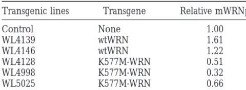
which we observed a 25% reduction of mWRNp. There was no relationship between the degree of expression of the transgenes and the extent of the changes in the expression of endogenous mWRNp. These data suggest that overexpression of either wtWRNp or K577M-WRNp increased or decreased, respectively, the expression of the endogenous mouse gene. Only the K577M-WRNp, however, produced detectable effects at the cellular level.
DISCUSSION
Our data show that expression of a putative dominant-negative human WRNp in transgenic mice conferred three cellular characteristics of WS. First, we docu-mented a significant decrease in replicative potential of fibroblast-like cells from tail skin. This is a particularly well-documented WS phenotype, having been reported by many laboratories and many patients (Martinet al.
Figure 4.—Clone size distribution of primary tail fibro- 1970, 1974; Tollefsbol andCohen 1984; Salk et al.
blasts. Clone sizes are expressed as the mean (6SE of the 1985a;Killet al. 1994). Second, we established hyper-mean) percentage of cells achieving at least the number of
sensitivity to a genotoxic agent, 4NQO, shown by two
cell divisions indicated. Between 200 and 300 colonies were
laboratories to be a characteristic of WS (Gebhartet
counted for each animal assayed. Solid circles represent the
al. 1988; Ogburn et al. 1997). That 4NQO sensitivity
results for cells from nontransgenic littermate controls (N5
14 mice), numbers in squares represent the results for cells may be a robust marker of WRN helicase deficiency is from mice expressing wtWRN (1, WL4139, N 5 8 mice; 2, suggested by the observation of cells from heterozygous WL4146, N 52 mice), and numbered circles represent the
carriers that exhibit a sensitivity intermediate between
results for cells from mice expressing K577M-WRN (1,
that of wild-type individuals and homozygous WS
pa-WL4128, N 58 mice; 2, WL5025, N 5 3 mice; 3, WL4998,
N51 mouse). tients (Ogburnet al. 1997). Finally, as shown for human
cells deficient in WRN, expression from the resident normal allele (in this case two murine alleles) is down-regulated.
not result in detectable differences in cellular replicative
There are at least two major mechanisms that may potentials. Overexpression of human wtWRNp had no
be responsible for the observed dominant-negative ef-effect on the cellular replicative potential when
com-fects of the mutant form of human WRNp used in our pared to that of littermate controls (P50.158).
experiments. Because many known helicases function Downregulation of the endogenous mouse WRNp in
as a multimeric complex, overexpression of a
dominant-K577M-WRN mice:It has been observed that
transcrip-negative mutant WRNp may cause the WS phenotype tion of reporter genes driven by human WRN promoters
was reduced toz40% when these constructs were intro-duced to WS cells (Wanget al. 1998). It has also been
shown that WRN mRNAs were reduced to less than half of wild-type levels in cells from WS heterozygotes (Yamabeet al. 1997). These findings are consistent with
the presence of a positive autoregulatory loop for WRN gene expression. We therefore performed Western blot assays to determine the expression of endogenous mWRNp levels in the tail fibroblasts used for the above studies. As shown in Figure 5, endogenous mWRNp levels were reduced in tail fibroblasts from K577M-WRN
transgenics. By contrast, they were elevated in fibroblasts Figure5.—Expression of endogenous mouse WRNp in pri-from wtWRN transgenics. Table 1 summarizes the ex- mary tail fibroblasts. Western analysis of nuclear fractions of tail fibroblasts in WL4139 (wtWRN transgenic line) and
pression of mouse WRNp in all lines of transgenics
in-WL4128 (K577M-WRN transgenic line) are shown.1,
trans-cluding two lines expressing wtWRNp and three lines
genic mice;2, littermate controls. (Top) Probed with
anti-expressing K577M-WRNp. The results were consistent human WRNp antibody, (middle) probed with anti-mouse in replicates, except for one experiment with a culture WRN antibody. (Bottom) The nonspecific bands in the mouse
WRNp blot serving as a loading control.
TABLE 1 suggest that the subcellular localization of WRNp is dic-tated by host factors. Murine cells may lack the ability Endogenous mouse WRN protein (mWRNp) levels in
to transport WRNp, either human or mouse, to the fibroblasts from mice expressing transgenic human
nucleoli. wild-type (wtWRN) or mutant WRN (K577M-WRN)
Embryonic stem (ES) cell lines with homozygous
dele-Transgenic lines Transgene Relative mWRNp tions of the third and fourth motifs of the helicase domains of mWRNp have been synthesized (Lebeland
Control None 1.00
Leder1998). The altered mWRNp retains the ATPase
WL4139 wtWRN 1.61
domain of the helicase and the exonuclease domains.
WL4146 wtWRN 1.22
These mice therefore may provide only a partial model
WL4128 K577M-WRN 0.51
WL4998 K577M-WRN 0.32 of WS. Because all known spontaneous mutations
ap-WL5025 K577M-WRN 0.66 pear to be null (their truncated products being
ex-cluded from the nucleus;Moseret al. 1999), it may be
Western analysis is shown in Figure 5. mWRNp levels were
necessary to eliminate the exonuclease function as well
normalized to nonspecific bands of the autoradiographs and
to the levels in littermate controls and expressed as the “Rela- as the helicase function to model WS. Our
dominant-tive mWRNp.” The averages of two to three experiments are negative model is more likely to impair both functions
shown. if, in fact, the dominant-negative effect results from
functional inactivation of the relevant multimeric com-plexes.
by disrupting the overall function of such a protein Our model may have certain complicating factors.
complex. An alternative possibility would be that Expression of a dominant-negative mutant could impair K577M-WRNp may generate a multimeric complex itself the functions of other related helicases, which could
and mouse WRNp may make the complex itself, and have at least partially complemented the loss of WRN
the K577M-WRNp complexes and the mouse WRNp helicase activity. We have not yet determined the degree
complexes compete for WRNp binding sites. If this is to which the introduced K577M-WRNp may affect the the case, the effect of K577M-WRNp would be dose expression of other related helicases such as BLM (Ellis dependent until reaching a plateau. Our data showed et al. 1995), human RecQL (PuranamandBlackshear that the effect of K577M-WRNp is independent of its 1994), human RecQ4 and RecQ5 (Kitaoet al. 1998), or expression levels. We therefore favor the first possibility. yet-unknown members of this family of proteins. It is also
Reduced endogenous mouse WRNp expression levels possible that overexpressed K577M-WRNp may “absorb”
might also be the mechanism producing WS phenotypes limiting cofactors necessary for the function of WRNp
in K577M-WRN mice. Cells from humans with heterozy- complex. There may exist some compensatory
mecha-gous WRN mutations express significantly lower WRN nisms mediated by other members of the RecQ family
mRNA than cells from normal individuals (Yamabeet of helicases when the WRNp function is absent, perhaps
al. 1997). This is thought to be the result of degradation particularly so in mice. If that is the case, knockout
of mutated mRNAs via a specific pathway (Jacobson of only the WRN protein might not be sufficient to
reproduce the complete phenotype; experiments de-andPeltz 1996), and the WRN promoter appears to
signed to limit such compensatory mechanisms might have a positive autoregulatory pathway (Wang et al.
result in a more robust model of the human disease. 1998). As a result, steady-state levels of WRN mRNA in
It will be important to carry out careful pathological the heterozygotes, most of which is presumably from
and pathophysiologic studies in aging cohorts of our the wild-type allele, are on average 30% of the controls
K577M-WRN transgenics. Many phenotypes, such as cat-(Yamabeet al. 1997). Cells from heterozygotes exhibit
aracts and neoplasms, may not become apparent until moderate hypersensitivity to 4NQO (Ogburn et al.
more advanced ages. It will be equally important to 1997), topoisomerase inhibitor I, and camptothecin
assess the role of background alleles, to quantitate fe-(Poot et al. 1999), although they exhibit no clinical
cundity, and to assess meiotic recombination rates once phenotypes.
the transgenic lines have been backcrossed to defined Transgenic lines expressing human wtWRNp showed
genetic backgrounds. increased expression of endogenous mWRNp, but only
marginally improved cell survival when challenged with We thank Dr. Steven D. Edland for his assistance in statistical analy-ses. This work was supported by grants from the National Institutes
4NQO. This may be because WRNp is not the limiting
of Health, R01 AG14446 (J.O.), R24 CA78088 (G.M.M.), P01 AG01751
factor in the functional pathway involved in survival of
(G.M.M.), and P30 AG13280 (Peter S. Rabinovitch).
4NQO challenge. Alternatively, the amount of wtWRN may not be sufficient to cause cellular biological effects
under the experimental conditions employed. LITERATURE CITED
The introduced hWRNp was localized to the
nucleo-Ellis, N. A., J. Groden, T. Z. Ye, J. Straughen, D. J. Lennonet al.,
plasm, where mWRNp had previously been shown to 1995 The Bloom’s syndrome gene product is homologous to
RecQ helicases. Cell 83: 655–666.
Epstein, C. J., G. M. Martin, A. L. SchultzandA. G. Motulsky, Martin, G. M., C. A. Sprague, T. H. NorwoodandW. R. Pender-grass,1974 Clonal selection, attenuation and differentiation 1966 Werner’s syndrome: a review of its symptomatology,
natu-ral history, pathologic features, genetics and relationships to the in an in vitro model of hyperplasia. Am. J. Pathol. 74: 137–154.
Martin, G. M., C. E. Ogburn, L. M. Colgin, A. M. Gown, S. D.
natural aging process. Medicine 45: 172–221.
Gebhart, E., R. Bauer, U. Raub, M. Schinzel, K. W. Ruprechtet Edlandet al., 1996 Somatic mutations are frequent and in-crease with age in human kidney epithelial cells. Hum. Mol.
al., 1988 Spontaneous and induced chromosomal instability in
Werner syndrome. Hum. Genet. 80: 135–139. Genet. 5: 215–221.
Matsumoto, T., A. Shimamoto, M. GotoandY. Furuichi,1997
George, J. W., R. M. Brosh, Jr.andS. W. Matson,1994 A dominant
negative allele of the Escherichia coli uvrD gene encoding DNA Impaired nuclear localization of defective DNA helicases in Wer-ner’s syndrome. Nat. Genet. 16: 335–336.
helicase II. A biochemical and genetic characterization. J. Mol.
Biol. 235: 424–435. Moser, M. J., J. Oshima and R. J. Monnat, Jr.,1999 Mutation update: WRN mutations in Werner syndrome. Hum. Mut. 13:
Goto, M.,1997 Hierarchical deterioration of body systems in
Wer-ner’s syndrome: implications for normal aging. Mechanisms 271–279.
Ogburn, C. E., J. Oshima, M. Poot, R. Chen, K. E. Huntet al., 1997
Aging Develop. 98: 239–254.
Gray, M. D., J. C. Shen, A. S. Kamath-Loeb, A. Blank, B. L. Sopher An apoptosis-inducing genotoxin differentiates heterozygotic car-riers for Werner helicase mutations from wild-type and
homozy-et al., 1997 The Werner syndrome protein is a DNA helicase.
Nat. Genet. 17: 100–103. gous mutants. Hum. Genet. 101: 121–125.
Poot, M., K. A. GollahonandP. S. Rabinovitch,1999 Werner
Gray, M. D., L. Wang, H. Youssoufian, G. M. MartinandJ. Oshima,
1998 Werner helicase is localized to transcriptionally active syndrome lymphoblastoid cells are sensitive to camptothecin-induced apoptosis in S-phase. Hum. Genet. 104: 10–14. nucleoli of cycling cells. Exp. Cell. Res. 242: 487–494.
Hogan, B., F. CostantiniandE. Lacy,1986 Manipulating the Mouse Puranam, K. L., andP. J. Blackshear,1994 Cloning and character-ization of RECQL, a potential human homologue of the
Esche-Embryo: A Laboratory Manual. Cold Spring Harbor Laboratory
Press, Cold Spring Harbor, NY. richia coli DNA helicase RecQ. J. Biol. Chem. 269: 29838–29845.
Salk, D., E. Bryant, H. Hoehn, P. JohnstonandG. M. Martin, Huang, S., B. Li, M. D. Gray, J. Oshima, I. S. Mianet al., 1998 The
premature aging syndrome protein, WRN, is a 39 to 59 exo- 1985a Growth characteristics of Werner syndrome cells in vitro. Adv. Exp. Med. Biol. 190: 305–311.
nuclease. Nat. Genet. 20: 114–116.
Imamura, O., K. Ichikawa, Y. Yamabe, M. Goto, M. Sugawaraet Salk, D., K. Au, H. HoehnandG. M. Martin,1985b Cytogenetics aspects of Werner syndrome. Adv. Exp. Med. Biol. 190: 541–546.
al., 1997 Cloning of a mouse homologue of the human Werner
syndrome gene and assignment to 8A4 by fluorescence in situ Smith, J. R., O. Pereira-SmithandE. L. Schneider,1978 Colony size distributions as a measure of in vivo and in vitro aging. Proc. hybridization. Genomics 41: 298–300.
Jacobson, A.,andS. W. Peltz,1996 Interrelationships of the path- Natl. Acad. Sci. USA 75: 1353–1356.
Suzuki, N., A. Shimamoto, O. Imamura, J. Kuromitsu, S. Kitao
ways of mRNA decay and translation in eukaryotic cells. Annu.
Rev. Biochem. 65: 693–739. et al., 1997 DNA helicase activity in Werner’s syndrome gene product synthesized in a baculovirus system. Nucleic Acids Res.
Kill, I. R., R. G. Faragher, K. LawrenceandS. Shall,1994 The
expression of proliferation-dependent antigens during the life- 25:2973–2978.
Tollefsbol, T. O., andH. J. Cohen,1984 Werner’s syndrome: an span of normal and progeroid human fibroblasts in culture. J.
Cell Sci. 107: 571–579. underdiagnosed disorder resembling premature aging. Age 7: 75–88.
Kitao, S., I. Ohsugi, K. Ichikawa, M. Goto, Y. Furuichiet al., 1998
Cloning of two new human helicase genes of the RecQ family: Wang, L., K. E. Hunt, G. M. MartinandJ. Oshima,1998 Structure and function of the human Werner syndrome gene promoter: biological significance of multiple species in higher eukaryotes.
Genomics 54: 443–452. evidence for transcriptional modulation. Nucleic Acids Res. 26:
Lebel, M.,andP. Leder,1998 A deletion within the murine Werner 3480–3485.
syndrome helicase induces sensitivity to inhibitors of topoisomer- Yamabe, Y., M. Sugimoto, M. Satoh, N. Suzuki, M. Sugawaraet
ase and loss of cellular proliferative capacity. Proc. Natl. Acad. al., 1997 Down-regulation of the defective transcripts of the Sci. USA 95: 13097–13102. Werner’s syndrome gene in the cells of patients. Biochem.
Bio-Marciniak, R. A., D. B. Lombard, F. B. JohnsonandL. Guarente, phys. Res. Commun. 236: 151–154.
1998 Nucleolar localization of the Werner syndrome protein Yu, C. E., J. Oshima, Y. H. Fu, F. Hisama, E. M. Wijsmanet al., 1996
in human cells. Proc. Natl. Acad. Sci. USA 95: 6887–6892. Positional cloning of the Werner’s syndrome gene. Science 272:
Martin, G. M., C. A. SpragueandC. J. Epstein,1970 Replicative 258–262. life-span of cultivated human cells. Effects of donor’s age, tissue,