ABSTRACT
HEINECKE, CHRISTINE LORAINE. Small Molecule Control over Biological Processes. (Under the direction of Dr. Christian Melander).
Myotonic Dystrophy Type 1 (DM1) is a multisystemic neuromuscular disorder characterized by a (CTG)n (n > 50) triplet repeat expansion in the 3’-UTR of the myotonic dystrophy protein kinase gene (DMPK). Transcription produces aberrant poly(CUG) expansions which sequester a family of alternative splicing regulators, the muscleblind proteins (MBNL1-3). In the absence of muscleblind, CUG-binding proteins mediate aberrant splicing leading to the disease pathology. We investigated cyclic peptides for their ability to disrupt a (CUG)54:MBNL1 interaction as a unified approach towards treating DM1. To this end, we performed a phage display selection using a cysteine constrained cyclic heptapeptide library against immobilized (CUG)54, which evolved four highly conserved cyclic peptides. In an effort to develop cellularly irreducible analogs of the cysteine constrained heptapeptides, we performed solid phase peptide synthesis substituting allyl glycine at the N- and C-terminal positions, which were subjected to ring closing metathesis. One of the selected peptides was shown to bind (CUG)54 and disrupt the (CUG)54:MBNL1 interaction characteristic of DM1 to a small extent.
Kinamycin D is a potent antitumor antibiotic; however, its biological mode of action is poorly understood. In order to further elucidate the mechanism by which kinamycin D mediates DNA damage, we employed DNA sequencing gel electrophoresis. We have demonstrated that under reducing conditions, acidic media promotes enhanced DNA cleavage. Additionally, our results indicate that kinamycin D is capable of generating reactive oxygen species under acidic pH in the presence of a reducing agent and that DNA cleavage is dependent on trace iron and hydroxyl radicals.
implicated in a variety of skeletal diseases. In an effort to target osteoclasts and inhibit osteoclastogenesis, we have synthesized 1,4-substituted triazole libraries. Qualitative analysis of osteoclast maturation when treated with our compounds indicates some members of our triazole libraries to be the most potent known inhibitors of osteoclastogenesis ever disclosed.
Small Molecule Control over Biological Processes
by
Christine Loraine Heinecke
A dissertation submitted to the Graduate Faculty of North Carolina State University
in partial fulfillment of the requirements for the Degree of
Doctor of Philosophy
Chemistry
Raleigh, North Carolina
2010
APPROVED BY:
_______________________ _______________________
David Shultz Jonathan Lindsey
_______________________ _______________________
Reza Ghiladi Christian Melander
DEDICATION
For my grandfather, William Gestwick
“My grandfather used to say nobody gives a damn anymore and the thing I remember most about him is the way he never let that stop him from giving a damn all the days of his life.”
For my mother, Deborah Heinecke
“I didn't listen to her because she was my mother and wouldn't know anything until I was much older.”
For my father, David Heinecke
“When I was young, I used to wear my father's shoes and stomp around, making the world a better place for us all, and today I see I'm not the only one who did that and those shoes aren't
anywhere big enough for who we are now.”
For my grandmother, Margaret “Ellen” Heinecke
“I wish you could have been there for the sun and the rain and the long, hard hills. For the sound of a thousand conversations scattered along the road. For the people laughing and
BIOGRAPHY
The author, Christine Loraine Heinecke, was born in Baltimore, MD on May 4, 1982 to Deborah and David Heinecke. She spent her childhood in Baltimore, MD and later in Glen Rock, PA. At the age of 14 she moved to Powhatan, Virginia where she graduated from high school. After high school graduation she attended James Madison University and received a B.S. in Chemistry with a concentration in Materials Science and was the proud recipient of the American Institute of Chemists Award. Her undergraduate research experience with Professor Barbara Reisner facilitated her appreciation of science and encouraged her to attend graduate school to further her scientific endeavors. In the fall of 2004, Christine began her graduate research studies at North Carolina State University under the advisement of Professor Christian Melander. After graduation, Christine began a postdoctoral research position at Colorado State University under the direction of Professor Christopher J. Ackerson.
TABLE OF CONTENTS
LIST OF TABLES...vii
LIST OF FIGURES...viii
LIST OF SCHEMES...x
LIST OF ABBREVIATIONS AND TERMS...xi
CHAPTER 1: A Unified Approach at Targeting Myotonic Dystrophy...1
1.1Introduction...1
1.1.1 DMPK Haploinsufficiency Hypothesis...2
1.1.2 RNA Gain of Function Hypothesis...3
1.2Previous Approaches to Targeting DM1...4
1.2.1 Dynamic Combinatorial Peptide Library Approach...4
1.2.2 Triaminotriazine-DNA Intercalator Approach ...5
1.3Our Approach at Targeting DM1 with Phage Display Libraries...6
1.3.1 Target Immobilization...8
1.3.2 Phage Display Selection...9
1.3.3 Synthesis of Cellularly Irreducible Analogs...12
1.3.4 Expression of MBNL Proteins ...13
1.3.5 Electromobility Shift Assays for (CUG)54:MBNL-N Interactions....14
1.4Conclusion ...17
1.5Experimental...18
References ...36
Appendix ...39
CHAPTER 2: Kinamycin D Mediated DNA Cleavage...46
2.1 Introduction...46
2.2.2 Electrophilic Activation Mechanism ...49
2.2.3 Reductive Activation Mechanism...49
2.3 Previous MOA Studies: Biomimetic Approaches ...51
2.3.1 Biomimetic DNA Cleavage Mediated by Kinamycin D ...51
2.3.2 Biomimetic DNA Cleavage Mediated by Kinamycin F ...52
2.4 Current MOA Examination of Kinamycin D Mediated DNA Cleavage ...53
2.5 Conclusion ...58
2.6 Experimental...59
References ...62
CHAPTER 3: Osteoclastogenesis Inhibition using Substituted 1,4-Triazoles...64
3.1 Introduction...64
3.2 Known Inhibitors of Osteoclastogenesis ...65
3.2.1 Bisphosphonates ...65
3.2.2 Selective Estrogen Receptor Modulators...66
3.3 Library Design ...68
3.3.1 Synthesis of Second Generation Triazole Library...69
3.3.2 Osteoclastogenesis Inhibition Screen of Second Generation Library... 71
3.3.3 Synthesis of Third Generation Library ...73
3.3.3.1 2,4,6-Trichlorobenzamide Third Generation Library...73
3.3.3.2 Osteoclastogenesis Inhibition Results of Third Generation C-Series Library ...74
3.3.3.3 Cyclohexylmethyl Third Generation Library ...76
3.3.3.4 Osteoclastogenesis Inhibition Results of Third Generation Series Library...77
3.4 Conclusion ...80
3.5 Experimental...80
References ...104
CHAPTER 4: Substituted 1,4-Triazole Modulation of Bordetella bronchiseptica
Biofilms...171
4.1 Bacterial Biofilms ...171
4.1.1 Anti-Biofilm Modulating Small Molecules ...172
4.2 Bordetella Genus...173
4.3 Library Design ...174
4.4 Biofilm Screening Results ...175
4.4.1 Nutrient Broth Mediated RB50 Screens ...176
4.4.2 Stainer Scholte Mediated RB50 Screens...177
4.5 Conclusion ...179
4.6 Experimental...179
References ...185
LIST OF TABLES
Table 1. Buffers assayed for EMSAs ...14
Table 2. Cytotoxicity profile of lomaiviticin A...47
Table 3. Evaluation of second-generation library by osteoclast formation stages...72
Table 4. Evaluation of third-generation C-series library by osteoclast formation stages...75
Table 5. Evaluation of third generation cyclohexylmethyl library by osteoclast formation Stages ...77
Table 6. RB50 biofilm inhibition results in NB at 100 µM...176
LIST OF FIGURES
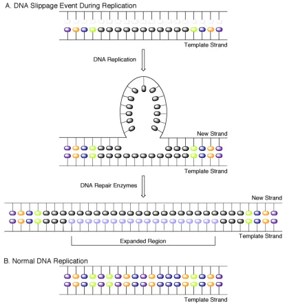
Figure 1. DNA slippage model for triplet repeat expansion disorders...2
Figure 2. RBDCL selected peptides ...5
Figure 3. Ligand L and Janus-wedge hydrogen bonding ...6
Figure 4. Phage display selection cycle...8
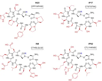
Figure 5. Cyclic peptides identified through phage display selection...12
Figure 6. (CUG)54:MBNL-N EMSAs...14
Figure 7. Competitive EMSAs for (CUG)54:MBNL-N with IN23...16
Figure 8. Representative kinamycins and lomaiviticins ...47
Figure 9. Feldman’s reductive activation mechanism ...50
Figure 10. Potential routes to DNA cleavage...52
Figure 11. DNA sequencing gel electrophoretic radiograms...54
Figure 12. DNA sequencing gel electrophoretic radiograms...56
Figure 13. Revised mechanistic rationale for DNA cleavage...58
Figure 14. Osteoclast maturation pathway...64
Figure 15. Common bisphosphonates and nitrogen-containing bisphosphonates ...66
Figure 16. Comparison of raloxifine, estradiol and estrogen receptor 1 ...67
Figure 17. First generation 2-aminoimidazole-triazole library for osteoclastogenesis Inhibition...68
Figure 18. Retrosynthetic analysis of triazole library...69
Figure 19. Complete second generation triazole library...71
Figure 20. Stages of osteoclast development for analysis of triazole library mediation of osteoclast formation ...72
Figure 21. C2 as a scaffold for third generation triazole libraries ...73
Figure 23. Comparison of cyclohexyl and cyclopentyl pendant groups on osteoclastogenesis
Inhibition...75
Figure 24. Third generation cyclohexylmethyl-triazole library...77
Figure 25. Comparison of various benzamide EWG substitution patterns on osteoclastogenesis inhibition ...78
Figure 26. Comparison of benzamide aliphatic substituents on osteoclastogenesis Inhibition...79
Figure 27. Most successful compounds from cyclohexylmethyl triazole library ...80
Figure 28. Stages of biofilm maturation ...172
Figure 29. Representative small molecules that inhibit biofilm formation ...173
Figure 30. Representative 2-aminoimidazole containing small molecules ...173
Figure 31. 1,4-Substitued triazole library for biofilm modulation screens...175
Figure 32. Dose response curve of B3 against RB50 ...177
LIST OF SCHEMES
Scheme 1. Synthesis of biotinylated (CUG)54 template for phage display...9
Scheme 2. Synthetic route to linear and cyclic peptides ...13
Scheme 3. Jebartanam’s oxidative activation hypothesis...48
Scheme 4. Dmietrienko’s supporting data for an electrophilic activation...49
Scheme 5. Feldman’s reductive activation supporting data...51
Scheme 6. Synthesis of azido precursors...70
Scheme 7. Synthesis of 1,4-substituted triazoles for second-generation library...70
Scheme 8. Synthesis of C-series third generation library...73
LIST OF ABBREVIATIONS AND TERMS
A alanine
Ac acetyl
2-AI 2-aminoimidazole AIBN azobisisobutyronitrile amp ampicillin
2-AIT 2-aminoimidazole-triazole AHL N-acylhomoserine lactone
aq aqueous
Ala alanine
Asn asparagine bd broad doublet
BP bisphosphonate
brine saturated aqueous sodium chloride bs broad singlet
C cysteine
CAT catalase
CTG cytosine-thymidine-guanosine CUG cytosine-uracil-guanosine CUG-BP CUG-binding protein cTnT cardiac troponin T Cys cysteine
d doublet
dt doublet of triplets dd doublet of doublets
DMAP 4-(dimethylamino)pyridine DM myotonic dystrophy DMF N,N-dimethylformamide
DMPK myotonic dystrophy protein kinase DMSO dimethyl sulfoxide
DNA deoxyribonucleic acid DTT dithiothreitol
EDTA ethylene diamine tetraacetic acid EMSA electrophoretic mobility shift assay EtBr ethidium bromide
EtOAc ethyl acetate EtOH ethanol
FLAG Fmoc-L-allylglycine
Fmoc Fluorenylmethyloxycarbonyl
Gln glutamine
GMPS guanosine monophosphorothioate GRL genome research laboratory GSH glutathione
GST glutathione S-transferase
h hour(s)
H histidine
His histidine
Hz hertz
IC50 inhibitory concentration: 50% IPTG isopropyl-β-thiogalactoside J coupling constant
K lysine
Kd dissociation constant LB Luria-Bertani media
Lys lysine
m multiplet
MBNL muscleblind protein
MBNL-FL full length muscleblind (MBNL1) MBNL-N N-terminal muscleblind
M-CSF macrophage colony stimulating factor
MeOH methanol
MOA mode-of-action MHz megahertz min minute(s)
NaAsc sodium ascorbate
NBP nitrogen-containing bisphosphonates NMR nuclear magnetic resonance spectrometry
Nu nucleophile
OD600 optical density at 600 nm OPG osteoprotegrin
P proline
PFU plaque forming unit
Q glutamine
q quartet
QS Quorum Sensing
RA rheumatoid arthritis
RANK receptor activator of nuclear factor κB RANKL receptor activator of nuclear factor κB ligand RBDCL resin bound dynamic combinatorial library RB50 Bordetella bronchiseptica wild type strain RCM ring closing metathesis
RT room temperature
RNA ribonucleic acid
s singlet
SDS-PAGE sodium dodecylsulfate-polyacrylamide gel electrophoresis SERM selective estrogen receptor modulator
SOD superoxide dismutase TBE Tris/boric acid/EDTA TIS triisopropylsilane TFA trifluoroacetic acid
TRAP tartrate resistant acid phosphatase Tris tris(hydroxymethyl)aminomethane UTR untranslated region
V volts
W watts
CHAPTER 1
A Unified Approach to Targeting Myotonic Dystrophy
1.1 Introduction
Myotonic dystrophy (DM) was first recognized as a multisystemic neuromuscular disorder in 1909.1, 2 DM is the most common form of adult onset muscular dystrophy with an incidence of 1:8000. Muscular dystrophy and myotonia are classic symptoms of DM; however, various superficially unrelated clinical features are also symptomatic of the disease as well. Some of the seemingly unusual phenotypes consistent with DM include cataracts, heart conduction defects, insulin resistance and specific endocrine system irregularities including premature frontal baldness and testicular atrophy.3-5 DM is inherited in an autosomal dominant fashion and as a progressively degenerative disease it exhibits anticipation through generations. Currently there are two well-established types of DM, myotonic dystrophy type 1 (DM1) and type 2 (DM2), each triggered by specific genetic modifications. The symptoms of DM2 are similar but typically mild in comparison to DM1, and there is no known congenital form of DM2. Additionally, the prevalence of DM1 far exceeds that of DM2.
Figure 1. DNA slippage model for triplet repeat expansion disorders.
1.1.1 DMPK Haploinsufficiency Hypothesis
been shown to be caused by a point mutation in the DMPK gene, suggesting that the multisystemic features of the disease are not caused by DMPK haploinsufficiency.13
1.1.2 RNA Gain of Function Hypothesis
Houseman and Singer later demonstrated that aberrant poly(CUG) transcripts of the mutant DMPK gene localized within nuclei, leading to general downregulation of DMPK. 14, 15 The nuclear accumulation of expanded CUG was proposed to account for the deleterious effects of the repeat expansion. This unprecedented RNA gain of function mechanism predicted that aberrant CUG transcripts exerted a trans-dominant effect capable of disrupting alternative splicing as well as other cellular events.16-20 This mechanism was further substantiated by the observation that an expanded repeat in the 3’-UTR of DMPK RNA inhibited myogenic differentiation of C2C12 myoblasts.21 In addition, transcription of an expanded CUG RNA was shown to generate a DM1 phenotype in transgenic mice when expressed in a gene entirely unrelated to DMPK.22
The toxic RNA gain of function hypothesis for DM1 pathology directed researchers to study the ability of proteins to bind expanded CUG transcripts. Initial explorations revealed that CUG-binding protein (CUG-BP) regulated the alternative splicing of cardiac troponin T (cTnT) by binding CUG. This splicing pattern was shown to be disrupted in DM1 cardiac and skeletal muscle tissues.17 However, CUG-BP activity was shown to increase in DM1, which did not support a trans-dominant effect of RNA-mediated inactivation of binding proteins. Furthermore, studies failed to exhibit co-localization of CUG-BP with aberrant CUG in nuclear foci.23 The inability of CUG to sequester CUG-BP suggested a secondary role for CUG-BP in the pathogenesis of DM1.
these three isoforms was recruited into mutant RNA nuclear foci in DM1 cells, which was presumed to disrupt their customary biological functions.26 Consequently, MBNL proteins became attractive targets for the toxic RNA gain of function exhibited after transcription of aberrant DMPK RNA. Further substantiation for this mechanism was achieved when MBNL1 knock-out mice developed myotonia, cataracts and splicing irregularities consistent with the DM1 phenotype.27 In addition, cell culture experiments involving siRNA depletion of MBNL1, MBNL2 and CUG-BP indicated that nuclear accumulation favored MBNL1.17, 20, 27 Complementary evidence revealed that upon MBNL1 co-localization with expanded CUG in nuclei, aberrant alternative splicing events were mediated by CUG-BP, supporting the earlier suggestion of a secondary role for CUG-BP in DM1 disease pathology.
1.2 Previous Approaches to Targeting DM1
The emergent data supporting the poly(CUG) gain of function mechanism via sequestration of MBNL promoted other researchers to investigate small molecule disruption of this interaction as well.
1.2.1 Dynamic Combinatorial Peptide Library Approach
(Quin/Pip)-(assuming commutative property transfer) and then identified 4 disulfide-crosslinked peptides capable of high affinity binding (Kd ~2 µM) to (CUG)109 via a filter binding assay (disulfides 3-3, 4-4, 2-4, 3-4). However, when the selected peptides were incubated with (CUG)109 in the presence of MBNL1, the maximum displacement measured through an enzyme complementation assay was only 50% even at concentrations of 100 µM. This result showed the first promise of small molecules and their ability to mediate the interaction between poly(CUG) and MBNL. However, the therapeutic use of these crosslinked peptides would be irrelevant due to the reduction of the disulfide linkage mediated by intracellular thiols, given that the individual peptides alone were unable to bind poly(CUG).
Figure 2. RBDCL selected peptides.
1.2.2 Triaminotriazine-DNA Intercalator Approach
a minor groove DNA intercalator, the joined molecule could target CUG or CTG through the major or minor groove due to a “stacking intercalator” effect. The designed ligand L was assayed for affinity and selectivity for T-T, C-C, G-G and A-A mismatches in [(CG)m(CXG)n(CG)m]2 hairpins and found to only bind to DNA with T-T mismatches. Furthermore, when at least two T-T mismatches were present, L bound DNA with high nanomolar affinities. This affinity was also translated to an RNA counterpart, where (CUG)4 was shown to bind L with a Kd of 0.43 µM using isothermal titration calorimetry. Furthermore, upon 1:1 binding of MBNL-N and (CUG)12, the addition of compound L disrupted the RNA-protein complex with IC50 values of 40 – 50 µM. Electromobility shift assays also indicated almost complete inhibition when treated with 250 µM compound L. This salient result renders compound L as the most promising lead for therapeutic intervention of DM1 known to date.
Figure 3. Ligand L and Janus-Wedge hydrogen bonding.
1.3 Our Approach at Targeting DM1 with Phage Display Libraries
intended alternative splicing events, which are aberrantly regulated in DM1. Given that peptides and proteins are endogenous RNA binding molecules, we chose to identify peptides capable of binding poly(CUG) selectively. Additionally, given that cyclic peptides have enhanced bioavailability, do not suffer entropic losses on binding and are less susceptible to protease degradation than their linear counterparts,31 we chose to investigate a library of cyclic peptides for their ability to bind poly(CUG).
Figure 4. Phage display selection cycle.
1.3.1 Target Immobilization
Preceding the selection, we first prepared a target RNA capable of immobilization. Professor Swanson (UFL) kindly provided an ampicillin resistant plasmid encoding (CTG)54 engineered into a DH5α E. coli cell line. The p(CTG)54 encoded cell line was amplified in
coupled with biotin-PEG2-maleimide to provide our immobilizable target for phage display (Scheme 1). The RNA target was then immobilized by exploiting the extremely high affinity biotin-avidin interaction using magnetic streptavidin beads.
Scheme 1. Synthesis of biotinylated (CUG)54 template for phage display selection.
1.3.2 Phage Display Selection
After target immobilization, we began performing selections using a commercially available cysteine constrained heptapeptide library with a complexity of 1.28 billion unique peptides (according to the literature provided with the library). During preliminary selection experiments, the library of phage was found to evolve the known HPQ (His-Pro-Gln) consensus motif specific for streptavidin within three rounds of positive selection. In order to prevent the selection and amplification of phage specific for binding streptavidin, we chose to perform alternating rounds of positive and negative selection. The addition of a negative selection round in each cycle confers a higher degree of stringency by removing any peptides or phage, which could bind the streptavidin beads and provide false positive results.
immobilized as well by association (Figure 4, Step 2). All nonbinding phage were then eliminated through numerous washing steps (Figure 4, Step 3). The RNA-bound phage were then eluted with an acidic solution of glycine (a general phage eluent, Figure 4, Step 4). The desired RNA-binding phage were then infected into E. coli and amplified (Figure 4, Step 5). After phage amplification, a negative selection round was employed to remove phage capable of binding streptavidin. During a negative round of selection the amplified phage library was incubated only with streptavidin beads (Figure 4, Step 6). Any phage that did not bind the streptavidin immobilization support were then removed and saved (Figure 4, Step 7), while the streptavidin beads and bead-binding phage were discarded. This was the final step for the selection cycle; at this point, these phage were then placed onto a subsequent selection cycle and this protocol was repeated until sequence homology was achieved.
The selection cycle we employed as described above begins with a round of positive selection. However, given our previous results of identifying streptavidin-binding phage in only three selection cycles we chose to also employ a parallel selection which began with a round of negative selection. The initial negative selection cycle was posited to immediately remove any streptavidin binding phage, which could complicate our selection results. This parallel selection was performed exactly as described above and in Figure 4, excepting only that this selection began with Step 6 (as illustrated) and then continued through the subsequent steps as delineated above.
library. During the early selection cycles, the total number of phage within the remaining pool decreased via the removal of non-CUG-binding phage; therefore, the number of PFUs decreased as expected. However, after the majority of nonbinding phage were removed, only CUG-binding phage remained in the pool and continued to be amplified through multiple cycles of selection. This result was indicated by a sharp increase the number of PFUs during later selection cycles.
1.3.3 Synthesis of Cellularly Irreducible Analogs
After the identification of poly(CUG)-binding cyclic peptides, we sought to investigate their ability to disrupt the poly(CUG)-MBNL interaction characteristic of DM1. However, cysteine constrained cyclic peptides undergo reduction to their linear forms mediated by cellular thiols. Although linear peptides are capable of binding RNAs, most literature suggests enhanced binding favorability for their cyclic counterparts.31 In addition, linear peptides are more susceptible to degradation by intracellular proteases. Therefore, we aimed to synthesize cellularly irreducible analogs of these cyclic peptides by employing ring-closing metathesis (RCM). This was achieved by replacing the first and last cysteine amino acids with an allyl glycine. Peptides were synthesized using the standard solid phase Fmoc protocol on a Rink amide MBHA resin (Scheme 2). Once the linear peptide chain had been synthesized, attempts were made to cyclize the peptide using RCM.
A former group member, Anne Basso was able to cyclize peptides of this length using Hoyveda-Grubbs 2nd Generation Catalyst; therefore, we chose to employ her optimized procedure for RCM. The long refluxing times and amount of catalyst necessary; however inhibited the amount of product isolated due to degradation and purification obstructions respectively. A typical loading of 500 mg resin generally yielded less than 5 mg crude solid after cleavage from the resin and subsequent precipitation. Furthermore, the only successful metathesis reaction was performed on IN23; which contains 3 proline residues. Given that purification of peptides after subjection to RCM procedures proved extremely difficult, we chose to test our methodology using linear analogs of the peptides identified during the selection. Although they would suffer an entropic loss upon folding into the active conformation for binding, we reasoned they should still be capable of binding the target RNA.
1.3.4 Expression of MBNL Proteins
Before we could assess the ability of these peptides to displace the poly(CUG):MBNL interaction we first needed to express the muscleblind protein and determine its binding capabilities toward (CUG)54. Professor Swanson also provided us with fusion constructs of GST::MBNL1::His and GST::MBNL-N::His (N-terminus AAs: 1 – 253) as plasmids engineered into a BL21 strain of E. coli. Expression of full-length MBNL1 proved extremely difficult due to the inherent instability of muscleblind. SDS-PAGE analysis indicated when
the protein was successfully expressed it would frequently degrade during affinity column purification or during storage even in the presence of protease inhibitors.
The N-terminal domain of MBNL1 (MBNL-N) has been identified as the region that poly(CUG) binds in vitro.32, 33 MBNL-N, in addition to full length MBNL1 (MBNL-FL), has been shown to cross-link with (CUG)54,19 form RNA-protein complexes with cTnT RNAs (Tnnt3/T5.1 and T5.45) and (CUG)54;32 however, the truncated protein was more likely than MBNL-FL to form unresolved complexes which remained in the wells of polyacrylamide gels during elecrophoresis.32 Additionally, MBNL-N has been shown to possess higher binding affinities than MBNL-FL and is more stable than the full-length protein. Therefore we chose to employ MBNL-N for use in binding reactions and electrophoretic mobility shift assays (EMSA) with (CUG)54.
The GST::MBNL-N::His encoded cell line was amplified in 2XYT supplemented with 500 µg/mL carbenicillin and protein expression was induced under IPTG control. After the cells were harvested and lysed, the N-terminal fusion construct was purified through GST-affinity chromatography and dialyzed into storage buffer supplemented with protease inhibitors. MBNL-N was then utilized in electromobility shift assays to determine the ability of our selected peptides to disrupt the poly(CUG):MBNL interaction.
1.3.5 Electromobility Shift Assays for (CUG)54:MBNL-N Interactions
in. Initial explorations of MBNL-N interactions with (CUG)54 indicated some binding in Buffer C (selection buffer); however, the alternative buffers were explored in an effort to enhance the binding constant, and consequently lower the necessary concentrations of MBNL-N needed to effect a binding event. These buffers showed little deviation in their ability to promote binding, thus Buffer C was used for the subsequent EMSAs for consistency given that the selection was performed in this buffer.
Table 1. Buffers assayed for EMSAs.
Buffer Components
A 175 mM NaCl, 20 mM Tris-HCl, 5 mM MgCl2, 1.25 mM BME, 12.5% glycerol, 2 mg/ml BSA, 0.1 mg/ml heparin
B 100 mM NaCl, 20 mM Tris-HCl, 5 mM MgCl2, 1 mg/ml BSA, 0.1 mg/ml heparin C 20 mM Tris-HCl, 100 mM KCl, 5 mM MgCl2, 0.1% Triton-X
After some optimization we were able to observe 100% complex formation between MBNL-N and (CUG)54, this is depicted in Figure 6 (Gel A) where as the concentration of MBMBNL-NL-MBNL-N is increased, the free RNA (lower band) starts to disappear; while a higher molecular weight band is formed indicating a binding event between the RNA and protein. The EMSA indicated that with our conditions 100% of (CUG)54 was bound with 4 µM MBNL-N, which was in agreement with literature results.
Figure 6. (CUG)54:MBNL-N EMSAs A. (CUG)54 + [MBNL-N]. B. (CUG)54 + [IN23L]: 30 min, + 234 nM
The next step in our investigation of the MBNL-poly(CUG) interaction was to assess the ability of our selected peptides to competitively disrupt the MBNL-N:(CUG)54 interaction. The first preliminary competitive gel shift experiment was performed with linear IN23 (IN23L). When increasing concentrations of IN23L were pre-incubated with (CUG)54 for 30 min and were then treated with 234 nM MBNL-N, we discovered as little as 286 nM IN23L was able to completely disrupt this interaction and release free RNA (Figure 6, Gel B). Unfortunately, this result could never be duplicated even after preparing every reaction component (MBNL-N, (CUG)54, buffer systems, etc.) again under strict sterile conditions.
While trying to repeat the result of this experiment we observed that upon increasing the peptide concentrations dramatically, IN23L inhibited the formation of the (CUG)54:MBNL-N complex to a small extent at 723 µM. To measure the observed effect, we counted the pixels of free RNA and the RNA-protein complexes in the gel. The percent of the lower band (e.g. free RNA) was as follows: (CUG)54 control = 92%, (CUG)54:MBNL-N control = 68%, 723 µM IN23L + (CUG)54:MBNL-N = 80%. Thus IN23L mediated a 12 percentage point increase in free RNA when compared to the RNA:protein control. Although this effect is not as dramatic as our initial competitive EMSA, the results of this assay nonetheless indicate that IN23L is capable of mildly disrupting this RNA-protein interaction (Figure 7, Gel A).
Although cyclic IN23 (IN23C) posed purification difficulties, we chose to test the ability of crude IN23C to disrupt the (CUG)54:MBNL-N interaction as well. Crude IN23C, however did not display any extent of (CUG)54:MBNL-N inhibition, even at extremely high peptide concentrations (2.2 mM). The results of this assay are shown in Figure 7, Gel B. Pixel counts were also measured for the competitive IN23C EMSA: the percent of the lower band (free RNA) was as follows: (CUG)54 control = 94%, (CUG)54:MBNL-N control = 73%, 2.2 mM IN23C + (CUG)54:MBNL-N = 63%. In fact, the competitive EMSA using higher concentrations of crude IN23C appeared to have a noticeable effect of decreasing free RNA. This effect is most likely due to RNA degradation from the remaining RCM impurities.
Although IN23L was demonstrated to be capable of disrupting the CUG54:MBNL-N interaction to some extent, unfortunately the affinity of IN23L for (CUG)54 was not strong enough to disrupt the RNA-protein interaction at therapeutically relevant concentrations. Perhaps the inclusion of MBNL-N during the selection could have increased the stringency enough to prompt the evolution of peptides capable of binding (CUG)54 with enough affinity to competitively displace muscleblind at lower concentrations. The selected peptides currently represent leads for future analog development.
1.4 Conclusion
peptide with a round initial conformation. We then assayed the peptides for their ability to disrupt the (CUG)54:MBNL1 interaction characteristic of DM1. One selected peptide was capable of binding (CUG)54 and was able to disrupt the (CUG)54:MBNL1 complex to a small extent. These cyclic peptides currently represent leads for further analog development.
1.5 Experimental
All chemicals were used without further purification. Transformed cell lines were obtained from the Maurice Swanson laboratory at UFL. All Fmoc protected peptides and resins were obtained from Peptides International. All other chemicals were supplied by Aldrich, unless otherwise noted. All buffer and media recipes are included in the Appendix.
Isolation of (CTG)54 plasmid from E. coli
The ampicillin resistant (ampr) p(CTG)54 was transformed into a DH5α strain of E. coli and
kindly provided by the Maurice Swanson Laboratory at the University of Florida at Gainesville. 50 mL modified LB media (10 g tryptone, 5 g yeast, 10 g NaCl) was dispensed into a sterile 250 mL Erlenmeyer flask doped with 10 mg/ml carbenicillin. A single colony was selected and inoculated for 16 – 20 hours at 37 ºC and 220 rpm in a shaking incubator. After the cellular density was large enough, the cells were centrifuged for 10 min at 10,000 x g; the supernatant was discarded and the plasmid was extracted using a Promega Pure Yield™ Plasmid Midi Prep DNA Purification System. The isolated DNA was resuspended in 600 µL nuclease free water and quantified by ultraviolet absorption at 260 nm where [DNA] µg/ml = A260 * 50 µg/mL * dilution factor. Plasmid DNA was stored in aliquots at -20 ºC.
BamHI Restriction Enzyme Digest of p(CTG)54
DNA was separated from the restriction enzyme by phenol/chloroform extraction. An equal volume of well-suspended phenol/chloroform/isoamyl alcohol (25/24/1) was added to the sample of DNA and vortexed at a low setting for five min. The mixture was then centrifuged for 5 min at 13.2 rcf and most of the top aqueous layer was carefully removed with a micropipette. The aqueous layer containing the DNA was then precipitated with 2.5 volumes of ice-cold ethanol and 1/3 volume 5 M NH4OAc for at least 30 min at -20 ºC. The samples were then centrifuged for 30 min at 4 ºC, 13.2 rcf, the supernatant was removed and the pellet was washed with 200 µL ice cold 70% ethanol. The sample was centrifuged briefly, the supernatant was removed and the pellet was allowed to air dry for at least 30 min at RT. The DNA was then resuspended in 50 µL nuclease free water.
Transcription of (CUG)54
EtBr intercalation was excised with a nuclease free scalpel and placed into a microfuge tube. Hot RNA was visualized on X-Ray film (Fuji Film); the gel (wrapped in cellophane) was exposed to the film for approximately 20 min before developing. After the film was developed it was aligned with the gel and the desired band of RNA was excised using a nuclease free scalpel and placed into a microfuge tube. The gel slices were gently crushed with a pipet tip and the transcripts were eluted overnight in 400 µL Gel Elution Buffer (500 mM NH4OAc, 1 mM EDTA pH 8.0, 0.1 % SDS). The following morning the samples were centrifuged for 10 min at 13.2 rcf and the liquid was carefully separated from the gel fragments. The transcripts were precipitated with 3 volumes of ice-cold isopropanol, placed at -20 ºC for at least 30 min and then centrifuged at 4 ºC, 13.2 rcf for 30 min. The supernatant was carefully removed and the pellet was washed with 200 µL of ice-cold 70% isopropanol and centrifuged briefly. The supernatant was removed and the pellet was allowed to air dry for at least 30 min at RT.
Cold RNA
Cold RNA was transcribed by combining 4 µL 5X transcription buffer (Promega), 2 µL 5 mM ATP, 2 µL 5 mM UTP, 2 µL 5 mM CTP, 2 µL 5 mM GTP, 0.5 µL RNAsin (40 U/µL), 3 µL nuclease free water, 1.5 µL T7 RNA polymerase (RNAP, 20 U/µL ) and 3 µL DNA template.
Hot RNA
Hot RNA was uniformly body labeled with 50 µCi α-32-P-UTP (MPBiomedicals) by
combining 4 µL 5X transcription buffer, 2 µL 5 mM ATP, 2 µL 5 mM CTP, 2 µL 5 mM GTP, 0.5 µL UTP, 5 µL α-32-P-ATP (3000 Ci/mmol) 0.5 µL RNAsin (40 U/µL), 3 µL DNA
Biotinylated RNA
Biotinylated RNA was transcribed by including an excess of 5’-guanosinemonophosporothioate (GMPS, Axxora LLC, G-018-10) during standard “cold” transcription. GMPS-(CUG)54 was transcribed with 17 µL nuclease free water, 20 µL 5X transcription buffer, 0.5 µL RNAsin, 8 µL 5 mM ATP, 8 µL 5 mM CTP, 8µL 5 mM UTP, 8 µL 5 mM GTP, 20 µL GMPS (2 mM final), 4.5 µL DNA (1 µg/mL), 2 µL T7 RNAP. 5’-GMPS(CUG)54 was then reacted with 25 mM Maleimide-PEG2-biotin (Pierce) in DMF for 2 hours and purified on a MWCO-10 microcon. Percent biotinylation was quantified by comparison of free RNA after a streptavidin-binding assay, where free (non-biotinylated) RNA was quantified by EtBr staining on an agarose gel and the concentration of biotinylated RNA determined by difference.
Expression and Isolation of GST::MBNL1::His
loading dye at 95 ºC for 5 min and run on 8% SDS-PAGE in 1X Tris-Glycine-SDS for 30 min at 200 V. The gel was then stained with Coomassie Brilliant Blue for 30 min and then destained by washing 4-5 times with Destain Buffer over the course of several hours. The visibility of a blue band at approximately 67 – 70 kDa was indicative of the full-length construct (~67 – 68 kDa). Western blotting analysis was also performed to ensure the fusion construct was isolated and intact. A nitrocellulose membrane was presoaked in Transfer Buffer, then a clean gel was placed on the surface of the nitrocellulose and transferred to the membrane at 100 V for 1 h. The gel was then removed, and the membrane was washed 3 times with TBST for 10 min. The membrane was then blocked with 5 mL of TBS + Casein for at least 1 h at 4 ºC. The blocking solution was then decanted and the membrane was washed 4 times with TBST for 15 min. A GST-HRP Conjugate Antibody was diluted 1:5000 in 10 mL of TBST and exposed to the membrane with gentle shaking for 1 h. The membrane was then washed 2 times for 2 min, 1 time for 15 min and 3 times for 5 min to removed any residual antibody. The membrane was then dried and developed using a 1:1 solution of luminol and peroxide developer (BioRad, Immunostar HRP) for 5 min. The membrane was wrapped in cellophane and imaged on a GE Storm™ 840 Chemiluminescent Detector. Pure protein samples were then dialyzed overnight at 4 ºC into Protein Storage Buffer using a 10,000 MWCO Dialysis Cassette (3 buffer changes of 1 L). Protein concentration was measured with the DC Protein Assay® (BioRad) using BSA as the standardization control.
Expression and Isolation of GST::MBNL-N::His
addition of IPTG (1 mM final) for 3 h. The cells were then centrifuged at 10,000 rpm, 4 ºC for 15 min. The supernatant was removed and the cells were placed at -20 ºC for at least 2 h. The cells were then thawed on ice and purified using the PIERCE B-PER® GST Spin Purification Kit. The GST::MBNL1::His fusion construct was bound to immobilized glutathione-agarose beads, washed 4 times with Tris-Glycine Buffer and eluted with free GSH. A small amount of eluate was denatured in loading dye at 95 ºC for 5 min and run on 8 % SDS-PAGE in 1X Tris-Glycine-SDS for 30 min at 200 V. The gel was then stained with Coomassie Brilliant Blue for 30 min and then destained by washing 4-5 times with Destain Buffer over the course of several hours. The visibility of a blue band at approximately 50 – 55 kDa was indicative of the N-terminal construct (~53 – 54 kDa). The protein was dialyzed overnight at 4 ºC into Protein Storage Buffer using a 3,000 MWCO Dialysis Cassette (4 buffer changes of 1 L). Protein concentration was measured using the DC Protein Assay® (BioRad) with BSA as the standardization control.
Negative Selection Protocol
sample was then centrifuged for 1 min, the supernatant was removed and immediately placed onto a positive selection or stored at 4 ºC overnight until a positive selection could be performed.
Positive Selection Protocol
Phage Amplification and Isolation
A tetr E. coli (ER2738) cell line was inoculated in 20 mL LB and incubated at 37 ºC, 200 rpm. While ER2738 was growing the selection procedure was performed. After approximately 4 h the neutralized phage solution from the positive selection (less 10 µL for titer) was infected into the ER2738 cell line. The cells were incubated for another 4 h at 37 ºC, 200 rpm. After amplification the cells were centrifuged for 10 min at 4 ºC, 10,000 rpm. The supernatant was then transferred to a new vessel and centrifuged again. The upper 80 % was transferred to a new vessel and 1/6 volume of PEG-NaCl Buffer was added. The phage were allowed to precipitate overnight at 4 ºC. The following morning the samples were centrifuged for 15 min at 4 ºC and 10,000 rpm. The supernatant was decanted and all residual liquid was removed with a micropipette. The pellet was suspended in 1 mL TBS and centrifuged at 10,000 rpm for 5 min at 4 ºC to remove any remaining debris. The supernatant was removed, transferred to a new tube and re-precipitated with 1/6 volume PEG-NaCl Buffer while at -20 ºC for 40 min. The sample was then centrifuged at 13.2 rcf for 10 min at 4 ºC, the supernatant was removed and the pellet suspended in TBSA. This solution comprised the amplified pool of phage, 10 µL of which were used for the amplified phage titer (to ensure amplification is successful) and the remaining pool was placed on the next round of negative selection.
Phage Titering
dilutions were plated on one plate, as the dilution increased another plate was added for easier plaque counting) were pre-warmed to 37 ºC. While the cells were growing, a 50 mL stock of Agarose Top was melted in the microwave and 3 mL aliquots were dispensed into sterile 10 mL culture tubes (one for each plate), the tubes were then placed in a 45 ºC water bath until needed. When the OD600 finally reached mid log phase, 200 µL of the culture was added to empty eppendorf tubes (one for each dilution). Then 10 µL of each serially diluted solution of phage in LB was added to the ER2738, quickly vortexed and incubated at RT for 5 min. The infected cells were then transferred to a 3 mL aliquot of Agarose Top, vortexed and immediately poured onto a pre-warmed LB Agar IPTG/XGal plate. The plate was tilted so the entire surface area was covered with Agarose Top and it was allowed to solidify over the course of 5 min at RT. After each dilution was plated, the plates were inverted and incubated overnight at 37 ºC. The following morning the numbers of blue plaques were counted and the phage titer was reported as plaque forming units (PFU) per 10 µL.
Isolation and Characterization of Binding Clones
General Procedure for Linear Peptide Synthesis
the ether was allowed to evaporate, the peptide was then suspended in 1 mL H2O, aliquoted into 1.5 mL microfuge tubes and frozen prior to lyophilization on a vacuum centrifuge.
General Procedure of RCM on Olefin-Terminated Peptides
Dichloroethane (DCE) was degassed with argon for 5 – 10 min prior to the addition of resin-bound peptide and then brought to reflux at 84 ºC. Hoveyda Grubbs 2nd Generation Catalyst was then added in 10 mol% portions over the course of 24 hours. Approximately 100 equivalents of DMSO was added to the mixture and allowed to reflux for 12 more hours. After the mixture was cooled to RT, the resin was filtered over a frit and washed with 5 mL DMSO, DMF, DCM and MeOH three times each. The peptide was then globally deprotected and cleaved from the resin simultaneously with the addition of a fresh mixture of TFA, TIS and H2O (98:1:1). The mixture was allowed to rock at RT for 2 hours, after which the beads were filtered off and the filtrate was reduced in vacuo to a volume of approximately 1 mL. Peptides were then precipitated out with the addition of cold ether (~1-2 mL) while sonicating. The suspension was then transferred to a 15 mL conical and centrifuged at 4,000 rpm for 15 min. The supernatant was decanted and the ether was allowed to evaporate. Three to four mL of H2O was added to the solid and vortexed to resuspend the peptide, the mixture, which was never fully soluble in water was then aliquotted into 1.5 mL microfuge tubes, frozen and lyophilized on a vacuum centrifuge.
General Procedure of HPLC Purification of RCM Treated Peptides
Synthesis of Rink-FLAG
Fmoc-L-allylglycine (FLAG) (300 mg, 0.890 mmol) was preactivated with HCTU (375 mg, 906 mmol) and DMAP (cat.) in 4 mL DMF and 1 mL DIEA for 5 min. It was then added to 500 mg Rink-Amide-MBHA Resin (0.44 meq/g) that had been washed with DCM (3 X 5 mL) and DMF (3 X 7 mL). The coupling reaction was rocked in a Solid Phase Peptide Synthesis reaction vessel for 5 hours. The liquid was then evacuated from the vessel and the resin was washed 4 times with 5 mL DMF. All free amines were then capped with the addition of acetic anhydride (1 mL, 10.6 mmol) in DMF (5 mL) and DIEA (1 mL) for 30 min. The liquid was evacuated from the vessel and the resin was washed with DMF (3 X 5 mL) and DCM (3 X 5 mL) and flushed with nitrogen until dry. Rink-FLAG was stored at 4 ºC until needed for the synthesis of all linear peptides.
Synthesis of Linear IN23 (allyl-Gly•Pro-Pro-Tyr-Ala-Pro-His-Ser•allyl-Gly)
period of 20 min. The liquid was then evacuated and the resin was washed with DMF (4 X 6 mL). Fmoc-Ser(tBu)OH (337 mg, 0.880 mmol) was preactivated with HCTU (331 mg, 0.800 mmol) in DMF (4 mL) and DIEA (1 mL) and allowed to couple for 1.5 hours. As outlined in the general peptide synthesis Fmoc deprotection and coupling were alternated until the desired peptide had been synthesized. Couple 2: Fmoc-His(Trt)OH (545 mg, 0.880 mmol) + HCTU (331 mg, 0.800 mmol). Couple 3: Fmoc-Pro-OH (297 mg, 0.880 mmol) + HCTU (331 mg, 0.800 mmol). Couple 4: Fmoc-Ala-OH•H2O (290 mg, 0.880 mmol) + HCTU (331
mg, 0.800 mmol). Couple 5: Fmoc-Tyr(tBu)OH (404 mg, 0.880 mmol) + HCTU (331 mg, 0.800 mmol). Couple 6: Fmoc-Pro-OH (297 mg, 0.880 mmol) + HCTU (331 mg, 0.800 mmol). Couple 7: Fmoc-Pro-OH (297 mg, 0.880 mmol) + HCTU (331 mg, 0.800 mmol). Couple 8: FLAG (297 mg, 0.880 mmol) + HCTU (331 mg, 0.800 mmol). After final Fmoc deprotection, about 100 mg resin was mixed with a freshly combined solution of TFA (3.92 mL), TIS (40 µL) and H2O (40 µL) for 2 hours. A white solid was isolated as described in the general method section. H1 NMR (300 MHz, DMSO) δ 8.96 (s, 1H), δ 8.21 – 7.70 (bm,
5H), δ 7.38 (s, 2H), 7.16 (s, 1H), 6.98 (d, 2H, J = 8.1 Hz) 6.61 (d, 2H, J = 7.8 Hz) 5.83 – 5.62
(bm, 2H), 5.21 – 4.99 (bm, 4H), 4.59 (bs, 4H), 5.59 – 4.35 (bm, 10 H), 4.28 – 4.05 (bs, 10 H), 3.71 – 3.36 (bm, 13H), 3.11 (m, 2H), 2.98 – 2.89 (bm, 3H), 2.72 (m, 2H), 2.46 – 2.32 (bm, 2H), 1.98 – 1.74 (bm, 16H), 1.29-1.14 (bm, 6H).
Synthesis of Linear IN8 (allyl-Gly•Thr-His-Ser-Leu-Gln-Leu-Gln•allyl-Gly)
mL). Fmoc-Gln(Trt)OH (537 mg, 0.880 mmol) was preactivated with HCTU (331 mg, 0.800 mmol) in DMF (4 mL) and DIEA (1 mL) and allowed to couple for 1.5 hours. As outlined in the general peptide synthesis Fmoc deprotection and coupling were alternated until the desired peptide had been synthesized. Couple 2: Fmoc-Leu-OH (311 mg, 0.880 mmol) + HCTU (331 mg, 0.800 mmol). Couple 3: Fmoc-Gln(Trt)OH (537 mg, 0.880 mmol) + HCTU (331 mg, 0.800 mmol). Couple 4: Fmoc-Leu-OH (311 mg, 0.880 mmol) + HCTU (331 mg, 0.800 mmol). Couple 5: Fmoc-Ser(tBu)OH (337 mg, 0.880 mmol) + HCTU (331 mg, 0.800 mmol). Couple 6: Fmoc-His(Trt)OH (545 mg, 0.880 mmol) + HCTU (331 mg, 0.800 mmol). Couple 7: Fmoc-Thr(tBu)OH (350 mg, 0.880 mmol) + HCTU (331 mg, 0.800 mmol). Couple 8: FLAG (297 mg, 0.880 mmol) + HCTU (331 mg, 0.800 mmol). After final Fmoc deprotection, about 100 mg resin was mixed with a freshly combined solution of TFA (3.92 mL), TIS (40 µL) and H2O (40 µL) for 2 hours. A white solid was isolated as described in the general method section. H1 NMR (300 MHz, DMSO) δ 8.96 (s, 1H), δ 8.43 (bs, 1H), δ
8.23 – 8.06 (bm, 6H), 7.94 (s, 2H), 7.80 – 7.60 (bm, 3H), 7.35 – 7.27 (bm, 4H), 7.12 – 7.09 (bm, 2H), 6.81 – 6.78 (bm, 2H), 5.76 – 5.58 (bm, 2H), 5.07 – 4.97 (bm, 4 H), 4.64 (bm, 1 H), 4.31 – 4.19 (bm, 6H), 4.00 – 3.98 (bm, 2H), 3.15 – 3.09 (bm, 2H), 2.88 (s, 2H), 2.72 (s, 2H), 2.45 – 2.27 (bm, 4H), 2.09 – 2.06 (bm, 4H), 1.87 – 1.55 (bm, 9H), 1.43 (s, 5H), 1.25 – 1.10 (bm, 4H), 1.08 (m, 2H), 0.96 (m, 4H), 0.93 – 0.82 (bm, 18H).
Synthesis of Linear IPN2 (allyl-Gly•Thr-Leu-Tyr-His-Lys-Asn-Ser•allyl-Gly)
period of 20 min. The liquid was then evacuated and the resin was washed with DMF (4 X 6 mL). Fmoc-Ser(tBu)OH (337 mg, 0.880 mmol) was preactivated with HCTU (331 mg, 0.800 mmol) in DMF (4 mL) and DIEA (1 mL) and allowed to couple for 1.5 hours. As outlined in the general peptide synthesis Fmoc deprotection and coupling were alternated until the desired peptide had been synthesized. Couple 2: Fmoc-Asn(Trt)OH (525 mg, 0.880 mmol) + HCTU (331 mg, 0.800 mmol). Couple 3: Fmoc-Lys(Boc)OH (537 mg, 0.880 mmol) + HCTU (331 mg, 0.800 mmol). Couple 4: Fmoc-His(Trt)OH (545 mg, 0.880 mmol) + HCTU (331 mg, 0.800 mmol). Couple 5: Fmoc-Tyr(tBu)OH (404 mg, 0.880 mmol) + HCTU (331 mg, 0.800 mmol). Couple 6: Fmoc-Leu-OH (311 mg, 0.880 mmol) + HCTU (331 mg, 0.800 mmol). Couple 7: Fmoc-Thr(tBu)OH (350 mg, 0.880 mmol) + HCTU (331 mg, 0.800 mmol). Couple 8: FLAG (297 mg, 0.880 mmol) + HCTU (331 mg, 0.800 mmol). After final Fmoc deprotection, about 100 mg resin was mixed with a freshly combined solution of TFA (3.92 mL), TIS (40 µL) and H2O (40 µL) for 2 hours. An off white solid was isolated as described in the general method section. H1 NMR (300 MHz, DMSO) δ 8.96 (s, 1H), δ 8.41
(m, 2H), 8.24 (bm, 4H), 8.00 – 7.94 (m, 3H), 7.80 (bs, 2H), 7.35 (s, 4H), 7.18 – 7.12 (bm, 4H), 7.01 (d, 2H, J = 6.4 Hz), 6.62 (d, 2H, J = 7.8 Hz), 5.69 (m, 2 H), 5.08 – 4.98 (bm, 4 H), 4.56 (m, 2H), 4.28 – 3.92 (bm, 14H), 3.15 – 3.09 (bm, 2H), 2.88 (s, 2H), 2.72 (s, 2H), 2.45 – 2.27 (bm, 4H), 2.09 – 2.06 (bm, 4H), 1.87 – 1.55 (bm, 9H), 1.43 (s, 5H), 1.25 – 1.10 (bm, 4H), 1.08 (m, 2H), 0.96 (m, 4H) 0.93 – 0.82 (bm, 18H).
Synthesis of Cyclic IN23
of 85 ºC. Hoveyda Grubbs 2nd Generation Catalyst ((1,3-Bis-(2,4,6-trimethylphenyl)-2-imidazolidinylidene)dichloro(o-isopropoxyphenylmethylene)ruthen-ium) was added in portions over the course of 24 hrs. Time = 0h, 4 mg, 6.4 µmol; 13h, 3 mg, 4.8 µmol; 15h, 5 mg, 8.0 µmol; 17h, 4 mg, 6.4 µmol; 19h, 2 mg, 3.2 µmol. After 24 hours, DMSO (500 µL, 7.04 mmol) was added and the system maintained reflux for a period of 12 hours. The reaction mixture was cooled to RT and the resin bound peptide was washed over a fritted filter (3 X 15 mL DMF, 3 X 15 mL DCM). During washing, the beads lost most of the original orange color and resembled a very flat brown color when dried under aspiration. The reaction product was then globally deprotected and cleaved from the resin with a fresh cocktail of TFA (3.92 mL, 52.9 mmol), TIS (40 µL, 195 µmol) and H2O (40 µL, 2.22 mmol) mixed for 2 h. The suspension was filtered over a fritted funnel and rinsed twice with TFA (2 x 1 mL) and the filtrate was concentrated in vacuo until approximately 1 mL of liquid remained. Approximately 3 mL of cold ether was added to precipitate out the peptide. The suspension was transferred to a 15 mL conical and centrifuged at 4,000 rpm for 15 min. The supernatant was then decanted and remaining ether was removed by evaporation.
EMSA of (CUG)54 and MBNL-N
Competitive EMSA of (CUG)54 and MBNL-N with Synthetic Peptides
REFERENCES
1. H. Steinert, Dtsch Z Nervenheilkd, 1909, 37, 58-104.
2. F. E. Batten and H. P. Gibb, Brain, 1909, 32, 187-205.
3. P. S. Harper, Myotonic Dystrophy, W. B. Saunders, London, 2001.
4. E. Gharehbaghi-Schnell, J. Finsterer, I. Korschineck, B. Mamoli and B. R. Binder, Wien Klin Wochenschr, 1998, 110, 7-14.
5. C. Thornton, Semin Neurol, 1999, 19, 25-33.
6. J. D. Brook, M. E. Mccurrach, H. G. Harley, A. J. Buckler, D. Church, H. Aburatani, K. Hunter, V. P. Stanton, J. P. Thirion, T. Hudson, R. Sohn, B. Zemelman, R. G. Snell, S. A. Rundle, S. Crow, J. Davies, P. Shelbourne, J. Buxton, C. Jones, V. Juvonen, K. Johnson, P. S. Harper, D. J. Shaw and D. E. Housman, Cell, 1992, 68, 799-808.
7. Y. H. Fu, A. Pizzuti, R. G. Fenwick, J. King, S. Rajnarayan, P. W. Dunne, J. Dubel, G. A. Nasser, T. Ashizawa, P. Dejong, B. Wieringa, R. Korneluk, M. B. Perryman, H. F. Epstein and C. T. Caskey, Science, 1992, 255, 1256-1258.
8. J. Buxton, P. Shelbourne, J. Davies, C. Jones, T. Vantongeren, C. Aslanidis, P. Dejong, G. Jansen, M. Anvret, B. Riley, R. Williamson and K. Johnson, Nature, 1992, 355, 547-548.
9. M. Mahadevan, C. Tsilfidis, L. Sabourin, G. Shutler, C. Amemiya, G. Jansen, C. Neville, M. Narang, J. Barcelo, K. Ohoy, S. Leblond, J. Earlemacdonald, P. J. Dejong, B. Wieringa and R. G. Korneluk, Science, 1992, 255, 1253-1255.
10. H. G. Harley, J. D. Brook, S. A. Rundle, S. Crow, W. Reardon, A. J. Buckler, P. S. Harper, D. E. Housman and D. J. Shaw, Nature, 1992, 355, 545-546.
11. S. J. Tapscott, Science, 2000, 289, 1701-1702.
13. B. Tian, R. J. White, T. B. Xia, S. Welle, D. H. Turner, M. B. Mathews and C. A. Thornton, Rna, 2000, 6, 79-87.
14. K. L. Taneja, M. Mccurrach, M. Schalling, D. Housman and R. H. Singer, J Cell Biol, 1995, 128, 995-1002.
15. B. M. Davis, M. E. McCurrach, K. L. Taneja, R. H. Singer and D. E. Housman, P Natl Acad Sci USA, 1997, 94, 7388-7393.
16. L. T. Timchenko, J. W. Miller, N. A. Timchenko, D. R. DeVore, K. V. Datar, L. J. Lin, R. Roberts, C. T. Caskey and M. S. Swanson, Nucleic Acids Res, 1996, 24, 4407-4414.
17. A. V. Philips, L. T. Timchenko and T. A. Cooper, Science, 1998, 280, 737-741.
18. X. H. Lu, N. A. Timchenko and L. T. Timchenko, Hum Mol Genet, 1999, 8, 53-60.
19. J. W. Miller, C. R. Urbinati, P. Teng-umnuay, M. G. Stenberg, B. J. Byrne, C. A. Thornton and M. S. Swanson, Embo J, 2000, 19, 4439-4448.
20. R. S. Savkur, A. V. Philips and T. A. Cooper, Nat Genet, 2001, 29, 40-47.
21. J. D. Amack, A. P. Paguio and M. S. Mahadevan, Hum Mol Genet, 1999, 8, 1975-1984.
22. A. Mankodi, E. Logigian, L. Callahan, C. McClain, R. White, D. Henderson, M. Krym and C. A. Thornton, Science, 2000, 289, 1769-1772.
23. S. Michalowski, J. W. Miller, C. R. Urbinati, M. Paliouras, M. S. Swanson and J. Griffith, Nucleic Acids Res, 1999, 27, 3534-3542.
24. M. Fardaei, K. Larkin, J. D. Brook and M. G. Hamshere, Nucleic Acids Res, 2001, 29, 2766-2771.
25. T. H. Ho, N. Charlet-B, M. G. Poulos, G. Singh, M. S. Swanson and T. A. Cooper, Embo J, 2004, 23, 3103-3112.
26. M. Fardaei, M. T. Rogers, H. M. Thorpe, K. Larkin, M. G. Hamshere, P. S. Harper and J. D. Brook, Hum Mol Genet, 2002, 11, 805-814.
28. P. C. Gareiss, K. Sobczak, B. R. McNaughton, P. B. Palde, C. A. Thornton and B. L. Miller, J Am Chem Soc, 2008, 130, 16254-16261.
29. B. R. McNaughton and B. L. Miller, Org Lett, 2006, 8, 1803-1806.
30. J. F. Arambula, S. R. Ramisetty, A. M. Baranger and S. C. Zimmerman, P Natl Acad Sci USA, 2009, 106, 16068-16073.
31. H. Kessler, Angew Chemie Int Ed, 1982, 21, 512-523.
32. Y. Yuan, S. A. Compton, K. Sobczak, M. G. Stenberg, C. A. Thornton, J. D. Griffith and M. S. Swanson, Nucleic Acids Res, 2007, 35, 5474-5486.
33. Y. Kino, D. Mori, Y. Oma, Y. Takeshita, N. Sasagawa and S. Ishiura, Hum Mol Genet, 2004, 13, 495-507.
Buffer and Media Recipes
0.7% Agarose Gel Mix: 1.4 g Agarose, 20 mL 10X TBE, 180 mL Millipore purified water. Heat in microwave until agarose has completely dissolved, allow to cool to about 45 ºC and add 50 µL ethidium bromide.
10% APS: 1 g Ammonium Persulfate, 10 mL Millipore purified water
1X Annealing Buffer: 66 mM NaCl, 6.7 mM MgCl2, 27 mM Tris-HCl pH 7.5
1X Protein Storage Buffer: 25 mM Tris, 192 mM Glycine, 1 mM EDTA, 50% glycerol, 0.02% NaN3
1X TBE Buffer: 89 mM Tris, 89 mM Boric acid, 2 mM EDTA
1X TE Buffer: 10 mM Tris, 1 mM EDTA
1X Tris-Glycine: 25 mM Tris, 192 mM Glycine, pH 8.3
6% RNA/DNA Purification Gel Mix: 240 g Urea (8 M), 75 mL 40% Acrylamide (19:1), 25 mL 10 X TBE, fill to 500 mL with Millipore purified water, place in 37 ºC water bath to dissolve urea and store at 4 ºC.
8% Native EMSA Gel Mix: 2 mL 40% Acrylamide (19:1), 0.5 mL 10X TBE, 7.5 mL Millipore purified water, mix well and polymerize using 100 µL 10% APS and 10 µL TEMED.
8% SDS-PAGE Gel Mix: 2 mL 40% Acrylamide (19:1), 1 mL 10X Tris-Glycine-SDS, 7 mL Millipore purified water, 100 µL 10% APS, 10 µL TEMED
Agarose Top: 20 g/ L LB, 1 g/L MgCl2 · 6 H2O, 7 g/L Agarose. Autoclave, store in 50 mL aliquots at RT.
Ampicillin Stock: 10 mg/mL in dd H2O. Filter sterilize, store at 4ºC.
Binding Buffer: 20 mM Tris-HCl pH = 7.6, 100 mM KCl, 5 mM MgCl2, 0.1 % Triton-X 100
Blocking Buffer: 100 mM NaHCO3 pH = 8.6, 5 mg/ml BSA, 0.02 % NaN3
Elution Buffer: 200 mM Glycine-HCl pH = 2.2, 1 mg/ml BSA, filter sterilize and store at 4 ºC
IPTG/XGal Solution: 1.25 g IPTG, 1 g XGal, 25 mL DMF, store in dark at -20 ºC
LB Medium: 10 g Bacto-Tryptone, 5 g Yeast Extract, 5 g NaCl
LB Medium Modified: 10 g Bacto-Tryptone, 5 g Yeast Extract, 10 g NaCl
LB-Agar Amp Plates: 100 mL LB Medium, 1.5 g Agar. Autoclave, cool to 55 ºC. Add 1 mL Ampicillin Stock. Wrap plates in parafilm, store at 4 ºC.
LB-Agar IPTG/XGal Plates: 100 mL LB Medium, 1.5 g Agar. Autoclave, cool to 55 ºC. Add 100 µL IPTG/XGal Solution. Parafilm, store at RT, in dark.
LB-Agar Tet Plates: 100 mL LB Medium, 1.5 g Agar, Autoclave, cool to 55 ºC. Add 100 µL Tetracycline Stock Solution. Parafilm, store at 4 ºC.
NaI Buffer: 10 mM Tris-HCl pH = 8.0, 1 mM EDTA, 4 M NaI. Store at RT in dark.
Neutralizing Solution: 1 M Tris-HCl pH = 9.1
PEG-8000 NaCl Buffer: 20 % (w/v) PEG 8000, 2.5 M NaCl, autoclave, store at RT
TBS Buffer: 50 mM Tris-HCl pH = 7.5, 150 mM NaCl
TBSA Buffer: 50 mM Tris HCl pH = 7.5, 150 mM NaCl, 0.02 % NaN3
CHAPTER 2
Kinamycin D Mediated DNA Cleavage
2.1 Introduction
The kinamycins (Figure 8) are a complex class of natural products characterized by an uncommon diazobenzo[b]fluorene skeleton. Upon isolation of kinamycins A – D from Streptomyces murayamaensis in 19701 these secondary metabolites were characterized as cyanobenzo[b]carbazoles.2 The structural assignments were later revised to diazobenzo[b]fluorenes only after attempts at total synthesis yielded spectral data that did not coincide with that of the isolated natural products.3, 4 This structural motif was recognized again after the isolation and characterization of the lomaiviticin natural products, which are glycosylated diazobenzo[b]fluorene dimers.5 This discovery promoted an intensified interest in the total synthesis and mechanism by which these classes of natural products elicit their biological activity.
Both families of natural products were discovered to possess potent antibiotic and antitumor activities. Upon isolation, kinamycins A – D were found to be effective antibiotics against gram-positive bacteria and to a lesser extent, gram-negative bacteria.1 Kinamycin A and C
possess anti-proliferative activity against Chinese hamster ovary (CHO) and K562 cell lines.6 In addition, previous work in this laboratory has shown that simple electronically tuned diazofluorene analogs possess antiproliferative activity against HeLa cells.7 Similarly potent biological activity is seen in the dimeric lomaiviticin family as well. A cytotoxicity profile5 of lomaiviticin A shown in Table 2 indicates a wide range of cancer cell lines that are effected by lomaiviticin A with inhibitory concentrations at 50% (IC50) ranging from 7 pM to
Figure 8. Representative kinamycins and lomaiviticins.
Table 2. Cytotoxicity profile of lomaiviticin A.5
Cell Line (ng/mL) IC50 Cell Line (ng/mL) IC50 Cell Line (ng/mL) IC50
colon cancer ovarian cancer brain cancer
MCF7 36 A2780DDP 6.5 T47D 25
CCL228 8.9 A2780S 0.01 B7474 37
SW948 62 HTB161 0.01 SKBR3 35
LS174T 0.97 prostate cancer lung cancer
HCT15 8.2 LNCAP 0.55 A549 4.8
CACO2 98 RC3 70 LX1 7.4
CX1 80 DU145 0.92 leukemia
COLO205 13 melanoma CCRFCEM 0.5
SW620 6.3 SKMEL303 10 HL60 0.9
2.2 Previous MOA Studies: Model Systems
The uniqueness and potency of the diazo containing natural products has attracted many theories regarding their biological mode of action (MOA). Upon the discovery of their DNA cleaving potential many researchers developed mechanistic proposals to account for this behavior. Early work included thermal and light induced decomposition and activation with cuprous salts. Although these explanations offered mechanistic insight into the behavior of diazo containing molecules, they could not explain the antibacterial and cytotoxic biological properties where these activators are not present.
2.2.1 Oxidative Activation Mechanism
Arya and Jebartanam originally speculated an oxidative activation to account for DNA damage when treated with diazo-containing small molecules. This hypothesis was tested by employing a plasmid relaxation assay by incubating pBR322 DNA with two model kinamycin substrates in the presence or absence of Cu(OAc)2, AgOAc, Tl(OAc)3 and
Hg(OAc)2. Both 9-diazofluorene 5 and β-napthylphenyl diazomethane 6 were observed to
generate DNA cleavage, but only when treated with cupric acetate. This result supported their mechanistic proposal that Cu(II) could oxidize the diazo substrate and through the loss
of N2 yield a fluorenyl acetate radical that could lead to DNA damage (Scheme 3). This
theory was supported by earlier work which indicated that the dimerization product of this radical, a fluoreneone pinacol diacetate, was the major reaction product of metal catalyzed decomposition of 5.8
2.2.2 Electrophilic Activation Mechanism
Dmietrienko offered the second kinamycin MOA postulate as a manifestation of the electrophilicity of the diazo group. Through comparison of calculated IR stretching frequencies for various diazo compounds they speculated a trend of enhanced diazonium ion character with the availability of an intramolecular hydrogen-bonding network. To this end, they tested the relative electrophilicities of the diazo group of isoprekinamycin (10) and 11 by treating them with β-naphthol and Cs2CO3. As shown in Scheme 4, both substrates were
subjects of nucleophilic attack; however, only 10 underwent H atom abstraction. They rationalized that the enhanced electrophilicity of the kinamycin diazo group could provide a substrate for nucleophilic attack by DNA purines, which upon decomposition would lead to radical formation and DNA strand scission through known pathways.9, 10
Scheme 4. Dmietrienko’s supporting data for an electrophilic activation.
2.2.3 Reductive Activation Mechanism
reduction to the corresponding semiquinone and concomitant loss of N2 to provide a
cyclopentenyl radical that could be capable of H atom abstraction to an orthoquinone methide and subsequent DNA base alkylation (Figure 9).
Figure 9. Feldman’s reductive activation mechanism.
They observed the single electron reductive chemistry of prekinamycin (16) upon treatment with the radical initiator AIBN and Bu3SnH in refluxing deuterobenzene (Scheme 5). To
their surprise, they did not isolate an orthoquinone methide unit, but a radically trapped deuterobenzene at the reactive center instead. Given, the very modest reactivity of benzene, they could not differentiate the mechanistic rationale (carbene, carbocation/orthoquinone methide equivalent or radical) for the reactive intermediate. They attempted to further elucidate the role of the reactive intermediate by employing selenium and sulfur nucleophiles as a supplement to their reaction conditions. One example is depicted below in Scheme 5 where prekinamycin derivative 17 was reacted with Se2Ph2 in benzene. The product
Scheme 5. Feldman’s reductive activation supporting data.
2.3 Previous MOA Studies: Biomimetic Approaches
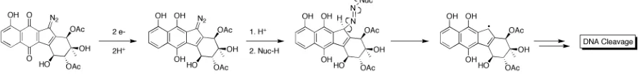
Although the postulates presented in connection with the diazo model studies described above had insightful rationale, none of them were directly applicable to a biological environment. The initial report disclosing the isolation of lomaiviticin A also revealed the ability of lomaiviticin A to cleave DNA under reducing conditions; however, experimental details were not presented.5 Given this result and considering components of the cellular environment, it seemed more reasonable that biological thiols (e.g. glutathione, GSH) were capable of inducing a 2H+/2e- reduction from diazoparaquinones to the corresponding hydroquinones to initiate the biological cascade of activity.
2.3.1 Biomimetic DNA Cleavage Mediated by Kinamycin D
hour periods. Control experiments using only GSH/DTT or kinamycin D demonstrated no evidence of DNA cleavage, supporting a 2H+/2e- reduction in vivo. Two mechanistic possibilities were proposed to account for the observed DNA cleavage mediated by a 2H+/2e -reduction of kinamycin D treated with GSH/DTT (Figure 10), which paralleled the previously proposed mechanisms. One possibility (Figure 10, Route A) proposed the resultant hydroquinone would be activated towards nucleophilic attack on the terminal diazo nitrogen, this nucleophilic adduct would then undergo homolytic bond cleavage to provide a carbon centered radical that could elicit DNA damage. The alternative possibility (Figure 10, Route B) proposed that protonation would promote the formation of an orthoquinone methide reactive intermediate with concomitant loss of N2.
Figure 10. Potential routes to DNA cleavage.
2.3.2 Biomimetic DNA Cleavage Mediated by Kinamycin F
Recently, in an extensive in vivo study,12 Hasinoff demonstrated that kinamycin F was also able to cleave DNA when treated with cellularly relevant concentrations of GSH. Furthermore, the DNA-cleavage exhibited upon treatment of kinamycin F with GSH was determined occur in an iron-, H2O2- and hydroxyl-radical-dependent manner. Kinamycin F