Pulmonary Tuberculosis Severity
Walid Ben-Selma and Jalel Boukadida
Laboratory of Microbiology and Immunology, Farhat Hached University Hospital, Sousse, Tunisia
The purpose of our study was to investigate the association between a functional single nucleotide polymorphism (SNP) in the
interleukin-23 receptor gene (
IL23R
; rs11209026, 1142 G
wild type¡
A
reduced function, Arg381Gln) and disease severity outcome in
pulmonary tuberculosis (TB) in the Tunisian population. SNP was investigated in a population of 168 patients with active
pul-monary TB (cases were stratified into patients with minimal/moderate lung involvement, i.e., patients with minimal/moderate
disease [Pmd], and patients with extensive lung involvement, i.e., patients with active disease [Pad]) and 150 healthy subjects.
Genotype analyses were carried out using the PCR-restriction fragment length polymorphism method. We have found that the
IL23R
reduced-function allele 1142A and genotypes AA and AG were overrepresented, especially in the Pad subgroup compared
with the control group (51% versus 18% [
P
ⴝ
10
ⴚ8], 33% versus 5% [
P
ⴝ
10
ⴚ8], and 36% versus 26% [
P
ⴝ
5
ⴛ
10
ⴚ3],
respec-tively). Additionally, comparison of the Pad and the Pmd groups showed that the A allele and AA genotype seemed to be
associ-ated with 2.79-fold (
P
ⴝ
4
ⴛ
10
ⴚ5) and 7.74-fold (
P
ⴝ
10
ⴚ5) increased risks of TB with minimal/moderate lung involvement,
respectively. Our results demonstrate that the reduced-function polymorphism 1142G
¡
A encoded by
IL23R
influences the
outcome of disease severity of active pulmonary TB in Tunisian patients.
T
uberculosis (TB) is a chronic infectious disease caused by
My-cobacterium tuberculosis
. It has been estimated that there are
8.8 million (range, 8.5 million to 9.2 million) incident cases of TB
and 1.1 million (range, 0.9 million to 1.2 million) deaths from TB
among HIV-negative people (
45
). Among those infected by
M.
tuberculosis
, only 5 to 10% develop clinical disease (
45
). Among
those individuals, some have identifiable risk factors, such as
ac-quired immunodeficiency disorders, old age, alcohol usage,
cor-ticosteroid consumption, diabetes mellitus, malnutrition, and
cigarette smoking (
3
,
4
). Additionally, studies of the concordance
rate of TB among monozygotic and dizygotic twins highlighted
the importance of host genetic factors in determining the
devel-opment of disease (
10
,
20
).
Cellular immunity plays an important role in controlling the
growth of
M. tuberculosis
. Thus, an effective host defense against
M. tuberculosis
infection requires the coordinated actions of both
the innate and adaptive immune systems (
19
). Interleukin-23
(IL-23) has been found to contribute to the development of Th1-like
CD4
⫹T-cell responses. The heterodimeric cytokine IL-23 is
se-creted by activated macrophages and dendritic cells (DCs) and
induces clonal expansion of memory CD4
⫹T cells (
30
,
35
). IL-23
is composed of a p40 subunit, shared with IL-12, and a unique p19
subunit, signaling through interleukin-12 receptor

(IL-12R

)
and a unique IL-23 receptor (IL-23R) chain (
34
). In addition to its
direct action on T cells, IL-23 induces the secretion of IL-12 and
gamma interferon (IFN-
␥
) by DCs
in vitro
(
6
). This suggests that
IL-23 has indirect involvement in the activation of
antigen-pre-senting cells (APCs). Studies with gene-deficient mice reveal that a
number of roles that were previously accredited to IL-12 may be
dependent on IL-23 (
15
). In
M. tuberculosis
infection, the absence
of the p40 subunit common to IL-12 and IL-23 results in more
marked susceptibility to
M. tuberculosis
infection than IL-12 p35
deficiency, suggesting an important role for IL-23 in
mycobacte-rial infections (
13
). Additionally, Van de Wetering et al. suggested
that the synergy of IL-23 with IL-18 is likely important in initiating
Th1 differentiation early in infection, whereas the synergy
be-tween IL-18 and IL-12 may be important in a further Th1 response
in subsequent stages of infection (
41
).
IL-23 is secreted by activated macrophages and DCs, induces
memory T-cell proliferation, and is the critical factor required for
T-cell IL-17 expression in response to bacterial challenge (
2
).
IL-23 exerts its activity through its receptor (IL-23R), expressed in
the Th17 subset of T lymphocytes (
44
). These newly characterized
CD4
⫹T cells were originally identified through their ability to
secrete high levels of the proinflammatory cytokine IL-17 upon
stimulation (
23
,
35
). Furthermore, IL-17 promotes neutrophilic
inflammation by upregulating CXC chemokines and
hematopoi-etic growth factors (
22
). Several recent studies have reported the
important role of IL-23 and IL-17 in the induction of a
neutro-phil-mediated protective immune response against extracellular
bacterial or fungal pathogens, such as
Escherichia coli
(
8
,
39
),
Kleb-siella pneumoniae
(
21
),
Porphyromonas gingivalis
(
47
),
Pseudomo-nas aeruginosa
(
18
),
Citrobacter rodentium
(
33
), and
Bacteroides
fragilis
(
11
).
In regard to
M. tuberculosis
infections, recent studies have
demonstrated a greater sensitivity in IL-12/IL-23 p40
⫺/⫺mice
than in IL-12 p35
⫺/⫺animals (
13
,
24
). Moreover, macrophages
rapidly express IL-23 when exposed to mycobacterial antigens,
suggesting an immune-stimulatory role for this cytokine during
infection (
5
,
43
). This finding was supported by Happel and
col-laborators (
22
). They reported that pulmonary administration of
adenovirus vectors expressing IL-23 can improve host resistance
to
M. tuberculosis
in wild-type mice (
22
). However, studies using
Received2 March 2012Returned for modification2 April 2012 Accepted1 June 2012
Published ahead of print13 June 2012
Address correspondence to Walid Ben-Selma, [email protected].
Copyright © 2012, American Society for Microbiology. All Rights Reserved.
doi:10.1128/CVI.00135-12
on August 17, 2020 by guest
http://cvi.asm.org/
IL-23-deficient mice have shown that the absence of IL-23 has
little or no effect on host resistance to
Toxoplasma gondii
,
Crypto-coccus neoformans
, and
M. tuberculosis
infection, unless IL-12 is
also absent (
27
,
29
,
31
). These studies suggest that, compared to
the dominant role of IL-12, the role of IL-23 in chronic infections
is more subtle. Recently, Khader and collaborators have reported
that IL-23 plays an essential role in chronic infection. They
showed that this cytokine is required for long-term control of
M.
tuberculosis
and B-cell-follicle formation in the infected lung (
26
).
IL-23 mediates its activity through IL-23R. IL-23 is a
heterodi-meric cytokine composed of a p19 subunit and a p40 subunit, with
the latter being shared with IL-12 (
12
,
25
), a Th1-promoting
cy-tokine (
29
,
40
) which has also been considered a candidate
sus-ceptibility gene in many autoimmune diseases. The IL-23 receptor
also shares a subunit, IL-12 receptor

1 (IL-12R

1), with the
IL-12 receptor, but it is the specific subunit of the IL-23 receptor,
named IL-23R. IL-23 stimulates the proliferation of Th17 cells, a
T-cell population which produces inflammatory cytokines such as
IL-17, tumor necrosis factor, and IL-6 (
28
).
Several polymorphisms within the IL-23R gene (
IL23R
; such as
the 1142G
¡
A polymorphism encoded by
IL23R
[
IL23R
1142G
¡
A]; rs11209026, 1142 G
wild type¡
A
reduced function, Arg381Gln) have
been associated with immune-related diseases, including
inflamma-tory bowel disease, psoriasis, and ankylosing spondylitis (
9
,
17
,
36
,
42
). However, to date, there have been no studies evaluating the
as-sociation between this polymorphism and the risk of development of
active TB in the world or in Tunisian patients.
In our study, we have investigated the association between
IL23R
(1142G
¡
A) and the risk of development of active
pulmo-nary TB and its severity in TB patients in Sousse, Tunisia, a region
characterized by a moderate TB prevalence (31 new cases per
100,000 population) and incidence (25 cases/100,000 population/
year) and a predominating
M. tuberculosis
strain (
46
).
MATERIALS AND METHODS
Study populations.One hundred sixty-eight patients with active pulmo-nary TB from Sousse, Tunisia, which is in the central region of the coun-try, were enrolled in this study (Table 1). One hundred fifty healthy blood donors (135 males and 15 females) were studied as controls.
Patients and healthy blood donors were selected over the period from January 2009 to June 2010. Individuals with a history of severe patholo-gies, including HIV infection, cardiovascular disease, asthma, or atopy autoimmune diseases, and cancer were excluded from the study. An in-formed written consent was obtained from all individuals prior to blood sampling. Moreover, our study was approved by the ethics committee of the Farhat Hached University Hospital.
Patients were recruited from the Pneumology Unit, CHU Farhat Hached, and the health care service, Sousse, Tunisia. Inclusion criteria for the patients in this group were determined according to the criteria de-fined by the American Thoracic Society (1).
Diagnosis of active pulmonary TB was based on clinical symptoms, the presence of acid-fast bacilli in sputum smears, and culture on Lowenstein-Jensen and Coletsos medium in all cases.
Pulmonary TB patients whose radiological lung tissue involvement (n⫽168) was available were further stratified into pulmonary patients with minimal (n⫽80)/moderate (n⫽43) disease or lung involvement (Pmd) and pulmonary patients with advanced disease and extensive lung involvement (Pad;n⫽45), according to non-HIV-related TB guidelines for disease classification (14,32).
All controls had the same ethnic and geographic origins and lived in the same city as the TB patients. The inclusion criteria for the control group were the absence of acute or chronic pulmonary disease, a negative history for TB, and proof of being healthy.
DNA extraction and genotyping.Genomic DNA was isolated from fresh whole blood-EDTA and buffy-coat lymphocytes of TB patients and controls using a Wizard Genomic DNA purification kit (Promega Corpo-ration, Madison, WI), according to the manufacturer’s instructions.
To genotype Arg381Gln (1142G¡A), a PCR-restriction fragment length polymorphism (RFLP) method was used as previously reported (42). Briefly, 100 ng of genomic DNA was added to 25l of a reaction mixture containing 1 mM each primer. The forward and reverse primers were 5=-CTTTTCTGGCAGGGTCATTTTG-3=and 5=-AAGTTGTTTCC TGGGGTAGTTGTG-3=, respectively. The remainder of the mixture con-sisted of 1⫻PCR GoTaq buffer (Promega), 1.5 mM MgCl2, 0.2 mM
deoxynucleoside triphosphates, and 1 U GoTaq Hot Start polymerase (Promega). The mixture was then initially subjected to 95°C for 10 min, followed by 40 cycles of denaturation for 30 s at 95°C, annealing for 1 min at 53°C, and extension for 1 min and 30 s at 72°C; final extension was for 7 min at 72°C. Amplifications were performed in a MyCycler thermal cycler (Bio-Rad). RFLP of the PCR product was used for the detection of TABLE 1Demographic and clinical data for tuberculosis patients and
controlsa
Study group No. of cases
Gender (no. M:no. F)
Mean (range) age (yr)
pTB 168 127:41 44 (14–78)
Pmd 123 100:23 39 (14–65)
Pad 45 27:18 45 (37–78)
Healthy 150 135:15 35 (24–55)
apTB, pulmonary tuberculosis; Pmd, pulmonary patients with minimum/moderate
lung involvement; Pad, pulmonary patients with extensive lung involvement; M, male; F, female.
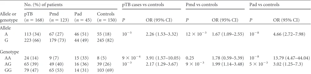
TABLE 2IL23R1142 G¡A allele and genotype frequencies in pulmonary tuberculosis cases and controlsa
Allele or genotype
No. (%) of patients pTB cases vs controls Pmd vs controls Pad vs controls
pTB (n⫽168)
Pmd (n⫽123)
Pad (n⫽45)
Controls
(n⫽150) P OR (95% CI) P OR (95% CI) P OR (95% CI) Allele
A 113 (34) 67 (27) 46 (51) 55 (18) 10⫺5 2.26 (1.53–3.32) 12⫻10⫺3 1.67 (1.09–2.55) 10⫺8 4.66 (2.72–7.98) G 223 (66) 179 (73) 44 (49) 245 (82)
Genotype
AA 24 (14) 9 (7) 15 (33) 8 (5) 9⫻10⫺4 3.91 (1.57–10.05) 0.25 1.78 (0.59–5.39) 10⫺8 13.79 (4.47–44.04) AG 65 (39) 49 (40) 16 (36) 39 (26) 10⫺3 2.17 (1.29–3.67) 9⫻10⫺3 1.99 (1.14–3.48) 5⫻10⫺3 3.02 (1.25–7.3) GG 79 (47) 65 (53) 14 (31) 103 (69)
apTB, pulmonary tuberculosis; Pmd, pulmonary patients with minimal/moderate lung involvement; Pad, pulmonary patients with extensive lung involvement.
on August 17, 2020 by guest
http://cvi.asm.org/
IL23R1142G¡A alleles. RFLP analysis was performed by incubating 5l of PCR product with 5 U of Hpy188I restriction endonuclease (New Eng-land BioLabs, MA) at 37°C for 3 h in a final restriction digestion volume of 20l. The restriction fragments (wild-type DNA yields fragments of 288, 103, 82, and 35 bp, whereas DNA containing the G1142A polymorphism yields fragments of 323, 103, and 82 bp) were separated by electrophoresis on 2% agarose gels (Sigma) containing ethidium bromide (0.5 mg/ml; Sigma) and observed under UV illumination using a Gel Doc XR system (Bio-Rad).
Statistical analysis.All genotypes were tested for Hardy-Weinberg equilibrium using a2test between the observed and expected numbers
separately in patients and controls (37).
Statistical analysis was performed by Epi Info (version 6.0) software (Centers for Disease Control and Prevention, Atlanta, GA). The associa-tions between the allelic/genotype frequencies and the clinical forms of TB, as well as the odds ratio for susceptibility to infection, were obtained by the2test. APvalue of⬍0.05 was considered statistically significant.
RESULTS
Hardy-Weinberg equilibrium.
In this study, evaluation of
Hardy-Weinberg equilibrium showed that the genotype
frequen-cies of the
IL23R
1142G
¡
A polymorphism were in
Hardy-Wein-berg equilibrium in the pulmonary TB, Pmd TB, and Pad TB
groups and healthy blood donors (
P
ⱕ
0.05).
Association of the
IL23R
1142G
¡
A reduced-function
poly-morphism with pulmonary tuberculosis.
We have used
PCR-RFLP to examine the status of the
IL23R
gene polymorphism
linked to G
wild-type¡
A
reduced-functionphenotypes.
The frequency distributions of different
IL23R
1142G
¡
A
genotypes are summarized in
Table 2
. We observed that the
IL23R
1142A (reduced-function) allele was significantly overrepresented
in the pulmonary TB group in comparison to the control group
(34% versus 18%; odds ratio [OR]
⫽
2.26, 95% confidence
inter-val [CI]
⫽
1.53 to 3.32) (
Table 2
). Moreover, when this group was
stratified into pulmonary patients with minimal/moderate lung
involvement (Pmd) and pulmonary patients with extensive lung
involvement (Pad), we found that the 1142A allele was
signifi-cantly more frequent in these two groups (27% versus 18% [
P
⫽
12
⫻
10
⫺3] and 51% versus 18% [
P
⫽
10
⫺8], respectively) (
Table
2
). Additionally, the 1142A allele seemed to be associated with the
increased risk of development of TB with minimal/moderate lung
involvement (OR
⫽
1.67, 95% CI
⫽
1.09 to 2.55) and TB with
extensive lung involvement (OR
⫽
4.66, 95% CI
⫽
2.72 to 7.98).
Three genotypes, AA, AG, and GG, were observed in the
dif-ferent TB and control groups (
Table 2
). The AA and AG genotypes
were significantly more frequent in pulmonary TB patients and
pulmonary patients with extensive lung involvement (Pad) than
in the control group (14% versus 5% [
P
⫽
9
⫻
10
⫺4] and 33%
versus 5% [
P
⫽
10
⫺8], respectively, for the AA genotype and 39%
versus 26% [
P
⫽
10
⫺3] and 36% versus 26% [
P
⫽
5
⫻
10
⫺3],
respectively, for the AG genotype). Additionally, these genotypes
seemed to be associated with an increased risk for development of
active pulmonary TB with extensive lung involvement (
Table 3
).
When the frequency distribution of different allele and
geno-types of the
IL23R
1142G
¡
A single nucleotide polymorphism
(SNP) was adjusted by gender in the Pmd, Pad, and control
groups, we found that (i) the A allele seemed to be associated with
an increased risk of development of TB with minimal/moderate
lung involvement (OR
⫽
1.67, 95% CI
⫽
1.02 to 2.74,
P
⫽
0.031)
and extensive lung involvement (OR
⫽
3.37, 95% CI
⫽
1.89 to
7.38,
P
⫽
2
⫻
10
⫺5) only in men (
Tables 4
and
5
) and (ii) men
harboring the AA genotype seemed to be at greater risk of
devel-opment of active TB with extensive lung involvement than women
(OR
⫽
12.06, 95% CI
⫽
2.97 to 51.4,
P
⫽
10
⫺4) (
Table 5
).
DISCUSSION
An increased number of association studies have implicated
polymorphisms located in promoter regions or coding regions
TABLE 3IL23R1142 G¡A allele and genotype frequencies in pulmonary patients with extensive lung involvement and pulmonary patients with minimal/moderate lung involvementa
Allele or genotype
No. (%) of patients
P OR (95% CI) Pad
(n⫽45) Pmd (n⫽123) Allele
A 46 (51) 67 (27) 4⫻10⫺5 2.79 (1.64–4.75) G 44 (49) 179 (73)
Genotype
AA 15 (33) 9 (7) 10⫺5 7.74 (2.54–24.2) AG 16 (36) 49 (40) 0.31 1.52 (0.63–3.67) GG 14 (31) 65 (53)
aPad, pulmonary patients with extensive lung involvement; Pmd, pulmonary patients
with minimal/moderate lung involvement.
TABLE 4IL23R1142G¡A allele and genotype frequencies in Pmd and control groups, by gendera
Allele or genotype
No. (%) of patients
Male and female Pmd vs male and female control cases
Male Pmd vs male control cases
Female Pmd vs female control cases
Pmd (n⫽123) Controls (n⫽150)
P OR (95% CI) P OR (95% CI) P OR (95% CI)
M⫹F M F M⫹F M F
Allele
A 67 (27) 46 (23) 21 (46) 55 (18) 41 (15) 14 (47) 12⫻10⫺3 1.67 (1.09–2.55) 0.031 1.67 (1.02–2.74) 0.93 0.96 (0.34–2.67) G 179 (73) 154 (77) 25 (54) 245 (82) 229 (85) 16 (53)
Genotype
AA 9 (7) 7 (7) 2 (9) 8 (5) 5 (4) 3 (20) 0.25 1.78 (0.59–5.39) 0.22b 2.27 (0.61–8.7) 0.58b 0.67 (0.04–10.72)
AG 49 (40) 32 (32) 17 (74) 39 (26) 31 (23) 8 (53) 9⫻10⫺3 1.99 (1.14–3.48) 0.08 1.68 (0.89–3.15) 0.3b 2.13 (0.32–14.36)
GG 65 (53) 61 (61) 4 (17) 103 (69) 99 (73) 4 (27) a
Pmd, pulmonary patients with minimal/moderate lung involvement; M, male; F, female. bFisher’s exact test.
on August 17, 2020 by guest
http://cvi.asm.org/
of cytokine receptor genes, such as the gamma interferon
re-ceptor (
10
,
20
) and interleukin-10 receptor, as we have recently
reported (
7
), to be host factors influencing the development of
active TB. This is the first study demonstrating that the
IL23R
1142G
¡
A functional polymorphism is associated with
in-creased susceptibility to active pulmonary TB and its severity in
Tunisian patients. In fact, our result showed that patients
car-rying the
IL23R
1142A allele or AA genotype had 2.79- and
7.74-fold increased risks of developing active TB with extensive
lung involvement, respectively.
Recently, Khader and collaborators have reported that IL-23
plays a crucial role in the long-term immune response against
M.
tuberculosis
(
26
). They showed that IL-23 is required for
long-term containment of
M. tuberculosis
, as well as the expression of
CXCL13 within and the maintenance of B-cell follicles within the
lung lesions. Additionally, they demonstrated that IL-17RA and
IL-22 were involved in B-cell-follicle development at distinct
times during infection and that IL-23 is necessary for the
expres-sion of both of these cytokines in the lung.
Many studies have revealed that several single nucleotide
poly-morphisms in the
IL23R
gene are associated with immune-related
diseases, including inflammatory bowel disease, psoriasis, and
an-kylosing spondylitis (
9
,
17
,
36
,
42
). The most studied SNP was
IL23R
R381Q. Sarin et al. indeed showed that the 381Q variant has
a reduced percentage of cells that secrete IL-17 and IL-22 in
re-sponse to IL-23 stimulation and reduced levels of STAT3
phos-phorylation and IL-17 and IL-22 production in T-cell subsets
(
38
). The group of Pidasheva et al. also showed reduced STAT3
phosphorylation in response to IL-23 in T cells with the 381Q
variant, although they did not find reduced numbers of IL-17- or
IL-22-producing cells (
36
). Sarin et al. showed that IL-23-induced
IFN-
␥
production is not affected in the 381Q variant (
38
), and
Pidasheva et al. showed that IFN-
␥
-producing cells are not
re-duced (
36
). Another group has also shown that the 381Q variant
of IL-23R is comparable to the wild-type variant in an
overexpres-sion system, when analyzing the ability to induce IFN-
␥
and IL-10
production (
16
). The heterogeneity of these results could be
re-lated to the cytokines investigated or the use of different cell types,
such as retrovirally transduced T-cell blasts (
16
) rather than
iso-lated primary CD8
⫹T cells expressing the endogenous wild type
or R381Q IL23R variant (
38
).
To our knowledge, no study in the world has investigated the
association between the
IL23R
R381Q reduced-function
poly-morphism and the risk of development of active pulmonary TB.
This is the first study demonstrating an association between the
IL23R
R381Q reduced-function polymorphism and pulmonary
active TB and its severity in the Tunisian population. Our result
has shown that patients carrying the A allele of the
IL23R
R381Q
reduced-function polymorphism had 2.26-, 1.67-, and 4.66-fold
increased risks of developing pulmonary TB, pulmonary TB with
minimal/moderate lung involvement, and pulmonary TB with
ex-tensive lung involvement, respectively. Additionally, comparison
of the Pad (advanced TB) and Pmd (mild to moderate TB) groups
showed that the A allele and AA genotype of the 1142G
¡
A
polymorphism seemed to be associated with 2.79- and 7.74-fold
increased risks of advanced disease and disease of
minimal/mod-erate severity, respectively. The A allele of the
IL23R
R381Q
poly-morphism corresponds to decreased IL-23-dependent IL-17 and
IL-22 production, which may impair the immune response
against
M. tuberculosis
infection, resulting in pulmonary TB
de-velopment.
Interestingly, in our study, the
IL23R
1142A allele appeared to
be associated with 1.67- and 3.37-fold increased risks of
develop-ment of active TB with minimal/moderate lung involvedevelop-ment and
extensive lung involvement, respectively, in males. Additionally,
men with the AA genotype appeared to be at a 12.06-fold
in-creased risk of development of the active form of pulmonary TB
with extensive lung involvement.
In conclusion, our study shows for the first time that the
IL23R
1142G
¡
A reduced-function polymorphism seems to be
associ-ated with an increased risk of development of a severe form of
active pulmonary TB. Further studies with both adult and
pediat-ric populations in ethnically diverse settings are needed to confirm
our findings.
ACKNOWLEDGMENTS
Financial support was provided by the Ministry of Higher Education, Scientific Research and Technology (UR02SP13) of Tunisia.
W.B.-S. performed the experiments and wrote the manuscript. J.B. conceived the project, supervised experiments, and revised the manu-script. Both W.B.-S. and J.B. read and approved relevant portions of the manuscript.
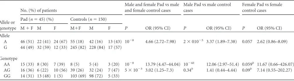
We state that we have no conflicts of interest. TABLE 5IL23R1142G¡A allele and genotype frequencies in Pad and control groups, by gendera
Allele or genotype
No. (%) of patients
Male and female Pad vs male and female control cases
Male Pad vs male control cases
Female Pad vs female control cases
Pad (n⫽45) (%) Controls (n⫽150)
P OR (95% CI) P OR (95% CI) P OR (95% CI)
M⫹F M F M⫹F M F
Allele
A 46 (51) 22 (41) 24 (67) 55 (18) 42 (16) 13 (43) 10⫺8 4.66 (2.72–7.98) 2⫻010⫺5 3.37 (1.89–7.38) 0.057 2.62 (0.86–8.09) G 44 (49) 32 (59) 12 (33) 245 (82) 228 (84) 17 (57)
Genotype
AA 15 (33) 8 (30) 7 (39) 8 (5) 5 (4) 3 (20) 10⫺8 13.79 (4.47–44.04) 10⫺4b 12.06 (2.97–51.4) 0.059b 11.67 (0.66–426.07)
AG 16 (36) 6 (22) 10 (56) 39 (26) 32 (24) 7 (47) 5⫻10⫺3 3.02 (1.25–7.3) 0.34b 1.41 (0.44–4.44) 0.09b 7.14 (0.55–202.27)
GG 14 (31) 13 (48) 1 (5) 103 (69) 98 (72) 5 (33) a
Pad, pulmonary patients with extensive lung involvement; M, male; F, female. bFisher’s exact test.
on August 17, 2020 by guest
http://cvi.asm.org/
REFERENCES
1.American Thoracic Society.2000. Diagnostic standards and classification of tuberculosis in adults and children. Am. J. Respir. Crit. Care Med. 161:1376 –1395.
2.Aujla SJ, Dubin PJ, Kolls JK.2007. Th17 cells and mucosal host defense. Semin. Immunol.19:377–382.
3.Barnes DS.2000. Historical perspectives on the etiology of tuberculosis. Microbes Infect.2:431– 440.
4.Beck JS, Lowe JG, Grange JM.1993. Corticosteroids as a risk factor for tuberculosis. Tuber. Lung Dis.74:413– 414.
5.Begum NA, et al. 2004. Mycobacterium bovis BCG cell wall-specific differentially expressed genes identified by differential display and cDNA subtraction in human macrophages. Infect. Immun.72:937–948. 6.Belladonna ML, et al.2002. IL-23 and IL-12 have overlapping, but
dis-tinct, effects on murine dendritic cells. J. Immunol.168:5448 –5454. 7.Ben-Selma W, Ben-Abderrahmen Y, Boukadida J, Harizi H. 2012.
IL-10R1 S138G loss-of-function polymorphism is associated with ex-trapulmonary tuberculosis risk development in Tunisia. Mol. Biol. Rep. 39:51–56.
8.Brereton CF, et al.2011. Escherichia coli heat-labile enterotoxin pro-motes protective Th17 responses against infection by driving innate IL-1 and IL-23 production. J. Immunol.186:5896 –5906.
9.Carvalho A, et al.2010. Prognostic significance of genetic variants in the IL-23/Th17 pathway for the outcome of T cell-depleted allogeneic stem cell transplantation. Bone Marrow Transplant.45:1645–1652.
10. Casanova JL, Abel L.2002. Genetic dissection of immunity to mycobac-teria. The human model. Annu. Rev. Immunol.20:581– 620.
11. Chung DR, et al.2003. CD4⫹T cells mediate abscess formation in intra-abdominal sepsis by an IL-17-dependent mechanism. J. Immunol. 170:1958 –1963.
12. Cooper AM, Khader SA.2007. IL-12p40: an inherently agonistic cyto-kine. Trends Immunol.28:33–38.
13. Cooper AM, et al.2002. Mice lacking bioactive IL-12 can generate pro-tective, antigen specific cellular responses to mycobacterial infection only if the IL-12 p40 subunit is present. J. Immunol.168:1322–1327. 14. Crofton J.1990. Clinical features of tuberculosis, p. 395– 421.InSeaton A,
et al (ed), Crofton and Douglas’s respiratory diseases. Blackwell Scientific, London, United Kingdom.
15. Cua DJ, et al.2003. Interleukin-23 rather than interleukin-12 is the critical cytokine for autoimmune inflammation of the brain. Nature421: 744 –748.
16. de Paus RA, van de Wetering D, van Dissel JT, van de Vosse E.2008. IL-23 and IL-12 responses in activated human T cells retrovirally trans-duced with IL-23 receptor variants. Mol. Immunol.45:3889 –3895. 17. Di Meglio P, et al.2011. The IL23R R381Q gene variant protects against
immune-mediated diseases by impairing IL-23-induced Th17 effector re-sponse in humans. PLoS One6:e17160. doi:10.1371/journal.pone. 0017160.
18. Dubin PJ, Kolls JK.2007. IL-23 mediates inflammatory responses to mucoid Pseudomonas aeruginosa lung infection in mice. Am. J. Physiol. Lung Cell. Mol. Physiol.292:L519 –L528.
19. Flynn JL.2004. Immunology of tuberculosis and implications in vaccine development. Tuberculosis84:93–101.
20. Gao L, Tao Y, Zhang L, Jin Q.2010. Vitamin D receptor genetic poly-morphisms and tuberculosis: updated systematic review and meta-analysis. Int. J. Tuberc. Lung Dis.14:15.
21. Happel KI, et al.2003. Cutting edge: roles of Toll-like receptor 4 and IL-23 in IL-17 expression in response to Klebsiella pneumoniae infection. J. Immunol.170:4432– 4436.
22. Happel KI, et al.2005. Pulmonary interleukin-23 gene delivery increases local T-cell immunity and controls growth of Mycobacterium tuberculo-sis in the lungs. Infect. Immun.73:5782–5788.
23. Harrington LE, et al.2005. Interleukin 17-producing CD4⫹effector T cells develop via a lineage distinct from the T helper type 1 and 2 lineages. Nat. Immunol.6:1123–1132.
24. Hölscher C, et al.2001. A protective and agonistic function of IL-12p40 in mycobacterial infection. J. Immunol.167:6957– 6966.
25. Hunter CA.2005. New IL-12-family members: IL-23 and IL-27, cytokines with divergent functions. Nat. Rev. Immunol.5:521–531.
26. Khader SA, et al.2011. IL-23 is required for long-term control of Myco-bacterium tuberculosis and B cell follicle formation in the infected lung. J. Immunol.187:5402–5427.
27. Khader SA, et al.2005. IL-23 compensates for the absence of IL-12p70 and is essential for the IL-17 response during tuberculosis but is dispens-able for protection and antigen specific IFN-gamma responses if IL-12p70 is available. J. Immunol.175:788 –795.
28. Kikly K, Liu L, Na S, Sedgwick JD.2006. The IL-23/Th(17) axis: thera-peutic targets for autoimmune inflammation. Curr. Opin. Immunol.18: 670 – 675.
29. Kleinschek MA, et al.2006. IL-23 enhances the inflammatory cell re-sponse in Cryptococcus neoformans infection and induces a cytokine pat-tern distinct from IL-12. J. Immunol.176:1098 –1106.
30. Langrish CL, et al.2004. IL-12 and IL-23: master regulators of innate and adaptive immunity. Immunol. Rev.202:96 –105.
31. Lieberman LA, et al.2004. IL-23 provides a limited mechanism of resis-tance to acute toxoplasmosis in the absence of IL-12. J. Immunol.173: 1887–1893.
32. Maher D, Chaulet P, Spinaci S, Harries A.1997. Treatment of tubercu-losis— guidelines for national programmes, p. 1–78. World Health Orga-nization, Geneva, Switzerland.
33. Mangan PR, et al.2006. Transforming growth factor-beta induces devel-opment of the T(H)17 lineage. Nature441:231–234.
34. Oppmann B, et al.2000. Novel p19 protein engages IL-12p40 to form a cytokine, IL-23, with biological activities similar as well as distinct from IL-12. Immunity13:715–725.
35. Park H, et al.2005. A distinct lineage of CD4 T cells regulates tissue inflammation by producing interleukin 17. Nat. Immunol.6:1133–1141. 36. Pidasheva S, et al.2011. Functional studies on the IBD susceptibility gene IL23R implicate reduced receptor function in the protective genetic vari-ant R381Q. PLoS One6:e25038. doi:10.1371/journal.pone.0025038. 37. Rodriguez S, Gaunt TR, Day IN.2009. Hardy-Weinberg equilibrium
testing of biological ascertainment for Mendelian randomization studies. Am. J. Epidemiol.169:505–514.
38. Sarin R, Wu X, Abraham C. 2011. Inflammatory disease protective R381Q IL23 receptor polymorphism results in decreased primary CD4⫹ and CD8⫹human T-cell functional responses. Proc. Natl. Acad. Sci. U. S. A.108:9560 –9565.
39. Shibata K, Yamada H, Hara H, Kishihara K, Yoshikai Y.2007. Resident Vdelta1⫹gammadelta T cells control early infiltration of neutrophils after Escherichia coli infection via IL-17 production. J. Immunol.178:4466 – 4472.
40. Trinchieri G.2003. Interleukin-12 and the regulation of innate resistance and adaptive immunity. Nat. Rev. Immunol.3:133–146.
41. Van de Wetering D, de Paus RA, van Dissel JT, van de Vosse E.2009. IL-23 modulates CD56⫹/CD3⫺NK cell and CD56⫹/CD3⫹NK-like T cell function differentially from IL-12. Int. Immunol.21:145–153. 42. Venegas M, et al.2008. IL-23R Arg381Gln polymorphism in Chilean
patients with inflammatory bowel disease. Eur. Cytokine Netw.19:190 – 195.
43. Verreck FA, et al.2004. Human IL-23-producing type 1 macrophages promote but IL-10-producing type 2 macrophages subvert immunity to mycobacteria. Proc. Natl. Acad. Sci. U. S. A.101:4560 – 4565.
44. Wilson NJ, et al.2007. Development, cytokine profile and function of human interleukin 17-producing helper T cells. Nat. Immunol.8:950 – 957.
45. World Health Organization.2011. Global tuberculosis control. World Health Organization, Geneva, Switzerland. http://www.who.int/tb /publications/global_report/en/.
46. World Health Organization.2011. Tuberculosis. World Health Organiza-tion, Geneva, Switzerland.http://www.who.int/tb/country/data/profiles/en /index.html.
47. Yu JJ, Ruddy MJ, Conti HR, Boonanantanasarn K, Gaffen SL.2008. The interleukin-17 receptor plays a gender-dependent role in host protection against Porphyromonas gingivalis-induced periodontal bone loss. Infect. Immun.76:4206 – 4213.