_____________________________________________________________________________________________________
*Corresponding author: Email: [email protected];
www.sciencedomain.org
Rare Case of Multiple Compound Odontomas with
Dens Invaginatus Associated with Impacted Primary
and Unerupted Permanent Maxillary Central Incisor
– A Case Report
Suzan Sahana
1*, Aron Arun Kumar Vasa
1, Srikanth Koya
2and J. Raghavendra Kumar
11
Department of Pedodontics and Preventive Dentistry, St. Joseph Dental College, Duggirala, Eluru-534003, Andhra Pradesh, India. 2
Department Pedodontics and Preventive Dentistry, Drs. Sudha & Nageshwar Rao Institute of Dental Sciences, Chinna Autapalli, Gannavaram – 521286, Andhra Pradesh, India.
Authors’ contributions
This work was carried out in collaboration between all authors. All authors read and approved the final manuscript.
Article Information
DOI: 10.9734/BJMMR/2016/20481 Editor(s): (1) Emad Tawfik Mahmoud Daif, Professor of Oral & Maxillofacial Surgery, Cairo University, Egypt. Reviewers: (1) Maen Mahfouz, Arab American University Jenin, Palestine. (2)Navroop Kaur Bajwa, Government Dental College, Patiala, India. Complete Peer review History:http://sciencedomain.org/review-history/11466
Received 29th July 2015 Accepted 26th August 2015 Published 19th September 2015
ABSTRACT
Aims: An Odontoma is usually found in the surrounding area of the unerupted permanent tooth bud
and rarely adjacent to the primary central incisor. Histopathological examination of the Odontoma revealed presence of surface invagination similar to Dens invaginatus.
Presentation of Case: A seven year-old healthy girl presented with an unerupted maxillary left
primary central incisor along with swelling of labial gingiva in that region. No history of traumatic injury was recorded. Radiological examination revealed impacted primary maxillary left central incisor and three irregular masses of calcified tooth like tissues.
Discussion: Odontomas are commonly occuring hamartomas associated with permanent dentition.
Odontomas are rarely associated with Primary dentition. Multiple Odontomas leading to impaction
of primary incisors is still rarer. This report presents a case of an impacted primary central incisor and unerupted permanent central incisor due to multiple odontomas, one of which was histologically diagnosed as multiple compound odontoma with Dens invaginatus. The Odontomas and impacted primary central were surgically removed to facilitate the eruption of permanent incisor.
Conclusion: This report presents a unique and rare case of multiple Odontomas leading to
impaction of primary tooth and unerupted permanent incisor. This report also proves Odontomas with deep surface invagination similar to Dens Invaginatus can be present.
Keywords: Compound odontoma; dens invaginatus; impaction; primary tooth.
1. INTRODUCTION
Odontomas occur more often in the permanent dentition and are rarely associated with Primary teeth. They were first reported by Oudet in 1821. Odontomas are developmental anomalies resulting from the growth of completely differentiated epithelial and mesenchymal cells that give rise to functional ameloblasts and odontoblasts. These cells in-turn form variable amounts of enamel, dentin, cementum and pulpal tissue of the odontoma. Based on morphology, they are classified as Compound and Complex Odontomas. Odontomas may cause disturbances in the eruption of teeth such as impaction, delayed eruption or retention of primary teeth as well as permanent teeth [1-3]
Dens invaginatus is a deep surface invagination of the crown or root that is lined by enamel [2].
This article describes a case of multiple Compound odontome associated with impacted maxillary left primary central incisor and unerupted left permanent maxillary central incisor. Histological examination of one of the odontome revealed presence of Dens invaginatus which is unique and has not been previously reported in the literature.
2. PRESENTATION OF CASE
A 7-year-old girl reported with the complaint of absence of upper left front tooth as well as presence of bulge in that region. Her dental history revealed unerupted primary left central incisor. There was no history of trauma nor relevant medical, or family history indicative of dental abnormalities.
On extra oral examination, the left side of the upper lip was found to be swollen. There was no regional lymphadenopathy. Intra-oral examination revealed non-tender swelling in relation to labial gingiva measuring about 1 cm in length and 1.5 cm in breadth, extending
mesiodistally from labial frenum up to 2 mm in front of the canine eminence (Fig. 1). On palpation, swelling was bony hard in consistency, non-tender, well defined, fixed to the underlying tissue and immobile. The mucosa appeared normal and erupting permanent right upper central incisor was also seen.
Fig. 1. Intraoral view showing unerupted maxillary permanent left central incisor, along
with bulge in that area
Intraoral periapical radiograph in relation to upper left central and lateral incisor (Fig. 2) revealed impacted primary upper left central incisor and three irregular masses of calcified tooth like tissues which were obstructing the eruption of permanent left upper central incisor. These calcified irregular masses were separated in few areas by a narrow radiolucent band with a smooth outer periphery. On the basis of clinical and radiographic examination a provisional diagnosis of odontoma was made. Maxillary occlusal radiograph was also taken to help in localizing these calcified masses which revealed the masses to be present labially.
The impacted primary left maxillary central incisor along with the three odontomes were carefully extracted (Fig. 3 & Fig. 4). The permanent left maxillary central incisor was left intact. Sharp bony margins were rounded off, the area was irrigated with povidone-iodine solution and the flap was placed back and held in position with the help of vicryl sutures and patient was kept on amoxicillin 250 mg TID for five days along with Ibuprufen 200 mg SOS. The patient was recalled monthly after healing of the surgical site to monitor the eruption of the permanent Incisor. One year follow-up revealed eruption of left permanent maxillary central incisor uneventfully.
Fig. 2. Intraoral periapical radiograph showing calcified structures (A,B,C), impacted primary central incisor (D), unerupted permanent central Incisor (E)
Macroscopically, the odontomes appeared like collection of tooth like structures of varying size and shape with enamel like tissue as cap. Out of the three odontomes, one [Fig. 3(C)] appeared like fusion of four odontomes and was sent for histo-pathological evaluation.
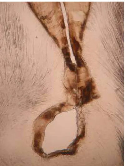
The histo-pathological sections revealed presence of enamel, dentin, and pulp like tissues (Fig. 5 & Fig. 6). The histo-pathological section also revealed presence of deep invagination of enamel like tissue which resembles “Dens
Invaginatus” or “Dens in Dente”. Based on histo-pathological findings a final diagnosis of “Compound odontoma with Dens invaginatus” was made.
Fig. 3. Calcified structures (specimens A,B,C) and Impacted primary central incisor (D) after
surgical removal
Fig. 4. Intraoral periapical radiograph showing unerupted permanent central Incisor
(E) and lateral incisor (F) after surgery
3. DISCUSSION
Neoplasm. When fully developed, odontomas consist chiefly of enamel and dentin with variable amounts of pulp and Cementum. The term odontome by definition alone refers to a tumor of odontogenic origin. In a broad sense, it means a growth with both epithelial and mesenchymal components exhibiting complete differentiation with the result that functional ameloblast and odontoblast form enamel and dentin [4].
Fig. 5. Decalcified eosin stained specimen ‘C’ showing dentin and pulp like tissue
Fig. 6. Ground section of specimen ‘C’ showing enamel, dentin like tissue and
dens-invaginatus
Clinically, odontomes are generally asymptomatic; discovered on a routine radiographic examination or when films are taken to determine the reason for failure of a tooth to erupt and they usually remain small, rarely exceeding the diameter of the tooth. Occasionally it does become large and may produce expansion of bone with consequent facial asymmetry. This is particularly true if dentigerous cyst develops around the odontome. Signs and symptoms associated consist of unerupted teeth or impacted teeth, retained deciduous teeth, swelling and incidence of infection [5].
Radiographically the Compound odontoma appears as a collection of tooth like structures of varying size and shape surrounded by a narrow radiolucent zone. The radiographic findings are usually diagnostic and the Compound odontoma is seldom confused with any other lesion. All the radiographic features, suggestive of Compound odontome are present in this case [6].
The Compound odontoma is a lesion in which all the dental tissues are represented in an orderly fashion so that there is at least superficial anatomic resemblance to teeth. In a Complex odontome, on the other hand although all the dental tissues are represented they are formed in such a rudimentary fashion that there is little or no morphological resemblance to normal tooth formation. Compound odontomas have a propensity for occurrence in the canine and incisor region more often in maxilla [1].
Histologically the Compound odontoma consists of multiple structures resembling small, single rooted teeth contained in a loose fibrous matrix.
Odontomes, when obstructing the eruption of permanant teeth are surgically managed. The permanent teeth are radilographically observed for spontaneous eruption, if it fails to erupt then it is surgically exposed to allow it to erupt. In some scenarios orthodontic banding with hook or Bonding can be done after surgical exposure and cross elastics are placed on opposing teeth to aid in eruption [3]. The complications of surgical management include Infection, non-eruption of Permanent teeth, Impaction or formation of dentigerous cyst.
mass, [7]which is not the same in this case [8]. whereas, multiple radio opaque structures were present in the anterior maxilla, which were separated by thin radiolucent band in few areas. Cystic odontome increase in size slowly and cause large expansion of bone, [6] which is not seen in our case.
Literature review suggests that radiographic examination should be performed in all pediatric patients who present with clinical evidence of delayed permanent tooth eruption or temporary tooth displacement, with or without history of previous dental trauma. Early diagnosis of odontomas allows adoption of a less-complex and less-expensive treatment and ensures better prognosis [9].
4. CONCLUSION
Presence of impacted primary maxillary central incisor and unerupted permanent maxillary central incisor due to multiple odontomas with Dens invaginatus makes this case a rare and unique occurrence.
CONSENT
All authors declare that ‘written informed consent was obtained from the patient (or other approved parties) for publication of this case report and accompanying images.
ETHICAL APPROVAL
All authors hereby declare that all experiments have been examined and approved by the appropriate ethics committee and have therefore been performed in accordance with the ethical standards laid down in the 1964 Declaration of Helsinki.
COMPETING INTERESTS
Authors have declared that no competing interests exist.
REFERENCES
1. Neville Damm, Allen Bouquot. Oral and maxillofacial pathology. Second edn. Saunders. 2004;631-632.
2. Shafer Hine, Levy. A text book of oral pathology. Fourth Edition; W.B. Saunders & Co. 1993;308-312.
3. Batra Puneet, Gupta Shweta, Rajan Kumar, Duggal Ritu, Hariparkash. Odontomes-diagnosis and treatment: A case report. JPFA. 2003;19:73-76.
4. Thoma KM, Goldman HM. Oral pathology, 5th edn. St Louis, The CV Mosby Company. 1960;1221-1222.
5. Kramer IRH, Pindborg JJ, Shear M. Histological typing of odontogenic tumour. WHO. International Histological Classification of Tumours; Second Edition; Berlin Springer. 1992;16-21.
6. White Pharaoh. Oral radiology - Principles and interpretation. Fourth Edition; Mosby. 2000;395-397.
7. Supriya Pande, Ganvir SM, Hazarey VK. Recurrent Odontome- A Rarity. JIDA 2003; 74:115-118.
8. Kharbanda OP, Saimbi CS, Kharbanda Renu. Odontome- A case report. JIDA. 1986;58:269-271.
9. Philipsen HP, Reichart PA, Praetorious F. Mixed odontogenic tumors & odontomas. Considerations on Interrelationship. Review of Literature and Presentation of 134 New Cases of Odontomas. Oral Oncol. 1997;33:86-99.
© 2016 Sahana et al.; This is an Open Access article distributed under the terms of the Creative Commons Attribution License (http://creativecommons.org/licenses/by/4.0), which permits unrestricted use, distribution, and reproduction in any medium, provided the original work is properly cited.
Peer-review history: