Original Article
Multiple stage-dependent roles for histone deacetylases
during amphibian embryogenesis: implications for
the involvement of extracellular matrix remodeling
SASHKO DAMJANOVSKI
1, LAURENT M. SACHS
1,2and YUN-BO SHI*
Unit of Molecular Morphogenesis, Laboratory of Molecular Embryology, National Institute of Child Health and Human Development, Bethesda, USA
ABSTRACT Histone acetylation has long been implicated in the regulation of gene expression. Recently, a number of histone acetyltransferase and histone deacetylase genes have been identified and cloned. Molecular studies have shown that these enzymes influence transcriptional regulation as components of cofactor complexes that interact with diverse transcription factors. However, relatively little is known about their function during development. Here, we make use of the ability to manipulate Xenopus laevis embryos in vitro to study the role of histone deacetylases in development. We first demonstrate that the histone deacetylase Rpd3 and its associated co-repressor Sin3A are coordinately expressed during embryogenesis. Rpd3 and Sin3A are known to be part of at least one large corepressor complex, which is involved in transcriptional regulation by many transcription factors, suggesting that deacetylase activity is important for embryogenesis through transcriptional regula-tion. Indeed, treating developing embryos with a specific histone deacetylase inhibitor, trichostatin A (TSA), leads to embryonic lethality with severe defects in the head and tail regions. Furthermore, the effects of TSA are stage-dependent with the severity of the defects decreasing when treatment is initiated at later stages. On the other hand, a sharp bend (kink) develops in the tail even when TSA treatment begins at tadpole hatching. We provide evidence that this tail defect may be in part due to the TSA-dependent inhibition of the expression of the matrix metalloproteinase gene stromelysin-3, which has been implicated in tail development through extracellular matrix remodeling.
KEY WORDS: Xenopus laevis, matrix metalloproteinase, histone acetylation, chromatin remodeling, morphogenesis.
0214-6282/2000/$20.00
© UBC Press Printed in Spain
www.ehu.es/ijdb
*Address correspondence to: Yun-Bo Shi. Unit of Molecular Morphogenesis, Laboratory of Molecular Embryology, National Institute of Child Health and Human Development, National Institute of Health, Building 18T, Room 106, Bethesda, MD 20892-5431, USA. FAX: 1 301 402 1323. e-mail: [email protected]
1Both authors contributed equally to this work.
2Present address: Laboratoire de Physiologie, MNHN, UMR CNRS 8572, 7 rue Cuvier, 75231 Paris Cedex 05, France.
Abbreviations used in this paper: TSA, trichostatin A; TR, thyroid hormone receptor; ST3, stromelysin 3; Col3, collagenase 3; xhh, Xenopus sonic hedgehog.
Introduction
The central theme of developmental biology has always been to understand how the fertilization of a single cell, the egg, gives rise to a complex organism. At molecular level, this involves determin-ing how various genes are activated or repressed at various developmental stages to effect cell fate determination and tissue patterning. A key factor in transcriptional regulation is believed to be the state of the local chromatin environment of the gene to be regulated (Wolffe, 1996). Chromatin structure can be influenced by both DNA and histone modifications such as DNA methylation, histone phosphorylation and acetylation as well as histone meth-ylation (Wolffe, 1996; Strahl and Allis, 2000).
It has been suggested that histone acetylation influences gene expression (Pogo et al., 1966; Hebbs et al., 1988; Wolffe, 1996;
acetyltransferase) activity and that transcriptional corepressors are complexed with histone deacetylases (Wade and Wolffe, 1997; Pazin and Kadonaga, 1997; Struhl, 1998; Ng and Bird, 2000).
One of the best-studied transcriptional co-repressor complexes contains the co-repressor Sin3 and deacetylase Rpd3. Sin3 can interact with the co-repressors SMRT and N-CoR, which in turn bind to a number of transcription repressors including Mad/Max and unliganded thyroid hormone receptors (TRs) (Heinzel et al., 1997; Nagy et al., 1997; Pazin and Kadonaga, 1997). Thus, specific local changes in histone acetylation through targeting histone deacetylases by transcription factors may serve as a mechanism for specific gene regulation during development. Com-pared to the extensive biochemical and molecular characterization of histone acetylation and deacetylation, relatively little is known about their developmental functions. Amphibian development is an excellent system with which one can investigate how histone acetylation influences gene expression during development. Ovipa-rous amphibian embryogenesis can be easily manipulated in vitro without any concerns of maternal effects as occurs with mamma-lian development, and amphibian embryos are easily accessible for molecular analysis.
Here we make use of Xenopus laevis development to investi-gate the developmental roles of histone deacetylases. We first determined the spatial and temporal expression of histone deacetylase Rpd3 and corepressor Sin3, which interacts with Rpd3. Using a specific chemical inhibitor of histone deacetylases, trichostatin A (TSA), we directly demonstrate the requirement for histone deacetylases in Xenopus embryogenesis. We further identify a potential downstream mediator of this effect, the matrix metalloproteinase stromelysin-3, in a specific developmental mor-phogenetic process.
Results
Rpd3 and Sin3 are co-localized during Xenopus embryogenesis
To gain insight into the possible role that histone acetylation may play in the regulation of developmental events, we focused on the involvement of histone deacetylases due to the availability chemical inhibitors of these enzymes for possible embryonic studies. One histone deacetylase, Rpd3, and its likely interacting partner in the
co-Fig. 1. Coordi-nated spatial and temporal expres-sion of histone deacetylase Rpd3 and corepressor Sin3. Digoxigenin labeled Rpd3 and Sin3 antisense mRNA was used as probes in whole mount analysis of their developmental expression in albino Xenopus embryos.
(A,B,C,D) Rpd3 and
(E,F,G,H) Sin3 ex-pression was exam-ined at the end of neuralation (stage 18/21, A,E), tailbud (stage 31/32,B,F), hatching stages (stage 35/36, C,G), and at the onset of feeding (stage 45, D,H). Both Rpd3 and Sin3 were abundantly ex-pressed, particu-larly in dorsal axial structures and in
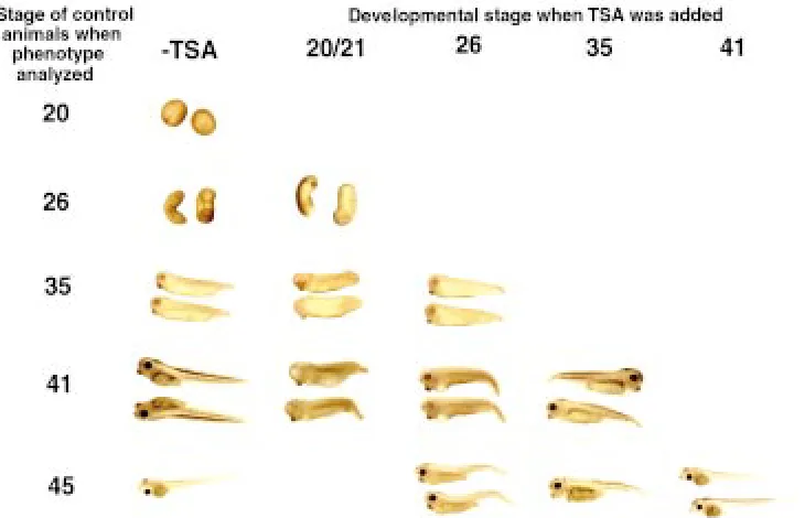
Fig. 2. Blocking histone deacetylases leads to stage-dependent developmen-tal defects. TSA was added to embryo or tadpole-rearing water at a concentration of 100 nM at various developmental stages as indicated and phenotypic effects were subsequently assayed when control ani-mals reached indicated stages. All em-bryos within a treatment group developed essentially identical phenotypes (also see Table 1). In general all treatment groups were delayed in their development, and treatment of embryos up to and including stage 26 resulted in developmental arrest and death when control embryos reached feeding stage. All treatments, except when started at stage 41 or later, resulted in kinked tails. Earlier treatments (starting prior to stage 26) also resulted in embryos with eye and head defects, ventral swell-ing, and impaired swimming ability. head and anterior structures. Their expression diminished by tadpole
stromelysin-repressor complexes, Sin3, have been cloned in Xenopus laevis (Wong et al., 1998; Vermaak, et al., 1999). Thus, we first performed whole mount in situ hybridization for both Rpd3 and Sin3 to determine their spatial and temporal expression profiles during embryogenesis (Fig. 1). Consistent with the idea that Rpd3 and Sin3 function together in a deacetylation complex, the expression profiles of both genes largely overlapped throughout embryogenesis. Both genes were found to be abundantly expressed in head and axial structures of embryos from early neurula to tailbud stages (Fig. 1 A,B for Rpd3, and Fig. 1 E,F for Sin3, respectively). Expression in anterior and axial structures was within the area of the embryo where most differentia-tion was occurring (as opposed to the medial and ventral endoderm where there were relatively few differentiating tissues at these stages). By hatching stage, when the differentiation of many axial structures was near completion, the levels of both Rpd3 and Sin3 in the dorsal axis decreased (Fig. 1 C,G). Similarly, as feeding begins and the tadpole entered a growth rather than differentiation stage of development, expression of both Rpd3 and Sin3 further decreased
(Fig. 1 D,H). Thus, histone deacetylase activity is likely to be playing an important role in regulating genes necessary for tissue differentia-tion and morphogenesis, especially head and axial development.
Inhibition of histone deacetylase activity causes stage-de-pendent developmental abnormalities
To investigate the function of histone deacetylases in embryogen-esis, we took advantage of the fact that one can inhibit this family of enzymes with chemical inhibitors. In particular, the drug trichostatin A (TSA), has been shown to specifically inhibit histone deacetylases both in vitro and in vivo (Yoshida et al., 1990, 1995; Finnin et al., 1999). Addition of TSA to embryo rearing water led to global and stage-dependent defects in embryos and tadpoles (Fig. 2). Notably most or all embryos within a treatment group developed identical phenotypes (Table 1). All embryos were delayed in their develop-ment. In addition, when treatment began prior and up to stage 35, developmental arrest occurred shortly and thereafter the embryos died (Table 1). Treatment with TSA at very early, pre-gastrulation
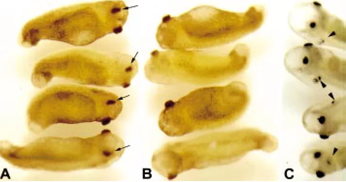
Fig. 3. TSA treatment prior to and at stage 20 results in severe head and eye defects.
Embryos treated with 100 nM TSA beginning at stage 18 displayed severe head and tail defects when examined when control sibling reached stages 35 (A and B) and 41 (C). The two sides of the same four embryos were pictured in A and B to demonstrate the pres-ence of only one pigmented structure, pre-sumable the rudiment of the eye, on only one side of the embryo (A, arrow) but not on the other (B). Note also the irregularly shaped swollen head, protruding cement gland, swol-len tail tip, and the lack of a prominent tail fin. By stage 41 (C), the swelling in the head is more pronounced and in addition to the single pigmented area present in earlier embryos, often an additional pigmented structure devel-ops on the same side (arrowhead).
TABLE 1
PHENOTYPES OF EMBRYOS TREATED WITH TSA AT VARIOUS DEVELOPMENTAL STAGES
Developmental TSA added stage 20/21 TSA added stage 26 TSA added stage 35 TSA added stage 41
stage of control embryos
26 50/50 largely normal
35 49/50 50/50
abnormal head and eye development; eye and cement gland abnormal; swollen thorax; cement gland structure and position; kinked, short tail kinked, short tail
41 49/50 48/50 40/45
delayed development; single pigmented area (eye?); delayed development; eyes abnormal; delayed development;
cement gland structure and position abnormal; cement gland structure and position abnormal; slight swollen head; swollen thorax; swollen head, thorax, ventral mesoderm; kinked, short tail swollen head, thorax, ventral mesoderm; kinked short tail
kinked, short tail 5/45 normal except for kinked, short tail
45 41/50 47/50 34/45 37/40
development arrest – death within 2 days delayed development; delayed development; normal head structures better; swollen cranium and swollen thorax; kinked short tail
thorax; kinked, short tail 10/45
normal except for kinked, short tail
47 41/50 25/45 30/40
development arrest – death within 3-4 days development arrest – death within 3-4 days normal 11/45 normal
stages led to severe developmental defects, including improper induction/formation of the axis, head and mesoderm (data not shown, Almouzni et al., 1994). All treatments initiated between stage 20 and stage 35 resulted in kinked tails, eye and head defects, and ventral swelling and edema. Eye and head defects were most severe in embryos treated at very early stages (stage 20, Table 1 and Fig. 2) and included the presence of only one pigmented structure (Fig. 3), presumable remnants of the eye, on only one side of the embryo (3A) but not on the other (3B). They also had deformed and swollen heads, protruding cement glands, and swollen tail tips (Fig. 3). When control siblings reached stage 41, the swelling in the heads of the treated embryos was more pronounced, and a second pigmented spot developed on the same side of the embryo (Fig. 3C). In general, these TSA-treated embryos were also characterized by ventral swellings, a ventral arching of the entire body axis, lack of a prominent tail fin, and poor or no heart development.
When TSA treatment began at stage 26-35, the embryos developed largely normal heads, eyes, and axes, but had delayed overall development. All embryos treated with TSA developed kinked tails. Most appeared to be arrested at stage 34-39 and died about 24 h after control embryos reached feeding stage (stage 45) or shortly after. Compared to TSA treatment beginning at stage 26, when TSA treatment began at stage 35, the severity of the tail bending was less and decreased further as the animal developed to about stage 45 (Fig. 2 and Table 1).
When treatment began at stage 41, the animals developed normally up to stage 47, without any noticeable morphological differences compared to control animals (Fig. 2 and Table 1), although they too died within 4 days of treatment (Table 1). On the other hand, TSA treatment of postfeeding stage (stage 45) tadpoles, e.g., at stage 55, for up to a few weeks had no observable effects on the animals. Thus, histone deacetylases are important for embryo-genesis and organoembryo-genesis but not essential for tadpole growth.
TSA treatment causes numerous tissue defects in embryos
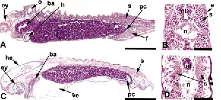
To examine the tissue defects underlying the gross morphologi-cal abnormalities, embryos were treated with TSA at stage 20 and fixed for histological analysis when control embryos reached hatching (stage 35). In contrast to control embryos (Fig. 4 A,B)
numerous internally defects were found as a result of TSA treat-ment (Fig. 4 C,D). Obvious defects included edemas and swellings in the head, tail and ventral region (Fig. 4C) and the poor organi-zation or differentiation of a number of tissues including the eyes, somites, and heart, branchial arches, tail fin. The swelling in the head and tail appeared to be due to a swelling within the anterior and posterior neural tube. The proctodeal opening and channel (Fig. 4 A,C) were also abnormal in the TSA treated embryos and appeared to, in part, result in the ventral bending of the tail seen at this stage. Major axial structures such as the notochord, neural tube and somites were present in TSA treated embryos, but their organization and morphology was abnormal (Fig. 4 B vs. D).
Tissue specific downregulation of matrix metalloproteinase gene stromelysin-3 by TSA is associated with the formation of a sharp bend (kink) in the tail
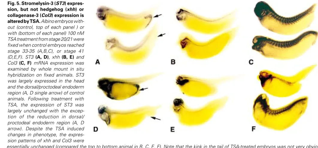
TSA is a highly specific drug against histone deacetylases (Yoshida et al., 1990, 1995). Thus, we reasoned that the morphological defects caused by TSA were likely due to specific changes in developmental gene expression. The lack of known TSA regulated genes in devel-opment prompted us to take a candidate gene analysis approach. Given the major defects in the head and axial structures, we focused on genes that are expressed in these regions and are likely involved in morphogenesis. We previously showed that the matrix metalloproteinase genes collagenase 3 (Col3) and stromelysin-3 (ST3) and the morphogen sonic hedgehog (xhh) were expressed in these regions during embryogenesis (Damjanovski et al., 2000 and unpublished observation). As these genes are likely involved in the development of these structures, we analyzed their expression by whole mount in situ hybridization on control and TSA treated em-bryos at stages 35 (Fig. 5A-C) and 41 (Fig. 5 D-F) (referring to the stages of control sibling animals). ST3 was found to be strongly expressed in the head and in the dorsal/proctodeal endoderm region (Fig. 5A, arrow, top animal) of the untreated embryo while Col3 mainly in the axial structures (Fig. 5C, top animal). The morphogen xhh was also found to be expressed in the head and the around the notochord (Fig. 5B, top animal). Following treatment with TSA the expression of ST3 was greatly downregulated in the dorsal/proctodeal endoderm region of the tail (Fig. 5A, arrow, bottom animal), although
Fig. 4. TSA treatment causes numerous tissue defects in embryos. Embryos treated without (control) or with TSA begin-ning at stage 20 were fixed when control embryos reached stage 35 and sectioned for histological examination. (A,C) Longitu-dinal and (B,D) cross sections were exam-ined of control (A,B,) and TSA treated (C,D) embryos. (A,B) Control embryos revealed well defined structures such as eye (ey), branchial arches (ba), otic vesicle (o), heart (h), well organized somites (s), tail fin (f), and narrow proctodeal channel (pc). (C)
it remained essentially unchanged in the rest of the embryo (Fig. 5A, bottom animal). On the other hand, despite the drastic embryonic deformation caused by TSA, the expression of xhh (Fig. 5B, bottom animal) and Col3 (Fig. 5C, bottom animal) was largely unaffected. This tissue specific downregulation of ST3 by TSA persisted up to stage 41 (Fig. 5 D, E, F for ST3, xhh, Col3, respectively), after which ST3 expression was repressed during normal development (Patterton et al., 1995).
To confirm the inhibition of ST3 expression in embryonic tail by TSA treatment as seen by whole mount in situ , semi-quantitative RT-PCR was then performed to compare the levels of ST3 expres-sion in stage 35 total embryos or tail region from control embryos and embryos treated with TSA from stage 20 (Fig. 6). Overall ST3 mRNA levels in the total embryo was not affected by TSA, however, there was a dramatic decrease in ST3 expression in the tail, in complete agreement with the findings of the whole mount in situ hybridization. Analysis of a control gene, the ribosomal protein L8 or rpl8, showed that an equal amount of total RNA was present in the control and TSA treated samples. Together, these results showed that the downregulation of ST3 expression in the tail region prior to stage 41 was temporally associated with TSA-induced bending in the tail. After stage 41, ST3 was downregulated in control tadpoles (Patterton et al., 1995), suggesting that it was no longer required for normal development. Consistently, TSA treat-ment at or after stage 41 did not cause tail bending (Fig. 2, Table 1, and data not shown).
Discussion
Embryogenesis involves a complex regulation of gene expres-sion in a tissue and developmental stage-dependent manner. To accomplish this, diverse families of transcriptional regulatory pro-teins are employed to activate or repress gene transcription in
various regions/tissues of the embryos. The organization of eucaryotic genome into complex yet poorly understood chromo-some argues that the nature of the chromatin will undoubtedly be an important factor in influencing the interactions of these regula-tors with their target genes. It has been well established that during development in many species, the structural proteins within the chromosome are known to change in both composition and chemi-cal modification (Poccia, 1986; Wolffe, 1991). As the structure backbone of the chromatin, histones play a critical role in develop-mental gene regulation. Histone acetylation is known to change during Xenopus embryogenesis (Dimitrov et al., 1993). Such changes are likely determined by the action of histone
Fig. 5. Stromelysin-3 (ST3) expres-sion, but not hedgehog (xhh) or collagenase-3 (Col3) expression is altered by TSA. Albino embryos with-out (control, top of each panel ) or with (bottom of each panel) 100 nM TSA treatment from stage 20/21 were fixed when control embryos reached stage 33-35 (A,B,C), or stage 41 (D,E,F). ST3 (A, D), xhh (B, E) and Col3 (C, F) mRNA expression was examined by whole mount in situ hybridization on fixed animals. ST3 was largely expressed in the head and the dorsal/proctodeal endoderm region (A, D single arrow) of control animals. Following treatment with TSA, the expression of ST3 was largely unchanged with the excep-tion of the reducexcep-tion in dorsal/ proctodeal endoderm region (A, D arrow). Despite the TSA induced changes in phenotype, the expres-sion patterns of xhh and Col3 were
essentially unchanged (compared the top to bottom animal in B, C, E, F). Note that the kink in the tail of TSA-treated embryos was not very obvious compared to that in Fig. 2. This is due to the whole mount in situ procedure. The protease digestions, high temperatures, and dehydration process caused all embryos to be dorsally curved, thereby diminishing the kinked tail phenotype associated with TSA.
acetyltransferases and histone deacetylases. However, the devel-opmental functions of the changes in histone acetylation are unclear. We have provided here two complementary pieces of evidence to support stage and tissue-dependent roles for histone deacetylases during vertebrate development. First, we have shown a correlation of the developmental expression profiles of histone deacetylase Rpd3 and its associated transcriptional corepressor Sin3 with development. Second, we have directly demonstrated a requirement for histone deacetylases during embryogenesis with inhibitor studies.
The temporal and spatial expression of Rpd3 and Sin3 corre-lates well with the observed defects by TSA treatment. For exam-ple, both Rpd3 and Sin3 are highly expressed in the anterior end or the head of the embryo at early stages. The addition of TSA to rearing water of early embryos has a profound effect on develop-ment in nearly 100% of the embryos. TSA treatdevelop-ment before gastrulation results in severe developmental abnormalities during which few head and axis differentiation events occur (data not shown). Addition of TSA at stage 20 results in deformations, which include prominent head swelling and ventral tail bending. On the other hand, such embryos and those treated with TSA at stage 26 do develop extensively, albeit at a slower rate. Their development is, however, arrested at the tailbud stages. These animals never appear to go past a stage which appears to be around stage 34-39 (as based on tail elongation) even after control sibling reach stage 47. After stage 41, the expression of both Rpd3 and Sin3 de-creases. Correspondingly, the animals can develop further in the presence of TSA, although all eventually die as well.
It should be pointed out that TSA may influence the function of proteins other than Rpd3 to affect embryogenesis. TSA is a highly specific inhibitor for histone deacetylases. Thus, its effects on embryonic development are likely through affecting histone deacetylases. On the other hand, multiple histone deacetylases are known to exist in vertebrates (Ng and Bird, 2000), it is quite possible that TSA inhibition of deacetylases other than Rpd3 also contributes to the observed effects. Studies of these other histone deacetylases, once they are cloned in frogs, should help to clarify their developmental roles.
Detailed analysis of the TSA-induced defects revealed that different regions/tissues of the embryo have different developmen-tal requirements for histone deacetylases. For example, TSA head and eye development requires histone deacetylase activity during early period of development (up to stage 26), while tail develop-ment appears to require the action of histone deacetylases up to stage 35. It is unclear how TSA treatment leads to such defects. It may alter early induction processes, such as the induction of lens differentiation by neural tissue on ectoderm (Zygar et al., 1998, Streit and Stern 1999). Molecularly, TSA is likely to regulate the expression of genes responsible for morphogenetic and remodeling events. It is very likely that one or more gene regulation steps downstream of histone deacetylases are involved to produce the observed morphological defects. Our analysis has provided evi-dence that one candidate gene important for tail development encodes the matrix metalloproteinase stromelysin-3.
Our previous studies have identified several candidate genes that are important for development during early tailbud stages. These include the matrix metalloproteinase genes stromelyin-3 (ST3) and collagenase–3 (Col3), and the morphogen sonic hedge-hog (xhh) (Stolow and Shi, 1995; Damjanovski et al., 2000, and unpublished observation). Whole mount in situ analyses of the
expression of these genes suggested that they are unlikely to be involved in TSA-induced embryonic defects with the exception of tail development. ST3 but not Col3 or xhh is expressed in the dorsal/proctodeal endoderm region of the tail, adjacent to the region where the kink forms upon TSA treatment. The downregulation of ST3 expression by TSA is thus correlated both spatially and temporally with the formation of the kinked tail. Further support for a role of premature downregulation of ST3 in TSA-induced tail kinking comes from the fact that TSA does not cause tail bending when added to embryo rearing water at or after stage 41, when ST3 is already downregulated even in control embryos (Patterton et al., 1995). As a matrix metalloproteinase, ST3 presumably participate in tail development by remodeling the extracellular matrix.
The exact mechanism by which TSA affects embryogenesis and ST3 gene expression is unclear. TSA is likely to influence ST3 expression through multiple gene regulation steps, possibly involv-ing the upregulation of a transcriptional repressor of ST3 gene. Several lines of evidence point to the involvement of histone deacetylases. First, TSA is a highly specific inhibitor of histone deacetylases (Yoshida et al., 1990, 1995). Second, at least one histone deacetylase, Rpd3, and its associated transcriptional corepressor Sin3 are highly expressed in regions where TSA has the greatest effects. Third, the downregulation of ST3 by TSA also occurs in a region where Rpd3 is expressed. The fact that ST3 is not affected in other parts of embryo indicates that the regulation of gene expression by TSA is also tissue-dependent, consistent with the observation that TSA causes developmental abnormali-ties in a tissue and stage-dependent manner.
The embryonic abnormalities that result from TSA treatment are difficult to interpret due to complex developmental interactions among different organs/tissues. The histological analysis of the TSA treated embryos shows poor notochord and neural develop-ment. These structures are themselves responsible for the proper induction and differentiation and patterning of other tissues, such as the neural crest, somites, etc. The future challenge is to identify the early events and the corresponding genes that are responsible for mediating the effects of TSA. In this regard, the possibility of manipulating the amphibian embryo means that this model will continue to be valuable for not only investigating the role of histone acetylation/deacetylation in regulating developmental gene ex-pression, but also dissecting out the functions of genes that are important for vertebrate development and the corresponding mecha-nisms.
Materials and Methods
In vitro fertilization of Xenopus eggs and Trichostatin-A treatment
Whole mount in situ hybridization
The protocol used was essentially that of Harland (1991). Briefly, albino Xenopus embryos were fixed with paraformaldehyde, rinsed with PBS and treated with proteinase K. Following refixation with paraformaldehyde, embryos were acetylated with acidic anhydride and prehybridized in hybridi-zation solution. Full length clone containing Xenopus Rpd3 (Wong et al., 1998), Sin3A (Vermaak, et al., 1999), ST3 (Patterton et al., 1995), Col3 (Brown et al., 1996), or hedgehog (Stolow and Shi 1995) cDNA in pBluescript ks(-) vector (Stratagene) was linearized with appropriate restriction en-zymes. The DNA was transcribed with T3 or T7 RNA polymerase using a Roche Genius 4 kit to generate digoxigenin labeled antisense and control, sense RNA probes. Hybridization was carried out at 60ºC overnight followed by high stringency washes with 0.2% SSC, 0.1% CHAPS at 60ºC (Damjanovski et al., 2000). An alkaline phosphatase-conjugated digoxigenin antibody was used for colorimetric reaction using NBT and BCIP. Embryos were then post-fixed in Bouin’s, dehydrated with methanol and cleared with benzyl benzoate and benzyl alcohol for photography.
Histological analysis
Embryos were fixed for 2 h with 4% paraformaldehyde in 1X PBS. The embryos were dehydrated through an ethanol series and xylene and then embedded into paraplast wax (Sigma). Following sectioning at 7 µm thickness, samples were left on a 42°C slide warmer overnight. They were then de-waxed in xylene and rehydrated through an ethanol series into water. The samples were stained 5 minutes in each Mayer’s hemotoxylin (Sigma) followed by Eosin Y (Sigma). They were dehydrated again with ethanol and xylene and finally mounted with Permount for photography.
RNA extraction and RT PCR
RNA from the whole embryos, or the 25% most posterior section (the tail) was extracted using RNAzol –B (Teltest Inc.) according to the manu-facturer’s instructions. Following resuspension in DEPC treated water and UV quantitation, reverse transcription was preformed using 2g total RNA. Briefly, RNA and a specific primer (the reverse primer, see below) for stromelysin-3 (ST3, Patterton et al., 1995) and another one for the control gene, the ribosomal protein L8 or rpl8 (Shi and Liang, 1994) were annealed in 10 µl at 65°C for 5 min followed by cooling down to room temperature. A mixture (10 µl) contained 5x first strand buffer (4 µl, Gibco-BRL), DTT (2 µl, 0.1 M, Gibco-BRL), dNTP mix (1 µl, 25 mM each, Pharmacia), RNAsin (0.1
µl, 10 U/µl, Gibco-BRL) and reverse transcriptase SuperScript™II (0.5 µl, 200 U/µl, Gibco-BRL) was added to the annealed RNA and primer solution. The mixture was incubated for 1 h at 42°C. Two µl of the resulting cDNA solution were used for PCR in 50 µl with 10x Ex Taq buffer (5 µl, Takara, Intergene), dNTP mix (8 µl, 2.5 mM each, Takara, Intergene), four primers (reverse and forward primers for ST3 and rpl8, 2 µl of 2 µM each, Gibco-BRL) and Ex Taq polymerase (0.5 µl, 5 U/µl, Takara, Intergene). PCR was performed for 30 cycles consisting of 94°C for 30 seconds, 55°C for 30 seconds and 72°C for 30 seconds. PCR using RNA without reverse transcription were performed was done as a control for genomic DNA contamination (data not shown). PCR products (10 µl) were electro-phoresed on 2% agarose gels and stained with ethidium bromide. The reverse and forward primers are : for ST3, 5’ GTTCATCCTGGAAAGCAG 3’ (1503 to 1482) and 5’ CCTGATGCATGCAAAACT 3’ (1038 to 1055, Patterton et al., 1995) ; for rpl8, 5’ GACGACCAGTACGACGA 3’ (749 to 732) and 5’ AAAGAGAAACTGCTGGC 3’ (586 to 602, Shi and Liang, 1994), respectively.
References
ALMOUZNI, G., KHOCHBIN, S., DIMITROV, S. and WOLFFE, A.P. (1994). Histone acetylation influences both gene expression and development of Xenopus laevis. Dev. Biol. 165: 654-669.
BROWN, D.D., WANG, Z., FURLOW, J.D., KANAMORI, A., SCHWARTZMAN, R.A., REMO, B.F. and PINDER, A. (1996). The thyroid hormone-induced tail resorption program during Xenopus laevis metamorphosis. Proc. Natl. Acad. Sci. USA. 93: 1924-9.
DAMJANOVSKI, S., PUZIANOWSKA-KUZNICKA, M., ISHUZUYA-OKA, A. and SHI, Y.B. (2000). Differential regulation of three thyroid hormone-responsive matrix metalloproteinase genes implicates distinct functions during frog embryogenesis. FASEB J. 14: 503-10.
DIMITROV, S., ALMOUZNI, G., DASSO, M. and WOLFFE, A.P. (1993). Chromatin transitions during early Xenopus embryogenesis: changes in histone H4 acetyla-tion and in linker histone type. Dev. Biol. 160: 214-27.
FINNIN, M.S., DONIGIAN, J.R., COHEN, A., RICHON, V.M., RIFKIND, R.A., MARKS, P.A., BRESLOW, R. and PAVLETICH, N.P. (1999). Structure of a histone deacetylase homologue bound to the TSA and SAHA inhibitors. Nature 401: 188-193.
GRUNSTEIN, M. (1997). Histone acetylation in chromatin structure and transcription. Nature 389: 349-352.
HARLAND, R.M. (1991). in situ hybridization: an improved whole-mount method for Xenopus embryos. Methods Cell Biol. 36: 685-695.
HEBBES, T.R., THORNE, A.W. and CRANE-ROBINSON, C. (1988). A direct link between core histone acetylation and transcriptionally active chromatin. EMBO J. 1988 7: 1395-1402.
HEINZEL, T., LAVINSKY, R.M., MULLEN, T.M., SODERSTROM, M., LAHERTY, C.D., TORCHIA, J., YANG, W.M., BRARD. G., NGO, S.D., DAVIE, J.R., SETO, E., EISENMAN, R.N., ROSE, D.W., GLASS, C.K. and ROSENFELD, M.G. (1997). A complex containing N-CoR, mSin3 and histone deacetylase mediates transcrip-tional repression. Nature 387: 43-48.
HONG, L., SCHROTH, G.P., MATTHEWS, H.R., YAU, P. and BRADBURY, E.M. (1993). Studies of the DNA binding properties of the histone H4 amino terminus. J. Biol. Chem. 268: 305–314.
IKEGAMI, S., OOE, Y., SHIMIZU, T., KASAHARA, T. TSURUTA, T., KIJIMA, M., YOSHIDA, M. and BEPPU, T (1993). Accumulation of multiacetylated forms of histones by trichostatin A and its developmental consequences in early starfish embryos. Roux’s Arch. Dev. Biol. 202: 144-151.
LEE, D.Y., HAYES, J.J., PRUSS, D. and WOLFFE, A.P. (1993). A positive role for histone acetylation in transcription factor access to nucleosomal DNA. Cell 72: 73-84.
NAGY, L., KAO, H.Y., CHAKRAVARTI, D., LIN, R.J., HASSIG, C.A., AYER, D.E., SCHREIBER, S.L. and EVANS, R.M. (1997). Nuclear receptor repression medi-ated by a complex containing SMRT, mSin3A, and histone deacetylase. Cell 89: 373-380.
NG, H.H. and BIRD, A. (2000). Histone deacetylases: silencers for hire. Trends Biochem. Sci. 25: 121-126.
NIEUWKOOP, P.D. and FABER, J. (1956). Normal table of Xenopus laevis. North Holland Publishing, Amsterdam.
NORTON, V.G., IMAI, B.S., YAU, P. and BRADBURY, E.M. (1989). Histone acetyla-tion reduces nucleosome core particle linking number change. Cell 57: 449-457.
PATTERTON, D., HAYES, W.P. and SHI, Y.-B. (1995). Transcriptional activation of the matrix metalloproteinase gene stromelysin-3 coincides with thyroid hormone-induced cell death during frog metamorphosis. Dev. Biol. 167: 252-262.
PAZIN, M.J. and KADONAGA, J.T. (1997). What’s up and down with histone deacetylation and transcription? Cell 89: 325-328.
POCCIA, D. and COLLAS, P. (1996). Transforming sperm nuclei into male pronuclei in vivo and in vitro. Curr. Top. Dev. Biol. 34: 25-88.
POGO, B.G., ALLFREY, V.G. and MIRSKY, A.E. (1966). RNA synthesis and histone acetylation during the course of gene activation in lymphocytes. Proc. Natl. Acad. Sci. USA. 55: 805-812.
SHI, Y.B. and LIANG, V.C. (1994). Cloning and characterization of the ribosomal protein L8 gene from Xenopus laevis. Biochim Biophys Acta. 1217: 227-8.
STOLOW, M.A. and SHI, Y.B. (1995). Xenopus sonic hedgehog as a potential morphogen during embryogenesis and thyroid hormone-dependent
metamor-phosis. Nucleic Acids Res. 23: 2555-62.
STRAHL, B.D. and ALLIS, C.D. (2000). The language of covalent histone modifica-tions. Nature 403: 41-45.
STREIT, A. and STERN, C.D. (1999). Neural induction. A bird’s eye view. Trends Genet. 15: 20-24.
STRUHL, K. (1998). Histone acetylation and transcriptional regulatory mechanisms. Genes Dev. 12: 599-606.
Functional analysis of the SIN3-histone deacetylase RPD3-RbAp48-histone H4 connection in the Xenopus oocyte. Mol Cell Biol. 19: 5847-60.
VETTESE-DADEY, M., GRANT, P.A., HEBBES, T.R., CRANE- ROBINSON, C., ALLIS, C.D. and WORKMAN, J.L. (1996). Acetylation of histone H4 plays a primary role in enhancing transcription factor binding to nucleosomal DNA in vitro. EMBO J. 15: 2508-2518.
WADE, P.A. and WOLFFE, A.P. (1997). Histone acetyltransferases in control. Curr Biol. 7: R82-R84
WOLFFE, A.P. (1991). Xenopus transcription factors: key molecules in the develop-mental regulation of differential gene expression. Biochem J. 278 (Pt 2): 313-24.
WOLFFE, A.P. and PRUSS, D. (1996). Targeting chromatin disruption: Transcription regulators that acetylate histones. Cell 84: 817-819.
WONG, J., PATTERTON, D., IMHOF, A., GUSCHIN, D., SHI, Y.B. and WOLFFE, A.P.
(1998). Distinct requirements for chromatin assembly in transcriptional repression by thyroid hormone receptor and histone deacetylase. EMBO J. 17: 520-34.
YOSHIDA, M., HORINOUCHI, S. and BEPPU, T. (1995). Trichostatin A and trapoxin: novel chemical probes for the role of histone acetylation in chromatin structure and function. Bioessays 17: 423-30.
YOSHIDA, M., KIJIMA, M., AKITA, M. and BEPPU, T. (1990). Potent and specific inhibition of mammalian histone deacetylase both in vivo and in vitro by trichostatin A. J. Biol. Chem. 265: 17174-9.
ZYGAR, C.A., COOK, T.L. and GRAINGER, R.M. Jr. (1998). Gene activation during early stages of lens induction in Xenopus. Development. 125: 3509-19.