Mechanisms of Aggresome Biogenesis: Ubiquitination, Transport, & Maintenance
AJ Keefe
Department of Biology, Big Bend Community College Moses Lake, Washington, 98837
Email: [email protected]
Keywords: Proteostasis, neurodegeneration, aggresome, HDAC6, p62, BAG3
Pages: 20
Body Word Count: 6170
Body Character Count Excluding Spaces: 37230 Figures: 3
Citations: 123
The author has declared no conflicts of interest The author received no funding for article preparation
HIGHLIGHTS:
● Perinuclear aggresomes serve a critical role in maintaining proteostasis.
● Lysine 63 linked polyubiquitination modifications direct misfolded proteins to the aggresome.
● HDAC6 and BAG3 are the primary adaptors for retrograde transport of misfolded proteins.
● Aggresomes are constructed by p62, which also mediates its autophagic clearance.
● Undegradable aggresomes aggravate protein misfolding and can cause other pathologies.
ABBREVIATIONS
Microtubule Organizing Center, MTOC; Peripheral Microaggregates, PMA; Parkinsons Disease, PD; Amyotrophic Lateral Sclerosis, ALS; Alzheimer's Disease, AD; Cystic
Fibrosis Transmembrane Receptor, CTFR;
Ubiquitin-Proteasome System, UPS; Lysine 63, K63; Bovine Serum Albumin, BSA; Defective Ribosomal Products, DRiPs; Deubiquitinase, DUB
ABSTRACT
Neurodegenerative diseases are universally marked by the accumulation of misfolded protein. Neurons respond to these proteostatic disturbances by sequestering, and thus inactivating, toxic misfolded proteins into a perinuclear organelle called the aggresome. The aggresome can be subsequently degraded in bulk by autophagy, a process termed aggrephagy. The formation of protein aggregates has historically been considered a spontaneous and unregulated process, but emerging research has instead discovered a diverse cohort of regulatory proteins that mediate protein aggregation. Chaperones are the first proteins to respond to misfolded proteins, and do so by recognizing the aberrant exposure of hydrophobic domains. When chaperones are unable to correctly refold proteins, their substrates are transferred to ubiquitin ligating machinery to catalyze polyubiquitination. Although ubiquitin chains typically direct proteins towards proteasomes, severe proteotoxic stress can overwhelm, or even directly inhibit, proteasomes. As an alternative to proteasomal degradation, misfolded proteins are redirected towards the mitotic organizing center (MTOC) and, following retrograde transport by dynein, are packaged and sequestered within an intermediate filament (IF) cage to form the aggresome. The biogenesis of the aggresome is thus a highly regulated event, and a better understanding of the mechanisms facilitating this process will provide critical insight into neurodegenerative disease.
____________________________________________________
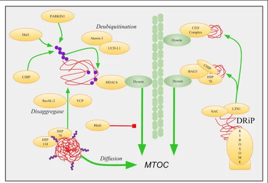
Figure 2 Retrograde Transport Of Peripheral Microaggregates. PMAs (red) are recognized by folding chaperones like HSP70 and HSP110. E3
ubiquitin ligases CHIP, PARKIN, and Hrd1 catalyze K63 linked ubiquitination (purple) of aggregates. VCP and RuvbL1/2 extract misfolded
proteins from larger aggregates. Deubiquitinases ataxin-3 and UCH-L1 cleave ubiquitin linkages to recruit and activate HDAC6, which then
mediates dynein retrograde transport. DRiPs are also an important source of misfolded protein, and are prepared for retrograde transport by the
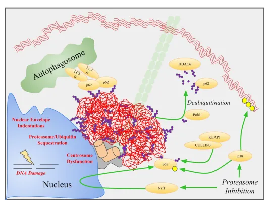
Figure 3 Aggresome Formation, Disposal, and Dysfunction. Aggresomes (red) are coated and actively aggregated by p62, which also recruits
LC3-II to promote aggrephagy. Deubiquitinases like Poh1 cleave K63 ubiquitin chains (purple) to recruit p62 and HDAC6. Proteasome inhibition
mediates this pathway by upregulating p62 through the Nrf1 transcription factor, and by p38 activation. P62 is subsequently activated by p38
phosphorylation (yellow) and KEAP1 ubiquitination. Aggresomes that fail to be degraded can cause centrosome dysfunction, DNA damage,
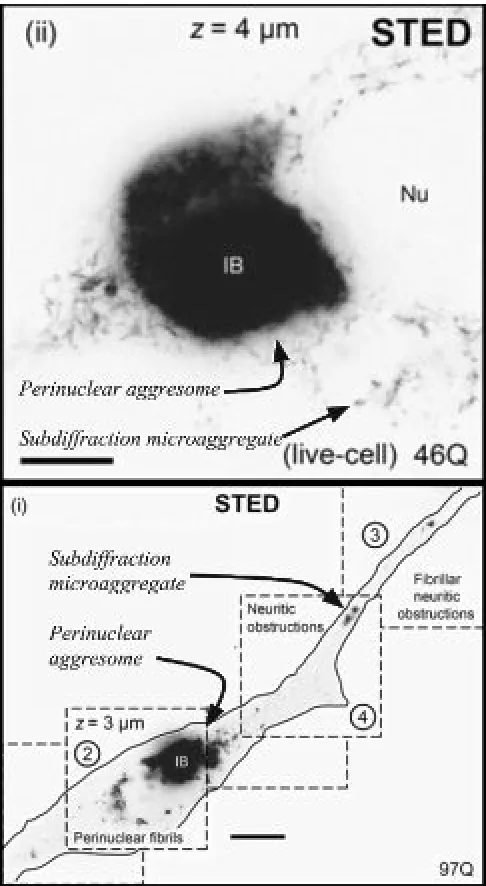
Figure 1 Subdiffraction Microscopy Reveals PMAs. Adapted from Delayed emergence of subdiffraction sized mutant
huntingtin fibrils following inclusion body formation by SJ Sahl
et al. 2016. Quarley Reviews of Biophysics. Nu =Nucleus. IB =
Inclusion Body. STED = Stimulated Emission Depletion. Images
Introduction
Protein folding occurs as the result of complex molecular interactions between amino acid side chains. However, subtle mutations in the genetic code, irregular biochemical conditions, or disturbances in the proteostatic networks that maintain protein integrity, can lead to the misfolding of protein (Reviewed in Balchin et al, 201 6) . The accumulation of soluble misfolded proteins can devastate cellular functioning through aberrant nonspecific interactions. Some of these interactions, mainly between exposed hydrophobic residues, can promote the polymerization of misfolded protein species into insoluble microaggregates. These peripheral microaggregates, or PMAs, are inefficiently disposed of by proteasomes, which require linearized peptide chains. Additionally, the sheer number of microaggregates may overwhelm the autophagic capacity of a cell. In order to mitigate these toxicities cells have developed regulated mechanisms of coalescing PMAs into relatively inert and benign membraneless organelles called aggresomes.
Aggresomes are transient perinuclear organelles formed from the piecemeal addition of smaller aggregates that can be degraded in bulk by aggrephagy. When this program is effectively activated, cells that are exposed to proteotoxic stressors, such as proteasome inhibition, misfolded protein overexpression, or autophagic deficiencies, will gradually accumulate perinuclear aggresomes at the base of microtubules called the Microtubule Organizing Center (MTOC) in a dynein-dependent manner (Johnston et al, 2002). Defects in aggresome biogenesis prevent the efficient disposal of PMAs, leading to their accumulation in axons and potentially obstructing axoplasmic transport or rupturing membranes ( Delay et al, 2014 Sahl et al, 2016 Relini et al, 2013 ). These challenges are compounded in neurons that, due to their post-mitotic nature, are unable to dilute misfolded proteins through cell division. In contrast to defects in aggresome formation, defects in aggresome
disposal typically manifest as persistent perinuclear aggresomes that may lead to DNA damage, nuclear membrane defects, and centrosome dysfunction. Despite this, aggresome formation is generally believed to confer significant cytoprotection, and thus the mechanisms required for its formation are critical for cell survival during proteostatic stress.
The majority of research into the mechanisms of aggresome biogenesis and disposal have been carried out in cell culture, and thus the physiological and disease relevance of such structures are not certain. Aggresomes are principally identified by the formation of a cell-specific intermediate filament (IF) cage, most commonly consisting of vimentin, at the location of the centrosome that encloses the proteinaceous aggresome. In composition, aggresomes are enriched in ubiquitin, folding chaperones (HSP70), and autophagic markers (p62). If the inclusion bodies observed in neurodegenerative disease share these characteristics, that is, a perinuclear IF enclosed aggregate enriched in ubiquitin, folding chaperones, and p62, then there is a good reason to believe they are indeed an aggresome or at least functionally related. Aggresome-like structures have been characterized in Parkinson's Disease (PD), composed primarily of either alpha-synuclein or PARKIN, Alzheimer's Disease (AD), composed of tau or amyloid beta, Amyotrophic Lateral Sclerosis (ALS), composed of SOD1 or TDP-43, prion diseases, composed of PrP, and in many other protein misfolding diseases (Reviewed in Lim et al, 2016). These various neurodegeneration-linked aggregates share many characteristics, and thus may also share conserved mechanisms of formation that could drastically improve our understanding of the aggregation process.
characterization. However, modern sub-diffraction microscopy methods have allowed researchers to visualize the pre-aggregation process in real time (Figure 1). Perhaps most startling was the discovery that PMAs can form naturally in untreated cells, but are rapidly deconstructed. PMAs composed of alpha-synuclein, for example, are relatively common, even in healthy cells, but their irreversible aggregation only becomes energetically favorable above a critical size, somewhere around 140 nm (Narayanan et al, 2017 ). This suggests that the purported toxicity of PMAs may be related to their solubilized state since microaggregates are generally not stable, although this could be dependent on the aggregating protein species. Theories suggesting solubilized monomeric proteins confer greater cytotoxicity than their aggregated counterpart is a common theme across protein misfolding diseases (Reviewed in Haass & Selko, 2007). Regardless, aggresomes function to mitigate such toxicity, and by understanding the mechanisms by which cells interact, transport, sequester, and dispose of protein aggregates represents a major topic of interest in protein misfolding disorders.
The aggresomal life cycle can be roughly divided into four stages; nucleation and processing of PMAs, retrograde transport, aggresome assembly and maintenance, and autophagic removal (aggrephagy). The initial nucleation of PMAs recruits folding chaperones, such as HSP70, that are often associated with E3 ubiquitin ligases, such as CHIP. Misfolded proteins unable to be refolded are typically polyubiquitinated with lysine 63 linked ubiquitin chains. These ubiquitin chains recruit desegregases, such as VCP, to remodel, expose, or extract ubiquitinated proteins from the microaggregates. The unique K63 linked ubiquitin chains are then recognized by dynein adaptor proteins, most notably HDAC6, for retrograde transport to the MTOC. The regulated nucleation of the aggresome is then facilitated by p62, a protein that binds K63 ubiquitinated protein and homo-oligomerizes. Aggresomes are sequestered in an IF enclosed cage and coated by an extensive assortment of proteostasis-related proteins such as p62, ubiquitin, chaperones, and E3 ubiquitin ligases. These proteins function to maintain
aggresome integrity, limit cytotoxicity, and, eventually, mediate its degradation through aggrephagy and proteasomes.
PERIPHERAL MICROAGGREGATES
Regulated Protein Aggregation by Ubiquitin Systems
multiple E3 ubiquitin ligases that are required for the aggresomal sequestration of certain misfolded protein species.
CHIP and PARKIN are among the two most well-studied E3 ubiquitin ligases that have been linked to aggresome formation. CHIP is commonly recognized for mediating the polyubiquitination of chaperone associated aggregates. Although CHIP polyubiquitinates many proteins, these typically result in proteasomal degradation as opposed to aggresome formation (Reviewed in Joshi et al, 2016 ). However, during proteostatic stress or proteasomal inhibition (induced by the small molecule MG132), CHIP ubiquitination can promote the formation of the aggresome (Figure 2). For example, CHIP promotes the aggregation of inducible Nitric Oxide Synthase, Cystic Fibrosis Transmembrane Conductance Regulator (CFTR), and Ataxin 1 ( Meacham et al, 2001 Choi et al, 2007 ). Similarly, the deletion of CHIP in mice expressing mutant aggregate-prone tau are unable to sequester toxic hyperphosphorylated tau in concentrated perinuclear aggregates, and instead have greatly increased levels of toxic soluble tau (Dickey et al, 2006).
PARKIN is another E3 ubiquitin ligase important in the recognition, ubiquitination, and targeting of PMAs to the aggresome (Figure 2). PARKIN, which can associate with and be activated by CHIP (Imai et al, 2002 ), colocalizes with aggresomes and PMAs, and loss of function mutations result in familial forms of Parkinson's Disease ( Olzmann et al, 2007 ).The loss of PARKIN results in the accumulation of either soluble misfolded protein species or PMAs, both of which are characteristic of defects in the transport of pre-aggresome aggregates (Chung et al, 2001 Yang et al, 2007 ). In addition, PD patients with mutations in PARKIN fail to form characteristic Lewy Bodies suggesting PARKIN is involved in directing alpha-synuclein to aggresomes in human disease ( Johanson et al, 2018 Lim et al, 2005 ). During MG132 induced proteasome inhibition, or other UPS dysfunctions, PARKIN mediated ubiquitination can facilitate the formation of the aggresome and has been demonstrated to direct various proteins including DJ-1, SOD1, synphilin-1, and TDP-43 to the aggresome ( Olzmann et al, 2007 Hebron et al, 2012 Olzmann et al, 2015 Yung et al,
2015). In order to promote aggresome formation, however, PARKIN itself must escape an autoinhibited conformation, which is coordinated by the PINK1 kinase. When localized to depolarized mitochondria, PINK1 is unable to translocate into the mitochondrial matrix, and instead forms a mitochondrial outer membrane-anchored kinase that phosphorylates ubiquitin and PARKIN. PARKIN not only requires PINK1 phosphorylation to escape an autoinhibited conformation but also requires phosphorylated ubiquitin for efficient substrate polyubiquitination (Reviewed in Pickrell et al, 2015 ). Thus PARKIN dependent aggresome biogenesis must also be contingent on the presence of depolarized mitochondria, otherwise PARKIN would, presumably, not be active.
expression of UBC13, an E2 ubiquitin ligase specific for K63 linkages, was capable of shifting PARKIN’s catalytic activity from K48 to K63 ubiquitin linkages (Lim et al, 2013). Interestingly, CHIP has also been shown to bind UBC13 and this promotes the catalysis of K63 ubiquitin chains, but its role in aggresome formation has not been specifically investigated (Zhang et al, 2005 Slotman et al, 2012 ). Although it is then tempting to suggest proteasome inhibition triggers the transcription of aggresome-promoting genes like UBC13, research has suggested that neither transcription or translation affects proteasome inhibition-induced aggresome formation (Meriin et al, 2012 ). Regardless of how proteasome inhibition alters the ubiquitin landscape, the catalysis of K63 ubiquitin linkages alone is still not sufficient to redirect misfolding proteins towards aggresomes, and additional modifications are required.
The retrograde transport of PMAs probably requires some degree of disaggregation or separation from their associated complexes, otherwise these misfolded cargoes may be too physically large for efficient transport. The disaggregation of peripheral aggregates prior to transport is achieved principally by the HSP70 chaperone in association with its cognate cochaperone HSP110 (Figure 2; Rampelt et al, 2012 Reviewed in Nillegoda et al, 2018 ). At least two other proteins have been implicated in this process; VCP and RuvbL1/2 (Figure 2). Both proteins are of the AAA-ATPase family and, in concert with various chaperones, are responsible for the ATP dependent extraction of ubiquitinated proteins from membranes or aggregates, followed by entrance into the proteasome or linkage to dynein for retrograde transport. In a recent high throughput screening experiment by Zaarur et al, the RuvbL1 and RuvbL2 homologs were discovered as two critical proteins in the formation of aggresomes in response to proteasomal inhibition. siRNA mediated knockdown of RuvbL1 and RuvbL2 reduced the percent of MG132 treated HeLa cells with aggresomes from ~80% to ~40%. ( Zaarur et al, 2015 ). The loss of RuvbL1/2 results in the abnormal accumulation of peripheral aggregates, consistent with an inability to retrogradely transport them to the
aggresome. The role of RuvbL1 in aggregate clearance has since been confirmed by a different research group ( Narayanan et al, 2017). A similar phenotype is observed following loss of VCP, an AAA-ATPase that contains the ability to bind ubiquitin (Kitami et al, 2006 ). VCP, which has been particularly well studied for its connection to various protein misfolding disorders, is critical not only for efficient aggresome formation, but also for the UPS, autophagy, and many other ubiquitin-related processes (Reviewed in Boom et al, 2018 ). In light of VCP’s pleiotropic activity, determining exactly how VCP contributes to aggresome formation has been difficult. However, the observation that VCP deleted cells are deficient in aggresome formation in response to stress and are instead riddled with peripheral aggregates, suggests VCP is essential to retrograde transport (Ju et al, 2008 Wojcik et al, 2004 ). This effect may be related to deficiencies in K63 based ubiquitin linkages, as pharmacological VCP inhibition reduces K63 ubiquitin abundance by 50% (Heidelberger et al, 2018 ). VCP probably does not catalyze or edit these ubiquitin chains itself, but instead likely functions to recruit the appropriate enzymes. VCP has indeed been characterized as a kind of ubiquitin “signaling hub”, in part due to its ability to bind numerous ubiquitin ligases and DUBs, including Hrd1 and ataxin-3, and its role in numerous ubiquitin-related processes ( Stach et al, 2017 Meyer et al, 2014). While localized to peripheral microaggregates, VCP and RuvbL1/2 probably function to extract, linearize, and facilitate the remodeling of ubiquitinated cargo in order to prime them for retrograde transport.
MICROAGGREGATE TRANSPORT
HDAC6 & BAG3 Facilitate Retrograde Transport
effectively and consistently blocks aggresome formation (Johnston et al, 2002 ). Misfolded proteins probably do not bind to dynein directly, but are instead linked to dynein through adaptors, most notably HDAC6 (Figure 2). Originally discovered as histone deacetylase, HDAC6 has emerged as a central protein in the formation of aggresomes. By harboring both a ubiquitin and dynein binding domain, HDAC6 functions as a dynein adaptor for the transport of ubiquitinated misfolded proteins towards the MTOC. The importance of this mechanism has been illustrated in multiple models of aggresome formation, and, in general, HDAC6 inhibition or deletion results in ~50% less aggresome positive cells and reduced aggresome size (Kowaguchi et al, 2003 Kalveram et al, 2008 Su et al, 2011 ). Recently, in a mouse model of microglial inflammation induced Tau aggregation, researchers Tseng et al discovered HDAC6 was essential in mediating the aggregation of Tau in vivo ( Tseng et al, 2017 ). The importance of HDAC6’s deacetylase activity in aggresome formation is controversial, but may also promote aggresome formation through the deacetylation of tubulin ( Jiang et al, 2008).
Mechanistic details of how HDAC6 links dynein and ubiquitinated cargo have been complicated by structural observations of HDAC6’s ubiquitin-binding domain, which does not actually bind to anchored ubiquitin chains, but instead binds free ubiquitin moieties by a C-terminal diglycine motif ( Ouyang et al, 2011 Turcu et al, 2006 ). In order to reconcile this contradiction, researchers have proposed the possibility that deubiquitinases could cleave ubiquitin moieties in order to bind and activate HDAC6. The ataxin-3 deubiquitinase was an attractive candidate due to its colocalization with both aggresomes and PMA structures. In addition, ataxin-3 can form a complex with VCP and HDAC6 proteins, and also has an apparent preference for editing K63 linked ubiquitin chains (Burnett et al, 2005 ). Wang et al demonstrated that ataxin-3 can indeed cleave K63 linked ubiquitin moieties to activate proximal HDAC6 proteins, and deletion or mutation of ataxin-3 disrupts aggresome formation (Figure 2; Wang et al, 2011 ). The UCH-L1 deubiquitinase has also been implicated in the activation of
HDAC6, and thus aggresome formation, by cleaving ubiquitin chains on misfolded protein (Yu et al, 2017).
Besides the deubiquitination of HDAC6 substrate, another important regulator of HDAC6 is the nucleocytoplasmic shuttling protein RanBPM. Researchers Salemi et al serendipitously discovered DNA damage induced the nuclear export and accumulation of RanBPM into HDAC6 positive aggresomes (Salemi et al, 2014). Knockdown of RanBPM following etoposide treatment, which induces double-stranded DNA breaks, reduced aggresome positive cells from 38% in controls to 18%, and HDAC6 positive aggresomes from 19% to 6%. Subsequent analysis revealed RanBPM associates with HDAC6 following DNA damage (or proteasome inhibition) and promotes aggresome formation, but it was not clear how RanBMP’s interaction with HDAC6 facilitated this process. Although HDAC6 has received the most attention regarding microaggregate transport, it is important to note that HDAC6 deletion only partially inhibits aggresome formation suggesting there are multiple mechanisms of aggresome transport. Indeed, emerging research has implicated several additional pathways in the transport of misfolded proteins towards the MTOC.
impaired aggresome formation (Fu et al, 2019 ). Following overexpression, BAG3 induced the perinuclear sequestration and eventual autophagic disposal of tau in vivo. BAG3 has been repeatedly linked to aggresome formation and could be therapeutically utilized to either promote, or inhibit, aggresome formation.
BAG3 promotes aggresome formation by functioning as a stress-induced co-chaperone in complex with the HSP70 chaperone. BAG3 binds to the ATPase domain of the HSP70 folding chaperone effectively inhibiting its folding activity and promoting aggresome formation. Pharmacological inhibition of chaperones, including HSP70, does indeed promote aggresome formation (Gedalya et al, 2011 Muchowski et al, 2000 Miyata et al, 2013). By inhibiting HSP70 refolding activity, BAG3 might promote the unloading of HSP70 clients onto dynein for retrograde transport. BAG3 facilitates this process by interacting with dynein, either via a C-terminal proline-rich domain or through the 14-3-3 adaptor protein (Xu et al, 2013). Additionally, BAG3 appears to compete with its homolog BAG1 for the same HSP70 binding domain. By displacing BAG1, which directs HSP70 bound substrate to the proteasome, BAG3 redirects HSP70 substrate to the aggresome ( Minoia et al, 2013 ). Since BAG3 expression is uniquely stress-induced, this may provide a mechanism by which proteostatic stressors like MG132 redirect HSP70 substrate towards aggresomes. BAG3 and HSP70 have also been captured in complex with CHIP, and this interaction was shown to promote aggresome formation (Zhang et al, 2011 ). BAG3 appears to be an important mediator in the retrograde transport of misfolded protein, possibly by bridging HSP70 and CHIP associated substrates to dynein for transport to the aggresomes.
More recently, BAG3 has been implicated in the preferential binding and targeting of defective ribosomal products (DRIPs) to aggresomes (Figure 2). DRiPs are dysfunctional and misfolded peptides associated with aberrant translation, such as the peptides produced during nonsense-mediated decay of mRNA. These defective peptides are normally degraded through the UPS but accumulate during
proteasomal inhibition and, even under basal conditions, represent about 12-15% of all newly synthesized protein ( Wang et al, 2013). In a series of experiments, Meriin et al show that BAG3 binds DRiPs in an HSP70 dependent manner, and the loss of ribosomal quality control machinery nearly completely blocks BAG3/HSP70 dependent aggresome formation (Meriin et al, 2018). For example, shRNA mediated knockdown of the ribosome-associated E3 ubiquitin ligase LTN1, or the ribosome-associated chaperones NACA/NACB, blocked the interaction between DRiPs and BAG3/HSP70 complexes and reduced the number of aggresome-positive cells from ~50% to ~5%. These results suggest that the ribosome quality control proteins may provide the source of misfolded proteins that BAG3/HSP70 recognize for transport to the aggresome. Recent work by Park et al elucidated a similar mechanism, whereby DRiPs are recognized by the CED complex, composed of CTIF, eEF1A1, and the dynein subunit DCTN1, and then retrogradely transported to the aggresome (Figure 2; Park et al, 2017 ). Loss of CTIF resulted in the dispersion of aggregates outside of aggresomes during MG132 treatment, consistent with defects in efficient transport. The recognition of DRIPs by BAG3 or the CED complex, and subsequent targeting to aggresomes, is likely an extension of ribosomal surveillance programs, but specific for the defective protein product derived from the initial translation of aberrant mRNA. Since certain mRNA species, such as those containing expanded CAG repeats, are inefficiently translated, DRiPs could potentially be a major source of misfolded protein in certain neurodegenerative diseases (Jain et al, 2017 Marti, 2016). Besides HDAC6, BAG3, and CED, one report also suggests the dynein subunit NDEL1 can directly bind and transport ubiquitinated cargo to the aggresome (Wan et al, 2012).
typically inactivated during starvation, this may explain how glucose deprivation induces aggresome formation, an effect observed in cardiomyocytes (Marambio et al, 2010 ). Future research will need to carefully assess the relative contribution of each mechanism to the aggresome formation in neurons, especially in animals models of disease.
Recent research by Lu et al has demonstrated, using super resolution microscopy, that polyglutamine aggregates reach the aggresome primarily through simple diffusion rather than active transport (Figure 2; Lu et al, 2018 ). According to their analysis only 11% of peripheral aggregates are colocalized with HDAC6, and only 3% appear to be directionally transported. These discoveries are in stark contrast to studies that have reported an essential role of microtubules and dynein for aggresome formation. Using mathematical models Lu et al propose that the initial nucleation of aggresomes requires active transport, such as those described here, but subsequent aggresome growth is driven by random diffusion. Furthermore, Lu et al discovered small immature aggresomes were primarily composed of newly synthesized protein, supporting theories positing DRiPs as a critical source of aggresome protein. This exciting research highlights the utility of super resolution microscopy and its potential to revolutionize our understanding of cellular processes.
AGGRESOME BIOGENESIS How p62 Aggregates the Aggresome
Once misfolded proteins have been retrogradely transported and delivered to the MTOC, they must be efficiently packaged and sequestered for aggrephagy. The essential role of p62 (SQSTM1) in aggrephagy is well understood, where it functions as an adaptor between LC3, on autophagosomes, and ubiquitin, on aggregates. Known as an autophagic cargo adaptor, p62 serves to localize and bridge the nucleating autophagosome to its substrate (Figure 3). In addition to aggrephagy, accumulating evidence also suggests that p62 is essential for the initial formation of the aggresome. The formation of aggresomes
is dependent on p62 expression, promoted by p62 overexpression, and highly colocalized with p62, illustrating the indispensability of p62 in aggresome formation ( Gal et al, 2007 Bjørkøy et al, 2005Pankiv et al, 2007).
aggresomes. During mitophagy, PARKIN is essential for the mitochondrial localization of both p62 and HDAC6 by generating K63 linked ubiquitin chains, and thus a similar mechanism could occur at aggresomes ( Lee et al, 2010 ). These K63 ubiquitin chains may then be released by various DUBs, which are indeed catalytically active at aggresomes, and thereby allow additional K63 chains to be generated ( Juenemann et al, 2018). For example, Nunduri et al have described an essential role for Poh1, a proteasome-bound deubiquitinase, in aggrephagy, where it functions to cleave and release K63 linked ubiquitin chains that then activate HDAC6, but could also recruit p62 (Figure 3; Nanduri et al, 2015 ). Similarly, the inhibition of other proteasome-associated DUBs by the small molecule b-AP15 induces cancer cell death by preventing cytoprotective aggresome formation, an effect that could be related to deficient p62 or HDAC6 activation (Hillert et al, 2018). It seems plausible, then, that various K63 ubiquitin ligases, in conjunction with deubiquitinases, coordinate the local saturation of K63 ubiquitin chains near aggresomes to recruit p62 en mass.
Recruitment of p62, driven by its UBA domain, is then followed by PB1 domain dependent oligomerization. PB1 domains facilitate protein interactions by binding other PB1 domains thereby promoting protein polymerization. The capacity for p62 to oligomerize is exemplified in autophagy-deficient cells, where the inability to turnover p62 by autophagy leads to its accumulation and aggregation driven by PB1 domain interactions (Wang et al, 2016 Sukseree et al, 2018 ). PB1 dependent oligomerization is critical for p62’s ability to stably bind ubiquitin and LC3B, and thereby initiate aggrephagy (Wurzer et al, 2015 ). In the context of aggresome formation, p62’s PB1 domain probably co-aggregates with aggresomes and promotes liquid-liquid phase separation. Supporting this notion, the PB1 domain of p62 is essential to p62’s localization to aggresomes, and its deletion prevents the formation of aggresomes (Cabe et al, 2018 ). Furthermore, the presence of polyubiquitinated proteins, or even unanchored free K63 ubiquitin chains, enhances PB1 dependent p62 aggregation (Herhaus et al, 2018 ). P62 oligomerization at aggresomes may
also be promoted by other autophagic cargo receptors such as ALFY, discovered to relocalize to aggresomes in a p62 dependent manner and facilitate aggresome formation (Clausen et al, 2010). Thus increased local concentrations of p62 at aggresomes, and the abundant presence of ubiquitinated proteins, both promote p62 oligomerization. Additionally, if PMA transport is indeed principally driven by diffusion, as described in Lu et al, then perhaps p62 could function to capture and then facilitate the absorption of PMAs into the aggresome. In sum, p62 facilitates aggresome formation by first localizing to aggregates through preferentially binding of K63 linked ubiquitin chains followed by regulated oligomerization, perhaps stabilizing the growing aggresome and priming it for aggrephagy.
ligase and was essential for aggregate formation following p62 upregulation (Figure 3). The phosphorylation of p62 by p38, and ubiquitination by KEAP1/Cullin3, are at least two critical modifications of p62, but many more have been documented.
THE AGGRESOME
Structural Insights & Functional Consequences
Mature aggresomes are wrapped in a
hyperphosphorylated intermediate filament (IF) enclosed cage (Figure 3). These cytoskeletal components are intricately linked to actin and microtubules and use their respective motor proteins to facilitate contractility (Helfand et al, 2004). Following proteotoxic stress, IFs coalesce around forming aggresomes in a microtubule-dependent manner entrapping both aggregates and various organelles including lysosomes and mitochondria (Zaarur et al, 2014 Matsumoto et al, 2018 Moriya et al, 2014 ). Periaggersomal lysosomes are critical for aggrephagy, and thus aggresome disposal, by fusing with terminal autophagosomes. The importance of mitochondria or other organelles ensnared within IF cages is not known. However, it is conceivable, for example, that mitochondrial periaggresomal clustering could promote PARKIN activation through the localization of active mitochondrial bound PINK1, and subsequent enrichment of local phospho-ubiquitin concentrations. Depolarized mitochondria, like misfolded proteins, are also retrogradely transported and sequestered around the nucleus, and thus these processes could complement each other (Narendra et al, 2010 ). Interestingly, IF cages appear to form before aggresomes, and can form in p62 knockout (and thus aggresome deficient) cells (Matsumoto et al, 2018). Like p62, IFs are also phosphorylated by p38, and this was shown to be essential for Mallory body formation (a type of liver aggresome) (Nan et al, 2006 ). Despite these discoveries, the importance of IF cages to aggresome formation have not been extensively studied, partly due to the lack of pharmacological IF inhibitors, and the functional redundancy of various intermediate filaments (Rogel et al, 2010).
Within the IF cages lies the aggresome, a bustling protein triage microenvironment teeming with ubiquitin ligating machinery, chaperones, proteasomes, and, most importantly, a concentrated population of misfolded proteins. High throughput analysis of aggresomes in neuroblastoma cells has revealed at least 500 unique proteins sequestered in MG132 induced aggresomes (Wilde et al, 2011). During transient stress, aggresomes are rapidly turned over through a combination of proteasomal degradation, which disposes of the accessible, soluble, and desegregated components (a process involving Ubiquilin-2) (Hjerpe et al, 2016 ), and aggrephagy, which is probably required for the internal insoluble fractions (Reviewed extensively by Lamark & Johansen, 2012). Human cells experience progressive deterioration in the efficacy of both degradative pathways as they age, and are simultaneously challenged by an increasing burden of misfolded proteins (Reviewed in Labbadia & Morimoto, 2014 ). This unsustainable trend between misfolded protein accumulation, and decreasing degradation capacity, eventually climax in the form of neurodegenerative disease. Should the machinery of aggresome formation remain intact, what challenges might these progressively expanding and persistent aggresomes present to neurons?
to repair double-stranded breaks, can be inactivated by aggresome sequestration (Liu et al, 2005 ). In addition, since histone ubiquitination represents a critical mechanism of DNA protein localization and repair, aggresomal sequestration of ubiquitin could starve cells of ubiquitin resulting in a chronically defective DNA damage response (Yehuda et al, 2017 ). Indeed, cells harboring aggresomes are depleted of ubiquitin and deficient in histone ubiquitination modifications. This potential mechanism of cytotoxicity is intriguing since a large body of research has centered around the relationship between neuronal DNA damage and various neurodegenerative diseases (Reviewed in Madabhushi et al, 2014 ). Aggresomes sequester other critical components of the ubiquitin protein system as well, including proteasomes and folding chaperones, potentially fueling a positive feedback cycle exacerbating protein misfolding (Figure 3; Guo et al, 2018 ). Supporting this notion, the presence of an aggresome does accelerate the formation of PMAs, which could be explained by the sequestration of machinery capable of thwarting or disaggregating PMAs (Figure 3; Sahl et al, 2016 ). Moreover, by forming at the MTOC, the presence of aggresomes has also been linked to the disruption of the centrosome and the nuclear membrane. The centrosome is critical to the formation of cilia and thus, not surprisingly, the accumulation of misfolded proteins around this organelle severely disrupts the formation and function of cilia (Figure 3; Iqbal et al, 2017 Lam et al, 2013 ). Aberrant cilia, like DNA damage, has been proposed to contribute to neurodegenerative processes including dysfunctional metabolic and autophagic signaling (Brown & Witman, 2014 Kaliszewski et al, 2015 ). Since olfactory neurons are uniquely reliant on cilia for sensory transduction, aggresome mediated ciliary defects would, in theory, manifest initially as olfactory sensory deficits, which is indeed among the first clinical symptoms of various neurodegenerative diseases. Centrosomes also periodically spawn “satellites”, large macromolecular complexes that crawl up and down microtubules to regulate activities such as autophagy and stress signaling (Reviewed in Joachim & Tooze, 2018 andVertii et al, 2016 ). Centriolar satellites might become
incorrigibly entangled within the intricate mesh of misfolded protein as they navigate through aggresomes. And finally, the aggresome’s perinuclear proximity has been linked to nuclear membrane distortions. The physiological effect of nuclear membrane abnormalities has been extensively studied in progerias resulting from mutations in the nuclear membrane support protein lamin A. These mutations disrupt transcriptional programs, chromatin organization, and various signaling pathways (Reviewed in Gruenbaum & Foisner, 2015 ). In HD, nuclear membrane abnormalities from perinuclear huntingtin aggregates have been linked to aberrant cell cycle signaling and nucleocytoplasmic shuttling defects (Figure 3; Liu et al, 2014 , Rosa et al, 2017 ). In summary, the proteostatic relief offered by aggresomes may only be temporary as abnormalities in DNA, cilia, or nuclear membranes eventually culminate in cell death.
Although great strides have been made in our understanding of aggresome biogenesis and disposal, many essential questions remain. Most importantly, are aggresomes between diseases truly the same structure and should they be treated as such? As the repertoire of proteins and pathways by which aggresomes are formed expands, the prospect of a single unified model of aggresome biogenesis breaks down. Despite this, there are several general mechanics conserved between aggresomes in neurodegenerative diseases, such as the importance of p62, ubiquitin, autophagy, or chaperones. A better understanding of aggresomes may be the key to deciphering the fundamental cellular dysfunction arising from protein misfolding, and could ultimately explain the pleiotropic nature of cellular pathology in neurodegenerative disease.
REFERENCES
1. An, H., & Statsyuk, A. V. (2015). An inhibitor of ubiquitin conjugation and aggresome formation.
Chemical Science,6(9), 5235-5245.
doi:10.1039/c5sc01351h
autophagy and has a protective effect on huntingtin-induced cell death. The Journal of Cell
Biology,171(4), 603-614. doi:10.1083/jcb.200507002
3. Boom, J. V., & Meyer, H. (2018). VCP/p97-Mediated Unfolding as a Principle in Protein Homeostasis and Signaling. Molecular Cell,69(2), 182-194. doi:10.1016/j.molcel.2017.10.028
4. Brown, J. M., & Witman, G. B. (2014). Cilia and Diseases. BioScience,64(12), 1126-1137. doi:10.1093/biosci/biu174
5. Burnett, B. G., & Pittman, R. N. (2005). The polyglutamine neurodegenerative protein ataxin 3 regulates aggresome formation. Proceedings of the
National Academy of Sciences,102(12), 4330-4335.
doi:10.1073/pnas.0407252102
6. Cabe, M., Rademacher, D. J., Karlsson, A. B., Cherukuri, S., & Bakowska, J. C. (2018). PB1 and UBA domains of p62 are essential for aggresome-like induced structure formation. Biochemical and
Biophysical Research Communications,503(4),
2306-2311. doi:10.1016/j.bbrc.2018.06.153
7. Cabe, M., Rademacher, D. J., Karlsson, A. B., Cherukuri, S., & Bakowska, J. C. (2018). PB1 and UBA domains of p62 are essential for aggresome-like induced structure formation. Biochemical and
Biophysical Research Communications,503(4),
2306-2311. doi:10.1016/j.bbrc.2018.06.153
8. Chin, L., Olzmann, J., & Li, L. (2008). Aggresome Formation and Neurodegenerative Diseases: Therapeutic Implications. Current Medicinal
Chemistry,15(1), 47-60.
doi:10.2174/092986708783330692
9. Choi, J. Y., Ryu, J. H., Kim, H., Park, S. G., Bae, K., Kang, S., . . . Lee, D. H. (2007). Co-chaperone CHIP promotes aggregation of ataxin-1. Molecular and
Cellular Neuroscience,34(1), 69-79.
doi:10.1016/j.mcn.2006.10.002
10. Chung, K. K., Zhang, Y., Lim, K. L., Tanaka, Y., Huang, H., Gao, J., . . . Dawson, T. M. (2001). Parkin ubiquitinates the α-synuclein–interacting protein, synphilin-1: Implications for Lewy-body formation in Parkinson disease. Nature Medicine,7(10), 1144-1150. doi:10.1038/nm1001-1144
11. Clausen, T. H., Lamark, T., Isakson, P., Finley, K. D., Larsen, K. B., Brech, A., . . . Johansen, T. (2010). P62/SQSTM1 and ALFY interact to facilitate the
formation of p62 bodies/ALIS and their degradation by autophagy. Autophagy,6(3), 330-344. doi:10.4161/auto.6.3.11226
12. Dickey, C. A., Yue, M., Lin, W., Dickson, D. W., Dunmore, J. H., Lee, W. C., . . . Petrucelli, L. (2006). Deletion of the Ubiquitin Ligase CHIP Leads to the Accumulation, But Not the Aggregation, of Both Endogenous Phospho- and Caspase-3-Cleaved Tau Species. Journal of Neuroscience,26 (26), 6985-6996. doi:10.1523/jneurosci.0746-06.2006
13. Fu, H., Possenti, A., Freer, R., Nakano, Y., Villegas, N. C., Tang, M., . . . Duff, K. E. (2018). A tau homeostasis signature is linked with the cellular and regional vulnerability of excitatory neurons to tau pathology.
Nature Neuroscience,22(1), 47-56.
doi:10.1038/s41593-018-0298-7
14. Gal, J., Ström, A., Kilty, R., Zhang, F., & Zhu, H. (2007). P62 Accumulates and Enhances Aggregate Formation in Model Systems of Familial Amyotrophic Lateral Sclerosis. Journal of Biological
Chemistry,282(15), 11068-11077.
doi:10.1074/jbc.m608787200
15. Gamerdinger, M., Kaya, A. M., Wolfrum, U., Clement, A. M., & Behl, C. (2011). BAG3 mediates chaperone-based aggresome-targeting and selective autophagy of misfolded proteins. EMBO Reports,12(2), 149-156. doi:10.1038/embor.2010.203
16. Gasset-Rosa, F., Chillon-Marinas, C., Goginashvili, A., Atwal, R. S., Artates, J. W., Tabet, R., . . . Lagier-Tourenne, C. (2017). Polyglutamine-Expanded Huntingtin Exacerbates Age-Related Disruption of Nuclear Integrity and Nucleocytoplasmic Transport.
Neuron,94(1). doi:10.1016/j.neuron.2017.03.027
17. Gruenbaum, Y., & Foisner, R. (2015). Lamins: Nuclear Intermediate Filament Proteins with Fundamental Functions in Nuclear Mechanics and Genome Regulation. Annual Review of Biochemistry,84(1), 131-164.
doi:10.1146/annurev-biochem-060614-034115
18. Guilbert, S. M., Lambert, H., Rodrigue, M., Fuchs, M., Landry, J., & Lavoie, J. N. (2018). HSPB8 and BAG3 cooperate to promote spatial sequestration of ubiquitinated proteins and coordinate the cellular adaptive response to proteasome insufficiency. The
FASEB Journal,32(7), 3518-3535.
19. Haass, C., & Selkoe, D. J. (2007). Soluble protein oligomers in neurodegeneration: Lessons from the Alzheimers amyloid β-peptide. Nature Reviews Molecular Cell Biology, 8(2), 101-112. doi:10.1038/nrm2101
20. Hebron, M. L., Lonskaya, I., Sharpe, K., Weerasinghe, P. P., Algarzae, N. K., Shekoyan, A. R., & Moussa, C. E. (2012). Parkin Ubiquitinates Tar-DNA Binding Protein-43 (TDP-43) and Promotes Its Cytosolic Accumulation via Interaction with Histone Deacetylase 6 (HDAC6). Journal of Biological Chemistry,288 (6), 4103-4115. doi:10.1074/jbc.m112.419945
21. Heidelberger, J. B., Voigt, A., Borisova, M. E., Petrosino, G., Ruf, S., Wagner, S. A., & Beli, P. (2018). Proteomic profiling of VCP substrates links VCP to K6-linked ubiquitylation and c-Myc function. EMBO
Reports,19(4). doi:10.15252/embr.201744754
22. Helfand, B. T. (2004). Intermediate filaments are dynamic and motile elements of cellular architecture.
Journal of Cell Science,117(2), 133-141.
doi:10.1242/jcs.00936
23. Herhaus, L., & Dikic, I. (2018). Ubiquitin-induced phase separation of p62/SQSTM1. Cell Research,28(4), 389-390. doi:10.1038/s41422-018-0030-x
24. Hillert, E., Brjnic, S., Mazurkiewicz, M., Larsson, R., Fryknäs, M., Swanton, L., . . . Darcy, P. B. (2018). Abstract 1859: Inhibition of proteasome deubiquitinase activity prevents cytoprotective aggresome formation in cancer cells. Cancer Research,78(13 Supplement), 1859-1859. doi:10.1158/1538-7445.am2018-1859
25. Hjerpe, R., Bett, J. S., Keuss, M. J., Solovyova, A., Mcwilliams, T. G., Johnson, C., . . . Kurz, T. (2016). UBQLN2 Mediates Autophagy-Independent Protein Aggregate Clearance by the Proteasome. Cell,166(4), 935-949. doi:10.1016/j.cell.2016.07.001
26. Hutt, D. M., Mishra, S. K., Roth, D. M., Larsen, M. B., Angles, F., Frizzell, R. A., & Balch, W. E. (n.d.).
27. Imai, Y., Soda, M., Hatakeyama, S., Akagi, T., Hashikawa, T., Nakayama, K., & Takahashi, R. (2002). CHIP Is Associated with Parkin, a Gene Responsible for Familial Parkinsons Disease, and Enhances Its Ubiquitin Ligase Activity.Molecular Cell,10(1), 55-67. doi:10.1016/s1097-2765(02)00583-x
28. Ben-Gedalya, T., Lyakhovetsky, R., Yedidia, Y., Bejerano-Sagie, M., Kogan, N. M., Karpuj, M. V., . . . Cohen, E. (2011). Cyclosporin-A-induced prion protein
aggresomes are dynamic quality-control cellular compartments. Journal of Cell Science, 124(11), 1891-1902. doi:10.1242/jcs.077693
29. Ito, H., Kamei, K., Iwamoto, I., Inaguma, Y., Garcia-Mata, R., Sztul, E., & Kato, K. (2002). Inhibition of Proteasomes Induces Accumulation, Phosphorylation, and Recruitment of HSP27 and -Crystallin to Aggresomes. Journal of
Biochemistry,131(4), 593-603.
doi:10.1093/oxfordjournals.jbchem.a003139
30. Iqbal, A., Baldrighi, M., Fleming, A., & Wilkinson, C. (2017). Aggresomes inhibit multiple functions of the centrosome. Mechanisms of Development, 145. doi:10.1016/j.mod.2017.04.029
31. Ito, H., Kamei, K., Iwamoto, I., Inaguma, Y., Garcia-Mata, R., Sztul, E., & Kato, K. (2002). Inhibition of Proteasomes Induces Accumulation, Phosphorylation, and Recruitment of HSP27 and -Crystallin to Aggresomes. Journal of
Biochemistry,131(4), 593-603.
doi:10.1093/oxfordjournals.jbchem.a003139
32. Jain, A., & Vale, R. D. (2017). RNA phase transitions in repeat expansion disorders. Nature,546(7657), 243-247. doi:10.1038/nature22386
33. Jiang, Q., Ren, Y., & Feng, J. (2008). Direct Binding with Histone Deacetylase 6 Mediates the Reversible Recruitment of Parkin to the Centrosome. Journal of
Neuroscience,28(48), 12993-13002.
doi:10.1523/jneurosci.2860-08.2008
34. Joachim, J., & Tooze, S. A. (2017). Control of GABARAP-mediated autophagy by the Golgi complex, centrosome and centriolar satellites. Biology of the
Cell,110(1), 1-5. doi:10.1111/boc.201700046
35. Johansen, K. K., Torp, S. H., Farrer, M. J., Gustavsson, E. K., & Aasly, J. O. (2018). A Case of Parkinson’s Disease with No Lewy Body Pathology due to a Homozygous Exon Deletion in Parkin. Case Reports in
Neurological Medicine,2018, 1-4.
doi:10.1155/2018/6838965
36. Johnston, J. A., Illing, M. E., & Kopito, R. R. (2002). Cytoplasmic dynein/dynactin mediates the assembly of aggresomes. Cell Motility and the Cytoskeleton,53(1), 26-38. doi:10.1002/cm.10057
Protein Quality Control Mechanism Implicated in Neurodegeneration and Aging?Frontiers in Molecular
Neuroscience,9. doi:10.3389/fnmol.2016.00093
38. Ju, J., Miller, S. E., Hanson, P. I., & Weihl, C. C. (2008). Impaired Protein Aggregate Handling and Clearance Underlie the Pathogenesis of p97/VCP-associated Disease. Journal of Biological
Chemistry,283(44), 30289-30299.
doi:10.1074/jbc.m805517200
39. Juenemann, K., Jansen, A. H., Riel, L. V., Merkx, R., Mulder, M. P., An, H., . . . Reits, E. A. (2018). Dynamic recruitment of ubiquitin to mutant huntingtin inclusion bodies. Scientific Reports,8(1). doi:10.1038/s41598-018-19538-0
40. Kaganovich, D., Kopito, R., & Frydman, J. (2008). Misfolded proteins partition between two distinct quality control compartments. Nature,454(7208), 1088-1095. doi:10.1038/nature07195
41. Kaliszewski, M., Knott, A. B., & Bossy-Wetzel, E. (2015). Primary cilia and autophagic dysfunction in Huntington’s disease. Cell Death &
Differentiation,22(9), 1413-1424.
doi:10.1038/cdd.2015.80
42. Kalveram, B., Schmidtke, G., & Groettrup, M. (2008). The ubiquitin-like modifier FAT10 interacts with HDAC6 and localizes to aggresomes under proteasome inhibition.Journal of Cell Science,121 (24), 4079-4088. doi:10.1242/jcs.035006
43. Kaneko, M., Koike, H., Saito, R., Kitamura, Y., Okuma, Y., & Nomura, Y. (2010). Loss of HRD1-Mediated Protein Degradation Causes Amyloid Precursor Protein Accumulation and Amyloid-Generation. Journal of Neuroscience,30(11), 3924-3932. doi:10.1523/jneurosci.2422-09.2010
44. Kapuria, V., Peterson, L. F., Fang, D., Bornmann, W. G., Talpaz, M., & Donato, N. J. (2010). Deubiquitinase Inhibition by Small-Molecule WP1130 Triggers Aggresome Formation and Tumor Cell Apoptosis.
Cancer Research,70(22), 9265-9276.
doi:10.1158/0008-5472.can-10-1530
45. Kawaguchi, Y., Kovacs, J. J., Mclaurin, A., Vance, J. M., Ito, A., & Yao, T. (2003). The Deacetylase HDAC6 Regulates Aggresome Formation and Cell Viability in Response to Misfolded Protein Stress. Cell,115(6), 727-738. doi:10.1016/s0092-8674(03)00939-5
46. Kitami, M., Kitami, T., Nagahama, M., Tagaya, M., Hori, S., Kakizuka, A., . . . Hattori, N. (2005). Dominant-negative effect of mutant valosin-containing protein in aggresome formation. FEBS Letters,580(2), 474-478. doi:10.1016/j.febslet.2005.12.044
47. Kobayashi, A., Tsukide, T., Miyasaka, T., Morita, T., Mizoroki, T., Saito, Y., . . . Yamamoto, M. (2011). Central nervous system-specific deletion of transcription factor Nrf1 causes progressive motor neuronal dysfunction. Genes to Cells,16 (6), 692-703. doi:10.1111/j.1365-2443.2011.01522.x
48. Labbadia, J., & Morimoto, R. I. (2014). Proteostasis and longevity: When does aging really begin? F1000Prime Reports, 6. doi:10.12703/p6-7
49. Lam, H. C., Cloonan, S. M., Bhashyam, A. R., Haspel, J. A., Singh, A., Sathirapongsasuti, J. F., . . . Choi, A. M. (2013). Histone deacetylase 6–mediated selective autophagy regulates COPD-associated cilia dysfunction.Journal of Clinical Investigation,123 (12), 5212-5230. doi:10.1172/jci69636
50. Lamark, T., & Johansen, T. (2012). Aggrephagy: Selective Disposal of Protein Aggregates by Macroautophagy. International Journal of Cell
Biology,2012, 1-21. doi:10.1155/2012/736905
51. Lee, J., Nagano, Y., Taylor, J. P., Lim, K. L., & Yao, T. (2010). Disease-causing mutations in Parkin impair mitochondrial ubiquitination, aggregation, and HDAC6-dependent mitophagy. The Journal of Cell
Biology,189(4), 671-679. doi:10.1083/jcb.201001039
52. Lee, Y., Chou, T., Pittman, S. K., Keith, A. L., Razani, B., & Weihl, C. C. (2017). Keap1/Cullin3 Modulates
p62/SQSTM1 Activity via UBA Domain
Ubiquitination. Cell Reports,20(8), 1994. doi:10.1016/j.celrep.2017.08.019
53. Lim, G. G., Chew, K. C., Ng, X., Henry-Basil, A., Sim, R. W., Tan, J. M., . . . Lim, K. (2013). Proteasome Inhibition Promotes Parkin-Ubc13 Interaction and Lysine 63-Linked Ubiquitination. PLoS ONE,8(9). doi:10.1371/journal.pone.0073235
54. Lim, J., & Yue, Z. (2015). Neuronal Aggregates: Formation, Clearance, and Spreading. Developmental
Cell,32(4), 491-501. doi:10.1016/j.devcel.2015.02.002
cell death in mouse and cell models of Huntingtons disease. Human Molecular Genetics,24(6), 1602-1616. doi:10.1093/hmg/ddu574
56. Liu, Y., Shevchenko, A., Shevchenko, A., & Berk, A. J. (2005). Adenovirus Exploits the Cellular Aggresome Response To Accelerate Inactivation of the MRN Complex. Journal of Virology,79(22), 14004-14016. doi:10.1128/jvi.79.22.14004-14016.2005
57. Lu, M., Boschetti, C., & Tunnacliffe, A. (2015). Long Term Aggresome Accumulation Leads to DNA Damage, p53-dependent Cell Cycle Arrest, and Steric Interference in Mitosis. Journal of Biological
Chemistry,290(46), 27986-28000.
doi:10.1074/jbc.m115.676437
58. Lu, M., Banetta, L., Young, L. J., Smith, E. J., Bates, G. P., Zaccone, A., . . . Kaminski, C. F. (2018). Live-cell super-resolution microscopy reveals a primary role for diffusion in polyglutamine-driven aggresome assembly. Journal of Biological Chemistry, 294(1), 257-268. doi:10.1074/jbc.ra118.003500
59. Madabhushi, R., Pan, L., & Tsai, L. (2014). DNA Damage and Its Links to Neurodegeneration.
Neuron,83(2), 266-282.
doi:10.1016/j.neuron.2014.06.034
60. Mao, J., Xia, Q., Liu, C., Ying, Z., Wang, H., & Wang, G. (2017). A critical role of Hrd1 in the regulation of optineurin degradation and aggresome formation.
Human Molecular Genetics,26(10), 1877-1889.
doi:10.1093/hmg/ddx096
61. Martí, E. (2016). RNA toxicity induced by expanded CAG repeats in Huntingtons disease. Brain
Pathology,26(6), 779-786. doi:10.1111/bpa.12427
62. Matsumoto, G., Inobe, T., Amano, T., Murai, K., Nukina, N., & Mori, N. (2018). N-Acyldopamine induces aggresome formation without proteasome inhibition and enhances protein aggregation via p62/SQSTM1 expression. Scientific Reports,8(1). doi:10.1038/s41598-018-27872-6
63. Meacham, G. C., Patterson, C., Zhang, W., Younger, J. M., & Cyr, D. M. (2001). The Hsc70 co-chaperone CHIP targets immature CFTR for proteasomal degradation. Nature Cell Biology,3(1), 100-105. doi:10.1038/35050509
64. Meriin, A. B., Zaarur, N., & Sherman, M. Y. (2012). Association of translation factor eEF1A with defective ribosomal products generates a signal for aggresome
formation.Journal of Cell Science,125 (11), 2665-2674. doi:10.1242/jcs.098954
65. Meriin, A. B., Narayanan, A., Meng, L., Alexandrov, I., Varelas, X., Cissé, I. I., & Sherman, M. Y. (2018).
Hsp70–Bag3 complex is a hub for
proteotoxicity-induced signaling that controls protein aggregation. Proceedings of the National Academy of
Sciences,115(30). doi:10.1073/pnas.1803130115
66. Meyer, H., & Weihl, C. C. (2014). The VCP/p97 system at a glance: Connecting cellular function to disease pathogenesis. Journal of Cell Science,127 (18), 3877-3883. doi:10.1242/jcs.093831
67. Minoia, M., Boncoraglio, A., Vinet, J., Morelli, F. F., Brunsting, J. F., Poletti, A., . . . Carra, S. (2014). BAG3 induces the sequestration of proteasomal clients into cytoplasmic puncta. Autophagy,10(9), 1603-1621. doi:10.4161/auto.29409
68. Moriya, S., Komatsu, S., Yamasaki, K., Kawai, Y., Kokuba, H., Hirota, A., . . . Miyazawa, K. (2014). Targeting the integrated networks of aggresome formation, proteasome, and autophagy potentiates ER stress-mediated cell death in multiple myeloma cells.
International Journal of Oncology,46(2), 474-486.
doi:10.3892/ijo.2014.2773
69. Muchowski, P. J., Schaffar, G., Sittler, A., Wanker, E. E., Hayer-Hartl, M. K., & Hartl, F. U. (2000). Hsp70 and Hsp40 chaperones can inhibit self-assembly of polyglutamine proteins into amyloid-like fibrils.
Proceedings of the National Academy of
Sciences,97(14), 7841-7846.
doi:10.1073/pnas.140202897
70. Muqit, M. M. (2003). Parkin is recruited into aggresomes in a stress-specific manner: Over-expression of parkin reduces aggresome formation but can be dissociated from parkins effect on neuronal survival. Human Molecular Genetics,13(1), 117-135. doi:10.1093/hmg/ddh012
71. Nan, L., Dedes, J., French, B. A., Bardag-Gorce, F., Li, J., Wu, Y., & French, S. W. (2006). Mallory body (cytokeratin aggresomes) formation is prevented in vitro by p38 inhibitor. Experimental and Molecular
Pathology,80(3), 228-240.
doi:10.1016/j.yexmp.2006.01.003
Chemistry,290(15), 9455-9464. doi:10.1074/jbc.m114.627950
73. Narayanan, A., Meriin, A. B., Sherman, M. Y., & Cisse, I. I. (2017). A First Order Phase Transition Underlies the Formation of Sub-Diffractive Protein Aggregates in Mammalian Cells. BioRxiv. doi:10.1101/148395
74. Narendra, D., Kane, L. A., Hauser, D. N., Fearnley, I. M., & Youle, R. J. (2010). P62/SQSTM1 is required for Parkin-induced mitochondrial clustering but not mitophagy; VDAC1 is dispensable for both.
Autophagy,6(8), 1090-1106.
doi:10.4161/auto.6.8.13426
75. Nillegoda, N. B., Wentink, A. S., & Bukau, B. (2018). Protein Disaggregation in Multicellular Organisms.
Trends in Biochemical Sciences,43(4), 285-300.
doi:10.1016/j.tibs.2018.02.003
76. Olzmann, J. A., Li, L., Chudaev, M. V., Chen, J., Perez, F. A., Palmiter, R. D., & Chin, L. (2007). Parkin-mediated K63-linked polyubiquitination targets misfolded DJ-1 to aggresomes via binding to HDAC6.
The Journal of Cell Biology,178(6), 1025-1038.
doi:10.1083/jcb.200611128
77. Olzmann, J. A., & Chin, L. (2008). Parkin-mediated K63-linked polyubiquitination: A signal for targeting misfolded proteins to the aggresome-autophagy pathway. Autophagy,4(1), 85-87. doi:10.4161/auto.5172
78. Ouyang, H., Ali, Y. O., Ravichandran, M., Dong, A., Qiu, W., Mackenzie, F., . . . Zhai, R. G. (2011). Protein Aggregates Are Recruited to Aggresome by Histone Deacetylase 6 via Unanchored Ubiquitin C Termini.
Journal of Biological Chemistry,287(4), 2317-2327.
doi:10.1074/jbc.m111.273730
79. Pankiv, S., Clausen, T. H., Lamark, T., Brech, A., Bruun, J., Outzen, H., . . . Johansen, T. (2007). P62/SQSTM1 Binds Directly to Atg8/LC3 to Facilitate Degradation of Ubiquitinated Protein Aggregates by Autophagy. Journal of Biological Chemistry,282 (33), 24131-24145. doi:10.1074/jbc.m702824200
80. Park, J., Park, Y., Ryu, I., Choi, M., Lee, H. J., Oh, N., . . . Kim, Y. K. (2017). Misfolded polypeptides are selectively recognized and transported toward aggresomes by a CED complex. Nature
Communications,8, 15730. doi:10.1038/ncomms15730
81. Peng, H., Yang, J., Li, G., You, Q., Han, W., Li, T., . . . Hu, R. (2017). Ubiquitylation of p62/sequestosome1 activates its autophagy receptor function and controls selective autophagy upon ubiquitin stress. Cell
Research,27(5), 657-674. doi:10.1038/cr.2017.40
82. Pickrell, A., & Youle, R. (2015). The Roles of PINK1, Parkin, and Mitochondrial Fidelity in Parkinson’s Disease. Neuron,85(2), 257-273. doi:10.1016/j.neuron.2014.12.007
83. Rampelt, H., Kirstein-Miles, J., Nillegoda, N. B., Chi, K., Scholz, S. R., Morimoto, R. I., & Bukau, B. (2012). Metazoan Hsp70 machines use Hsp110 to power protein disaggregation. The EMBO Journal,31(21), 4221-4235. doi:10.1038/emboj.2012.264
84. Reilly, R., Mroz, M. S., Dempsey, E., Wynne, K., Keely, S. J., Mckone, E. F., . . . Coppinger, J. A. (2017). Targeting the PI3K/Akt/mTOR signalling pathway in Cystic Fibrosis. Scientific Reports,7(1). doi:10.1038/s41598-017-06588-z
85. Relini, A., Marano, N., & Gliozzi, A. (2013). Misfolding of Amyloidogenic Proteins and Their Interactions with Membranes. Biomolecules,4(1), 20-55. doi:10.3390/biom4010020
86. Reyes-Turcu, F. E., Horton, J. R., Mullally, J. E., Heroux, A., Cheng, X., & Wilkinson, K. D. (2006). The Ubiquitin Binding Domain ZnF UBP Recognizes the C-Terminal Diglycine Motif of Unanchored Ubiquitin.
Cell,124(6), 1197-1208. doi:10.1016/j.cell.2006.02.038
87. Rogel, M. R., Jaitovich, A., & Ridge, K. M. (2010). The Role of the Ubiquitin Proteasome Pathway in Keratin Intermediate Filament Protein Degradation.
Proceedings of the American Thoracic Society,7(1),
71-76. doi:10.1513/pats.200908-089js
88. Rubio, N., Verrax, J., Dewaele, M., Verfaillie, T., Johansen, T., Piette, J., & Agostinis, P. (2014). P38MAPK-regulated induction of p62 and NBR1 after photodynamic therapy promotes autophagic clearance of ubiquitin aggregates and reduces reactive oxygen species levels by supporting Nrf2–antioxidant signaling. Free Radical Biology and Medicine,67, 292-303. doi:10.1016/j.freeradbiomed.2013.11.010
89. Sahl, S. J., Lau, L., Vonk, W. I., Weiss, L. E., Frydman, J., & Moerner, W. E. (2015). Delayed emergence of subdiffraction-sized mutant huntingtin fibrils following inclusion body formation. Quarterly Reviews of
90. Sahl, S. J., Lau, L., Vonk, W. I., Weiss, L. E., Frydman, J., & Moerner, W. E. (2015). Delayed emergence of subdiffraction-sized mutant huntingtin fibrils following inclusion body formation. Quarterly Reviews of
Biophysics,1-13. doi:10.1017/s0033583515000219
91. Salemi, L. M., Almawi, A. W., Lefebvre, K. J., & Schild-Poulter, C. (2014). Aggresome formation is regulated by RanBPM through an interaction with HDAC6. Biology Open,3(6), 418-430. doi:10.1242/bio.20147021
92. Schulz-Schaeffer, W. J. (2010). The synaptic pathology of α-synuclein aggregation in dementia with Lewy bodies, Parkinson’s disease and Parkinson’s disease dementia. Acta Neuropathologica,120(2), 131-143. doi:10.1007/s00401-010-0711-0
93. Seibenhener, M. L., Babu, J. R., Geetha, T., Wong, H. C., Krishna, N. R., & Wooten, M. W. (2004). Sequestosome 1/p62 Is a Polyubiquitin Chain Binding Protein Involved in Ubiquitin Proteasome Degradation.
Molecular and Cellular Biology,24(18), 8055-8068.
doi:10.1128/mcb.24.18.8055-8068.2004
94. Sha, Z., Schnell, H. M., Ruoff, K., & Goldberg, A. (2018). Rapid induction of p62 and GABARAPL1 upon proteasome inhibition promotes survival before autophagy activation. The Journal of Cell
Biology,217(5), 1757-1776. doi:10.1083/jcb.201708168
95. Slotman, J. A., Almeida, A. C., Hassink, G. C., Robert H. A. Van De Ven, Kerkhof, P. V., Kuiken, H. J., & Strous, G. J. (2012). Ubc13 and COOH Terminus of Hsp70-interacting Protein (CHIP) Are Required for Growth Hormone Receptor Endocytosis. Journal of
Biological Chemistry,287(19), 15533-15543.
doi:10.1074/jbc.m111.302521
96. Stach, L., & Freemont, P. S. (2017). The AAA ATPase p97, a cellular multitool. Biochemical Journal,474(17), 2953-2976. doi:10.1042/bcj20160783
97. Su, M., Shi, J., Yang, Y., Li, J., Zhang, Y., Chen, J., . . . Liu, C. (2011). HDAC6 regulates aggresome-autophagy degradation pathway of α-synuclein in response to MPP -induced stress. Journal of Neurochemistry,117(1), 112-120. doi:10.1111/j.1471-4159.2011.07180.x
98. Sukseree, S., László, L., Gruber, F., Bergmann, S., Narzt, M. S., Nagelreiter, I. M., . . . Eckhart, L. (2018). Filamentous Aggregation of Sequestosome-1/p62 in Brain Neurons and Neuroepithelial Cells upon Tyr-Cre-Mediated Deletion of the Autophagy Gene
Atg7. Molecular Neurobiology,55(11), 8425-8437. doi:10.1007/s12035-018-0996-x
99. Takahashi, M., Kitaura, H., Kakita, A., Kakihana, T., Katsuragi, Y., Nameta, M., . . . Fujii, M. (2018). USP10 Is a Driver of Ubiquitinated Protein Aggregation and Aggresome Formation to Inhibit Apoptosis. IScience,9, 433-450. doi:10.1016/j.isci.2018.11.006
100.Tan, J. M., Wong, E. S., Kirkpatrick, D. S., Pletnikova, O., Ko, H. S., Tay, S., . . . Lim, K. (2007). Lysine 63-linked ubiquitination promotes the formation and autophagic clearance of protein inclusions associated with neurodegenerative diseases. Human Molecular
Genetics,17(3), 431-439. doi:10.1093/hmg/ddm320
101.Tan, J. M., Wong, E. S., Kirkpatrick, D. S., Pletnikova, O., Ko, H. S., Tay, S., . . . Lim, K. (2007). Lysine 63-linked ubiquitination promotes the formation and autophagic clearance of protein inclusions associated with neurodegenerative diseases. Human Molecular
Genetics,17(3), 431-439. doi:10.1093/hmg/ddm320
102.Tseng, J., Xie, L., Song, S., Xie, Y., Allen, L., Ajit, D., . . . Cohen, T. J. (2017). The Deacetylase HDAC6 Mediates Endogenous Neuritic Tau Pathology. Cell
Reports,20(9), 2169-2183.
doi:10.1016/j.celrep.2017.07.082
103.Vertii, A., Hehnly, H., & Doxsey, S. (2016). The Centrosome, a Multitalented Renaissance Organelle.
Cold Spring Harbor Perspectives in Biology,8(12).
doi:10.1101/cshperspect.a025049
104.Volpicelli-Daley, L. A., Gamble, K. L., Schultheiss, C. E., Riddle, D. M., West, A. B., & Lee, V. M. (2014). Formation of α-synuclein Lewy neurite–like aggregates in axons impedes the transport of distinct endosomes.
Molecular Biology of the Cell,25(25), 4010-4023.
doi:10.1091/mbc.e14-02-0741
105.Wan, Y., Yang, Z., Guo, J., Zhang, Q., Zeng, L., Song, W., . . . Zhu, X. (2012). Misfolded Gβ is recruited to cytoplasmic dynein by Nudel for efficient clearance.
Cell Research,22(7), 1140-1154.
doi:10.1038/cr.2012.41
106.Wang, C., Chen, S., Yeo, S., Karsli-Uzunbas, G., White, E., Mizushima, N., . . . Guan, J. (2016). Elevated
p62/SQSTM1 determines the fate of