International Journal of Research (IJR)
e-ISSN: 2348-6848, p- ISSN: 2348-795X Volume 2, Issue 08, August 2015
Available at http://internationaljournalofresearch.org
Astrocytoma (pituicytoma) of the posterior pituitary
Mohamed AR Arbab
1,; Sawsan Aldeaf
2& Ahmed M Hassan
31. Department of Surgery, Faculty of Medicine, University of Khartoum. 2. National Center of Neurological Sciences, Khartoum.
3. Institute of Endemic Diseases, University of Khartoum
*Correspondence to: - Mohamed AR Arbab M.D, PhD Department of Surgery,
Faculty of Medicine, University of Khartoum P. O. Box, 13456 code: 1111 Khartoum, Sudan
Tel: 249-0912351544
E-mail: [email protected]
Abstract
Astrocytoma of the posterior pituitary gland (pituicytoma) has been considered as rare tumors. In this paper two cases of pituicytoma were reported. The clinical presentation in both cases was that of acute deterioration of neurologic symptoms. The brain imaging in both cases showed sellar tumors with supra sellar extension and cystic components.
Key words: Pituicytoma; cystic component; supra-sellar extension
Introduction
The neurohypophysis, or posterior pituitary, is an inferior extension of the hypothalamus and lies in close proximity to the anterior pituitary. The neurohypophysis is composed of specialized glial cells (pituicytes) together with their axons; these cells play a supportive role for the axons of large vasopressin and oxytocin-producing neurons which cell bodies reside in the hypothalamus (Hatton GI et al 1984). These anatomic facts render the neurohypophysis a potential site for tumors that arise from glial cells. Intrasellar granular cell tumors are known to arise from the neurohypophysis ( Liss L and Kahn EA 1957), while low-grade spindle cell tumors are considered to be rare ( Hurley TR et al 1994), (. Rossi ML et al 1987).
( Scothorne CM. 1955), (Brat DJ et al 2000). Other tumors include metastases, choristomas, craniopharyngiomas, teratomas, hamartomas, schawanomas, and germinomas ( Maartens NF, et al 2003). In this communication, we report two cases of pituicytoma; in one patient the presentation was acute as a result of a vascular insult. The relevant literature is reviewed and discussed.
Case report 1
A 19- year-old female university student presented as an emergency case with high grade fever, generalized throbbing headache , right retro orbital pain, deteriorating vision ,photophobia and projectile vomiting that have been progressing few days prior to presentation. This was followed soon by neck pain, retrocolis and generalized tonic clonic convulsions with loss of consciousness.
International Journal of Research (IJR)
e-ISSN: 2348-6848, p- ISSN: 2348-795X Volume 2, Issue 08, August 2015
Available at http://internationaljournalofresearch.org
Laboratory tests showed a total white cell count of 4500 with normal differential; the hemoglobin was 13.3gm/dl. Renal profiles urea, serum creatinine, sodium and potassium levels were normal and X-ray of the cervical spine was normal. Hormonal assay of T3, T4, TSH and prolactin showed normal values.
An immediate VP shunt was done and the patient was started on phenytoin medication 100 mg tid. The patient showed remarkable improvement, the consciousness level picked to normal, the headaches ceased, vomiting and seizure attacks stopped. The vision showed partial improvement. The patient was then prepared for the definite surgical procedure which was done through right sub frontal approach. A solid soft capsulated mass with suprasellar cystic component was resected. The cystic part contained xanthochromic fluid suggestive of old hematoma. Both optic nerves as well as the chiasma were freed. The patient had uneventful postoperative recovery. Her clinical status showed even better progress as the vision improved. New hormonal assessment was within normal values.
Microscopic examination of the mass showed a tumor composed of spindle shaped cells arranged in fascicles and astrocytes. The nuclei had stippled chromatin.
Histological examination of the tumor specimen showed features suggestive of posterior pituitary astrocytoma (Fig.2).
Case 2
A 19 -years old boy, presented to Neurosurgery referral clinic with history of chronic headache, blurring vision and unsteady gait few days before his admission. He developed deterioration in the level of consciousness. On examination the Glasgow coma scale was 12, both pupils were dilated and sluggishly reactive to light. MRI brain showed heterogeneous lobulated mass in the suprasellar region resulting in lateral ventricular dilatation (Fig.3). An urgent ventriculo peritoneal shunt was done. Post operatively the level of consciousness picked up to 15. Decision for excision of the tumor was taken; however, the patient did not show up till two years later.
Evaluation of the patient at this stage showed a conscious completely blind patient, fundal
examination showed bilateral optic atrophy. The patient was taken to the theater and through right sub frontal craniotomy the tumor was totally excised. The mass was found soft with necrotic areas and a cystic component that contained xanthochromic fluid.
Histopathology of the specimen revealed a tumor composed of spindle shaped cells with dark nuclei in a fibrillary matrix. Several Rosenthal fibers and hyaline globules are seen. The tumor shows focal necrosis. (Fig.4)
Discussion
International Journal of Research (IJR)
e-ISSN: 2348-6848, p- ISSN: 2348-795X Volume 2, Issue 08, August 2015
Available at http://internationaljournalofresearch.org
pituitary prone to vascular insults. The acute onset of symptoms in case I could be attributed to bleeding within the tumor. This sudden increase in the size of the tumor had resulted both in the visual deterioration and fever which reflects hypothalamic dysfunction. The hydrocephalus is a result of acute upward expansion of the mass and subsequent pressure of the third ventricle. These cases to our knowledge constitute the fourth reported case in the literature of pituitary astrocytoma with Suprasellar extension (3, 4, and 6) and the second with a cystic component (6).
MRI findings imaging are usually that of a focal well-circumscribed solid posterior intrasellar mass with suprasellar extension without a bulky pituitary stalk. The tumor manifests low signal intensity on T1WIs, low to intermediate intensity on T2WIs, and fairly homogenous enhancement on post contrast T1WIs.
In conclusion astrocytoma of the posterior pituitary, though, rare has to be considered in the differential diagnosis of sellar masses.
References:
1. Brat DJ, Scheithauer BW, Staugaaitis SM, Holtzman RNN, Morgello S, Burger PC. Pituicytoma: a distinctive low-grade glioma of the neurohypophysis. Am J Surg Pathol 2000; 24:362-368.
2. Hatton GI, Perimutter LS, Salm AK, Tweedle CD. Dynamic neuronal-glial interactions in hypothalamus and pituitary: implications for control of hormone synthesis and release. Peptides 1984; 5:121-138
3. Hurley TR, D'Angelo CM, Clasen RA, Wilkinson SB, Passavory RD. Magnetic resonance imaging and pathological analysis of a pituicytoma: case report. Neurosurgery 1994; 35:314-317.
4. Liss L, Kahn EA. Pituicytoma: tumor of the sella turcica: a clinicopathological study. J Neurosurg 1957; 15:481-488
5. Maartens NF, Ellegala DB, Vance ML, et al. Intrasellar schannomas: report of two cases. Neurosurgery 2003; 52: 1200-1206.
6. Rossi ML, Bevan JS, Esiri MM, Hughes JT, Adams CBT. Pituicytoma (pilocystic astrocytoma): case report. J Neurosurg 1987; 67:768-772.
International Journal of Research (IJR)
e-ISSN: 2348-6848, p- ISSN: 2348-795X Volume 2, Issue 08, August 2015
Available at http://internationaljournalofresearch.org
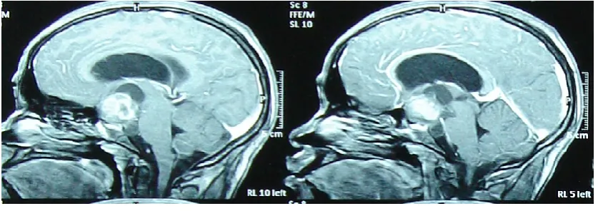
Fig.1
MRI images (case I) of the brain show a sellar mass with suprasellar extension, a central area of necrosis (red arrow) and a cystic part (white arrow) indenting the third ventricle and resulting in hydrocephalus.
International Journal of Research (IJR)
e-ISSN: 2348-6848, p- ISSN: 2348-795X Volume 2, Issue 08, August 2015
Available at http://internationaljournalofresearch.org
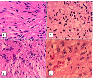
Fig. 2
A. Fusiform and oval cells with dark hyper chromatic nuclei in a fibrillary background. B. the cell nuclei vary in size in this field. C. Cells with dark nuclei in a fibrillary matrix. . D. In this field the tumour cells show better differentiated astrocytes (H&Ex40)
B
A C
International Journal of Research (IJR)
e-ISSN: 2348-6848, p- ISSN: 2348-795X Volume 2, Issue 08, August 2015
Available at http://internationaljournalofresearch.org
Fig. 3