Control of Vulval Competence and Centering in the Nematode
Oscheius
sp. 1 CEW1
Sophie Louvet-Valle´e,*
,1Irina Kolotuev,
†Benjamin Podbilewicz
†and Marie-Anne Fe´lix*
,2*Institut Jacques Monod, CNRS-Universite´s Paris 6 et 7, 75251 Paris Cedex 05, France and†Department of Biology, Technion-Israel Institute of Technology, Haifa 32000, Israel
Manuscript received August 27, 2002 Accepted for publication October 10, 2002
ABSTRACT
To compare vulva development mechanisms in the nematodeOscheiussp. 1 to those known in Caenorhab-ditis elegans, we performed a genetic screen for vulva mutants inOscheiussp. 1 CEW1. Here we present one large category of mutations that we callcov, which affect the specification of the Pn.p ventral epidermal cells along the antero-posterior axis. The Pn.p cells are numbered from 1 to 12 from anterior to posterior. In wild-typeOscheiussp. 1 CEW1, the P(4–8).p cells are competent to form the vulva and the progeny of P(5–7).p actually form the vulva, with the descendants of P6.p adopting a central vulval fate. Among the 17 mutations (defining 13 genes) that we characterize here, group 1 mutations completely or partially abolish P(4–8).p competence, and this correlates with early fusion of the Pn.p cells to the epidermal syncytium. In this group, we found a putative null mutation in thelin-39HOM-C homolog, the associated phenotype of which could be weakly mimicked by injection of a morpholino againstOsp1-lin-39in the mother’s germ line. Using cell ablation in a partially penetrant competence mutant, we show that vulval competence is partially controlled by a gonadal signal. Most other mutants found in the screen display phenotypes unknown inC. elegans. Group 2 mutants show a partial penetrance of Pn.p competence loss and an abnormal centering of the vulva on P5.p, suggesting that these two processes are coregulated by the same pathway inOscheiussp. 1. Group 3 mutants display an enlarged competence group that includes P3.p, thus demonstrating the existence of a specific mechanism inhibiting P3.p competence. Group 4 mutants display an abnormal centering of the vulval pattern on P7.p and suggest that a specific mechanism centers the vulval pattern on a single Pn.p cell.
T
HE phenotypic analysis of mutants obtained in ge- and is extremely common in the soil (Delattre and netic screens after mutagenesis provides informa- Fe´lix2001c;Fe´lixet al. 2001).tion at two levels: (i) on the mechanism of the process The nematode vulva is formed during postembryonic under study, as used, for example, in developmental development from vulval precursor cells in the ventral genetics, and (ii) on the evolutionary potential of the epidermis, called Pn.p cells (the posterior daughters of population, by exploring the mutational phenotypic Pncells; Figure 1). InC. elegans, the six cells P3.p–P8.p neighborhood. The comparison of phenotypes ob- are competent to form vulval tissue, as a result of their tained by a similar genetic screen in different species expression of the HOM-C genelin-39(Clarket al. 1993; therefore reveals both the evolution of the underlying Wanget al. 1993). The vulva is normally formed by the mechanisms and the difference in evolutionary poten- progeny of only three of these cells, with P6.p adopting tial between the two populations. a central (1⬚) fate and P5.p and P7.p adopting lateral The nematode Caenorhabditis elegans is used as a ge- (2⬚) fates; P(3,4,8).p adopt a nonvulval (3⬚) fate. The netic model system; one of its best-studied develop- 1⬚/2⬚/3⬚terminology of fates corresponds to their hier-mental features is the formation of its egg-laying organ, archy of replacement within the competence group the vulva (Wang andSternberg 2001). We previously (HorvitzandSternberg1991). Each fate can be dis-introducedOscheiussp. 1 CEW1 as another nematode tinguished by several features, in particular the division genetic model system (Fe´lix et al. 2000b; Dichtelet pattern of the Pn.p cell. The nonvulval 3⬚ fate can be
al. 2001) that displays a different vulval precursor cell distinguished from that of more anterior or posterior patterning mechanism (Fe´lix and Sternberg 1997) Pn.p cells because P(3,4,8).p divide once in the L3 stage and then fuse with the large epidermal syncytium that surrounds the animal (called hyp7), whereas P(1,2,9–
Sequence data from this article have been deposited with the 11).p do not divide and already fuse in the L1 stage
EMBL/GenBank Data Libraries under accession no. AJ431730. with hyp7. Within the vulval competence group, the
1Present address:UMR 7622, CNRS-Universite´ Paris 6, 9 quai
Saint-three fates are patterned by a combination of a graded
Bernard, 75252 Paris Cedex 05, France.
induction through epidermal growth factor/Ras signal-2Corresponding author:Institut Jacques Monod, Tour 43, 2 place
Jus-sieu, 75251 Paris Cedex 05, France. E-mail: [email protected] ing from the gonadal anchor cell and oflin-12/Notch
are affected in vulval divisionsdov-1todov-16(Dichtelet al.
signaling between the Pn.p cells (Wang andSternberg
2001). To distinguish phenotypic categories of vulva mutants
2001).
inOscheiussp. 1, we call the mutants that are affected in the
Vulval development in Oscheius sp. 1 CEW1 differs competence or centering of the vulvacovmutants and reserve in several ways from that of C. elegans: (i) The vulval dovfor those affected in thedivisionsof thevulva (instead of developmentof thevulva as mentioned inDichtelet al. 2001).
competence group is reduced to P(4–8).p; (ii) the same
Genetic complementation analyses were performed as
de-spatial pattern of vulval precursor fates is obtained by
scribed inDichtelet al. (2001). Complementation tests could
two successive inductions from the anchor cell, the first
not be performed with mf70 because its vulva defect is so
specifying a vulvalvs.a nonvulval fate in P(5–7).p and strong that it could not be mated into and we could not get the second an innervs. an outer (1⬚vs.2⬚) vulval sub- males to mate; we therefore did not assign a gene number to lineage in P6.p daughters; and (iii) the cell division it (however, after its molecular characterization, we could check that it defines a different locus than other mutants of
pattern corresponding to the 3⬚and 2⬚sublineages
dif-its group; seeresults). All mutations appear recessive, with
fers from those ofC. elegans(SommerandSternberg
the exception of two mutants with a vulval centering on P7.p:
1995;Fe´lixandSternberg1997; Figure 1).
cov-10(mf55)is semidominant (heterozygotes showⵑ12% of
Because of these differences in vulval development centering on P7.p) andcov-11(mf71)either is semidominant between the two species, we performed a screen for (to a similar degree) or shows a maternal effect (these two possibilities could not be distinguished in this mutant because
egg-laying (Egl)-defective mutants inOscheiussp. 1. We
heterozygous males are sterile).
previously described a large class of mutations that
spe-Nomarski microscopy and laser ablations:The worms were
cifically affect the number of divisions of the vulval
pre-mounted on agar pads as described in Wood (1988) and
cursor cells and not their overall fate (Dichtel et al. observed by Nomarski optics with a⫻100 objective on a Zeiss 2001); such mutants were not found in similarC. elegans Axioskop. Cells were ablated as described in Epstein and Shakes(1995), using a Photonic Instruments laser system.
screens (Fergusonet al. 1987; Wang andSternberg
Permeabilization, fixation, and immunofluorescence
stain-2001). Here we describe a second large class of
muta-ing of worms:To fix and permeabilize the worms, the
Finney-tions found in the screen, which affect the
antero-poste-Ruvkun protocol (FinneyandRuvkun1990) was used with
rior specification of the Pn.p cells, namely their vulval paraformaldehyde (Fluka) at a final concentration of 2%. The competence and/or the centering of the vulval pattern procedures were followed in 1.5-ml eppendorf tubes. The (a third smaller class of mutants, which affect the level immunofluorescence staining of the epithelial cell adherens junctions was performed with the monoclonal antibody MH27
of vulval induction, will be described elsewhere; M.-L.
(FrancisandWaterston1991) and nuclei were stained with Dichtel and M.-A. Fe´lix, unpublished data). We call
propidium iodide (PodbilewiczandWhite1994; Podbile-this class ofOscheiussp. 1 mutants covforcompetence wicz1996;Sharma-Kishoreet al. 1999).
and/or centeringof thevulva. The stained worms were analyzed using an MRC-1024 laser Within this large class of mutants, we further distin- confocal scanning microscope (Bio-Rad, Hempstead, UK) with the objective Nikon Plan Apo 60⫻/1.40 (Sharma
-guish four groups of mutants (Figure 2): (1) mutants
Kishoreet al. 1999;Shemeret al. 2000). The projection
pic-with a reduced vulval competence, among which is an
tures were obtained from 10–25 serial optical sections,
col-Osp1-lin-39mutant; (2) mutants with a reduced vulval
lected every 0.4–0.5 mm, in the z-axis. For a more precise
competence as well as centering of the vulva pattern visualization of the details, tilt and rotation projections were (1⬚fate) on P5.p; (3) mutants with a vulval competence performed using the Laser Sharp Processing program (Bio-Rad). We relate to the cell contacts and cell fusions according
group enlarged to P3.p; and (4) mutants with a vulval
to the appearance or vanishing of the adherens junctions
pattern centered on P7.p. The phenotypes that we
boundaries marked by MH27 (Shemer and Podbilewicz found in Oscheiussp. 1 show interesting similarities to
2000).
and differences from those found in C. elegans. More- Genomic library:A genomic library ofOscheiussp. 1 CEW1 over, we show that a gonadal signal can maintain compe- was constructed in the phage FIX II/XhoI (Stratagene, La tence of the Pn.p cells inOscheiussp. 1. Jolla, CA) according to the manufacturer’s instructions, after partial Sau3A digestion of CEW1 genomic DNA. A total of 150,000 plaques from the primary library were further ampli-fied to yield the genomic library.
MATERIALS AND METHODS
Molecular cloning of theOscheiussp. 1 CEW1lin-39gene: The homeodomain of the Oscheius sp. 1 CEW1lin-39 gene Nematode culture:The wild-type reference strain ofOscheius
was amplified by RT-PCR. Total RNA from the CEW1 strain sp. 1 (Fe´lix et al. 2001) used in these studies is the strain
was first prepared using the RNAXEL kit (Eurobio). Reverse CEW1. The nematodes were cultured onEscherichia coliOP50
transcription was performed for 1 hr at 42⬚using the MMLV as described forC. elegans(Wood1988). Cultures were usually
reverse transcriptase (GIBCO BRL, Gaithersburg, MD) and grown at 25⬚, except for matings, which were performed at
oligo(dT) as a primer. The homeodomain was then amplified 23⬚(Dichtelet al. 2001).
by PCR using the degenerate primers 5⬘-CGTCAGMGTACTG Genetic screen and crosses: The mutagenesis ofOscheius
CNTAYAC-3⬘and 5⬘-CATGCKACKRTTYTGRAACCA-3⬘(as in sp. 1 CEW1 and the screen for egg-laying mutants were
de-EizingerandSommer1997), subcloned into pGEM-T (Pro-scribed inDichtelet al. (2001). Themf89mutant was
subse-mega, Madison, WI), sequenced, and used as a probe for screen-quently isolated by M. Delattre in a F1clonal screen. All
muta-ing the Oscheiussp. 1 genomic library, according to standard tions were obtained after ethyl methanesulfonate mutagenesis,
procedures (Maniatiset al. 1989). A positive phage was puri-except formf79,mf80, mf85, andmf89, which were obtained
fied and digested withNotI and the insert (ⵑ20 kb in size) after trimethylpsoralen-ultraviolet irradiation (TMP-UV)
mu-was purified and subcloned into pBluescript, yielding plasmid tagenesis.
of theOsp1-lin-39gene and was sequenced on one strand until 2.6 kb downstream of the stop codon. All exons and the last three introns were then sequenced on both strands.
The intron-exon structure was first analyzed using PCR on anOscheiussp. 1 mixed-stage cDNA library (Fe´lixet al. 2000b). The 5⬘part of the cDNA was amplified using the M13 reverse primer in the cloning vector and a primer on the reverse strand in the homeodomain (in exon 4). The 3⬘ part was amplified with a forward primer at the start of exon 2 and a reverse primer downstream of the stop codon in the 3⬘ untranslated region. From the few clones sequenced, alterna-tive splicing was apparent at the 5⬘ends of exons 3 and 6. We therefore directly sequenced (without cloning) a RT-PCR product that was amplified from poly(A)-RNA and that cov-ered the last five exons. The major splice form is presented in Figure 5, but the two other forms at the 5⬘ends of exons 3 and 6 could be found in the sequence in the form of double and then triple peaks. First, the alternatively spliced form at the beginning of exon 3 adds a CAG, transforming the protein sequence into SNSTGPR (instead of SNSSPR). Second, the alternatively spliced form at the beginning of exon 6 removes the nine nucleotides (GACTTTCAG) that correspond to amino acids DFQ after the end of the homeodomain. Interest-ingly, in both cases, the alternatively spliced form can be well aligned with theC. elegans sequence. The alignment of the protein sequence to C. elegans and Pristionchus pacificus was initially performed using Clustal W and further refined by hand.
Morpholino injection:A morpholino against the
Osp1-lin-39 gene (sequence GACAAAGTCAGTCGGAGAAGTCATC
spanning the translational start site; Gene Tools, Philomath, OR) was injected into the syncytial germ line ofOscheiussp.
1 CEW1 adult hermaphrodites at a concentration in the injec- Figure 1.—Vulval formation inOscheiussp. 1 CEW1. The tion needle of 2 mmor 0.2 mm. The injected animals were vulva is formed by the progeny of the ventral epidermal cells transferred to new plates at several time points after injection. P5.p, P6.p, and P7.p. InOscheiussp. 1 CEW1 as inC. elegans, At 2 mm, the morpholino caused sterility of the injected adult P6.p adopts a central 1⬚ vulval sublineage (solid symbols), after 1 day, as did injection of the standard control morpholino whereas P5.p and P7.p adopt a lateral 2⬚ vulval sublineage (CCTCTTACCTCAGTTACAATTTATA). The vulval pheno- (shaded symbols). P4.p and P8.p usually adopt a nonvulval types were scored on progeny that were laid 5–20 hr after fate (open) and their progeny fuse with the epidermal
syncy-injection. tium hyp7 that surrounds most of the animal; however, they
Double-stranded RNA against Osp1-lin-39 was synthesized are able to replace P(5–7).p and to adopt a vulval fate. P(4–8).p
in vitrofrom the T7 and Sp6 transcription sites of a plasmid thus form the vulval competence group inOscheiussp. 1 CEW1. (pMA44) containing a RT-PCR clone ofOsp1-lin-39exons 2–5 The three different Pn.p sublineages within the vulval compe-into the pGEM-T vector (Promega). Injection of this double- tence group are specified by two successive inductions by the stranded RNA (1 mg/ml) had no effect (as for other genes gonadal anchor cell (AC) on the Pn.p cells and their daugh-inOscheiussp. 1 CEW1;Delattre2001). Injection of siRNAs ters. The subsequent division pattern of each cell is given againstOsp1-lin-39(6 mg/ml of the sense oligo GGACGACA at the bottom. The sublineages of Pn.p granddaughters are GUGACAAAGAGACCGUU annealed with the antisense oligo abbreviated as follows: s, fuses with the epidermal syncytium; AACGGUCUCUUUGUCACUGUCGUCC; Xeragon, Hunts- u, remains undivided; t, divides transversally (left-right). After ville, AL) also did not result in any phenotype. fusion of cells with the same terminal fate, the progeny of Polymorphism in theOsp1-lin-39gene:A molecular polymor- P(5–7).p give rise to six vulval rings, lettered vulA to vulF phism in theOsp1-lin-39gene was found by amplifying intron (I. KolotuevandB. Podbilewicz, unpublished results). 2 in four differentO.sp. 1 wild strains (DelattreandFe´lix
2001c;Fe´lixet al. 2001) and digesting it with several restriction enzymes. APvuI site was present in the reference strain CEW1
and absent in the strain JU149 (from Madagascar). For analysis a vulval fate whereas P4.p and P8.p adopt a nonvulval of genetic linkage of thecov-1(mf53)andcov-2(mf57)mutations fate and become part of the epidermal syncytium after toOsp1-lin-39, this polymorphic region was amplified using
two divisions (noted “ssss” for syncytial; Figure 1). By
primers 5⬘-AGTGGGTCAGTTGCTCCAC-3⬘ and 5⬘-GCTCAT
contrast, the noncompetent cells, P(1–3).p and P(9–
GAGTCTAACTTTCT-3⬘ on single F2 progeny of a cross
be-tween the mutant and JU149 and then subjected toPvuI diges- 11).p, do not divide and fuse with the epidermal
syncy-tion. tium (noted “S”; Table 1). Therefore, a change in
divi-sion number may correspond to a change in cell competence. We found several mutants in which several
RESULTS
or all of P(4–8).p adopt the S fate (groups 1 and 2 in Figure 2).
Mutants with a reduced vulval competence group:
The strongest phenotype is shown in group 1 mutants The vulval competence group is formed by the P(4–8).p
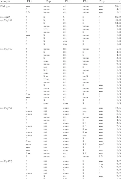
TABLE 1
Mutants with reduced Pn.p competence
Genotype P4.p P5.p P6.p P7.p P8.p n
Wild type ssss uuuu tttt uuuu ssss 95/100
S uuuu tttt uuuu ssss 4/100
S S uuuu tttt uuuu ssss 1/100
cov-(mf70) S S S S S 25/25
cov-1(mf53) S S S S S 40/83
S S tttt S S 34/83
S uuuu tttt uuuu S 3/83
S U U tttt uuuu S 1/83
S uuuu tttt S S 1/83
ssss S tttt uuuu S 1/83
S S tttt uuuu S 1/83
S S uttt S S 1/83
S S ttss S S 1/83
cov-2(mf57 ) S uuuu tttt uuuu S 5/33
S S tttt S S 4/33
S uuuu tttt S S 4/33
S S tttt uuuu S 3/33
S ssuu tttt uuuu S 2/33
S uuuu tttt uuss S 2/33
S ssss tttt S S 1/33
S S S tttt uuuu S 1/33
S ssuu tttt S S 1/33
S S ss tttt uu S S 1/33
S U uu tttt ssss S 1/33
S uuuu tttt uuuu S 1/33
S S tttt S ssss 1/33
S suuu tttt uuuu ssss 1/33
S uuuu tttt uuuu ssss 1/33
S ss uuuu tttt S S 1/33
ssss suuu tttt uuus S 1/33
S S S S S 1/33
S ssuu uuss S S 1/33#
cov-3(mf79) S tttt uuuu ssss ssss 12/50
uuuu tttt uuuu ssss ssss 9/50
uuuu tttt uuuu S ssss 6/50
S uuuu tttt uuuu ssss 4/50
S uuuu tttt S ssss 4/50
S tttt uuuu S S ssss 3/50
uuuu tttt uuuu S S ssss 2/50
S tttt uuuu S ss ssss 1/50
uuuu tttt uuuu S ss ssss 1/50
uuuu tttt ssss ssss ssss 1/50
S tttt uuuu S ssss 1/50
S uu tttt uuuu S ssss 1/50
ssuu tttt uuus S S ssss* 1/50
ssss tttt uuuu S S 1/50
ssss uutt ttuu S ssss 1/50
S ss uuuu tttt S S ssss 1/50
S uuuu tttt uuuu S S 1/50
cov-4(sy493) S tttt uuuu S ssss 3/24
S tttt uuuu S S ss 2/24
S tttt uuuu S S S 2/24
S tttt S S S 2/24
uuuu tttt uuuu S S 2/24
S S tttt S ssss 2/24
S S S S S ssss 1/24
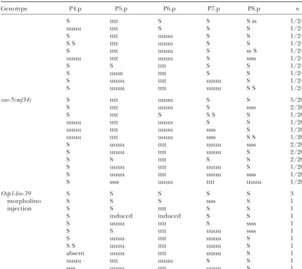
TABLE 1
(Continued)
Genotype P4.p P5.p P6.p P7.p P8.p n
S tttt S S S ss 1/24
uuuu tttt S S S 1/24
S tttt uuuu S S 1/24
S S tttt uuuu S S 1/24
S tttt uuuu S ss S 1/24
uuuu tttt uuuu S ssss 1/24
S S tttt S S 1/24
S uuut tttt S S 1/24
S uuuu tttt uuuu S 1/24
S uuuu tttt uuuu S S 1/24
cov-5(mf34) S tttt uuuu S S 5/20
S tttt uuuu S ssss 2/20
S tttt S S S S 1/20
uuuu tttt uuuu S S 1/20
uuuu tttt uuuu ssss S 1/20
uuuu tttt uuuu ssss S S 1/20
S uuuu tttt uuuu ssss 2/20
S uuuu tttt uuuu S 2/20
S S tttt S S 2/20
S uuuu tttt uuuu S 1/20
S uuuu tttt uuuu ssss 1/20
S ssss uuuu tttt uuuu 1/20
Osp1-lin-39 S S S S S 3
morpholino S S S ssss S 1
injection S S tttt S S 1
S induced induced S S 1
S uuuu tttt S ssss 1
S S tttt uuuu ssss 1
S uuuu tttt uuuu S 1
S S uuuu tttt uuuu S 1
absent uuuu tttt uuuu S 1
uuuu tttt uuuu S S 1
ssss uuuu tttt uuuu S 1
The division pattern and fates of Pn.p cells were scored in individual L4 stage hermaphrodites cultured at 25⬚. The sublineages of Pn.p cells, their daughters, or granddaughters are indicated from anterior to posterior, as follows. t, transverse division (left-right) of a Pn.p granddaughter; u, undivided granddaughter; s, nonvulval (“syncytial,” although we do not directly score for fusion in this table). A capital letter indicates the absence of one or two rounds of divisions. S, no division; S S, one division; S ss/ss S, one division followed by the division of one of the daughters; U, vulval fate with only one round of division. The right column indicates the number of animals observed. Incov-1(mf53)mutants, some Pn.p cells cannot be found (especially in the anterior region, apparently because the Pnmothers do not migrate); the Pn.p cell below the anchor cell is referred to as P6.p, but could be another Pn.p cell if P6.p were absent. Somecov-4(sy493)animals are also missing some Pn.p cells and only animals displaying all Pn.p cells were taken into account in this table. cov-2(mf79)mutants sometimes present a small posterior vulval invagination (formed by P6.p or P7.p progeny) in addition to the main invagination.cov-3andcov-4mutants show bright posterior intestinal granules. For the
lin-39morpholino injection, only those 13/228 animals displaying an abnormal vulval phenotype are shown; in addition, 35/228 animals showed no (or a single) division of P4.p or P8.p. After injection of the control morpholino, 7/139 animals had an undivided P4.p (a percentage that is similar to untreated CEW1;Dichtel et al. 2001), and none showed stronger defects. #, this animal displayed a gonad development problem. *, in this animal, P8.p progeny appeared posterior to P9.p.
the animals are fully vulvaless.cov-1(mf53)mutants show less penetrant and, in addition, the vulval 1⬚ fate is mis-centered on P5.p. This relatively large group is com-a similcom-ar phenotype, except thcom-at one Pn.p cell adopts
a central vulval fate in about one-half of the animals.cov- posed of seven mutants that define three genes,cov-3,
cov-4, andcov-5 (Figure 2). The P5.p centering defect
2(mf57)mutants show an even less penetrant phenotype
(Table 1). is highest incov-3mutants (Table 1 and legend to Table
Figure2.—Oscheiussp. 1 mutants with an altered competence and/or center-ing of the vulva. Four phenotypic catego-ries are distinguished among mutations that result in an altered vulval compe-tence and/or centering. Pn.p cell fates are schematized for each group of mu-tants. Symbols are as in Figure 1. Squares represent noncompetent cells. The dif-ferent complementation groups are shown on the right, with different alleles given in parentheses. When two alternative cell fates are observed, the most frequent is indicated above the less frequent.
appears highest incov-4(sy493)andcov-5(mf34)mutants case always moves close to the anchor cell (Table 2). This experiment suggests that the anchor cell or another cell and the P7.p defect appears highest incov-4(sy493)
mu-tants. of the gonad could influence the competence of the
Pn.p cells. To test this hypothesis, gonad ablations were To study the differences in Pn.p competence between
the wild type and these Cov mutants, we scored the time performed at different times of development. In wild-type animals, gonad ablation in the L1 stage leads to of fusion of the Pn.p cells to the epidermal syncytium
by staining the worms with the MH27 antibody, which the adoption of the 3⬚ sublineage (ssss) by P(4–8).p whereas, incov-1(mf53)andcov-2(mf57)mutants, all the recognizes cell junctions. In wild-typeOscheiussp. 1, as
inC. elegans, P(1,2,9–11).p fuse with an antero-posterior cells adopt an S fate (Table 2). After ablation of the anchor cell (or of the two anchor cell precursors) in chronology in the late L1 stage to the early L2 stage
(Figure 3, A and C). P3.p fuses later, in the late L2 stage the early L2 or L3 stages, one-fourth of the animals have at least one cell that adopts a ssss sublineage. In contrast, (data not shown). In the cov-(mf70) mutant, P(3–8).p
also fuse in the late L1 stage/early L2 stage, like P(1,2,9– gonad ablation in cov-3(mf79)mutants has little effect on cell competence, except possibly for P4.p (Table 2). 11).p in the wild type (Figure 3, B and D). However, in
ⵑ15% (13/98) of the animals, one central cell remains These results suggest that the competence of Pn.p cells is regulated by two mechanisms: an autonomous unfused until adulthood and sometimes (3/98 stained
animals) appears to divide once (Figure 4C). In thecov- cell specification mechanism and a gonadal cell signal that can be uncovered in mutant contexts such ascov-1 1(mf53) mutant, the P(4–8).p cells that do not divide
fuse in the late L1 stage, and in half of the animals, the andcov-2.
To test whether the group 2 mutants had the same central cell divides and gives rise to central (vulE and
vulF) vulval rings (“tttt” lineage), which form a minimal effect as gonad ablations incov-1(mf53), we constructed double mutants between cov-1 and group 2 mutants. functional vulva (Figures 1 and 4B).
In contrast to previously described mutants such as The cov-1(mf53); cov-3(mf79) and the 1(mf53); cov-4(sy493) double mutants both show a strong Vulvaless
dov-4 (in which P4.p and P8.p do not divide, but are
competent;Dichtelet al. 2001), group 1 and group 2 phenotype: 40/41 and 23/23 animals show no division/ induction of any Pn.p cell (with no enhancement of the mutants have fewer than three induced cells, in a
pat-tern that suggests an altered competence (rather than Pn.p generation defect ofcov-1). Therefore, the number of competent Pn.p cells is greatly reduced compared to an altered division or induction): For example, a Pn.p
cell may not divide whereas it is immediately adjacent to that of either single mutant. Thus, loci defined by group 2 mutants may act in the maintenance of Pn.p compe-that adopting the 1⬚fate. Moreover, the MH27 stainings
show an early fusion of the Pn.p cells prior to the time tence by a gonadal signal, whereas those defined by group 1 mutants may act in an automonous cell specifi-of vulval fate induction (Figure 3). Thus, these mutants
actually define genes that play a role in Pn.p vulval cation mechanism.
cov-(mf70)displays a deletion in theOsp1-lin-39gene:
competence.
We made use of the incomplete penetrance of some In C. elegans, expression of the HOM-C gene lin-39in P(3–8).p is necessary to prevent the fusion of P(3–8).p of these mutants to investigate a possible role of the
gonad in the competence of Pn.p cells to later receive to the hyp7 syncytium in the L1 stage (Clarket al. 1993;
Wang et al. 1993) and for P(3–8).p to adopt a vulval the anchor cell inductive signal. Incov-1(mf53)mutants,
one-half of the animals have one induced cell that is fate downstream of Ras activation in the L3 stage ( Clan-dinin et al. 1997; MaloofandKenyon1998). LIN-39 always located below the anchor cell. When this cell is
Figure3.—Pn.p cell fusion to the epidermal syncytium in wild-type andOsp1-lin-39(mf70)mutants. Cell fusion is scored by the disappearance of cell junctions that are revealed by immunofluorescence with the MH27 antibody (in black). An-terior is to the left, posAn-terior to the right. (A) Wild-type CEW1 in late L1; lateral view. P1.p and P2.p have fused with the epidermal syncytium. At this stage, P9.p or P10.p or both have
Figure4.—Vulval structures in wild type,Osp1-lin-39(mf70), sometimes already fused. (B)Osp1-lin-39(mf70)mutant in late and
cov-1(mf53)mutants. Staining is as in Figure 3, in the late L1; lateral view. P(1–5).p have fused (typical pattern at this L4 stage. Anterior is to left, posterior to the right. (A) Wild-stage). (C) Wild type in mid L2; ventral view. P(3–8).p cells type CEW1; lateral view. The different vulval rings are visible remain unfused. (D)Osp1-lin-39(mf70)mutant in mid L2; ven- at the end of organogenesis. (B)cov-1(mf53)mutant; lateral tral view. No Pn.p cell contours can be seen. view. The reduced vulva is composed of the two inner rings
vulE and vulF that are connected to the uterus. (C) Osp1-lin-39(mf70)mutant; ventral view. This is a rather unusual animal
in vulval cells that fail to fuse to hyp7 (Shemer and in which two cells (which are likely the progeny of P6.p) are still unfused in the L4 stage.
Podbilewicz 2002). Because a loss-of-function muta-tion in theCel-lin-39gene results in a phenotype resem-bling that ofcov-(mf70)inO.sp. 1, we wondered whether
beginning of intron 2 and a primer 1.5 kb downstream thelin-39homolog could be mutated incov-(mf70)(and
of the stop codon, a band ofⵑ600 bp could be amplified possibly other mutants). We isolated thelin-39homolog
inmf70worms and sequenced. The breakpoints of the ofOscheius sp. 1 by RT-PCR of the homeodomain
fol-mf70deletion correspond to an AGA triplet found 483 lowed by screening of a genomic library and named it
bp after the start of intron 2 andⵑ1.2 kb downstream
Osp1-lin-39according to previous nomenclature (Eizinger
of the stop codon. This 3.5-kb deletion thus removes and Sommer 1997). We confirmed the intron-exon
exons 3–6 and therefore the whole homeodomain (Fig-structure by sequencing RT-PCR products (Figure 5)
ure 5A). Injection of a morpholino directed against the and found two splice variants at the beginning and the
5⬘end of theOsp1-lin-39coding sequence (see
materi-end of the homeodomain (seematerials and
meth-als and methods) caused a weakmf70-like phenotype
ods). The alignment of the putative protein with the
in the progeny of injected mothers (Table 1), thus con-homologs fromC. elegansand from the other nematode
firming that the molecular deletion in Osp1-lin-39 is
P. pacificusis shown in Figure 5B. The homeodomain
the cause of the vulval competence phenotype ofmf70. is highly conserved, as well as the hexapeptide upstream
Although we cannot rule out that a second mutation of the homeodomain. The N-terminal is the most
diver-plays a role in the loss of Pn.p competence in the
cov-gent part of the protein. The putative C-terminal
mito-(mf70)mutant since it was never outcrossed, on the basis gen-activated protein (MAP) kinase docking site (FXFP)
of the strongest phenotypes observed with the morpho-that is present in the C. elegans LIN-39 and absent in
lino and the molecular deletion of the homeodomain
P. pacificus(GrandienandSommer2001) is also present
inmf70, we conclude that thecov-(mf70)mutant is likely inO.sp. 1 (FGFP). However, the MAP kinase
phosphory-to be a null allele ofOsp1-lin-39. lation site (S/TP) is absent (no proline), rendering it
Since we could not perform complementation experi-unlikely to be phosphorylated by MAP kinase inO.sp. 1.
ments withcov-(mf70), we tested whethercov-2(mf53)or The Osp1-lin-39homeodomain could not be
ampli-cov-3(mf57)could be alleles ofOsp1-lin-39. We made use fied by PCR incov-(mf70)mutant worms, suggesting that
of themfP1polymorphism found in the second intron it could be deleted. The putative deletion was then
ofOsp1-lin-39in the wild strain JU149vs.our reference further mapped using various primers along the
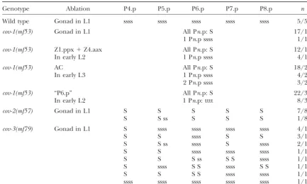
TABLE 2
Cell ablations in mutants with reduced vulval competence
Genotype Ablation P4.p P5.p P6.p P7.p P8.p n
Wild type Gonad in L1 ssss ssss ssss ssss ssss 5/5a
cov-1(mf53) Gonad in L1 All Pn.p: S 17/18
1 Pn.p ssss 1/18
cov-1(mf53) Z1.ppx⫹Z4.aax All Pn.p: S 12/16
In early L2 1 Pn.p ssss 4/16
cov-1(mf53) AC All Pn.p: S 18/25
In early L3 1 Pn.p ssss 4/25
2 Pn.p ssss 3/25
cov-1(mf53) “P6.p” All Pn.p: S 22/30
In early L2 1 Pn.p: tttt 8/30
cov-2(mf57) Gonad in L1 S S S S S 7/8
S S ss S S S 1/8
cov-3(mf79) Gonad in L1 S ssss ssss ssss ssss 4/14
S S ssss S S 3/14
S S ss ssss S ssss 2/14
S S ssss ssss ssss 1/14
S S S ss S S ssss 1/14
S ssss S S ssss S S 1/14
S S S S ssss ssss 1/14
ssss ssss ssss ssss ssss 1/14
The animals were scored during the L4 stage to determine the cell division pattern and fate. We do not directly score for cell fusion in this table. Abbreviations are as in Table 1. Z1.ppx and Z4.aax are ventral uterine cells, one of which will become the anchor cell (Kimbleand Hirsh1979). Incov-1(mf53)mutants, several Pn.p cells are missing, so the identification of each Pn.p cell is not possible (the ablated “P6.p” is the cell located below the gonad). Pn.p cells move posteriorly after gonad ablation incov-3(mf79)animals. AC, anchor cell.
aData fromDichtelet al. (2001).
5A). Neither cov-2(mf53) norcov-3(mf57) was found to vulval pattern is centered on P6.p in species of the family Rhabditidae (to which Caenorhabditis and Oscheius be genetically linked toOsp1-lin-39. These two mutations
thus define two other genes affecting Pn.p competence. belong) and of the family Diplogastridae (to which Pris-tionchus belongs;SommerandSternberg1995;
Som-Mutants with a vulval competence group enlarged to
P3.p:InOscheiussp. 1 CEW1, P3.p does not divide whereas mer 1997), whereas it is centered between P6.p and
P7.p in the more distant families Panagrolaimidae and P4.p divides twice at the end of the L3 stage in most
animals (SommerandSternberg1995;Delattreand Cephalobidae (SternbergandHorvitz1982;Fe´lixet al. 2000a).
Fe´lix2001c). Moreover, P4.p is part of the vulval
com-petence group and is able to replace P(5–7).p (Dichtel We described above a large group of mutants with a reduced competence group and an abnormal centering
et al. 2001), whereas P3.p is not part of the vulval
compe-tence group: It does not divide and does not acquire a of the vulva pattern on P5.p (group 2). Additionally, we found three mutants with a normal competence vulval fate after ablation of P(4–8).p in the L1 stage
(Table 3, last column). group but a vulval pattern centered on P7.p (group 4 in Figure 2; Table 4) or even sometimes on P8.p, in We found four mutants ofOscheiussp. 1 in which P3.p
divides twice, with penetrances of 50–100% (group 3 in which case the vulva lacks a posterior 2⬚ fate because P9.p is not competent. These mutants also show a slight Figure 2; Table 3). More anterior or posterior Pn.p
cells are not affected [except that additional cells were increase in vulval induction (strongest formf71), which may be partially explained by the anchor cell being observed in the P1.p region in 4/50 cov-9(mf89)
ani-mals]. Each mutation defines a complementation group, positioned between P6.p and P7.p at the time of the first induction. Indeed, a few animals with a vulval pattern which we name cov-6 to cov-9. If P(4–8).p are ablated
in the L1 stage in thecov-8(mf54)orcov-9(mf89)mutants, centered between two Pn.p cells were observed (see legend to Table 4). The three mutations apparently P3.p is able to adopt a vulval fate (Table 3). Thus, these
mutations correspond to an enlargement of the vulval complement and thus define three genes, cov-10, cov-11, andcov-12.
competence group to P3.p.
Figure 5.—Molecular analysis of theOscheiussp. 1lin-39 homo-log. (A)Osp1-lin-39genomic struc-ture. The homeodomain is shown in black. The size of each intron is indicated below. The drawing is not to scale for intronic sequences and the 3⬘endpoint of the mf70
deletion. (B) Alignment of the full LIN-39 protein sequences of three rhabditid nematodes. Conserved residues between species are indi-cated by shaded boxes, intron po-sitions by small solid squares. The cDNA sequence of Osp1-lin-39
is available under accession no. AJ431730.
pattern, either on P5.p (group 2) or on P7.p (group 4), displaced posteriorly relative to the Pncells at hatching, suggesting an embryonic defect. In group 2 mutants the anchor cell is positioned in the L4 stage immediately
dorsal to the cells adopting a central 1⬚ fate. Is the (pattern centered on P5.p and loss of Pn.p compe-tence), the misalignment of the anchor cell with P5.p anchor cell mispositioning responsible for a change in
vulval centering in these mutants? We first looked at occurs later, in the L2 and the L3 stages, close to the time of the first induction. A greater competence of the time of hatching (before anchor cell birth) to see
whether the gonad primordium was in its normal posi- P5.p to adopt a vulval or central vulval fate may be responsible for this change in vulval centering in the tion relative to the Pncells. In the group 4cov-12(mf85)
mutant, the gonad was located more posteriorly than group 2 mutants. in the wild type, but in the group 2 cov-3(mf35) and
cov-3(mf80)mutants, the gonad position was normal at
DISCUSSION
hatching (Table 5). Following a time course of gonad/
anchor cellvs.Pn.p position in these two latter alleles, We have described 17 mutants of vulval competence and centering inOscheiussp. 1, which define 12covloci it appears that P6.p is correctly located below the gonad
in the late L1 stage, but by the end of the L2 stage the and the Osp1-lin-39 gene. We grouped together these antero-posterior specification mutants affecting compe-anchor cell is sometimes displaced above P5.p (or P5.p
is displaced below the anchor cell), although not yet at tence and centering of the vulva for two reasons: first, because we (unexpectedly) found a large number of a frequency compatible with the final pattern (Table
5). Thus, the two groups of mutants appear to show an mutations that affect both and, second, because the number of competent cells and the precision of vulval alteration of vulva centering for two distinct reasons. In
TABLE 3
Mutants of P3.p fate
P3.p S S, % P3.p vulval
Genotype P3.p ssss Sss or ssS penetrance competence
Wild type 0/100 0/100 0 0/8
cov-6(sy460) 26/50 1/50 54 ND
cov-7(sy466) 74/100 7/100 81 ND
cov-8(mf54) 49/100 2/100 51 10/11
cov-9(mf89) 50/50 0/50 100 10/10
The division pattern of P3.p was scored in the L4 stage on worms cultured at 25⬚. P3.p competence was tested by ablation of P(4–8).p in the L1 stage (before Pn.a division) and scored positive if P3.p adopted a vulval fate. Abbreviations are as in Table 1. Thecov-6(sy460)andcov-8(mf54)mutants show a weak egg-laying-defective phenotype. Thecov-7(sy466)mutant shows a partially penetrant temperature-sensitive sinistral pheno-type (DelattreandFe´lix2001a) and a gonad morphology defect leading to partial sterility. ND, not deter-mined.
discuss their phenotypes in relation to vulval develop- cell death (in the Pn.a lineages) or for suppressors of Multivulva mutations (Clarket al. 1993). Thebar-1and mental mechanisms in Oscheius sp. 1, compare them
with those found in C. elegans, and assess their conse- several other similar mutations with a lesser penetrance than that of lin-39were found in a screen for animals quence for the phenotypic evolvability of the species.
Vulval developmental mechanisms in Oscheius sp. 1 with a protruding vulva (Eisenmannet al. 1998;
Eisen-mann andKim2000), which occurs (as inOscheiussp.
and comparison withC. elegans:We previously described
vulva mutants ofOscheiussp. 1 that affect the Pn.p divi- 1) when an incomplete vulva is formed from only one or two Pn.p precursor cells. Strangely, although null sion pattern specifically (dovmutants); this class is
al-most completely absent in C. elegans (Dichtel et al. mutations in these genes are homozygous viable (except for apr-1) and display a high penetrance of egg-laying 2001). Conversely, mutants affecting vulval induction
form the most numerous class of vulva development defects, they were not found in the original Egl screen, even though it was estimated to be close to saturation for mutants inC. elegans, but are very rare inOscheiussp. 1
(M.-L. Dichtel and M.-A. Fe´lix, unpublished data). such viable mutations (FergusonandHorvitz1985). Thus, the comparison of frequency of occurrence of The other frequent categories of vulva mutants found
inOscheiussp. 1 are described here and concern vulval phenotypes in the two different species should be taken with some caution. We will review each group succes-competence and/or centering. The paucity of mutants
affecting Pn.p competence in the first extensive screens sively.
Within the group 1 mutants (loss of Pn.p compe-for Egl mutants (HorvitzandSulston 1980;Trent
et al. 1983) is a bizarre historical feature ofC. elegans tence), the phenotype of theOsp1-lin-39(mf70)mutant closely resembles that of aC. elegans lin-39null mutant: vulva development studies. The lin-39locus was found
only later, in screens either for an abnormal pattern of All Pn.p cells (except P12.p) stay undivided. Thelin-39
TABLE 4
Mutants with posterior vulva centering
% penetrance of
Genotype P7.p centering P8.p centering posterior centering Hyperinduced
Wild type 0/100 0/100 0 0/100
cov-10(mf55) 23/51 0/51 45 3/51
cov-11(mf71) 37/88a 1/88 43 21/88
cov-12(mf85) 83/135 9/135 68 1/135
The vulva pattern was scored in the L4 stage.
aOne animal had a vulval pattern centered between P6.p and P7.p, and one between P7.p and P8.p. Animals
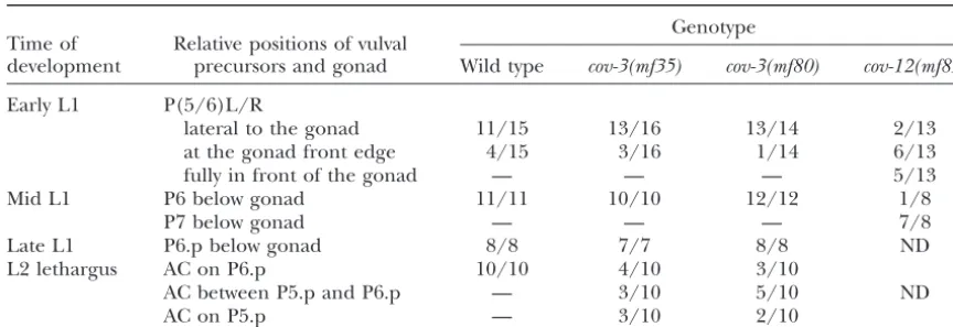
TABLE 5
Gonad position in mutants with altered vulval centering
Genotype Time of Relative positions of vulval
development precursors and gonad Wild type cov-3(mf35) cov-3(mf80) cov-12(mf85)
Early L1 P(5/6)L/R
lateral to the gonad 11/15 13/16 13/14 2/13
at the gonad front edge 4/15 3/16 1/14 6/13
fully in front of the gonad — — — 5/13
Mid L1 P6 below gonad 11/11 10/10 12/12 1/8
P7 below gonad — — — 7/8
Late L1 P6.p below gonad 8/8 7/7 8/8 ND
L2 lethargus AC on P6.p 10/10 4/10 3/10
AC between P5.p and P6.p — 3/10 5/10 ND
AC on P5.p — 3/10 2/10
The position of Pnor Pn.p cells relative to the gonad or the anchor cell (AC) was scored at different time points during larval development. “Early L1” is before the rotation of Pncells. “Mid L1” is after their antero-posterior alignment and before their division. “Late L1” is after their division. The penetrance of vulval centering on P5.p is high in the twocov-3alleles (17/18mf35animals and 15/18mf80animals) andⵑ68% in thecov-12(mf85)mutant (see Table 4).
(mf70)mutation is very likely to be a null allele since it the gonad. This signal may act by repressing cell fusion, likelin-39and the regulators of cell fusionref-1andref-2
deletes the whole homeodomain. However, unlike in a
C. elegans lin-39null mutant, at least one Pn.p cell stays inC. elegans(AlperandKenyon 2001, 2002). Depen-dence of cell competence on intercellular signaling has unfused until adulthood inⵑ15% of the animals (but
it usually does not divide and it does not form vulval been suggested inC. elegansby the fact that a hypomor-phic mutation in the LET-23 receptor (which acts in tissue). Thus, in Oscheiussp. 1, LIN-39 may be not be
absolutely necessary to prevent fusion, and it is required receiving the anchor cell inductive signal) and gonad ablation enhance the effect of thebar-1mutation (
Eisen-for vulval competence. We have found that the few cells
that evade cell fusion do not usually divide. Thus, LIN-39 mann et al. 1998).
The group 2 mutants are the most frequent inOscheius
inOscheiussp. 1 may be required for cell proliferation
as was recently found inC. elegans (Shemer andPod- sp. 1 (Figure 2) and display an Egl defect; however, such a phenotype with an altered centering on P5.p, in
bilewicz2002). The conserved role of lin-39 in
pre-venting Pn.p cell fusion and division in C. elegans and addition to a Pn.p competence defect, has not been found in C. elegans. The anterior vulva displacement
Oscheiussp. 1 contrasts with its role in inhibiting Pn.p
cell death in the more distantly related nematode P. has been found only at low penetrance in Lon (long) mutants (Sternberg and Horvitz 1986), and the
pacificus (Eizinger and Sommer 1997; Sommer et al.
1998). Oscheius sp. 1 group 2 mutants are not long. Incov-3
mutants, the gonad appears at its normal position at Although they do not map to the Osp1-lin-39locus,
the phenotypes of the other group 1 mutants resemble hatching and the anchor cell aligns only with P5.p late during the L2 or L3 stages (Table 5). P5.p thus appears hypomorphicCel-lin-39mutants (Clandininet al. 1997),
except that P4.p is not affected in the latter. Thecov-1 more competent to adopt a 1⬚sublineage than P6.p and the vulva pattern most often remains incomplete on and/orcov-2mutations may define regulators of
Osp1-lin-39expression in the L1 stage. the anterior side because P4.p is not competent in this mutant. The higher penetrance of defects in P7.p vs.
A specific feature of the control of Pn.p fusion and
competence that is unraveled in Oscheius sp. 1 is its P8.p (Table 1) is also surprising. In our screen, this combination of phenotypes has been found at three dependence on the gonad incov-1 and cov-2 mutants
(Table 2). As was previously found in thedov-1(sy543) distinct loci, with four alleles for one locus (including two obtained after TMP-UV mutagenesis), suggesting mutant for Pn.p cell divisions (Dichtelet al. 2001), this
signaling from the gonad is apparent only in a mutant that they reflect the loss of function of a pathway. Since group 2 mutations have an enhancing effect on Pn.p context and does not show in wild-type animals,
proba-bly because of redundant processes ensuring Pn.p cell competence loss incov-1mutants that is similar to gonad ablation, it is possible that they define an intercellular competence. In other words, competence (or the
would act redundantly in wild-type animals since gonad terior to lin-39 that is expressed in P(7–11).p ( Clan-dininet al. 1997), and atⵑ8% penetrance in mutants ablation has no effect in this context).
In C. elegans, some components of the Wnt pathway for theC. elegans even-skippedhomologvab-7(50% pene-trance in theP. pacificus vab-7mutants;Jungblutand act in maintaining lin-39 expression in the L2 stage
(although no ligand, receptor, or defined cell interac- Sommer2001). Here we describe three mutants, which cause up to 68% posterior displacement of the vulval tion has been demonstrated). In mutants of this
path-way, the affected cells fuse during the late L2 stage, like 1⬚fate (Table 4). As inP. pacificus vab-7mutants ( Jung-blutandSommer 2001), this posterior vulva shift ap-P3.p in wild type. TheC. elegans bar-1/Armadillomutant
is mostly affected for P3.p and P4.p (but less for P8.p; pears to result from aberrant gonad positioning at hatching (Table 5). None of the three Oscheius sp. 1
Eisenmannet al. 1998). Other loci, such asmig-14/mom-3,
egl-18, and sem-4, show a mutant phenotype similar to mutants shows the bobbed tail phenotype that is char-acteristic of C. elegans and P. pacificus vab-7 mutants
bar-1(EisenmannandKim2000). Tissue-specific
inacti-vation of the apr-1/APCgene causes L1 fusion of P(3– (Ahringer1997;JungblutandSommer2001). Inter-estingly, the two-step mechanism of vulval induction in 8).p, also with a higher penetrance for the distal cells
(Hoieret al. 2000). It is possible that group 2 mutations Oscheiussp. 1 could allow the 1⬚sublineage to be shared between daughters of two different Pn.ps (as happens define a Wnt pathway.
The phenotype of the group 3 mutants corresponds for P6.pp and P7.pa in Panagrolaimus sp.; Fe´lix and
Sternberg 1997). However, we find only a few such to an evolutionary change within the family
Rhabditi-dae: InC. elegans, P3.p divides in one-half of the animals animals (Table 4), suggesting the existence of a mecha-nism of centering on one Pn.p, for example, by align-(in the reference strain N2; Sulston and Horvitz
1977) and can adopt a vulval fate upon removal of P(4– ment of the anchor cell with a Pn.p cell before its division. In summary, the group 1 mutants resemble the C.
8).p (Sternberg and Horvitz 1986; Delattre and
Fe´lix 2001c), whereas in Oscheius sp. 1, P3.p fuses in eleganscompetence mutants, such aslin-39, and demon-strate the existence of a gonadal signal that maintains the late L2 stage, does not divide, and cannot replace
P(4–8).p. Two mutants, cov-6(sy460) and cov-8(mf54), vulval competence inOscheiussp. 1; the many group 2 mutants do not resemble anyC. elegans(norP. pacificus) show a 50% occurrence of P3.p division similar to that
of the C. elegansreference strain N2, whereas the cov- mutants and suggest that Pn.p competence and center-ing of the 1⬚fate are coregulated by the same pathway;
9(mf89) mutant shows a complete penetrance of P3.p
division (Table 3), as was noticed in C. elegans by G. the group 3 mutants resemble the C. eleganswild-type phenotype and demonstrate the existence of a specific Jongeward in lin-22/Hairy mutants (mentioned in
WrischnikandKenyon1997). Because of the variabil- developmental mechanism repressing P3.p competence in Oscheius sp. 1; the group 4 mutants have not been ity of P3.p division, a phenotype of loss or gain of P3.p
division/competence is much more difficult to screen found at such high penetrance inC. elegansand suggest the existence of a mechanism for centering of the vulva for inC. elegansthan inOscheiussp. 1. In our
mutagene-sis, these mutants were first picked because of their low- on a single Pn.p cell inOscheiussp. 1.
Comparison with phenotypes found in wild
popula-penetrance Egl phenotype and were easily found in a
secondary Nomarski screen. tions: The high number of mutations affecting vulval divisions described in Dichtel et al. (2001) demon-During development, P3.p and P4.p (or at least their
respective mothers) are initially equivalent. The 12 Pn strates the high mutability of Oscheius sp. 1 CEW1 to these phenotypes. Correspondingly, we found many cells (the Pn.p mother cells) are found at hatching as
six symmetric left-right pairs and subsequently align similar variations in vulval divisions in wild populations ofOscheiussp. 1 and in closely related species, whereas along the ventral midline, in a random orientation for
the P(3/4)L/R cell pair as for most Pncell pairs inC. such mutants and wild variations do not exist withinC. elegans(DelattreandFe´lix2001b,c). Here we review
elegans(SulstonandHorvitz1977). InOscheiussp. 1,
the left and right cells of the P(3/4)L/R cell pair also the occurrence of phenotypes in wild strains corre-sponding to that of the Cov mutants.
appear equivalent before rotation (Sommerand
Stern-berg 1995). They (or their daughters) must later ac- Animals with a loss of competence such as found in group 1 and group 2 mutants are not found in wild quire different fates, since only P4.p is competent and
divides. The group 3 mutants clearly demonstrate the isolates, which is likely due to a negative selection pres-sure acting on egg-laying-defective animals.
existence of a specific mechanism of inhibition of P3.p
competence that results in a vulval equivalence group By contrast, occasional centering of the vulva pattern on P5.p or P7.p occurs in wild-type animals of both of five rather than six cells.
The phenotype of the group 4 mutants (centering of species (Delattre and Fe´lix 2001c). Such noise in centering of the vulval pattern constitutes a selection the vulval pattern on P7.p) has also not been specifically
screened for in C. elegans, but has been observed at pressure for a five-cell competence group (one more competent cell on each side of the vulva). It is not clear low penetrances in some mutant contexts: at ⵑ25%
in the nematodeOscheius/Dolichorhabditissp. CEW1. Genetics157:
Oscheiussp. 1 acting to include or exclude the sixth cell
183–197.
P3.p from the competence group. This character evolves Eisenmann, D. M., andS. K. Kim, 2000 Protruding vulva mutants within each genus. A few animals with divided P3.p can identify novel loci and Wnt signaling factors that function during
Caenorhabditis elegansdevelopment. Genetics156:1097–1116.
be found in some Oscheiussp. 1 strains and in closely
Eisenmann, D. M., J. N. Maloof, J. S. Simske, C. KenyonandS. K.
related species in about one-fourth of the animals Kim, 1998 The-catenin homolog BAR-1 and LET-60 Ras coor-(DelattreandFe´lix2001c). We show here that a muta- dinately regulate the Hox genelin-39duringCaenorhabditis elegans
vulval development. Development125:3667–3680.
ble mechanism specifies a distinct noncompetent fate
Eizinger, A., and R. J. Sommer, 1997 The homeotic genelin-39 in P3.p compared to P4.p (whereas we did not find any and the evolution of nematode epidermal cell fates. Science278: mutants abolishing specifically P4.p or P8.p compe- 452–455.
Epstein, H. F., and D. C. Shakes (Editors), 1995 Caenorhabditis tence). The existence of such a mechanism suggests that
elegans: Modern Biological Analysis of an Organism. Academic Press,
some evolutionary pressure acts (perhaps indirectly) to San Diego.
reduce the competence group to five rather than six Fe´lix, M.-A., andP. W. Sternberg, 1997 Two nested gonadal induc-tions of the vulva in nematodes. Development124:253–259.
cells in Oscheius sp. 1. It will be interesting to study
Fe´lix, M.-A., P. De Ley, R. J. Sommer, L. Frisse, S. A. Nadleret the polarity of change in vulval competence group size al., 2000a Evolution of vulva development in the Cephalobina within the genus Oscheius and the family Rhabditidae. (Nematoda). Dev. Biol.221:68–86.
Fe´lix, M.-A., M. DelattreandM.-L. Dichtel, 2000b Comparative
Some nematodes such asPanagrolaimusspp. (family
Pa-developmental studies usingOscheius/Dolichorhabditissp. CEW1
nagrolaimidae) have a vulval competence domain that (Rhabditidae). Nematology2:89–98.
is not larger than the cells actually adopting a vulval Fe´lix, M.-A., A. VierstraeteandJ. Vanfleteren, 2001 Three bio-logical species related to Rhabditis (Oscheius) pseudodolichura fate and may rely on a greater precision of centering
Ko¨rner in Osche, 1952. J. Nematol.33:104–109.
(Sommer and Sternberg 1996; Fe´lix et al. 2000a). Ferguson, E., andH. R. Horvitz, 1985 Identification and charac-Thus, the combination of the number of competent terization of 22 genes that affect the vulval cell lineages of
Caeno-rhabditis elegans.Genetics110:17–72.
cells and of the precision of vulval centering is important
Ferguson, E. L., P. W. SternbergandH. R. Horvitz, 1987 A
for the reproducibility of vulva formation. genetic pathway for the specification of the vulval cell lineages
ofCaenorhabditis elegans. Nature326:259–267. We are very grateful to Marie-Laure Dichtel for great help with the
Finney, M., andG. Ruvkun, 1990 Theunc-86gene product couples screens, to Marie Delattre for finding themf89mutant, and to both
cell lineage and cell identity in Caenorhabditis elegans.Cell63:
of them for comments on the manuscript. We thank Evelyne Pierre
895–905. for help with genomic library construction and freezing many strains.
Francis, R., andR. H. Waterston, 1991 Muscle cell attachment We thank Paul Sternberg in whose lab the screen was started and inCaenorhabditis elegans.J. Cell Biol.114:465–479.
Christophe Antoniewski, Jean-Louis Bessereau, and Ralf Sommer for Grandien, K., andR. J. Sommer, 2001 Functional comparison of advice and discussions. This work was supported by a Human Frontier the nematode Hox genelin-39inC. elegansandP. pacificusreveals Science Program collaborative grant to M.-A. F. and B.P., the Centre evolutionary conservation of protein function despite divergence
of primary sequence. Genes Dev.15:2161–2172. National de la Recherche Scientifique (France), and the Association
Hoier, E. F., W. A. Mohler, S. K. KimandA. Hajnal, 2000 The pour la Recherche sur le Cancer.
Caenorhabditis elegansAPC-related geneapr-1is required for epi-thelial cell migration andHoxgene expression. Genes Dev.14:
874–886.
Horvitz, H. R., andP. W. Sternberg, 1991 Multiple intercellular
LITERATURE CITED signalling systems control the development of theC. elegansvulva.
Nature351:535–541.
Ahringer, J., 1997 Maternal control of a zygotic patterning gene
Horvitz, H. R., andJ. E. Sulston, 1980 Isolation and genetic char-inCaenorhabditis elegans.Development124:3865–3869.
acterization of cell-lineage mutants of the nematode
Caenorhab-Alper, S., andC. Kenyon, 2001 REF-1, a protein with two bHLH
ditis elegans.Genetics96:435–454. domains, alters the pattern of cell fusion inC. elegansby regulating
Jungblut, B., andR. J. Sommer, 2001 The nematodeeven-skipped
Hox protein activity. Development128:1793–1804.
homologvab-7regulates gonad and vulva position inPristionchus
Alper, S., andC. Kenyon, 2002 The zinc finger protein REF-2
func-pacificus.Development128:253–261. tions with the Hox genes to inhibit cell fusion in the ventral
Kimble, J., andD. Hirsh, 1979 Post-embryonic cell lineages of the epidermis ofC. elegans.Development129:3335–3348.
hermaphrodite and male gonads inCaenorhabditis elegans.Dev.
Clandinin, T. R., W. S. KatzandP. W. Sternberg, 1997
Caenorhab-Biol.70:396–417.
ditis elegansHOM-C genes regulate the response of vulval
precur-Maloof, J. N., andC. Kenyon, 1998 The Hox genelin-39is required sor cells to inductive signal. Dev. Biol.182:150–161.
duringC. elegansvulval induction to select the outcome of Ras
Clark, S. G., A. D. ChisholmandH. R. Horvitz, 1993 Control of
signaling. Development125:181–190. cell fates in the central body region ofC. elegansby the homeobox
Maniatis, T., E. F. FritschandJ. Sambrook, 1989 Molecular
Clon-genelin-39.Cell74:43–55.
ing: A Laboratory Manual. Cold Spring Harbor Laboratory Press,
Delattre, M., 2001 Variants de de´veloppement et e´volution chez
Cold Spring Harbor, NY. les ne´matodes: e´tudes sur la chiralite´ de P11/12 et sur le lignage
Mohler, W. A., G. Shemer, J. J. del Campo, C. Valansi, E. Opoku -des cellules pre´curseurs de la vulve. Ph.D. Thesis, Universite´ de
Serebuohet al., 2002 The type I membrane protein EFF-1 is Paris 6, Paris.
essential for developmental cell fusion. Dev. Cell2:355–362.
Delattre, M., andM.-A. Fe´lix, 2001a Evolution and development
Podbilewicz, B., 1996 ADM-1, a protein with metalloprotease- and of a variable left-right asymmetry in nematodes: the handedness
disintegrin-like domains, is expressed in syncytial organs, sperm, of P11/P12 migration. Dev. Biol.232:362–371.
and sheath cells of sensory organs inCaenorhabditis elegans.Mol.
Delattre, M., andM.-A. Fe´lix, 2001b Microevolutionary studies in
Biol. Cell7:1877–1893. nematodes: a beginning. Bioessays23:807–819.
Podbilewicz, B., andJ. G. White, 1994 Cell fusions in the
devel-Delattre, M., andM.-A. Fe´lix, 2001c Polymorphism and evolution
oping epithelia ofC. elegans.Dev. Biol.161:408–424. of vulval precursor cell lineages within two nematode genera,
Sharma-Kishore, R., J. G. White, E. SouthgateandB.
Podbilew-CaenorhabditisandOscheius.Curr. Biol.11:631–643.
icz, 1999 Formation of the vulva in Caenorhabditis elegans: a
Dichtel, M.-L., S. Louvet-Valle´e, M. E. Viney, M.-A. Fe´lix and
Shemer, G., and B. Podbilewicz, 2000 Fusomorphogenesis: cell Sternberg, P. W., andH. R. Horvitz, 1982 Postembryonic nongo-nadal cell lineages of the nematodePanagrellus redivivus: descrip-fusion in organ formation. Dev. Dyn.218:30–51.
tion and comparison with those ofCaenorhabditis elegans.Dev.
Shemer, G.,andB. Podbilewicz, 2002 LIN-39/Hox triggers cell
Biol.93:181–205. division and represses EFF-1/fusogen-dependent vulval cell
fu-Sternberg, P. W., andH. R. Horvitz, 1986 Pattern formation during sion. Genes Dev.15:3136–3141.
vulval development inCaenorhabditis elegans.Cell44:761–772.
Shemer, G., R. KishoreandB. Podbilewicz, 2000 Ring formation
Sulston, J., andH. R. Horvitz, 1977 Postembryonic cell lineages drives invagination of the vulva inC. elegans: Ras, cell fusion and
of the nematodeCaenorhabditis elegans.Dev. Biol.56:110–156. cell migration determine structural fates. Dev. Biol.221:233–248.
Trent, C., N. TsungandH. R. Horvitz, 1983 Egg-laying defective
Sommer, R. J., 1997 Evolutionary changes of developmental
mecha-mutants of the nematodeCaenorhabditis elegans. Genetics104:
nisms in the absence of cell lineage alterations during vulva
forma-619–647. tion in the Diplogastridae (Nematoda). Development124:243–251.
Wang, B. B., M. M. Muller-Immergluck, J. Austin, N. T. Robinson, Sommer, R. J., andP. W. Sternberg, 1995 Evolution of cell lineage
A. Chisholmet al., 1993 A homeotic gene cluster patterns the and pattern formation in the vulval equivalence group of
rhab-anteroposterior body axis ofC. elegans.Cell74:29–42. ditid nematodes. Dev. Biol.167:61–74.
Wang, M., andP. W. Sternberg, 2001 Pattern formation during
Sommer, R. J., andP. W. Sternberg, 1996 Apoptosis and change
C. elegansvulval induction. Curr. Top. Dev. Biol.51:189–220. of competence limit the size of the vulva equivalence group in
Wood, W. B., 1988 The Nematode Caenorhabditis elegans. Cold Spring
Pristionchus pacificus: a genetic analysis. Curr. Biol.6:52–59. Harbor Laboratory Press, Cold Spring Harbor, NY.
Sommer, R., A. Eizinger, K.-Z. Lee, B. Jungblut, A. Bubecket al., Wrischnik, L. A., andC. J. Kenyon, 1997 The role of lin-22, a 1998 The Pristionchus HOX gene Ppa-lin-39 inhibits pro- hairy/Enhancer of split homolog, in patterning the peripheral grammed cell death to specify the vulva equivalence group and nervous system ofC. elegans.Development124:2875–2888. is not required during vulval induction. Development125:3865–