Scholarship@Western
Scholarship@Western
Electronic Thesis and Dissertation Repository
5-1-2014 12:00 AM
American Ginseng Modulation of Immune Function and
American Ginseng Modulation of Immune Function and
Phytochemical Analysis
Phytochemical Analysis
Chike G. Azike
The University of Western Ontario Supervisor
Dr. Edmund MK Lui
The University of Western Ontario
Graduate Program in Pharmacology and Toxicology
A thesis submitted in partial fulfillment of the requirements for the degree in Doctor of Philosophy
© Chike G. Azike 2014
Follow this and additional works at: https://ir.lib.uwo.ca/etd
Part of the Pharmacology, Toxicology and Environmental Health Commons
Recommended Citation Recommended Citation
Azike, Chike G., "American Ginseng Modulation of Immune Function and Phytochemical Analysis" (2014). Electronic Thesis and Dissertation Repository. 2069.
https://ir.lib.uwo.ca/etd/2069
(Thesis format: Integrated Article)
by
Chike G Azike
Graduate Program in Pharmacology and Toxicology
A thesis submitted in partial fulfilment of the requirements for the degree of
Doctor of Philosophy
The School of Graduate and Postdoctoral Studies The University of Western Ontario
London, Ontario, Canada
Abstract
The relationship between American ginseng immunostimulatory and immunoinhibitory
effects and the unique bioactive fractions of its different extracts namely aqueous (AQ) and
alcoholic (ALC) extracts was investigated. AQ extract up-regulated the production of nitric
oxide (NO), tumour necrosis factor-α (TNF-α) and interleukin-6 (IL-6), while ALC extract
did not upregulate macrophage function. ALC extract but not AQ extract suppressed
lipopolysaccharide (LPS) induced macrophage NO and TNF-α production.
Macrophage-stimulating activity of the AQ extract was inhibited in the presence of ALC extract.
Fractionation of AQ extract revealed that its crude polysaccharides (PS) are the only
immunostimulatory phytochemical. Fractionation study of ALC extract showed that its
macromolecule and ginsenoside fractions contribute to the extract’s immunoinhibitory effect.
ALC extract which was devoid of PS, was immunoinhibitory whereas the AQ extract which
contained PS, was immunostimulatory. These effects may be considered as the paradoxical
immunomodulatory actions of ginseng.
Recent studies suggest that ginseng PS also suppress induced proinflammatory responses.
Investigation was performed ex vivo and in vivo to determine whether American ginseng
roots polysaccharides (AGRPS) stimulates basal innate immune function and at the same
time can suppress LPS proinflammatory response. An in vitro mechanistic study was used to
identify the fractions responsible for AGRPS immunobioactivities. Orally administered
AGRPS exerted immunostimulation and suppressed LPS immune response under basal and
LPS proinflammatory conditions ex vivo and in vivo. Similar AGRPS immunostimulatory
and 50 - 100 kDa.
The intestinal absorption of orally administered immunomodulatory AGRPS is yet to be
ascertained. Absence of a method to analyze ginseng PS created the need for a novel method
to investigate the intestinal absorption of orally administered unlabeled AGRPS into systemic
circulation. Perchloric acid-protein precipitation of plasma and high performance size
exclusion chromatography (HPSEC) with right angle light scattering detection was used as
novel approach to analyze AGRPS in plasma of rats after oral administration of AGRPS.
Outcome of this study indicates that orally administered immunomodulatory AGRPS is
absorbed from the gastrointestinal tract into systemic circulation.
Keywords: American ginseng, aqueous extract, alcoholic extract, phytochemical,
polysaccharides, macromolecules, immunostimulation, suppression of LPS induced
immunologic response, ex vivo, in vivo, in vitro, gastrointestinal tract absorption, plasma
CO-AUTHORSHIP
Published Chapters of this thesis have been marked as so on the title page of that chapter.
Experimental work and initial manuscript preparation was performed by Chike G Azike who
also received considerable aid from colleagues and supervisors as follows:
CHAPTER 3:
1. Chike G Azike, Paul A Charpentier, Jirui Hou, Hua Pei, Edmund MK Lui (2011). The Yin
and Yang actions of North American ginseng root in modulating the immune function of
macrophages. Chinese Medicine, 6, 1-12.
2. Chike G Azike, Paul A Charpentier and Edmund MK Lui (2014). Novel macromolecules
of American ginseng root Alcoholic extract suppresses Lipopolysaccharide induced
production of inflammatory mediators in vitro. Manuscript in preparation.
The RAW 264.7 (ATCC TIB 67) murine macrophage cell lines were provided by Dr. Jeff
Dixon. Hua Pei lyophilized the ginseng extracts and maintained the murine macrophages
(Raw 264.7) culture. PolyAnalytik London Ontario, Canada performed the gel permeation
chromatography of American ginseng extracts. Dr. Edmund MK Lui and Dr. Paul A
Charpentier supervised the project and aided in manuscript preparation. The preparation of
American ginseng polysaccharides, fractionation of American ginseng extracts via size
CHAPTER 4:
1.Edmund MK Lui, Chike G Azike, José A Guerrero-Analco, Sherif J Kalda, Ahmad A
Romeh, Hua Pei, John T Arnason, Paul A Charpentier (2012). Bioactive Polysaccharides
of American Ginseng Panax quinquefolius L. in Modulation of Immune Function:
Phytochemical and Pharmacological Characterization. The Complex World of
Polysaccharides. InTech Publications, Chapter 19, 513-534.
2. Chike G Azike, Paul A Charpentier and Edmund MK Lui (2014). Stimulation and
Suppression of Innate Immune Function by American Ginseng Polysaccharides:
Biological Relevance and Identification of Bioactives. Manuscript in preparation.
Dr. Edmund MK Lui, Dr. Paul A Charpentier and Dr. John T Arnason supervised the project
and aided in manuscript preparation. Dr. José A Guerrero-Analco, Sherif J Kalda and Chike
G Azike performed the preparation, ion exchange chromatography, size exclusion
chromatography and ultrafiltration of American ginseng polysaccharides. Ex vivo, in vivo and
CHAPTER 5:
1. Chike G Azike, Paul A Charpentier, William Z Xu, Ahmad A Romeh and Edmund MK
Lui (2014). Analysis of Intestinally Absorbed American Ginseng Polysaccharides in
Plasma by High Performance Size Exclusion Chromatography. Manuscript in preparation.
Dr. William Z. Xu and Ahmad A. Romeh provided guidance in the selection of columns and
maintenance of High Performance Size-Exclusion Chromatography (HPSEC) instrument. Dr.
Edmund MK Lui and Dr. Paul A Charpentier supervised the project and aided in manuscript
preparation. Administration of American ginseng polysaccharides to rats, collection &
processing of plasma samples, HPSEC instrumental analysis of American ginseng
DEDICATION
This Thesis is dedicated to God Almighty, the LORD Most High, the great King of all the
ACKNOWLEDGMENTS
My infinite appreciation, glory and thanks goes to the Holy one of Israel God Almighty,
precious Redeemer, King and Lord Jesus Christ, Helper and Comforter Holy Spirit who have
seen me through all my endeavors in life, including bringing this project to a successful and
victorious end.
I sincerely thank my supervisors Dr. Edmund MK Lui and Dr. Paul Charpentier and
members of my supervisory committee Dr. Rommel Tirona, Dr. Dave Freeman, Dr. Sung
Kim and staff of the Department of Physiology and Pharmacology University of Western
Ontario for all their invaluable guide and effort in completing this PhD Thesis.
I would like to express my appreciation and gratitude to all members of the Azike family for
your immeasurable love, care, prayers and support.
Finally, I wish to acknowledge funding support from Ontario Ginseng Research &
Innovation Consortium (OGRIC) funded by the Ministry of Research & Innovation, Ontario
Research Funded Research Excellence program for the project ‘New Technologies for
Table of Contents
Abstract………..ii
Co-Authorship Statement………...iv
Dedication ……….vii
Acknowledgments………..viii
Table of Contents……….…..ix
List of Figures………xx
List of Tables……….xxvi
List of Appendices……….xxvii
Chapter 1
Chapter 1 INTRODUCTION AND LITERATURE REVIEW…….………...1
1. Introduction...2
1.1 Medicinal Plants...2
1.1.1 The Use of Medicinal Plants in Traditional (Herbal) Medicine……….…..2
1.1.2 The Use of Medicinal Plants in Drug Discovery ………...4
1.1.3 Quality, Safety and Efficacy of Herbal Products from Medicinal Plants ………....5
1.2 Ginseng...10
1.2.1 Ginseng Bioactives...12
1.2.1.1 Ginsenosides...12
1.2.1.2 Polysaccharides...16
1.3 Immunomodulation...18
1.3.1 Macrophage-mediated Innate Immunity...19
1.3.2 Modulation of Macrophage Function as a Target for Immunotherapy………..24
1.3.3 Ginseng Modulation of Immune Function...25
1.4 Phytochemical Analysis of Ginseng Polysaccharides………...27
1.5 Absorption of Orally Administered Ginseng polysaccharides ………...29
1.6 Summaryand Knowledge Gaps to be addressed ……….…...33
Chapter 2
Chapter 2 SPECIFIC AIMS AND HYPOTHESES……….……53
2.1 SPECIFIC AIMS 1………...………….54
2.2 SPECIFIC AIMS 2………....55
2.3 SPECIFIC AIMS 3………...……….57
2.4 References………..59
Chapter 3 Chapter 3 IN VITRO IMMUNOSTIMULATORY AND ANTI-INFLAMMATORY EFFECTS OF AMERICAN GINSENG AQUEOUS AND ALCOHOLIC EXTRACTS………...………..………63
3.1 Introduction………...………..………...64
3.2 Materials ……….……….…….…...66
3.2.1 Ginseng and its extracts………..………..………….66
3.3 Methods………..……..……….……...66
3.3.1 Preparation of the AQ, ALC and Crude PS ginseng extracts…….………...66
3.3.2 Chromatography of ginseng extracts………...67
3.3.2.2 Size Exclusion Chromatography for PS analysis...68
3.3.2.3 Fractionation of the ALC extract by Molecular Weight………..………..….…...68
3.3.3 Cell culture..………...………...69
3.3.4 Cell treatment…..………70
3.3.4.1 Immuno-stimulatory effect...………..…….70
3.3.4.2 Immuno-suppression of LPS-induced effect………...70
3.3.4.3 Suppression of AQ extract-induced Macrophage NO stimulation by ALC extract………70
3.3.4.4 Quantification of NO, TNF-α and IL-6………...70
3.4 Statistical analysis……….………71
3.5 Results………...72
3.5.1 Immunostimulatory effect of the AQ and ALC ginseng extracts in macrophages in vitro……….………...72
3.5.2 Effect of the AQ and ALC ginseng extracts on LPS-stimulated production of NO and TNF-α in macrophages in vitro……….………...74
3.5.3 Suppression of the AQ ginseng extract-induced immunostimulation by the ALC ginseng extract………...76
3.5.4 Sephadex G-75 Chromatography of the AQ and ALC ginseng extracts …………...78
3.5.5 Immunobioactive Fraction(s) of the AQ ginseng extract………...80
3.5.7 Effect of the ginseng ALC extract, its macromolecules and ginsenoside
fraction on LPS stimulated macrophage production of NO,
TNF-α and IL-6………...84
3.5.8 Size Exclusion Chromatographic Analysis of AQ and ALC ginseng extracts and their fractions………...…………91
3.6 Discussion………..94
3.7 Conclusion………...97
3.8 References………..98
Chapter 4 Chapter 4 STIMULATION AND SUPPRESSION OF INNATE IMMUNE FUNCTION BY AMERICAN GINSENG POLYSACCHARIDES: BIOLOGICAL RELEVANCE AND IDENTIFICATION OF BIOACTIVES..……….103
4.1 Introduction………..104
4.2 Materials………...………...……107
4.2.1 Ginseng test materials………..………….107
4.2.2 Chemicals and biological………..…………..……..107
4.4 Methods……….………...108
4.4.1 Preparation of Aqueous and AGRPS Extracts………..……...……108
4.4.2 Ex vivo and In vivo Pharmacological Evaluation……….109
4.4.2.1 Ex vivo study to evaluate immunostimulation and suppression of LPS response………...109
4.4.2.2 In vivo study to evaluate immunostimulation and suppression of LPS response ………...………..109
4.4.2.3 Quantification of NO and TNF-α…...………110
4.4.2.4 Lung Histopathological Studies…...………..110
4.4.3 Ion exchange Chromatography of AGRPS………...110
4.4.4 Size Exclusion Chromatography via Superdex G-200 Fractionation of Acidic PS………111
4.4.5 Fractionation of Acidic PS via Ultrafiltration………..…111
4.4.6 In vitro Pharmacological Evaluation………....112
4.4.6.1 Cell culture….………...……….………112
4.4.6.2 Cell treatment………...………..113
4.4.6.2.1 Immunostimulatory effect……….……….………...113
4.4.6.2.2 Indirect Suppression of LPS immunological response………...…113
4.5 Statistical analysis………...………..………...113
4.6.1 Ex vivo Immunomodulatory Effect of AGRPS and AQ extracts...114
4.6.2 In vivo Immunomodulatory Effect of AGRPS and AQ extracts..……...117
4.6.3 Mechanistic Identification of immunobioactive components of AGRPS...121
4.6.3.1 Ion exchange chromatography of AGRPS…………..………..………121
4.6.3.2 Size Exclusion Chromatography of Acidic PS……...………..……….123
4.6.3.3 In vitro immunostimulatory effect of of AGRPS and AQ extracts, Acidic and Neutral fractions isolated from AGRPS………..…...125
4.6.3.4 In vitro immunostimulatory effect of different molecular weight fractions isolated from Acidic PS..………...………..…………...………..132
4.6.3.5 In vitro suppression of LPS-induced stimulation by AGRPS and AQ extracts, Acidic and Neutral fractions isolated from AGRPS………..……...………135
4.6.3.6 In vitro suppression of LPS-induced stimulation by different molecular weight fractions isolated from Acidic PS.…....…...………...…142
4.7 Discussion………...……...145
4.8 Conclusion……….……...152
Chapter 5
Chapter 5 ANALYSIS OF INTESTINALLY ABSORBED
AMERICAN GINSENG POLYSACCHARIDE IN PLASMA BY
HIGH PERFORMANCE SIZE EXCLUSION CHROMATOGRAPHY………...162
5.1 Introduction………...…163
5.2 Materials and Methods……….166
5.2.1 Materials………...……166
5.2.2 Animals…...………...166
5.2.3 Methods……….167
5.2.3.1 HPSEC Method Validation……….………...167
5.2.3.1.1 Specificity and Determination of AGRPS Molecular Weight…..………..167
5.2.3.1.2 AGRPS Calibration Curve (Linearity)………..………..167
5.2.3.1.3 Accuracy and Precision.………...………...168
5.2.3.1.4 Sensitivity.………...169
5.2.3.1.5 Recovery………...169
5.2.3.2 Animal studies.………..………170
5.2.3.2.1 Determination of the absorption of AGRPS following oral administration……….170
oral administration………..170
5.2.3.2.3 Determination of the absorption of CVT-E002TM following oral Administration………170
5.2.3.2.4 Sample collection and processing to determine AGRPS plasma profile……….……….171
5.2.3.2.5 Enzymatic Hydrolysis of plasma samples………..………171
5.2.3.3 Size-Exclusion Chromatography Analysis………...172
5.2.3.3.1 Instrumentation and Chromatographic Conditions……..………..172
5.3 Statistical analysis……….………...173
5.4 Results………..…………....174
5.4.1 HPSEC Method Validation (Chromatographic Performance)…………...………..174
5.4.1.1 Specificity and Determination of AGRPS Molecular Weight………..…….174
5.4.1.2 AGRPS Calibration Curve (Linearity)………..…………176
5.4.1.3 Accuracy and Precision………...………...………178
5.4.1.4 Recovery………....178
5.4.1.5 ensitivity……….178
5.4.2 Determination of the absorption of orally administered AGRPS into……….179
systemic circulation in rats 5.4.3 Verification of Polysaccharide peak in the plasma of AGRPS treated rats…..……...183
with AGRPS
5.4.5 Determination of the absorption of orally administered AQ extract………194
into systemic circulation in rats 5.4.6 Determination of the absorption of orally administered CVT-E002TM into systemic circulation in rats………...196
5.5 Discussion……….………...198
5.6. Conclusion……….…………...202
5.7 References……….………...203
Chapter 6 Chapter 6 DISCUSSION AND CONCLUSIONS………...210
6.1 Summary and Discussion………...……….211
6.1.1 Chapter Three: In vitro immunostimulatory and Anti-inflammatory Effects of American Ginseng Aqueous and Alcoholic Extracts………211
6.1.2 Chapter Four: Stimulation and Suppression of Innate Immune Function by American Ginseng Polysaccharides: Biological Relevance and Identification of Bioactives ………...213
6.1.3 Chapter Five: Analysis of Intestinally Absorbed American Ginseng
Chromatography………214
6.2 Research Significance……….………..…….217
6.3 Future Directions……….…………...218
6.4 Conclusions………...221
6.5 References………..………222
APPENDICES………...231
LIST OF FIGURES
Figure Title Page
1.1 Summarized Scientific Approach of Evaluating Herbs and 8
Herbal products
1.2 Chemical Structures of Panaxadiol ginsenosides 14
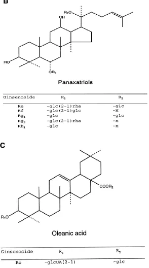
1.3 Chemical Structures of Panaxatriol ginsenosides and Oleanic acid 15
(nonsteroidal saponin)
1.4 Chemical Structures of Ginseng Polysaccharides 17
1.5 LPS and Polysaccharides initiation of Innate Immune response 24
via TLR-4 Signaling pathway
1.6 Gastrointestinal absorption of orally administered biochemical 32
compounds into systemic circulation
3.1 Immunostimulatory effects of the (a) AQ and (b) ALC ginseng extracts 73
on 24 hr macrophage production of (i) NO, (ii) TNF-α and (iii) IL-6
3.2 Effect of the (a) AQ and (b) ALC ginseng extracts on LPS-stimulated 24 hr 75
macrophage production of (i) NO and (ii) TNF-α
3.3 ALC ginseng extract suppressed up-regulation of macrophage 77
NO production by the AQ ginseng extract
3.4 Sephadex G-75 (47×2.5 cm) chromatographic fractionation 79
3.5 Immunostimulatory effect of Fraction I and III of the AQ, PS extracts of 81
ginseng
3.6 Effect of Fractions I (Mw = ~ 73 kDa) and III (Mw = ~ 37 kDa) of the 83
ALC extract on LPS-stimulated 24 hr macrophage production of NO
3.7 Effect of the ALC ginseng extract, its macromolecules and ginsenoside 85-86
fraction on LPS-stimulated 24 hr macrophage production of NO
3.8 Effect of the ALC ginseng extract, its macromolecules and ginsenoside 87-88
fraction on LPS-stimulated 24 hr macrophage production of TNF-α
3.9 Effect of the ALC ginseng extract, its macromolecules and ginsenoside 89-90
fraction on LPS-stimulated 24 hr macrophage production of IL-6
3.10 Visual comparison of the chromatograms obtained from size exclusion 92
chromatography of crude PS, AQ and ALC extracts of ginseng
3.11 Summarized identification of polysaccharides in Fraction I (Mw = ~73 kDa) 93
of the AQ extract, PS extract, AQ extract and ALC extract of ginseng by
size exclusion chromatography with right angle light scatteringdetection
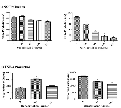
4.1 Orally administered AGRPS extract: (A) elevated NO production 115
and (B) reduced LPS-stimulated NO production ex vivo in cultured alveolar
Macrophages
4.2 Orally administered AQ extract: (A) elevated NO production and (B) 116
macrophages
4.3 Immunostimulatory and suppression of LPS induced immunological effects 118
of AGRPS or AQ extract treatment in vivo
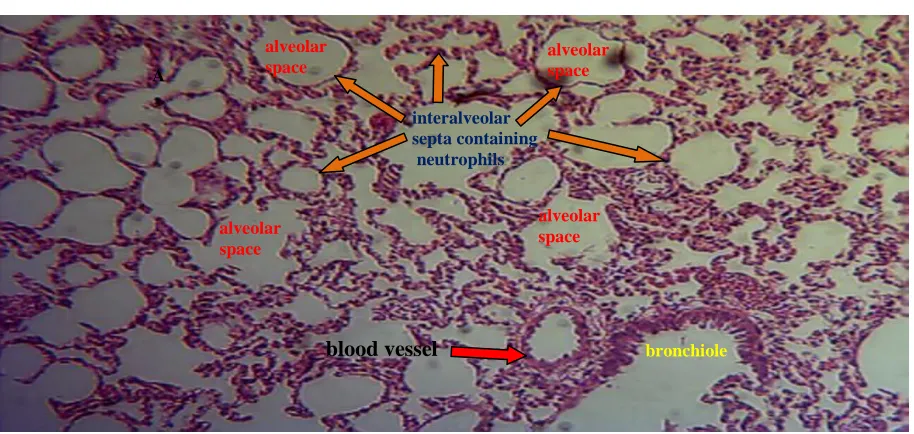
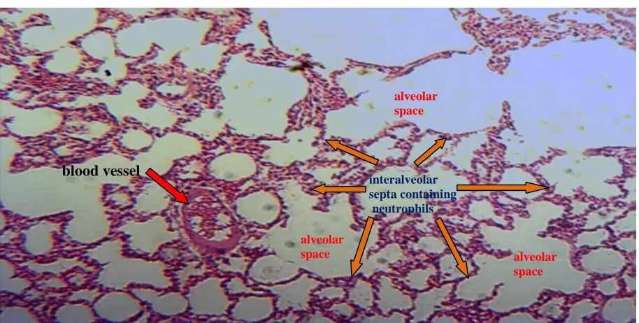
4.4 Lung histopathology of Saline (Control) and LPS treated rats 119
(magnification x40)
4.5 Effect of AGRPS and AQ extracts on LPS induced histopathology 120
alterations in lung (magnification x40)
4.6 DEAE-cellulose Ion exchange Column (40 × 2.5 cm) chromatographic 122
fractionation of the AGRPS into neutral PS fraction and acidic PS
fraction
4.7 Superdex G-200 size exclusion column (40 × 2.5 cm) 124
chromatographic fractionation of acid PS
4.8 Immunostimulatory effects of AGRPS and AQ extracts, 126-127
acidic and neutral PS fractions on 24 hours murine macrophage
production of (A) NO and (B) TNF-α in vitro
4.9 Immunostimulatory effects of AGRPS and AQ extracts, 128-129
acidic and neutral PS fractions on 24 hours rat alveolar macrophage
production of (A) NO (nitrite) and (B) TNF-α in vitro
4.10 Comparison of composite mixture (12% acidic PS fraction and 130-131
of NO (nitrite) in vitro
4.11 Immunostimulatory effects of acidic PS fractions ≥100 kDa, 133-134
50 - 100 kDa, 30 - 50 kDa, 10 - 30 kDa on 24 hours
macrophage production of NO in vitro
4.12 Effects of AGRPS and AQ extracts, acidic and neutral PS fractions 136-137
on LPS-stimulated 24 hours murine macrophage production of
(A) NO (nitrite) and (B) TNF-α in vitro
4.13 Effects of AGRPS and AQ extracts, acidic and neutral PS fractions 138-139
on LPS-stimulated 24 hours rat alveolar macrophage production
of (A) NO (nitrite) and (B) TNF-α in vitro
4.14 Comparison of composite mixture (12% acidic PS fraction and 140-141
30% neutral PS yield), acidic PS, neutral PS fractions on LPS-stimulated
24 hours macrophage production of NO (nitrite) in vitro
4.15 Effects of acidic PS fractions ≥100 kDa, 50 - 100 kDa, 30 - 50 kDa, 143-144
10 - 30 kDa, on LPS-stimulated 24 hours macrophage production of
(A) NO (nitrite) and (B) TNF-α in vitro
5.1 Chromatograms obtained from high performance size exclusion 175
chromatography analysis of (A) blank plasma (B) 2000 μg/mL AGRPS
and (C) blank plasma spiked with 2000 μg/mL AGRPS
chromatographic analyses of plasma samples
5.3 Chromatograms obtained from high performance 180
size exclusion chromatography of AGRPS analyte (11.22 mL)
and its biotransformed end product (16.03 mL) in plasma samples
collected from rats orally fed with 125 mg/kg single dose of AGRPS for
(A) 0.5 hr and (B) 4 hr
5.4 Chromatograms obtained from high performance size exclusion 182
chromatography of AGRPS analyte (11.22 mL) and its biotransformed
endproduct (16.03 mL) in plasma samples collected from rats treated IP with
125 mg/kg single dose of AGRPS for (A) 0.5 hr and (B) 4 hr
5.5 Chromatograms obtained from high performance size exclusion 185
chromatography analysis after (A) 1 hr (B) 8 hr and (C) 24 hr incubation
of AGRPS solution with pectinase enzyme
5.6 Chromatograms obtained from high performance size exclusion 186
chromatography analysis after (A) 1 hr, (B) 8 hr and (C) 24 hr
incubation of AGRPS solution with cellulase enzyme
5.7 Chromatograms obtained from high performance size exclusion 188
chromatography analysis after 24 hr incubation of rat plasma obtained
from 4 hr oral AGRPS treatment (A) without pectinase enzyme
5.8 Chromatograms obtained from high performance size exclusion 189
chromatography analysis after 24 hr incubation of rat plasma obtained
from 4 hr IP AGRPS treatment (A) without pectinase enzyme and
(B) with pectinase enzyme
5.9 Plasma concentration–time curve of AGRPS in rats after 0.5–8 hr 192
(A) oral and (B) IP administration of 125 mg/kg single doses of AGRPS
5.10 Chromatogram obtained from high performance size exclusion 195
chromatography of rat plasma sample collected 24 hr on the
last day after a 6 days oral treatment with 125 mg/kg of AQ extract
5.11 Chromatograms obtained from high performance size exclusion 197
chromatography of (A) CVT-E002TM (11.22 mL) and its biotransformed
end products (14.00 mL and 16.03 mL) in rat plasma sample collected
24 hr on the last day after a 6 days oral treatment with 125 mg/kg of
CVT-E002TM and (B) 2000 µg of CVT-E002TM
LIST OF TABLES
Table Title Page
LIST OF APPENDICES
Title Page
Appendix 1: Copyright permission for Chapter 1 232
Appendix 2: Copyright permission for Chapter 3 234
Appendix 3: Copyright permission for Chapter 4 235
Appendix 4: Standard curve of pullulan polysaccharide
Log Molecular weight versus Retention volume for
Determination of molecular weight for Chapter 5 237
Appendix 5: Calculation of AGRPS Maximum Plasma Concentrations 238
LIST OF ABBREVIATIONS
Abbreviation Full Name
ALC Alcoholic
AGRPS American Ginseng Root Polysaccharides
AP-1 Activator protein 1
AQ Aqueous
AUC Area Under the Concentration–time curve
BAL Broncho Alveolar Lavage
CAM Complementary or Alternative Medicine
Cmax Maximum plasma Concentration
COX-2 Cyclooxygenase-2
Da Dalton
DEAE Diethylaminoethyl
DC Dendritic cells
ELISA Enzyme-Linked Immunosorbent Assay
ERK1/2 Extracellular signal-regulated kinases 1/2
GAP Good Agricultural Practice
GFC Gel Filtration Chromatography
GRPS Ginseng root polysaccharides
HPLC High Performance Liquid Chromatography
HPSEC High Performance Size Exclusion Chromatography
IKK Inhibitory Kappa Kinase
iNOS inducible Nitric Oxide Synthase
IR Infrared
IRAKs Interleukin-1 receptor-associated kinases IL-6 Interleukin-6
JNKs Jun N-terminal kinases
kDa kilo Dalton
LPS Lipopolysaccahride
LC Liquid Chromatography
MAPK Mitogen-Activated Protein Kinase
MyD88 Myeloid Differentiation factor 88
Mw Molecular weight
NF-κB Nuclear Factor kappa B
NK Natural Killer
NHPs Natural Health Products
NMR Nuclear Magnetic Resonance
OGRIC Ontario Ginseng Research & Innovation Consortium
PAMPs Pathogen-Associated Molecular Patterns PI3K Phosphoinositide 3-kinase
PRRs Pattern Recognition Receptors
PBS Phosphate-Buffered Saline
PGE2 Prostaglandin E2
PS Polysaccharides
PPD Protopanaxadiol
PPT Protopanaxatriol
RI Refractive Index
SD Standard Deviation
SEC Size Exclusion Chromatography
TAK1 Transforming growth factor-beta Activated Kinase 1
TIR Toll/IL-1 receptor
TIRAP TIR domain-containing adaptor protein
TLRs Toll-like receptors
TLR-4 Toll-like receptor 4
TRAF6 TNF receptor associated factor 6
TRAM TRIF-related adaptor molecule
Tmax Time to attain Cmax
TM Traditional medicine
TNF-α Tumor Necrosis Factor-alpha
UV Ultraviolet
Chapter 1
1. Introduction
1.1 Medicinal Plants
1.1.1 The Use of Medicinal Plants in Traditional Medicine
Our experience with medicinal plants can be traced back to biblical times as described in the
book of Ezekiel chapter 44 verse 12 which states ‘and the leaf thereof for medicine’ [1]. Since
ancient times mankind has utilized the properties of plants not just for food and shelter, but also
for health and well-being’ [2]. Early empirical observations served as the basis for the use of
plants as prophylactic and therapeutic agents in herbal medicine [3]. Traditional medicine can be
described as the combination of knowledge and practice used for diagnosing, preventing, or
curing disease which relies exclusively on past experience and observation handed from
generation to generation, verbally or in writing [4]. Traditional medicine (TM) covers a wide
variety of medicinal plants and their concoctions which vary from country to country and region
to region. Practice of herbal medicine is often referred to as "complementary" or "alternative"
medicine (CAM) in some countries. Since the 1990s its use has surged in many developed and
developing countries as Eastern and Western medicines intersect [5].
Traditional Chinese, East Indian Ayurveda, Native American and African medicine are examples
of different systems of traditional medicine, with the philosophy and practices of each being
influenced by the prevailing diseases, environments for plant growth, and geographic areas
within which they first evolved. Generally, in each of these regions, a common philosophy
evolved with a holistic approach to life, i.e. equilibrium of the mind, body, and the environment,
The use of medicinal plants is a central part of all these systems of traditional medicine in which
a restoration of health, rather than on treating a particular ailment or disease from which the
patient is suffering is usually the main goal [8-9].
A medicinal plant (herb) is any plant which, in one or more of its organs or parts (e.g. leaf,
flower, fruit, seed, stem, wood, bark, root, rhizome, juice, gum, fixed oil, resin) contains a
variety of substances that can be used for therapeutic purposes or which are precursors for the
synthesis of useful drugs [4].
Scientists from academic institutions and industrial firms have turned to plants as a source of
either new drugs or of compounds from which more efficacious or less toxic drugs can be
developed based on their long-standing use in traditional medicine [10]. The World Health
Organization (WHO) has estimated that 80% of the world’s rural population relies on TM’s use
of plant extracts or their active principles for their primary health care needs. Despite tremendous
progress in synthetic chemistry, some 25% of prescription medicines are still derived either
directly or indirectly from plants [2, 11-12].
A 2010 survey shows that 73% of Canadians regularly take natural health products (NHPs),
Health Canada defines NHPs as: vitamins and minerals, herbal remedies, homeopathic
medicines, traditional medicines such as traditional Chinese medicines, probiotics, or other
products like amino acids and essential fatty acids. NHPs must be safe to use as over-the-counter
1.1.2 The Use of Medicinal Plants in Drug Discovery
Of the 252 drugs considered as basic and essential by the WHO, 11% are exclusively of plant
origin and a significant number are synthetic drugs obtained from natural precursors [14].
Examples of important drugs obtained from plants are taxol which is isolated from Taxus (T.
brevifolia and T. bacata), artemisinin from Artemisia annua, digoxin from Digitalis spp., quinine
and quinidine from Cinchona spp., vincristrine and vinblastine from Catharanthus roseus,
atropine from Atropa belladonna and morphine and codeine from Papaver somniferum [2].
About 6% of higher plant species (angiosperms and gymnosperms) have been screened for
biological activity and 15% of them have been reported to be phytochemically active [14]. Since
only 5–15% of the higher plants have been systematically investigated for the presence of
bioactive compounds, nature’s biodiversity remains largely unexplored [15].
Different approaches to drug discovery using higher plants include: chemical screening,
biological assays, biological activity reports (e.g., ecology based), or ethnomedical (traditional
medicine) use of plants [10]. A diversified approach to drug discovery involves the observation,
description, and experimental investigation of indigenous medicinal plants and their biological
activities. It is based on botany, chemistry, biochemistry, pharmacology, and many other
disciplines that contribute to the discovery of natural products that are pharmacologically active
[16]. Of the more than 120 pure drugs derived from plants in current commercial use, three
quarters were discovered through scientific investigations of traditional uses [17]. Phytochemical
and biological characterization of plants used in traditional herbal medicine is an important tool
in the scientific research of potential medicinal agents which can be used for the prevention, risk
The goals of using plants as sources of therapeutic agents are to: a) isolate bioactive compounds
for use as drugs; b) produce bioactive compounds that can be used for synthesis to produce
entities of higher activity and/or lower toxicity, c) use agents as pharmacologic tools and d) use
the whole plant or part of it as a herbal medicine [16].
Interest in drugs of plant origin is due to several reasons, namely, conventional medicine can be
inefficient (e.g. side effects and ineffective therapy), incorrect use of synthetic drugs results in
side effects and other problems, a large percentage of the world’s population does not have
access to conventional pharmacological treatment, and NHPs are considered harmless [2].
Phytomedicines exists as extracts which are concentrated preparations of a liquid, powdered, or
viscous consistency that are ordinarily made from dried plant parts (the crude drug) by
maceration or percolation [19].
1.1.3 Quality, Safety and Efficacy of Herbal Products from Medicinal Plants
As the world’s market demand for herbal products from medicinal plants increases, quality
control is of critical necessity to ensure that they are of consistent composition (quality), safety,
and efficacy. Medicinal plants do not supply its phytochemicals with a consistent, composition,
and unlike chemical drugs with a single entity, they contain multiple components. Factors such
as soil, climate, species, age, geographical origin, cultivation method, processing (harvesting,
storage, and manufacturing techniques), and contamination (pesticide, microbial and heavy
metal) can affect the composition, safety, and efficacy of medicinal plants and their NHPs
[19-20]. When species are harvested from wild plant populations, collectors may inadvertently
confuse desired species with close relatives, or even with unrelated species that are superficially
difficult to differentiate, especially when the financial incentive is great [21].
The use of good agricultural practice (GAP) and good manufacturing practice (GMP) is
encouraged by health regulatory agencies to ensure consistent quality from the point of
cultivation and harvesting of medicinal plants until the stage of processing and final delivery of
the NHPs [22]. The WHO has published general guidelines for GMP procedures which can be
employed for the manufacture of plant derived herbal products involving; the raw material
production, botanical taxonomic identification to assure species identification; the processing
and manufacturing stage, and analytical procedures similar to those employed for the
manufacture of conventional drugs to assure quality and purity by appropriate protocols [23-25].
Ensuring quality can be controlled in part by removing plant material that does not meet strict
quality standards and limiting further processing of plants that are not sufficiently consistent in
their relevant constituents. Thus, quality control of plant derived NHPs begin with the selection
and mixing of the herbal raw materials [19].
A major challenge when using herbal products is being able to consistently formulate a product
via the identification of a particular “marker compound” which is believed to be responsible for
the physiological effect [26]. A reported study on selected commercial ginseng products
prepared from Panax ginseng and Panax quinquefolius marketed as botanical supplements in
North America in the 1995-1998 period showed that the ginsenoside contents of P. ginseng and
P. quinquefolius products ranged from 0.00% to 13.54% and from 0.009% to 8.00%,
respectively, and close to 26% of these products did not meet label claims [27-28].
The percentage of a particular marker for a particular plant derived NHPs varies from product to
compounds are chemicals that are merely characteristic components of the herb in question and
may not have been tested for their actions or therapeutic efficacy in pharmacologic test models
or in clinical studies. Chemical characterization (extraction, fractionation, purification and
structural identification) are only meaningful when the purified and identified marker
compounds (fractions) have the same or similar biological activity as the plant or its extract [18].
Proper in vitro and in vivo assay methods need to be established for each step of the fractionation
and purification process [29]. Only those with sufficient activity should be used for further
purification.
The presence of multiple active compounds in herbal products can provide either synergistic or
antagonistic effects that may not be achievable by any single-component. These complex
interactions can present a unique challenge for bioactivity-guided fractionation, because the
relative activity of fractions may decrease with greater purity and may even be lost entirely [8].
The activity of the individual active components of botanicals can be assessed by recombining
the fractions after separation followed by confirmation of biological activity [29]. Consistency of
botanical therapeutic products can only be achieved when the active marker compounds are
Figure 1.1 - Summarized Scientific Approach of Evaluating Herbs and Herbal products [30]. GAP Certified Herbs or
Herbal Mixture
Pharmacognostic Identification and Authentication
Preparation Process and Technologies
CE, HPLC, ICP, IR, MS, UV Chromatographic and
Spectroscopic Determination (and Characterization) of phytochemical constituents to obtain physico-chemical discriminant indices
Qualitative and
Quantitative Biological
and Toxicological (In
vitro and In vivo) Assays to obtain
bioactive discrimination indices
Recognition Ability
Prediction Ability Pattern Recognition,
Logic Analysis and Classification into various Categories
Assessment Ability
Quality, efficacy and safety assured herbs and herbal products Working Plant Extracts
Quality control of plant derived NHPs involve finger printing of phytochemical markers to
ensure consistency in quality, safety and efficacy for consumers. Depending on the technology
and solvent used, chromatographic and spectrometric techniques can generate chemical
chromatographs and spectra that characterize the multicomponent active principle as uniquely as
a fingerprint. Phytochemical fingerprinting of test samples with authentic reference standards is a
fundamental quality control step for ensuring consistency. This is particularly true when little is
known about the relation of these constituents to actions and efficacies or about the
quantitative/qualitative makeup of the remaining components [19].
The initial challenge in the quality control process is isolating and analyzing medicinal plants
and their NHPs due to the complexity of their sample matrices and the presence of multiple
phytochemical components. Isolation of active constituents from medicinal plants serves as a
means by which constituents can be processed into safe medicinal products tailored towards
obtaining a quality product that has a consistent, uniform composition [31]. The nature of the
solvent and of the extraction and drying processes critically affects the phytochemical
composition of the finished product. Polar compounds are soluble in water, while lipophilic
constituents are soluble in alcohol. For example an aqueous extract of medicinal plant such as
ginseng will have a different spectrum of ingredients than an extract that has been isolated using
ethanol [19].
Novel technologies are required for separating and isolating these phytochemicals before they
are characterized using physicochemical techniques. A high quality chemical library of reference
standards is vital for research in the structure-activity relationship, and investigation of the
mechanism of NHPs [14, 18]. Some reference compounds are commercially available, such as
addition, insufficient availability of phytochemical reference standards such as ginseng
polysaccharides adds another level of difficulty and challenge to evaluate these components from
medicinal plants. Chromatographic or spectral phytochemical characterization is used and
accepted worldwide for the evaluation of medicinal plants and their NHPs. This fingerprinting
enables the detection and quantitation of desired markers present and for the assessment of the
pattern of the phytochemicals [19].
There is also a belief that plant derived NHPs are inherently safe without side effects when
properly used at normal therapeutic doses. However, these NHPs may also have undesirable side
effects and herb–drug or herb–herb interactions are possible [8]. Where safety information is
lacking on any medicinal plants being contemplated for NHPs, relevant research must be
performed prior to its employment [20]. The WHO has established guidelines for such studies
[33]. Adverse events, including drug-herb interaction must also be monitored to promote a safe
integration of efficacious herbal medicine into conventional medical practices [20]. Hence there
is a need to screen medicinal plants such as ginseng to identify novel bioactives for the
promotion of human health.
1.2 Ginseng
The name ginseng comes from the Chinese words ‘‘Jen Sheng,’’ meaning ‘‘man-herb,’’ because
of the humanoid shape of the root or rhizome of the plant, which is the part of the plant most
commonly used for extraction. The ginseng plant is a deciduous perennial belonging to the
Family Araliaceae and genus Panax. The genus name of ginseng "Panax" is derived from the
Greek pan (all) akos (cure), meaning "cure-all" or ‘‘all healing,’’ which describes the traditional
different species of ginseng which have being identified all over the world. The most commonly
used species of ginseng are P. ginseng (Asian ginseng) and P. quinquefolius (American ginseng).
P. ginseng has been used in the Orient for thousands of years, while P. quinquefolius has been
used by Native Americans for at least hundreds of years [9, 34-44].
In 1714 while at St Louise, Canada (near Montreal), Father Lafitau stumbled across ginseng
growing at the site of a new house with an appearance that accurately fit the ginseng roots Father
Jartoux (French Jesuit missionary) described to him in a letter in 1709 from China [36].
American ginseng was first introduced in the “New Compilation of Materia Medica” in 1757.
There are three kinds of American ginseng namely; cultivated, simulated wild, and wild.
American ginseng is also cultivated in some Asian countries, like China. As a perennial herb,
most wild populations of American ginseng thrive in the upland, north and east-facing woods
where shade and loamy soils are typical [40]. American ginseng has been harvested in North
America primarily for export to Asia since the 1700s and is highly valued for its medicinal and
herbal use [41]. The quantity of wild ginseng was not sufficient to meet the demand, so
experiments on cultivation were undertaken in 1878 and was achieved in Fabius, New York by
George Stanton, a retired tinsmith turned to farmer. Cultivation of Panax ginseng in Asia started
around 11 B.C. by transplantation of wild ginseng. In 1122 A.D., ginseng cultivation was also
attempted through the propagation of transplanted ginseng from seeds [36]. American ginseng is
distributed in the eastern temperate forest areas of North America; Ontario and Quebec in
Canada, Minnesota, Wisconsin, Oklahoma and Georgia in the United States of America [40].
Ginseng is prepared and used in several ways: as fresh ginseng (sliced and eaten, or brewed in a
tea), white ginseng (peeled and dried), red ginseng (peeled, steamed, and dried), extract (tincture
Medicine (TCM), Asian ginseng is believed to have a ‘warm’ or ‘yang’ property, while
American ginseng has a ‘cool’ or ‘ying’ characteristic [37]. Ginseng products are often referred
to as “tonics,” a term that has been replaced by “adaptogen”. The term “adaptogen” can be
defined as an agent that increases resistance to physical, chemical, and biological stress and
builds up general vitality, including the physical and mental capacity for work [43]. Ginseng root
is used as a medicinal plant in traditional herbal medicine to improve overall health, restore the
body to balance, help the body to heal itself, and to reduce stress [37, 44].
Ginseng’s broad spectrum of biological activity has been attributed to its multiple bioactive
components namely ginsenosides, polysaccharides, peptides, polyacetylenic alcohols, and fatty
acids. These known active phytochemicals are present in most ginseng species, although there
can be slight variation in different species [39, 44]. Scientific studies have shown that ginseng
exhibits a wide range of beneficial pharmacological effects including immunomodulation,
anti-tumor, anti-oxidation, anti-depression, hypoglycemia, inhibition of gastric lesions, attenuation of
leptin-induced cardiac hypertrophy, heart protection against ischemia and reperfusion injury,
prevention of glucose-induced oxidative stress, prevention of diabetic nephropathy, retinopathy
and cardiomyopathy [38, 41, 45-59].
1.2.1 Ginseng Bioactives
1.2.1.1 Ginsenosides
Ginsenosides belong to a family of steroids with a four trans-ring rigid steroid skeleton that
shares a unique triterpenoid saponin structure of the dammarane type (Figure 1.2). More than
100 different ginsenosides have been isolated from the roots, leaves, stems, flower buds, and
Ginsenosides differ from one another by the type of sugar moieties, sugar number, and site of
sugar attachment at positions C-3, C-6, or C-20. The structural isomerism and stereoisomerism,
the number and site of attachment of hydroxyl groups, and available modified side chain at C-20
also increase their diversity. Ginsenosides from ginseng are divided into several groups.
Protopanaxadiol (PPD) and protopanaxatriol (PPT) groups are the main constituents; while
ocotillol and oleanane groups are minor ones [37, 39-40]. The PPD group has sugar moieties
attached to the β-OH at C-3 and/or C-20, and the PPT group has sugar moieties attached to the
α-OH at C-6 and/or β-α-OH at C-20. The ocotillol group has a five- membered epoxy ring at C-20,
and the oleanane group has a modified C-20 side chain [40]. Ginsenoside Rb1, Re, Rd, Rc, Rg1,
and Rb3 are the six major saponins in American ginseng, and the variability in individual
ginsenosides and total ginsenoside amount in different commercial products of American
ginseng may be responsible for different or even opposing reported pharmacological activities,
which is in part associated with natural variations such as climate, geographical location and
cultivation length conditions [40]. Quality control of ginseng products will ensure consistency in
quality, safety and efficacy for consumers. Ginseng extracts (aqueous and alcoholic) contain
relative variable amount of ginsenoside components [54]. Ginsenosides have been reported to
exert numerous pharmacological activities including immunomodulatory, oxidant,
Figure 1.3 - Chemical Structures of Panaxatriol ginsenosides and Oleanic acid (nonsteroidal
1.2.1.2 Polysaccharides
Plant polysaccharides are biopolymers of various monosaccharides linked together through
glycosidic bonds, resulting in complex structures. Plants polysaccharides are usually isolated by
hot water extraction of the plant material, after which they are purified and precipitated with
alcohol from the water extract [63]. It is noteworthy that, in comparison with other biopolymers
such as proteins and nucleic acids, polysaccharides offer the highest capacity for carrying
biological information because they have the greatest potential for structural variability [64].
Scientific studies have demonstrated that plant polysaccharides such as water-soluble
polysaccharides used in NHPs are non-toxic and exhibit a number of beneficial biological
activities, including immunostimulation, anti-tumor, wound-healing, hematopoietic,
radioprotective, anti-ulceric and anti-atherosclerotic effects [65-66].
Ginseng polysaccharides are biopolymers formed from a complex chain of monosaccharides rich
in L-arabinose, galactose, L-rhamnose, galacturonic acid, glucuronic acid and
D-galactosyl residue linked together through glycosidic bonds, resulting in complex
macromolecular architectures [46, 63, 67]. Their molecular weights range from 3500 to
2,000,000 Da [47, 67-72] which contribute to their diverse physicochemical properties and
biological activities. CVT-E002™ (sold commercially as COLD-FX®) is a herbal product rich in
poly-furanosyl-pyranosyl-saccharides extracted by an aqueous method from the root of
American ginseng as described in its patent [67-68]. The process of preparing ginseng fraction
CVT-E002 comprises: combining American ginseng dried ground root with a first solvent
comprising an alcohol in a proportion of about 7 to 9 ml of first solvent per gram of ginseng. The
resulting solution is then heated at a temperature of about 80 to 100° C for a time period of about
Thereafter, the first ginseng solution is separated to produce an alcohol/ginseng solution and a
first ginseng residue. Thereafter, the first ginseng residue is combined with water in a proportion
of about 7 to 9 ml of water per gram of ginseng residue. The ginseng residue solution is then
heated at a temperature of about 80 to 100° C for a time period of about 2 to 4 hours, to produce
a ginseng residue solution. Thereafter, the ginseng residue solution is separated to produce a
second ginseng residue and an aqueous extract solution containing a ginseng extract. The
aqueous extract solution is then dried to produce ginseng fraction CVT-E002 [68].
1.3 Immunomodulation
The immune response of a host is its key defense and surveillance system capable of eradicating
invading infectious microorganisms (bacteria, fungi, virus, and protozoa) and maintains
homeostasis required for a normal healthy condition. The immune system is divided into the
innate and adaptive (specific) immunity which can distinguish between foreign pathogenic
micro-organisms and the body’s cells and tissues via surface cell receptors that can recognize
toxic surface pathogen-associated molecular patterns (e.g. lipopolysaccharides of Gram negative
bacteria) as foreign [73]. Components of the innate immunity are macrophages, natural killer
cells (NK-cells), dendritic cells (DC), granulocytes (neutrophils, basophils, eosinophils),
proinflammatory mediators e.g. cytokines (tumor necrosis factor, interleukins), reactive oxygen
/nitrogen species (e.g. nitric oxide) and prostanoids (e.g. prostaglandins). While that of the
adaptive (acquired or specific) immunity include T lymphocytes, B lymphocytes and antibodies
[74].
Compounds that are capable of interacting with the immune system to up-regulate or
biologic response modifiers [75]. A broad spectrum of such compounds are still being
investigated and characterized. Biopolymers like plant polysaccharides which have long been
believed to have benign biologic properties, have recently been shown to act as biological
response modifiers. They can either up-regulate or down-regulate specific aspects of biological
responses of the host [65-66]. A current significant problem is the rise of microbial infections
and their resistance to synthetic antimicrobial agents and naturally derived antibiotics. Unlike the
use of antibiotics and antimicrobials to kill pathogens, a key immunotherapeutic strategy to
address this challenge will be to identify bioactive agents which can interact with the host
immune response defense to enhance its ability to fight diseases and infections or neutralize
immunotoxic response.
1.3.1 Macrophage-mediated Innate Immunity
Macrophages, first described by Metchnikoff in the 1880s, are large mononuclear phagocytic
cells, which are derived from monocytes which originate from haematopoietic stem cells in the
bone marrow. Monocytes differentiate further into macrophages as they leave the blood and
enter the tissue. In tissues, a small number of macrophages differentiate under the influence of
cytokines and, depending on the tissue type, they may become osteoclasts (bone), kupffer cells
(liver), microglia (brain), alveolar macrophages (lung) and peritoneal macrophages (peritoneum)
[76]. Fast acting macrophage-mediated innate immunity is the first line of defense in identifying,
neutralizing, destroying and removing microbial pathogens and influencing the subsequent slow
adaptive immune response. Macrophages act by means of a number of different mechanisms: (a)
directly via phagocytosis by destroying bacteria, parasites, viruses and tumor cells; (b) indirectly
can activate other cells; (c) as accessory cells, by processing antigen and presenting digested
peptides to T lymphocytes; and (d) by repairing tissue damage [65, 77].
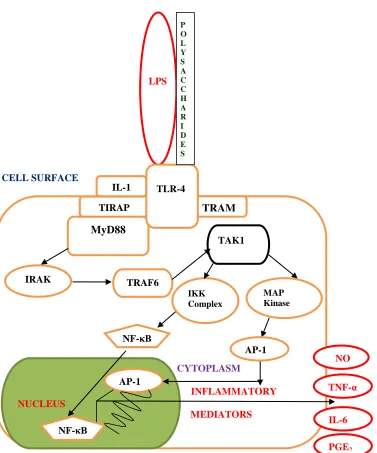
The pathogen-associated molecular patterns (PAMPs) of pathogens (bacteria to fungi, protozoa, and viruses) are the targets of innate immune recognition with the help of their pattern recognition receptors (PRRs) which can distinguish between self (host) and pathogens via their PAMPs [78]. During activation of macrophage function by plant polysaccharides or lipopolysaccharide (LPS) endotoxin (a PAMP) from microbial infection caused by Gram negative bacteria, plant polysaccharides or LPS ligand binds to the transmembrane PRRs such as Toll-like receptors (TLRs) of innate macrophage cells which induce down-stream intracellular
events [79]. As seen in Figure 1.5, the recognition and binding of plant polysaccharides or LPS
ligand by Toll-like receptor 4 (TLR-4) leads to the recruitment of various cytoplasmic TIR
(Toll/IL-1 receptor) domain-containing adaptors such as MyD88 (myeloid differentiation factor
88), TIRAP (TIR domain-containing adaptor protein) and TRAM (TRIF-related adaptor
molecule) [80]. MyD88 recruits IRAKs (interleukin-1 receptor-associated kinases) which then
activates TRAF6 (TNF receptor associated factor 6), leading to the activation of TAK1 (transforming growth factor-beta activated kinase 1). At the point of TAK1 activation, the
signaling pathway bifurcates. One limb of the pathway leads to TAK1 activating the IKK
(inhibitory kappa kinase) complex which phosphorylates and degrades IκB, resulting in the
release and translocation of NF-κB (nuclear factor kappa B) transcription factor from the
cytoplasm into the nucleus. Activated NF-κB binds to the promoters of diverse proinflammatory
mediators including TNF-α, interleukin-6 (IL-6), and enzymes such as iNOS (inducible nitric
oxide synthase) and COX-2 (cyclooxygenase-2), thereby activating the transcription and
of the MAPK (mitogen-activated protein kinase) kinases such as p38, JNKs (Jun n-terminal
kinases) and ERK1/2 (extracellular signal-regulated kinases 1/2), which leads to the activation of
AP-1 (activator protein 1) transcription factor that induces the transcription of proinflammatory
mediators [79, 80]. The expression of Tα, IL-6, iNOS and COX-2 induced by activated
NF-κB transcription factor results in the production of TNF-α, IL-6, nitric oxide (NO) and
prostaglandin E2 (PGE2) respectively. NF-κB is highly activated in inflammatory disease
conditions such as endotoxemia [82]. LPS induced macrophage overproduction of TNF-α, IL-6,
NO and PGE2 have been shown to play critical roles in the pathological process of many
inflammatory diseases, including endotoxemia and septic shock [83-90]. The presence of a large
amount of LPS endotoxin in the bloodstream, as observed during severe gram-negative bacterial
infections or as caused by translocation of enterobacteria from the gut, induces excessive
macrophage stimulation with uncontrolled production of proinflammatory mediators and
cytokines. This endotoxin is harmful and leads to endotoxemia with dramatic pathophysiological
reactions such as fever, leukopenia, tachycardia, tachypnea (acute respiratory failure),
hypotension, disseminated intravascular coagulation, myocardial dysfunction and multi-organ
CELL SURFACE
Figure 1.5 – LPS and Plant polysaccharides initiation of Innate Immune response via TLR-4
Signaling pathway [78-81].
CYTOPLASM
INFLAMMATORY
MEDIATORS
TRAM
TRAF6
MyD88
TAK1
MAP Kinase
NUCLEUS
NF-κB
AP-1
IKK Complex
AP-1 TIRAP
NF-κB
TNF-α NO
IL-6
IRAK
LPS
IL-1 TLR-4
P O L Y S A C C H A R I D E S
LPS is a glycolipid that constitutes the major portion of the outermost membrane of
Gram-negative bacteria and is essential for bacterial growth and survival [97]. It is a complex,
negatively charged molecule composed of a polysaccharide chain called the O-specific chain and
a lipid moiety referred to as lipid A. The latter is the actual toxic moiety of the LPS molecule and
contains phosphate groups shown to be essential for its immunostimulatory activity [98].
The interaction of lipid A moiety of LPS with macrophages is important and subsequent cellular
activation results in the release of systemically active proinflammatory molecules, which in turn
mediates systemic toxicity. LPS is an extremely potent toxin: macrophages can be activated at
concentrations of LPS as low as 1 pg/mL [97]. LPS endotoxin exerts its profound immunotoxic
effects by stimulating host cells (mainly monocytes/ macrophages, but also endothelial cells,
smooth muscle cells, and neutrophils) to produce and release proinflammatory mediators and
cytokines (NO, IL-6 and TNF-α). The presence of high amounts of LPS leads to the release of
these proinflammatory mediators and cytokines in large quantities, resulting in the described
pathophysiological reactions [91]. The suppression of LPS immunotoxic (anti-inflammatory)
effects of various immunomodulation compounds have been evaluated using both in vitro and in
vivo models [99-101]. The ability of an agent to inhibit LPS-induced macrophage overproduction
of proinflammatory mediators is called an immunosuppressive (anti-inflammatory) effect, while
the ability of an agent to stimulate or enhance macrophage production of proinflammatory
mediators is called an immunostimulatory effect. Antibiotics capable of killing Gram negative
bacteria are currently used in the management of endotoxin mediated inflammatory diseases,
although the continued rise in antibiotic resistance and mortality from infections caused by these
organisms has led to investigation of beneficial therapies aimed at inhibiting or neutralizing the
endotoxins, has been investigated and found to neutralize many endotoxic activities (including
lethality), and was more protective than antibodies to core LPS. Unfortunately, the neurotoxicity
and nephrotoxicity of polymyxin B limits its potential as a therapeutic agent [102]. Hence a less
toxic anti-LPS agent would be highly desirable as a therapeutic (prophylactic or curative) agent.
Therapies directed at the neutralization of proinflammatory mediators or LPS that are promising
in experimental models have been largely ineffective in clinical trials. Therefore, the
development of new therapies is of major interest [103-105].
1.3.2 Modulation of Macrophage Function as a Target for Immunotherapy
The innate immunity (e.g. macrophage function) of a host which is responsive to LPS is also a
known target for plant polysaccharides which are biological response modifiers. Plant
polysaccharides can up-regulate macrophages production of proinflammatory mediators to fight
infection, they can also neutralize or suppress immunotoxic response by down-regulating
macrophage production of proinflammatory mediators [65, 75]. Bioactive compounds which can
inhibit LPS endotoxin from triggering excess macrophage production of NO, TNF-α and IL-6
proinflammatory mediators will be very useful in the prevention and treatment of inflammatory
diseases such as endotoxemia. New approaches to the prophylaxis of diseases like endotoxemia
can be based on comprehensive blockade of the LPS signaling pathways in macrophages. The
immune response to LPS can take a number of different forms (immunostimulation and
immunosuppression). Low doses of endotoxin have been known since the 1940s to induce a state
of tolerance (desensitization), in which the immune response to subsequent LPS challenge is
altered. This alteration is characterized by suppression of proinflammatory mediators and
upregulation of anti-inflammatory mediators [106]. Mouse macrophages as well as human
challenge. The key readout for tolerance in these cells was the drastic reduction of TNFα
production as compared to the cells exposed to LPS only once [107].
Interestingly, bioactive polysaccharides have been shown to exhibit both macrophage-mediated
immunostimulatory and suppression of induced proinflammatory effects [46, 65, and 108-109].
A possible mechanism by which plant polysaccharides suppress LPS-induced macrophage
stimulatory effect may be through their ability to desensitize immune cells (e.g. macrophages)
from LPS toxic stimulation, similar to the tolerance ability of LPS pre-exposure to desensitize
subsequent LPS challenge. Bioactive polysaccharides can serve as useful prophylactic and
therapeutic agents for immune and inflammatory diseases. The ability to neutralize LPS
immunotoxicity is a desired requirement for future immunobioactive compounds that can be
used as a prophylactic against endotoxemia. Ginseng polysaccharides may provide such
immunobioactive compounds. Evaluation of an herb like ginseng provides an opportunity for the
discovery of novel agents that can combat disease conditions mediated by LPS such as
endotoxemia.
1.3.3 Ginseng Modulation of Immune Function
Different immunomodulatory effects of ginseng have been reported, including both
immunostimulatory and immunosuppressive effects [110-119]. Aqueous (AQ) and alcoholic
(ALC) ginseng extracts have been reported to exert immunostimulatory [53, 110-113] and
immunoinhibitory [45, 114-118] effects respectively. On the contrary, ginseng AQ extract has
also been reported to possess immunoinhibitory effects [119]. The basis for the apparent
paradoxical immunomodulatory effects is unclear but may be attributed to different experimental
![Figure 1.1 - Summarized Scientific Approach of Evaluating Herbs and Herbal products [30]](https://thumb-us.123doks.com/thumbv2/123dok_us/7770992.1279514/40.612.116.469.58.671/figure-summarized-scientific-approach-evaluating-herbs-herbal-products.webp)
![Figure 1.2 - Chemical Structures of Panaxadiol ginsenosides [39].](https://thumb-us.123doks.com/thumbv2/123dok_us/7770992.1279514/46.612.85.370.82.550/figure-chemical-structures-of-panaxadiol-ginsenosides.webp)
![Figure 1.4 - Chemical Structure of Ginseng Polysaccharides [69].](https://thumb-us.123doks.com/thumbv2/123dok_us/7770992.1279514/49.612.74.538.85.428/figure-chemical-structure-ginseng-polysaccharides.webp)