DOI: 10.1534/genetics.110.122994
Differential Maintenance of DNA Sequences in Telomeric
and Centromeric Heterochromatin
P. G. DeBaryshe and Mary-Lou Pardue
1Department of Biology, Massachusetts Institute of Technology, Cambridge, Massachusetts 02139
Manuscript received September 9, 2010 Accepted for publication October 23, 2010
ABSTRACT
Repeated DNA in heterochromatin presents enormous difficulties for whole-genome sequencing; hence, sequence organization in a significant portion of the genomes of multicellular organisms is relatively unknown. Two sequenced BACs now allow us to compare telomeric retrotransposon arrays fromDrosophila melanogastertelomeres with an array of telomeric retrotransposons that transposed into the centromeric region of the Y chromosome.13 MYA, providing a unique opportunity to compare the structural evolution of this retrotransposon in two contexts. We find that these retrotransposon arrays, both heterochromatic, are maintained quite differently, resulting in sequence organizations that apparently reflect different roles in the two chromosomal environments. The telomere array has grown only by transposition of new elements to the chromosome end; the centromeric array instead has grown by repeated amplifications of segments of the original telomere array. Many elements in the telomere have been variably 59-truncated apparently by gradual erosion and irregular deletions of the chromosome end; however, a significant fraction (4 and possibly 5 or 6 of 15 elements examined) remain complete and capable of further retrotransposition. In contrast, each element in the centromere region has lost$40% of its sequence by internal, rather than terminal, deletions, and no element retains a significant part of the original coding region. Thus the centromeric array has been restructured to resemble the highly repetitive satellite sequences typical of centromeres in multicellular organisms, whereas, over a similar or longer time period, the telomere array has maintained its ability to provide retrotransposons competent to extend telomere ends.
T
HE wealth of genome sequences now available has revealed much about genome organization and how this organization has evolved. These sequences have greatly extended our understanding of the ways in which transposable elements have added to and shaped eukaryotic genomes (Slotkinand Martienssen2007; Bourque2009; Cordauxand Batzer2009). For most eukaryotes, however, a significant portion of the genome is still poorly understood. This portion is the hetero-chromatin, which makes up about one-fifth of the human genome and a one-third of the Drosophila genome (Hoskinset al.2007). Heterochromatin is very rich in transposable element sequences, and it would be especially interesting to understand how these se-quences are related to the properties that distinguish heterochromatin from euchromatin. However, the large number of highly repeated and rapidly evolving DNA sequences in heterochromatin presents problems for accurately assembling sequences long enough to give a complete picture of the organization of these elements and their possible roles in specific heterochromatic regions such as centromeres and telomeres.We are studying the three non-LTR retrotransposons that maintain the length of Drosophila telomeresHeT-A,
TART, andTAHRE. These elements transpose by means of a poly(A)1
RNA that is reverse-transcribed directly onto the end of the chromosome (Pardueet al.2005). Successive transpositions form long arrays of head-to-tail repeats. These repeats are analogous to the repeats that telomerase adds in most other organisms except that the Drosophila repeats are copies of retrotransposons, which are three orders of magnitude longer than the repeats added by telomerase. Recently, we analyzed a BAC and a finished scaffold of equal quality containing sequence from two Drosophila melanogaster telomeres. These sequences gave us our first overview of both the organization of transposable elements within a telomere (Georgeet al.2006) and, now, the mechanisms by which these telomeres are maintained.
Although the telomeric retrotransposons appear capable of transposing only onto chromosome ends— either natural telomeres or broken chromosomes— HeT-ADNA was found to hybridize to the centromere region of theD. melanogaster Y chromosome (Agudoet al.1999) and later shown to colocalize with antibody to centro-mere-specific histone on the Y chromosomes of other members of themelanogasterspecies subgroup (Berloco et al. 2005). Thus, HeT-A-related sequences appear to 1Corresponding author:Massachusetts Institute of Technology, 31 Ames
St., Cambridge, MA 02142. E-mail: [email protected]
have been maintained at the centromere of the Y for .13 MY, even though the structure of the chromosome has diverged so that the Y is now metacentric in some species and telocentric in others. Mendez-Lago et al. (2009) have recently reported sequence of a BAC from thisD. melanogasterY centromere cluster.
The assembled sequences of the telomeric BAC and the centromeric BAC give the first opportunity to examine arrays long enough to allow us to analyze the organization, maintenance, and evolution of these retrotransposon arrays in two different heterochromatic environments. Comparison of the telomeric and cen-tromeric sequences reveals that HeT-A arrays in telo-meric heterochromatin are maintained very differently from those in centromeric heterochromatin; each is structured in ways that appear to be compatible with their different roles at the telomere and the centromere. It is frequently said that transposable elements have done much to shape eukaryotic genomes; in these cases, the genome is shaping the elements.
MATERIALS AND METHODS
Sequences analyzed: The BAC-containing telomere se-quence from the 4R telomere is AC010841. The XL telomere sequence is CP000372, a scaffold made by directed finishing of the sequence including the most distal gene on XL,CG17636, and extending some 20 kb into the telomere array. The BAC-containing sequence from the Y centromere region is BACR26J21 (Mendez-Lagoet al. 2009). The canonicalHeT-A
element is 23Zn-1 (U06920, bp 1015–7097); For Drosophila canon-ical sequences, seehttp://flybase.org/static_pages/downloads/ FB2010_09/transposons/transposon_sequence_set.embl.txt.gz.
Definition of sequence included inHeT-A/TARTtelomere arrays: We define HeT-A/TART telomere arrays as the se-quence on the chromosome end that is distal to the most distal
HeT-A or TARTelement disrupted by insertion of a non-telomere transposable element. The disrupted element marks the end of the transition zone, the proximal end of the telomere where nontelomere elements have invadedHeT-A
and TARTsequences. HeT-A Tags are recognizable 39-most sequences ofHeT-A that are transferred to the 59 end of a downstream element in the course of HeT-A transcription. They can form long ‘‘strings’’ on the 59 end of complete elements; each complete element in these arrays carries a Tag string. A fifth Tag string lies just 59of the transition zone on 4R where its parent element has apparently been invaded by a 1360 transposon.
The most interior Tags in long strings can become too decayed to be recognized and are indistinguishable from the 59UTR of the element to which they are attached; by default, we include these decayed Tags in the 59UTR. An exhaustive search for Tags was made during final annotation of the 4R and XL sequences after it had been recognized just how important their role might be in telomere maintenance. Using lowered stringency Blastn, we searched for terminal fragments ofHeT-A’s well-conserved 30-nucleotide 39terminus in short overlapping regions of the 59end of eachHeT-Aand the 39end of its upstream neighbor. (Only searches of short sequences identified all Tags.) Our search protocol ended the search at the last clearly defined Tag found, even if another could be seen within the nearby 59UTR. This procedure was adopted
because short TA(n) or TTA(n) (n,5) abound within the AT-rich 59UTR. Also, estimating conservatively, because it is likely that they are the result of replication slippage long after they moved into the interior of the telomere, we omit all but the first of several consecutive short sequences that make up the proximal end of the Tag string on XL. Not unexpectedly, Tag length is not distributed normally. In this article, we find it most descriptive to report mean lengths and their 95% confidence intervals. For most calculations, we omit elements truncated by cloning at the end of 4R and XL.
End erosion and terminal deletion data processing: Anal-ysis was performed using Excel and JMP 8 (SAS Institute, Cary, NC).
Dot-plot analysis:Dot plots comparing the canonicalHeT-A
with the sequences in the BACs were compared by the National Center for Biotechnology Information (NCBI) BLAST (bl2seq) using the default parameters for somewhat similar sequences (blastn). This procedure gives adequate alignment of the canonical sequence with sequences of all knownHeT-Asubfamilies. More stringent BLAST parameters show less sequence in the alignment.
RESULTS AND DISCUSSION
HeT-Aelements in telomeric heterochromatin
Sequences analyzed: Our comparison is based on a telomeric BAC from 4R, plus sequence from directed finishing of a scaffold from the telomere of chromosome XL, reported previously, but with some revisions in annotation. Both the 4R and the XL sequences begin in their assembled chromosome and extend into the telomere, thus showing the precise relationship between these telomere arrays and the rest of the genome (Georgeet al.2006). Neither sequence extends to the distal end of the telomere, but together they give nearly 100 kb of telomericHeT-A/TARTarray (76 kb from 4R and 20 kb from XL). Importantly, both include the most proximal elements of the array. Because telomere elements are added sequentially, the most proximal elements must be the oldest; thus, these arrays present the most accurate history available of D. melanogaster
telomere maintenance.
Neither the 4R nor XL sequences have nontelomeric elements in the telomeric array except for a small transition zone at the proximal edge of the array where there are some fragments of nontelomeric elements. We do not include these transition zones in our discussion of telomere arrays. Figure 1 gives an overview of the organization ofHeT-Asequences in the telomere array and transition zone of 4R. The sequence of this segment of the BAC is compared to the sequence of the canonical
HeT-A, 23Zn-1, which is diagrammed on thex-axis of the dot plot. Although none of the elements in the array belong to the same subfamily as the canonical element, all show linear similarity except for small gaps or repetitions in the 39 UTR. Each complete or partial
elements that have mildly truncated 59 UTRs are grouped with partial elements because we do not know whether they contain 59 UTR sequence required for transposition competence.
HeT-Aelements are much more abundant thanTART
elements in all D. melanogaster genomes that we have analyzed. Consistent with this, there are very fewTART
elements in the 4R array and none in the shorter XL sequence. Because we are making comparisons with a segment of the centromere array that consists entirely of
HeT-A, we have not includedTARTdata in the following analyses and discussion. Empty space in Figure 1 con-tains complete or partialTARTs. The third (very rare) telomeric retrotransposon,TAHRE, is also omitted from discussion because it is not found in any of the arrays considered here.
Each telomere array is a chronological record of events at the end of the chromosome: The studies reported here analyze the 4R and XL data to investigate sequence management within an intact Drosophila telomere. These sequences present a unique opportu-nity: the BAC and the sparser, but still useful, finished scaffold data are the first long sequences that give the exact order of elements in the telomere and their relationship to the rest of the genome. This relationship is informative becauseHeT-A,TART, andTAHRE trans-pose only onto the ends of chromosomes so that the order of elements in an array reflects the order of transposition onto the chromosome, with the oldest elements at the proximal end. Rearrangements and imprecise recombination are ruled out by the large-scale integrity of all the elements present; the full-length elements evidence replicative capability and even the partial elements are simply 59-truncated with no evi-dence of sequence rearrangement or decay.
The sequence at the 59end of each internal element is a record of the sequence that remained on a terminal retrotransposon when that element was capped by a newly transposing element and thus demoted to an internal position in the telomere. Comparing that capped sequence with the 59end of its presumed RNA template gives an estimate of the amount of sequence lost while that element served as the end of the chromosome. (This is a maximum estimate because some sequence could have been lost in transposition.)
The only measurements of the rate of end erosion and the addition of new elements on Drosophila chromosomes have come from broken chromosomes that have lost all telomeric and subtelomeric DNA. Such chromosomes shortened gradually by70 nucleotides per fly generation (Levis1989; Biessmannet al.1990; Mikhailovskyet al. 1999), calculated to represent an average loss of 2–3 nt per cell generation (Biessmann et al.1990). The rate at which a broken end is healed by addition ofHeT-Avaried from,2310 5to 23 10 3,
depending on the background genotype (Kahn et al. 2000). Thus, broken chromosome ends show a fairly regular slow loss of end sequence accompanied by infrequent addition of large retrotransposons;HeT-Ais
6 kb;TARTandTAHREare 10–12 kb.
The 4R and XL sequences give us the first opportunity to analyze the turnover of sequence on established telomeres. For this analysis we first discuss two indicators of sequence loss in the telomere arrays: (1) the Tags (see
59 Tags reveal slow sequence erosion of the telomere end) of nonessential sequence on the 59end of eachHeT-ARNA and (2) the distribution of complete and 59truncated elements in the telomere array.
These data fall into two classes: (1) qualitative obser-vations that describe the nature of the processes govern-Figure 1.—Analysis ofHeT-A-related
ing telomere maintenance and renewal (these observa-tions strongly constrain models based on numerical analysis) and (2) quantitative statistical analyses that help in distinguishing and judging competing conclu-sions. As discussed below, we conclude that maintenance of established telomeres involves at least three processes acting in concert to maintain relatively stable condi-tions: relatively short-range terminal erosion, long-range terminal deletion, and irregular transpositions.
59Tags reveal slow sequence erosion of the telomere end: Tags are short sequences added to the 59end of
HeT-ARNA byHeT-A’s unusual promoter that initiates transcription within the 39UTR of the upstream element (Danilevskayaet al.1997; Traverseet al.2010). The resulting Tag of upstream sequence becomes a de facto extension of the 59UTR and is reverse-transcribed with the rest of the RNA when the element transposes, providing expendable sequence to buffer the loss of essential 59 sequence from chromosome end erosion. Part or all of a Tag can be eroded while it forms the end of the telomere (Figure 2A). When a new element transposes to cap the chromosome end, erosion of the capped Tag is halted, leaving a partial Tag. Repeated transcription of an element will add a new Tag to any already on its 59end. Thus, an element can have a 59 string of variably truncated Tags—evidence that it has transposed multiple times (Figure 2B). Tags are the hallmark of complete HeT-As, which carry a string of them. We note that there is evidence to suggest that erosion can at times continue into the 59 UTR (see
Distribution of 59-truncated elements in the telomere array provides evidence of sporadic terminal deletions).
Tags are the best indicators of nucleotide loss on the end of an established telomere. There is only now a statistically robust sample of Tags and, because Tags are born in discrete short lengths, their end erosion is direct-ly measurable. (It is frequentdirect-ly convenient to differentiate between a Tag’s 39-oligoA sequence, its ‘‘Tag-tail,’’ and its more 59sequence, the ‘‘bare Tag’’). The initial length of each bare Tag is determined by its transcription start site, which can be 93, 62, or 31 nt from the 39 end of the element serving as a promoter; the 39-oligoA of the promoting element then forms the tail of this new Tag (Traverseet al.2010). Thus the longest Tag should have a 93-nt ‘‘bare Tag’’ plus its tail; the Tags in our arrays are all much shorter than this (Figure 2A).
If end erosion is regular and averages70 nt per fly generation, as found for broken ends, most Tags should last,1 generation, but complete elements typically have several Tags in various states of erosion (Figure 2B). Strings of partially eroded Tags indicate that these expendable sequences provide enough protection to allow a significant number of elements to survive with intact 59ends.
Tag properties: On average, our Tags are surprisingly short (Figure 2A). Their median length, including the oligoA tail, is 11 nt, mean of 14.0 with the 95% confidence
interval (C.I.) of the mean 10.7–17.3 nt. Furthermore, the very shortest Tags are overrepresented (18% have the sequence TAAA but contain ,5% of the total Tag sequence), suggesting that the rate of sequence loss is reduced as Tags are eroded toward their oligoA tail. There is also one very long Tag (68 nt) that is a distinct outlier; the next longest is 38 nt. The paucity of long Tags may be evidence that the two distant transcription starts, 93 and 62, are very rarely used. Alternatively, the longest Tags may be subject to more severe erosion; in fact, there may be an accumulation at the transition to bare lengths31 nt. The evidence for this conjecture is too weak to assert with confidence.
We note that the narrow limit on Tags per string (five to nine) and Tag string length (69–161 nt) indicates that erosion is regulated to balance transposition because we find neither intact HeT-As without Tags nor strings that Figure2.—HeT Tags in telomere arrays on 4R and XL. Tag
have grown without limit, as they would if not effectively pruned.
HeT-AoligoA tails (mean 8.3 nt, 95% C.I. 4.6–12.1 nt) are very short compared toTARToligoA tails (18.3 nt, range 13–23 nt). The facts thatHeT-AoligoA tails give rise to Tags and that TART does not utilize Tags to protect its 59end suggest thatHeT-AoligoA length is one adaptation to help control the overall length ofHeT-A
Tag strings. WithTART-like tails, the average Tag and Tag string were more than twice as long,
These analyses show that erosion of the telomere end is more complex than the relatively regular loss de-scribed by studies of broken chromosomes. It also shows that many, perhaps all, new transpositions occur before the terminal Tag has been completely eroded. In the maintenance of the extreme ends of chromosomes, there are at least two stochastic processes at work: relatively regular erosion of Tags and a mechanism that protects the very shortest ones. However, it is important to recognize that Tag erosion is nevertheless a relatively closely regulated process.
Distribution of 59-truncated elements in the telo-mere array provides evidence of sporadic terminal deletions: Within experimental uncertainties, the rate of terminal erosion measured from Tag lengths approx-imates that measured on broken chromosomes. In contrast, sequence loss from 59-truncated elements is on a much larger scale. There are 11 truncatedHeT-As in these arrays. All contain the 39-most 150 nt (which are almost completely conserved among HeT-A subfami-lies), but they differ significantly in the amount of sequence lost from the 59end (Figure 3). Their lengths are broadly distributed between 5892 and 241 bp, showing no correlation with position in the array. The longest two of these HeT-As have partial 59 UTRs, the next longest three contain partial ORFs, and all others have only 39UTR sequence. All have enough 39UTR to provide promoter activity for a downstream neighbor,
although the 274-bp element would provide only weak activity.
If the sequence loss on a telomere occurs at the gradual rate measured for broken ends, and the 11 truncated elements here were unprotected by 59 cap-ping Tags, then they would have resided on the end of the telomere for periods ranging from one fly genera-tion (the longest truncated element) to 81 fly gener-ations (the shortest element). Because the Tag analysis shows that elements frequently remain on the extreme end of the chromosome for less than one fly generation it seems unlikely that many of the more truncated elements result from gradual end erosion. Instead, we favor the idea that truncation can result from terminal deletion. These terminal deletions may occur at many places within the array, leading to occasional rebuilding of all or part of the array.
Sequence loss from the longest truncated element (asterisk in Figure 1) falls within the range of sequence loss measured by Tag erosion, suggesting that it was produced by the same erosional process, rather than by terminal deletion. With much lower probability, the same might be true of the second longest (asterisk in Figure 1). It is also possible that one or both of these elements are transposition-competent since each retains part of its 59UTR. (Unlike the typical non-LTR element,
HeT-A does not have its promoter in the 59UTR; thus these two elements have not lost essential promoter sequence.) However, 59UTR sequence might also have other functions, such as directing second-strand synthe-sis during transposition. Until more is known about activities of the 59UTR we cannot know whether either of these elements is competent to produce new HeT-A
transpositions. Therefore, we do not include them as complete elements.
Although Drosophila telomeres have telomere-spe-cific retrotransposons rather than telomerase, their telomeres appear to be functionally analogous to telo-meres in other organisms. The use of occasional terminal deletions to maintain telomere length is another point of similarity with other organisms. The first evidence that terminal deletion is used to regulate telomere length came from studies of terminal rapid deletion in budding yeast (Li and Lustig 1996; Bucholc et al. 2001; Lustig 2003). More recently, mammalian telomeres have been shown to utilize a similar mechanism (Wang et al. 2004; Pickett et al. 2009). These examples show that loss of long segments of telomeres can be part of the regulation of telomere length. We suggest that similar rapid losses are also utilized by Drosophila telomeres, although the mecha-nism of the deletion may be different. Terminal dele-tions in mammals are most likely due to homologous recombination between their short, identical telomere repeats. It is less likely that such recombination is a major cause of deletion in the more complex repeats of Drosophila telomeres.
Figure3.—Complete and 59-truncatedHeT-As in telomere
It is possible that some HeT-A elements become truncated during the process of transposition (Traverse et al. 2010). However, we suggest that most truncated elements in the arrays result from terminal deletion. Evidence of repeated loss of complete telomere arrays, discussed below, suggests that large terminal deletions are not uncommon, especially if we consider that any terminal deletions not reaching into subtelomere regions would almost certainly have escaped detection.
Although Drosophila telomeres may not undergo terminal deletion by homologous recombination, there is evidence that they do undergo loss of complete telomere arrays and then rebuild by addition of telomere retrotransposons. The evidence is perhaps more convinc-ing because some of it is a by-product of investigations directed, not at chromosome ends, but at P-element expression.P-elements frequently insert in subtelomeric sequences, and three inserts on the tips of X (Marinet al. 2000), 3R (Sheenand Levis1994), and 2L (Golubovsky et al.2001) were found to have terminal deletions that removed the telomere array and part of theP-element inserted in telomere-associated sequences. In most cases, it appears that theP-element did not cause the deletion. Further evidence of long terminal deletions comes from studies of subtelomeric regions (Walteret al.1995; Kern and Begun2008). These regions have high levels of gene presence/absence polymorphism not seen in the adja-cent euchromatin. At least some of this structural poly-morphism has been shown to be due to terminal deletions that have been healed by transposition ofHeT-A (Kern and Begun2008). Early studies oflethal (2) giant larvae, near the 2L tip (Walter et al. 1995), and a recent, more extensive study of the tip of 3L (Kernand Begun 2008) have revealed deletions similar to those found in the
P-element studies mentioned above.
Telomere arrays contain an unexpected number of complete elements with coding regions that show no signs of sequence decay:If terminal sequence loss and new transpositions were relatively regular continuous processes, it might be expected that each element in the array would undergo approximately the same amount of 59-truncation. As reported earlier (Georgeet al.2006), this is not what the arrays show. CompleteHeT-A and
TARTelements are overrepresented in the 4R and XL telomere arrays. (This includes two complete TARTs making up 24.6 kb of the 4R array. For simplicity they are not shown in Figure 1 and will not be considered here.) The 4R telomere sequence has three completeHeT-A
elements, and the most proximal element in the XL array is also complete. None of these elements shows evidence of sequence decay. Each has a complete 59 UTR with an associated cluster of Tags, indicating that it has transposed several times.
HeT-Aelements have a single ORF. It encodes a Gag protein involved in localization to telomere regions and apparently is required for transposition (Rashkova et al.2002, 2003; Pardueet al.2005). Complete coding
regions in theHeT-Aelements of the 4R and XL arrays identify fourHeT-A subfamilies whosegaggenes range from 2766 to 2856 nt (Georgeet al.2006). Most of the difference is in a length polymorphic region near the N terminus of the protein. None of these polymorphisms interrupt the reading frame, and all subfamilies share conserved sequences involved in specific interactions with other telomeric Gags (Fulleret al.2010).
Even the three truncated HeT-A gag genes in these arrays show no degradation except loss of 59sequence. and although some gag sequences lie in the most proximal, and therefore the oldest, part of the 4R and XL telomeres, they show no sign of sequence decay, even though they should no longer be under selection for function. This suggests that these arrays turn over more frequently than other chromosomal regions.
This study suggests that at least three processes may be operating in telomere arrays—slow erosion, termi-nal deletion, and irregular transposition: Like Tag erosion, transposition cannot be purely stochastic. It must be regulated in concert with end erosion and relatively frequent terminal deletion to control telo-mere length in response to environmental cues, to preserve a reservoir of replicatively competent HeT-A elements, and to balance the fact that each transposi-tion adds orders of magnitude more sequence than the loss measured from Tag erosion (HeT-A, 6 kb, orTART
orTAHRE, 10–12 kb).
It appears that telomere retrotransposons have two major functions. They provide telomere-specific DNA, analogous to the telomere-specific repeats produced by telomerase. They also maintain a population of func-tional elements capable of adding to the transposon array. The first function can be fulfilled by truncated elements, but the second function requires that some elements escape 59truncation and sequence decay. Our studies show that the 59 Tags could provide protection against gradual terminal erosion, at least for elements that do not remain in the terminal position very long. Terminal deletions could maintain a more regular telomere length in spite of the very long additions added by each transposition. Perhaps more importantly, they would remove decayed elements, allowing replacement by transposition-competent elements when the deleted telomere is regenerated by new transpositions.
We propose that together the processes of slow erosion, terminal deletion, and irregular transposition maintain an environment that forms telomeric hetero-chromatin and also assures a supply of new HeT-A
elements competent for maintenance of chromosome length by telomere-specific transposition.
HeT-Aelements in centromeric heterochromatin
Y chromosome .13 MYA (Berloco et al. 2005). This conserved localization suggests that theHeT-Acluster has some role at the centromere, possibly forming the kinet-ochore, affecting sister-chromatid cohesion, or maintain-ing the heterochromatic environment.
Mendez-Lagoet al. (2009) suggest that the sequence initially consisted of nine telomere retrotransposons in a typical telomeric head-to-tail array. Five retrotranspo-sons, fourHeT-As, and oneTARTat the distal end on the telomere were all extremely 59-truncated. The remain-der of the founremain-der array consisted of four completeHeT-A
elements, numbers 6, 7, 8, and 9 in their notation (see Table 1). This founder sequence could have been either a Y chromosome telomere that moved to the interior by an inversion or a segment of telomere from another chromosome inserted into the Y, which has a record of ac-cepting sequence from other chromosomes (Koerich et al.2008).
The proposed nine-element founder sequence that moved into the Y would have been30 kb long. In the Y chromosome, it has grown into a large sequence cluster: the BAC contains 159 kb of Y chromosome sequence, and theHeT-Acluster is truncated by cloning on both ends of the BAC. Part of the growth of the cluster is due to the insertion of members from seven families of nontelomeric transposable elements. Nevertheless, the majority of the expansion has come from amplification of regions within the original array. Mendez-Lagoet al. (2009) propose that this amplification came about by a series of events involving different sections of the founder sequence.
The amplifications divided the telomere sequence into two different kinds of arrays (Figure 4A). The severely truncated elements formed a 3.1-kb repeat that has been amplified to make up .100 kb of relatively homoge-neous simple sequence repeats of the type classified as satellite DNA. This section is named the 18HT satellite.
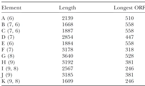
The four completeHeT-Aelements underwent a series of head-to-tail amplifications of different regions within their array to give 10 elements and an 11th element trun-cated by the cloning procedure. These elements, with the transposable elements inserted in them, now make up a complex set of repeats that stretches over.60 kb of the BAC. We refer to this region as theHeT-Aarray and designate the current set of elements as A–K (see Table 1 for their ancestral derivation as proposed by Mendez -Lagoet al.2009).
During their time on the Y chromosome, theseHeT-A
sequences have been conserved very differently from theHeT-As in telomere arrays. An overview of sequence from the centromeric BAC (Figure 4) shows thatHeT-A
sequences are much more fragmented than they are in the telomeric BAC (compare Figure 4A with Figure 1A). Amplification events are not seen in telomere arrays: In telomere arrays, each element is uniquely defined by combinations of subfamily sequence, 59truncation, pres-ence or abspres-ence of Tag sequpres-ences, and length of the 39
oligoA tail. These characters allowed Mendez-Lagoet al. (2009) to identify elements amplified in the centromeric sequence. Similar amplifications have not been detected in telomere regions. Thus the amplification events pro-vide the first epro-vidence that the HeT-A array is being maintained differently in its centromeric position.
Full-lengthHeT-A elements in the centromeric array have undergone extensive internal deletions: The 10 centromeric elements derived from ancestral intact ele-ments 6–9 have undergone extensive sequence changes. Our analysis of this sequence (Figure 4B) shows that each of these centromeric elements has lost 40% or more of its sequence, and none would encode the Gag protein thought to be necessary forHeT-Atransposition. In the original report (Mendez-Lagoet al.2009), these elements are described as decayed; however, our com-parisons withHeT-Asuggest that the changes are perhaps more accurately described as restructuring rather than decaying because they are not entirely random.
The four dot plots (Figure 4B) comparing individual centromeric elements to canonicalHeT-Agive a repre-sentative view of the changes in this array. In contrast to the telomere elements where loss is always from the 59 end, each centromere element has several large in-ternal deletions scattered through its sequence. There has been little rearrangement of the remaining se-quence, most of which is collinear with the canonical
HeT-A and has relatively few inversions and rearrange-ments. Surprisingly, the only regions that are conserved in every centromere element are the extreme 59and 39 ends.
TABLE 1
Elements in the Y chromosomalHeT-Aarray
Element Length Longest ORF
A (6) 2139 510
B (7, 6) 1668 558
C (7, 6) 1887 558
D (7) 2854 447
E (6) 1884 558
F (7) 3178 318
G (8) 3640 528
H (9) 3192 381
I (9, 8) 2567 246
J (9) 3185 381
K (9, 8) 1609 246
Elements are designated by single letters in their order on the BAC. Numbers in parentheses following the letter show the ancestral elements identified by Mendez-Lago et al.
Comparisons of these elements show that many deletions are shared with siblings derived from the same amplification. Thus these 10 elements and the partial element have become a complex array of repeats. Because amplification events apparently involved more than a single unit, higher orders of repeat arise, as can be seen in the full-length view in Figure 4A. A more specific example of a higher-order repeat, elements H, I, J, and K, is shown in Figure 4B. H and J are nearly identical while the alternating elements I and K are also nearly identical but quite different from their flanking elements (H and J).
As a result of both sequence loss and nucleotide changes, theseHeT-Aelements have lost much of their protein-coding capacity. The longest open reading frames in these elements range from 246 to 558 nt while the shortest completeHeT-A gaggene is 2766 nt.
The centromeric HeT-A array contains several non-telomeric transposable elements: At the telomere, nontelomeric elements are found only in small transi-tion zones at the junctransi-tion between the telomere array and the rest of the chromosome; elements in the transition zones are only fragments. In contrast, the centromeric BAC contains nontelomeric elements from Figure 4.—(A) Analysis
ofHeT-A-related sequences in the HeT-A array in the centromeric region of the Y chromosome. Sequence of 76 kb of the cluster in the sequenced BAC is com-pared with the sequence of the canonical 6-kb com-plete HeT-A exactly as the 4R telomere sequence was compared in Figure 1. TheHeT-Asequence is dia-grammed on the y-axis, showing the 59 UTR, ORF, and 39 UTR. The dotted line near the left end of the plot marks the division between the sequence am-plified in the 18HT satellite (first few repeats shown on the left) and the sequence of the ancestral complete
HeT-A elements (on the right). These HeT-As gave rise to 11 elements, but the 39end of the last ele-ment was cut off by clon-ing. Eleven 59ends can be detected as small frag-ments reaching the bottom line and the 10, 39ends as small fragments reaching the top line. These frag-ments are more easily seen in the higher magnifica-tion of B. Gaps in the ele-ments indicate significant loss of sequence. The hori-zontal gaps contain se-quence of nontelomeric transposable elements. (B) Comparisons of representative elements from the centromericHeT-Aarray with the canonical HeT-A. Dot plots of individual elements were made as in A except that sequence of invading transposable elements was removed from each element. Note that here theHeT-Aelement is on thex-axis, and the centromere elements are on they-axis. For description and order of elements, see Table 1. Element A now has three transposable elements inserted. Their sequence was removed before the dot plot, but some unidentified sequence remains. This element shows more sequence rearrangement than the others. There are two inversions (lines of opposite slant), and one of the inverted fragments is moved out of the collinear position. Element E was derived by amplification of A (Mendez-Lagoet al.2009), but both elements have undergone changes since the amplification.
seven different families, three in the 18HT sequences and four in the HeT-A array. In the HeT-A array, one element, the non-LTR retrotransposonF, arrived early in the expansion of the sequence and was included in two of the amplification events. More recent arrivals,
mdg1,diver, and1731, are present as single copies. The LTR retrotransposon1731is especially interest-ing. It is completely intact and its two LTR sequences are identical. This element is a member of the 1731
subfamily in which thegagandpol reading frames are fused to produce an ORF of 3852 nt (Kalmykovaet al. 1999). This entire reading frame is open and has 100% nucleotide identity with other elements in the database. Therefore, this element is potentially active. Other studies have suggested that there is at least one active
1731 on the Y chromosome. The 1731 fused Gag-Pol protein is expressed in testis (Kalmykovaet al.2004), where Y chromosomes are genetically active. Also, polytenized1731Y sequence has been found in salivary glands (Junakovicet al. 2003). Thus the1731 in the centromere array may be involved in these activities.
The centromere array has been shaped into clusters resembling the repeated sequences found in centro-meric heterochromatin: As discussed above, the main-tenance of HeT-A at the Y centromere has been dramatically different from the maintenance ofHeT-A
at telomeres. As a result, the ancestral telomere has given rise to two different types of clustered repeats: the more uniform 18HT satellite and a complex set of repeats derived from amplifications of several regions of internally deleted elements. Both types of repeats are head-to-tail arrays, rather than the palindromes that are abundant in protein-coding regions on human and chimpanzee Y chromosomes (Hugheset al.2010). Both types of repeats are also similar to repeated sequences that characterize the heterochromatic centromere regions in chromosomes of multicellular organisms (Sullivanet al. 2001; Sun et al. 2003; Schueler and Sullivan 2006; Pertileet al.2009).
Because centromeres in multicellular organisms are determined epigenetically (Sullivanet al.2001; Malik and Henikoff 2009), it is not possible to identify centromeres by sequence alone. Nevertheless, this Y cluster is similar in size, repeated sequence structure, and presence of transposable elements to the only functionally characterized centromere in Drosophila, the X chromosome centromere (Sunet al.2003). The similarities between the Y cluster and the X centromere do not extend to the level of the nucleotide sequence. Because the Y chromosome is the one chromosome that does not pair normally with its homolog, it is not surprising that the Y does not share centromeric sequences with the homolog. Y-specific centromere sequences could well be either a cause or an effect of this nonpairing behavior. [The mouse Y centromere is a known example of Y-specific centromere sequences (Pertileet al.2009).]
There are reports that some characteristics of repli-cation and/or repair in heterochromatin differ from those in euchromatin (Andersonet al.2008; Pengand Karpen 2008). We suggest that the structure of these HeT-A-derived clusters might in part be determined by mechanisms preferentially used in pericentric regions, in addition to being driven by selection for function. For example, repeated amplification of portions of this sequence is an effective way of providing the rapid evolution that has been noted for centromere regions (Henikoffand Malik2002).
The strong conservation of the 39-most end ofHeT-A
in both 18HT and the more complex repeats in the Y centromeric complex may also be driven by function. There is growing evidence that RNA transcripts may be important for the formation of heterochromatin and for some aspects of centromere function (Savitskyet al. 2006; Slotkin and Martienssen 2007; Lee 2009; Malik and Henikoff 2009). Start sites for both the sense and the antisense transcripts of HeT-Alie in the conserved 39region; sense-strand start sites are found 93, 62, and 31 nt upstream of the 39oligoA (Danilevskaya et al. 1997; Maxwell et al. 2006), while multiple an-tisense starts have been found 220 to 190 nt from the oligoA (Shpizet al.2009). Thus it is possible that the 39 region is conserved to direct transcription from this sequence. Northern blots show several transcripts,1 kb in length with sequence homology to the 39 region (Danilevskaya et al. 1999). Some of these RNAs are found only in males and might be products of the Y centromere. However, we have also found 39fragments of HeT-Ain several intergenic regions on the Y, so the origin of the transcripts will require further study.
We thank Madeleine Crosby, Harvard University, for aid and advice while performing final annotation of the 4R and XL sequences. This work was supported by National Institutes of Health grant GM50315 to M.-L. P.
LITERATURE CITED
Agudo, M., A. Losada, J. P. Abad, S. Pimpinelli, P. Ripollet al.,
1999 Centromeres from telomeres? The centromeric region of the Y chromosome of Drosophila melanogaster contains a tan-dem array of telomeric HeT-A- and TART-related sequences. Nu-cleic Acids Res.27:3318–3324.
Anderson, J. A., Y. S. Songand C. H. Langley, 2008 Molecular
population genetics of Drosophila subtelomeric DNA. Genetics
178:477–487.
Berloco, M., L. Fanti, F. Sheen, R. W. Levisand S. Pimpinelli,
2005 Heterochromatic distribution of HeT-A- and TART-like se-quences in several Drosophila species. Cytogenet. Genome Res.
110:124–133.
Biessmann, H., S. B. Carterand J. M. Mason, 1990 Chromosome
ends in Drosophila without telomeric DNA sequences. Proc. Natl. Acad. Sci. USA87:1758–1761.
Bourque, G., 2009 Transposable elements in gene regulation and
in the evolution of vertebrate genomes. Curr. Opin. Genet. Dev.19:607–612.
Bucholc, M., Y. Parkand A. J. Lustig, 2001 Intrachromatid
Cordaux, R., and M. A. Batzer, 2009 The impact of
retrotranspo-sons on human genome evolution. Nat. Rev. Genet.10:691–703. Danilevskaya, O. N., I. R. Arkhipova, K. L. Traverseand M. L.
Pardue, 1997 Promoting in tandem: The promoter for
telo-mere transposon HeT-A and implications for the evolution of ret-roviral LTRs. Cell88:647–655.
Danilevskaya, O. N., K. L. Traverse, N. C. Hogan, P. G. Debaryshe
and M. L. Pardue, 1999 The two Drosophila telomeric
trans-posable elements have very different patterns of transcription. Mol. Cell. Biol.19:873–881.
Fuller, A. M., E. G. Cook, K. J. Kelleyand M.-L. Pardue, 2010 Gag
proteins of Drosophila telomeric retrotransposons: collaborative targeting to chromosome ends. Genetics184:629–636. George, J. A., P. G. Debaryshe, K. L. Traverse, S. E. Celnikerand
M. L. Pardue, 2006 Genomic organization of the Drosophila
telomere retrotransposable elements. Genome Res. 16:1231– 1240.
Golubovsky, M. D., A. Y. Konev, M. F. Walter, H. Biessmannand
J. M. Mason, 2001 Terminal retrotransposons activate a
subte-lomeric white transgene at the 2L telomere in Drosophila. Genet-ics158:1111–1123.
Henikoff, S., and H. S. Malik, 2002 Centromeres: selfish drivers.
Nature417:227.
Hoskins, R. A., J. W. Carlson, C. Kennedy, D. Acevedo, M. Evans
-Holmet al., 2007 Sequence finishing and mapping of
Drosoph-ila melanogaster heterochromatin. Science316:1625–1628. Hughes, J. F., H. Skaletsky, T. Pyntikova, T. A. Graves, S. K. Van
Daalenet al., 2010 Chimpanzee and human Y chromosomes
are remarkably divergent in structure and gene content. Nature
463:536–539.
Junakovic, N., D. Fortunati, M. Berloco, L. Fantiand S. Pimpinelli,
2003 A subset of the elements of the 1731 retrotransposon fam-ily are preferentially located in regions of the Y chromosome that are polytenized in larval salivary glands of Drosophila mela-nogaster. Genetica117:303–310.
Kahn, T., M. Savitskyand P. Georgiev, 2000 Attachment of HeT-A
sequences to chromosomal termini in Drosophila melanogaster may occur by different mechanisms. Mol. Cell. Biol.20:7634– 7642.
Kalmykova, A., C. Maisonhaute and V. Gvozdev, 1999
Ret-rotransposon 1731 in Drosophila melanogaster changes retrovi-rus-like expression strategy in host genome. Genetica107:73–77. Kalmykova, A. I., D. A. Kwon, Y. M. Rozovsky, N. Hueber, P. Capy et al., 2004 Selective expansion of the newly evolved genomic variants of retrotransposon 1731 in the Drosophila genomes. Mol. Biol. Evol.21:2281–2289.
Kern, A. D., and D. J. Begun, 2008 Recurrent deletion and gene
presence/absence polymorphism: telomere dynamics dominate evolution at the tip of 3L inDrosophila melanogasterandD. simu-lans.Genetics179:1021–1027.
Koerich, L. B., X. Wang, A. G. Clark and A. B. Carvalho,
2008 Low conservation of gene content in the Drosophila Y chromosome. Nature456:949–951.
Lee, J. T., 2009 Lessons from X-chromosome inactivation: long
ncRNA as guides and tethers to the epigenome. Genes Dev.
23:1831–1842.
Levis, R. W., 1989 Viable deletions of a telomere from a Drosophila
chromosome. Cell58:791–801.
Li, B., and A. J. Lustig, 1996 A novel mechanism for telomere size
control in Saccharomyces cerevisiae. Genes Dev.10:1310–1326. Lustig, A. J., 2003 Clues to catastrophic telomere loss in mammals
from yeast telomere rapid deletion. Nat. Rev. Genet.4:916–923. Malik, H. S., and S. Henikoff, 2009 Major evolutionary transitions
in centromere complexity. Cell138:1067–1082.
Marin, L., M. Lehmann, D. Nouaud, H. Izaabel, D. Anxolabehere et al., 2000 P-element repression inDrosophila melanogasterby a naturally occurring defective telomeric P copy. Genetics 155:
1841–1854.
Maxwell, P. H., J. M. Beloteand R. W. Levis, 2006 Identification
of multiple transcription initiation, polyadenylation, and splice sites in the Drosophila melanogaster TART family of telomeric retrotransposons. Nucleic Acids Res.34:5498–5507.
Mendez-Lago, M., J. Wild, S. L. Whitehead, A. Tracey, B. De
Pablos et al., 2009 Novel sequencing strategy for repetitive
DNA in a Drosophila BAC clone reveals that the centromeric re-gion of the Y chromosome evolved from a telomere. Nucleic Acids Res.37:2264–2273.
Mikhailovsky, S., T. Belenkayaand P. Georgiev, 1999 Broken
chromosomal ends can be elongated by conversion in Drosoph-ila melanogaster. Chromosoma108:114–120.
Pardue, M. L., S. Rashkova, E. Casacuberta, P. G. Debaryshe, J. A.
Georgeet al., 2005 Two retrotransposons maintain telomeres
in Drosophila. Chromosome Res.13:443–453.
Peng, J. C., and G. H. Karpen, 2008 Epigenetic regulation of
het-erochromatic DNA stability. Curr. Opin. Genet. Dev.18:204–211. Pertile, M. D., A. N. Graham, K. H. Choo and P. Kalitsis,
2009 Rapid evolution of mouse Y centromere repeat DNA be-lies recent sequence stability. Genome Res.19:2202–2213. Pickett, H. A., A. J. Cesare, R. L. Johnston, A. A. Neumannand
R. R. Reddel, 2009 Control of telomere length by a trimming
mechanism that involves generation of t-circles. EMBO J. 28:
799–809.
Rashkova, S., S. E. Karam, R. Kellumand M. L. Pardue, 2002 Gag
proteins of the two Drosophila telomeric retrotransposons are targeted to chromosome ends. J. Cell Biol.159:397–402. Rashkova, S., A. Athanasiadis and M. L. Pardue, 2003
In-tracellular targeting of Gag proteins of the Drosophila telomeric retrotransposons. J. Virol.77:6376–6384.
Savitsky, M., D. Kwon, P. Georgiev, A. Kalmykovaand V. Gvozdev,
2006 Telomere elongation is under the control of the RNAi-based mechanism in the Drosophila germline. Genes Dev.20:
345–354.
Schueler, M. G., and B. A. Sullivan, 2006 Structural and
func-tional dynamics of human centromeric chromatin. Annu. Rev. Genomics Hum. Genet.7:301–313.
Sheen, F. M., and R. W. Levis, 1994 Transposition of the LINE-like
retrotransposon TART to Drosophila chromosome termini. Proc. Natl. Acad. Sci. USA91:12510–12514.
Shpiz, S., D. Kwon, Y. Rozovskyand A. Kalmykova, 2009 rasiRNA
pathway controls antisense expression of Drosophila telomeric retrotransposons in the nucleus. Nucleic Acids Res.37:268–278. Slotkin, R. K., and R. Martienssen, 2007 Transposable elements
and the epigenetic regulation of the genome. Nat. Rev. Genet.
8:272–285.
Sullivan, B. A., M. D. Blower and G. H. Karpen, 2001
Deter-mining centromere identity: cyclical stories and forking paths. Nat. Rev. Genet.2:584–596.
Sun, X., H. D. Le, J. M. Wahlstrom and G. H. Karpen,
2003 Sequence analysis of a functional Drosophila centromere. Genome Res.13:182–194.
Traverse, K. L., J. A. George, P. G. Debarysheand M. L. Pardue,
2010 Evolution of species-specific promoter-associated mecha-nisms for protecting chromosome ends by Drosophila Het-A te-lomeric transposons. Proc. Natl. Acad. Sci. USA107:5064–5069. Walter, M. F., C. Jang, B. Kasravi, J. Donath, B. M. Mechleret al.,
1995 DNA organization and polymorphism of a wild-type Dro-sophila telomere region. Chromosoma104:229–241.
Wang, R. C., A. Smogorzewskaand T. DeLange, 2004
Homol-ogous recombination generates T-loop-sized deletions at human telomeres. Cell119:355–368.