CHROMOSOME ABERRATIONS INDUCED BY X-RAYS
KARL SAX
Arnold Arboretum, Harvard University, Jamaica Plain, Massachusetts Received June 3, 1938
INTRODUCTION
INCE the discovery by MULLER and STADLER that X-rays induce
S
mutations in animals and plants, a new field has been developed in experimental genetics. This work on radiation genetics has been reviewed by MULLER (1932), HANSON (1933), OLIVER (1934), STADLER, GOOD- SPEED, GOWEN, et al. (DUGGAR 1936), STUBBE (1937), and TIMOFEEFF-RESSOVSKY (1937). The genetic results show that (I) the mutation rate increases directly with dosage, (2) the X-ray effect is not delayed or
indirect, (3) there is no temperature coefficient, (4) differential suscepti- bility is found in different stages of development, (5) the X-rays cause translocations, inversions, and deletion of chromosome segments, (6) the induced mutations are not distributed entirely a t random in the chromo- somes,
(7)
there is no differential effect of the various wave lengths in the X-ray range, and (8) the gene string is already partially split in Drosophila sperm and in Zea pollen grains.A direct cytological analysis of X-ray effects has confirmed some of the results obtained by genetic methods, but most of the cytological work has dealt with the nature of the chromosome rearrangements and the time of splitting of the chromonema. There is still no critical evidence regarding the relation between dosage and chromosome aberrations, the effect of temperature on chromosome susceptibility to radiation, differential sus- ceptibility at various times during the meiotic and mitotic cycles, the mechanism involved in translocation and inversion, and the time of chro- monema doubling. An analysis of X-ray effects on chromosomes of Trades- cantia microspores has solved some of these problems.
MATERIALS
Microspores of Tradescantia were used for the study of X-ray effects on chromosome behavior. The meiotic and mitotic cycle in microspore formation is well known, the chromosomes are large, and certain species flower throughout the year in the greenhouse. During the summer months the meiotic cycle from earliest prophase to the tetrad stage covers about one week, and a similar period is required for microspore development up to the time of nuclear division. The nucleus of the newly formed micro- spore remains in the resting stage for about five days, and is in the pro- phase stage for at least one day before nuclear division occurs. The length
X-RAY INDUCED ABERRATIONS 495
of the meiotic and mitotic cycles is increased during the winter months and may be increased to two weeks for each cycle. All experiments were done with a clonal line of a Tradescantia reflexa hybrid, which has six pairs of chromosomes and one pair of fragments. Flowering stalks were cut off and kept in a glass of water during radiation and for several days to a week more while the microspores were being examined. When the micro- spores were to be examined for a period of several weeks after irradiation, the potted plants were subjected to X-rays.
The source of the X-rays was a Coolidge tube with a tungsten target. The line voltage was 1 2 0 a t IO ma, and the secondary voltage was 160 kv. No screen was used, and the target distance was about 75 cm. At this distance the tube delivered about 2 5 r per minute. The dosage used ranged from 75 to zoo r for the analysis of types of chromosome aberrations.
CYTOLOGICAL OBSERVATIONS
A few observations were made a t meiosis, but most of the data were based on microspore chromosomes. A few hours after raying, the meiotic cells show clumping of the chromosomes and fusion of homologous chro- matids. The terminal association of chromosomes is not accompanied by fragments a t either the first (fig. 1') or the second (fig. 2 ) meiotic division.
Many sub-terminal associations are found, especially a t the second meiotic anaphase, but free fragment chromosomes were not observed. Twenty- four hours after raying, many chromosome bridges and free fragments were found at anaphase of both meiotic divisions.
The mitotic division in the microspore also shows a clumping of the chromosomes shortly after irradiation. At four to six hours after raying, about half the anaphase figures show terminal or subterminal fusion of sister chromosomes (figs. 3 and 4). Occasionally there are free fragments or evidence of unequal chromatid interchange, but these are rare. I n no case were fusions or interchanges found between non-homologous chro- matids or chromosomes during the first seven hours after irradiation. These early fusions following X-ray treatment appear to involve the chromosome envelope, and although fragments are released by breakage a t points of fusion in some figures, the fusion of sister chromatids at these stages is not of primary significance.
When moderate doses of X-rays are given to microspores the metaphase and anaphase figures can be analyzed a t any time after irradiation. During the first 24 hours after raying, most of the breaks involve only one of the two chromatids (figs. 5, 6, 8, 9, and IO), but chromatid breaks have been
observed as late as 7 2 hours after raying. Achromatic lesions also are
496 KARL SAX
frequent, and seem to be caused by breaks which have not released the distal ends of the chromatids (fig. 6).
The chromatid breaks may be single and release the distal end of a chromatid, or they may involve two chromatids, one from each of two chromosomes. A single break may produce an acentric fragment, or the break may be incomplete and produce an achromatic lesion (fig. 6). The double breaks may produce either reciprocal interchange of chromatids, or chromatid fusion accompanied by fused chromatid fragments. The re- ciprocal chromatid interchanges usually are equal or nearly equal (figs. 9 and IO), although unequal interchange of chromatid arms does occur
(fig. 8). The ends of two broken chromatids may fuse to form a dicentric chromatid and release an acentric fused fragment (figs. 9 and IS). I n
practically all cases of chromatid fusion an acentric fragment is released. The ends of the fragment are the normal ends of the two broken chro- matids, and a t the point of breakage the two chromatid fragments are fused.
DESCRIPTION OF PLATES
Camera lucida drawings of meiotic and microspore chromosomes a t various times after X-ray treatment. Acetocarmine preparations of Tradescantia reflexa hybrid. Magnification X 900.
EXPLANATION OP PLATE I
FIGURE I . Meiotic anaphase. Terminal fusion of chromatids. No fragments. 150 r. 3 hrs. FIGURE 2. Second meiotic anaphase. Terminal and sub-terminal fusion of chromatids. No
FIGURE 3 . Anaphase in microspore. Fusion of chromatids. 75 r. 6 hrs.
FIGURE 4. Anaphase in microspore. Terminal fusion of chromatids. Translocation between sister chromatids. 75 r. 6 hrs.
FIGURE 5 . Microspore metaphase. Two chromatid breaks, and a chromosome break followed by fusion of broken ends of sister chromatids. zoo r. 1 2 hrs.
FIGURE 6 . Microspore anaphase. Complete and incomplete chromatid breaks, and a dicentric chromosome resulting from sister chromatid fusion after a chromosome break. 100 r. 19 hrs.
FIGURE 7. Microspore metaphase. A chromosome break followed by sister chromatid fusion.
100 r. 6 hrs.
FIGURE 8. Microspore metaphase. A chromatid and a chromosome break in the same chro- mosome. The unequal chromatids of two chromosomes are the result of unequal reciprocal chro- matid translocations. 100 r. 17 hrs.
FIGURE 9. Microspore metaphase. Chromatid exchange and chromatid fusion. Also a simple chromosome break. 7s r. 24 hrs.
fragments. 150 r. 6 hrs.
FIGURE IO. FIGURE 11.
FIGURE 12.
FIGURE 13.
mosome break. FIGURE 14.
100 r. 28 hrs.
FIGURE 1 5 .
180 r. 4s hrs.
. - .
Microspore metaphase. Chromatid and chromosome breaks. IOO r. 24 hrs. Microspore metaphase. Chromatid ring formation. 75 r. 2 4 hrs.
Microspore-early anaphase. Chromatid and chromosome breaks. IOO r. 24 hrs. Microspore anaphase. Dicentric chromosome and fused fragment following chro-
100 r. 24 hrs.
X-RAY INDUCED ABERRATIONS 497
1
498 KARL SAX
Few chromatid breaks are found 48 hours after raying, and none from the fourth to the ninth day. During the winter months, the microspores examined 9 or I O days after raying were X-rayed at late stages in meiosis
or very early in microspore development. Although no chromatid breaks were found between the fourth and eighth day after irradiation, a few were found on the ninth day. Most of these were in a single microspore, where two single breaks and two chromatid fusions were found at metaphase (fig. 23). A single chromatid fusion was also found a t anaphase in another cell (fig. 24).
Chromosome breaks, with both chromatids broken a t the same locus, were found a t all times after raying the microspore. During the first 24
hours all the chromosome breaks are single. The break releases the distal end of the chromosome arm, and the broken ends of sister chromatids invariably fuse to form a U-shaped acentric fragment and a pair of sister chromatids fused a t one end. The first chromosome break was observed 6 hours after raying (fig.
7).
As the broken chromosome divides at anaphase, the fused ends form a bridge (figs. 6, 12, and 13). The distal ends of thebroken chromatids always fuse to form a single fragment. The size of the fragment varies considerably, but no bridge has been observed without a fragment.
Single chromosome breaks are found less frequently after the second day following irradiation. At this time there is no fusion of the ends of broken chromatids, and only pairs of chromatid bridges are found. The distal fragments appear as paired rods (figs. 2 2 , 23, and 24).
Breaks in two chromosomes may be followed by reciprocal interchange or by chromosome fusion with the release of a fragment. The reciprocal interchanges are difficult to detect, presumably because they are approxi- mately equal, but unequal interchanges have been observed. The fusion of broken ends of different chromosomes may produce also a dicentric chromosome and a pair of chromatid fragments. Each fragment chromo- some is composed of the ends of two non-homologous chromosomes fused together a t the point of the break. As the dicentric chromosomes separate a t anaphase, they may separate freely, or form two bridges, or interlock, depending on the amount of relational coiling between centromeres (figs. 16 and 17). Chromosome bridges are always accompanied by chromosome fragments. The size of the fragment may be no longer than the width of a chromatid or may be as long as a normal chromosome (figs. 16 and IS). Occasionally the break and fusion occur so near the centromeres that the duality of the centromeres in the dicentric chromosome can not be differ- entiated. The released fragment is then as long as two normal arms (figs.
18 and 2 0 ) .
X-RAY INDUCED ABERRATIONS 499
ring chromosomes. At anaphase the ring chromosomes may separate freely, or open out into a single dicentric ring, or they may be interlocked (figs. 19, 20, and 21). Ring chromosomes induced by raying the resting
microspore nucleus are always accompanied by fragments. Premeiotic irradiation has produced a ring chromosome a t the microspore division with no visible fragment. Evidently the loss of a small fragment is not always lethal.
The irradiation of meiotic cells produces a high degree of microspore sterility, but some microspores do develop. These microspores, even though they include only the more viable cells, show a large proportion of breaks. Every chromosome may be broken, but if no fragments are lost, the chromosomes develop normally (fig. 24). Occasionally diploid
microspores are produced after irradiation, and these also have many chromosome aberrations (fig. 26). These microspores were produced from
meiotic cells which were irradiated a t interphase or during the second meiotic division.
Most of the aberrations induced by X-rays are chromosome fragmenta- tions and fusions, but other abnormalities are found occasionally. The anaphase chromosomes may not be distributed equally to the poles, and all chromosomes may pass to the same pole. Monocentric spindles are rare, and only five were observed in the thousands of anaphase figures studied (fig. 30). The centromeres of some chromosomes and chromatids seem to be inactive in chromosome orientation at metaphase and anaphase. The inactive chromosomes may be acentric fragments which have lost the centromeres by chromosome fusion (fig. 18); but the unequal distri- bution of daughter chromosomes t o the poles and occasional lagging chromosomes a t anaphase (fig. 28) suggest that a centromere may be
inactivated or prevented from dividing by X-ray treatment. I n one microspore the chromosomes had developed to an early metaphase stage with no visible split in the chromosomes (fig. 29).
The sequence of appearance of various types of aberrations is of interest in an analysis of the nature of breaks and fusions of chromosomes. After the terminal fusions of chromosomes are past and the more significant aberrations appear, only chromatid and chromosome breaks are observed during the first 2 4 hours. For example, at
1 7
hours after irradiation a t IOO r, 21 chromatid fragments and IO chromosome fragments were foundwithout a single dicentric chromatid or chromosome. These and other data show that the breaks are not dependent on previous fusions.
500 KARL SAX
with a dose of 2 0 0 r, there were 69 dicentric chromosomes, I I ring chromo-
somes, and 11 single distal fragments. Thus about 86 percent of the fusions
are between different chromosomes, and only 14 percent are between the two arms of the same chromosomes. Single breaks without fusion con- stituted about 1 2 percent of the aberrations which could be detected. I n
another series of observations made at corresponding times after irradia- tion, the proportion of single breaks was about
17
percent (table 2 ) . The relatively small percentage of single breaks suggests that a broken end of a chromosome has a strong tendency to fuse with another broken end, and that broken ends usually reunite after a single break.It has been assumed by a number of cytologists that an X-ray “hit” can break only a single chromonema at a given locus, and that the occur- rence of chromosome breaks proves the existence of a single chromonema at the time of irradiation. But we find both chromosome and chromatid breaks in the same division figure, or even in the same chromosome, and a t various times after irradiation,-from
7
to 7 2 hours (table I). It seemshighly improbable that the splitting of the chromosomes is so variable in different chromosomes of the same cell or that the time of the split may vary from
7
to 7 2 hours before the chromosomes reach the metaphasestage. The evidence seems conclusive that both chromatids may be broken at the same time by a single X-ray “hit.”
EXPLANATION OF PLATE 2
FIGURE 16. Microspore anaphase. Chromosome breaks fcllowed by fusion to produce dicen- tric chromosomes and acentric fragments. Two types of separation-locked and free. zoo r. 8 days.
FIGURE 1 7 . Microspore anaphase. Dicentric chromosome with crossed chromatids. zoo r.
3 days.
FIGURE 18. Microspore-early anaphase. The pair of chromatid fragments presumably re- leased by breaks and fusions of two chromosomes very near the centromeres. ZOO r. 6 days.
FIGURE 19. Microspore anaphase. Free separation of ring chromatids. zoo r. 19 days. FIGURE 2 0 . Microspore anaphase. Dicentric ring chromosome. Long acentric fragment re- FIGURE 21. Microspore anaphase. Locked ring chromatids. zoo r. 7 days.
FIGURE 22. Microspore anaphase. Single chromosome break which released almost entire
FIGURE 23. Microspore metaphase. Chromatid breaks and fusions induced during meiosis FIGURE 24. Microspore metaphase. Numerous chromosome breaks induced a t meiosis. 100 r.
FIGURE 25. Microspore anaphase. Chromatid fusion of non-sister chromatids. zoo r. 9 days. FIGURE 26. Diploid microspore metaphase. Three dicentric and one ring chromsome. 200 r. FIGURE 27. Microspore anaphase. Dicentric and ring chromosomes. 1200 r. 5 days.
FIGURE 28. Microspore anaphase. Apparent inactivation of the centromere of one chromo- FIGURE 29. Microspore metaphase with no chromosome split. roo r. 1 1 days.
FIGURE 30. Monocentric spindle. 180 r. 4 days.
leased by breaks and fusion of two chromosomes a t or near centromere. 180 r. 7 days.
arm of one chromosome. 100 r. 9 days. or very early in microspore. 100 r. g days.
1 1 days.
9 days.
501
X-RAY INDUCED ABERRATIONS
5 0 2 KARL SAX TABLE I
Duration of chromatid breaks.
75 to 2 0 0 r
HOURS AFTER CHROMATID CHROMOSOME
RAYING BREAKS BREAKS %
5 7 '7 24 48 72 96 I 1 IO 2 2 3 90 I 5 6 21 0 0 3 I 1 0 81 61 41 all 0 I 2 2 5 3 2 48 80 87 IO0
If
chromosome breaks and fusions occur at every locus which is hit by the X-rays, they should be distributed a t random along the chromosomes. But if secondary factors are involved, such as torsion of the chromosomes or the relative positions or differential contraction of the chromosomes, then the breaks may be localized. An analysis of the position of breaks and fusions has been made, using cells which were rayed while the chromo- somes were in the resting stage. The data are shown in table 2.TABLE 2
Loci of chromosome breaks. 150r. 4-7 days after raying. Length of fragment in relation to chromosome bridge or shortened arm.
SINGLE BREAKS E X C H A N G E B R E A K S
__ -- -
N % N %
Break near centromere 38 5 7 28 50
Break near center of arm I 7 2 5 I9 34
Break near distal end of arm 12 I8 9 16
The position of the breaks and fusions was determined from the relative length of the released fragments compared with the broken chromosome arms, or the distance between centromeres in the dicentric chromosomes. I n about half the aberrant chromosomes the break had occurred in the proximal third of the chromosome arm. Breaks were less frequent in the central region of the chromosome arms and still less frequent at the distal ends of the chromosomes. Simple breaks and exchange breaks show about the same frequency of distribution a t the various loci.
Organisms a t different stages of development show a differential SUS-
X-RAY INDUCED ABERRATIONS 503
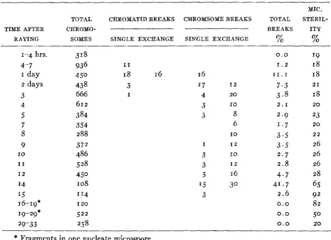
of chromosome aberrations at various times in the meiotic and mitotic cycles. Two series of observations were made, one with plants which had received a dose of 75 r, and the other with plants which had been sub- jected to 150 r. Both series showed similar results, but more data were obtained from the 150 r series. The data are shown in table 3.
TABLE 3
Series 17. J a n . I O . X 1 5 0 r . Greenhouse plants.
TOTAL CHROMATID BREAKS CHROMSOME BREAKS
- -
TIME AFTER CHROMO-
RAYING SOMES SINGLE EXCHANGE SINGLE EXCHANGE
1-4 hrs. 318
4- 7 936 I 1
I day 4 50 18 16 16
2 days 43 8 3 '7 I 2
3 666 I 4 2 0
4 612 3 I O
5 384 3 8
7 354 6
8 288 10
9 372 I I 2
I O 486 3 10
I 1 528 3 I 2
1 2 450 5 16
14 108 '5 30
I 5 '14 3
16-19* I 2 0
19-29* 522 29-33 258
* Fragments in one nucleate microspore.
MIC. TOTAL STERIL- BREAKS ITY
% %
0.0 19
1 . 2 18
1 1 . 1 18
7 . 3 21
3 . 8 18
2 . 9 23 1 . 7 2 0
3 . 5 2 2
3 . 5 26 2 . 7 26
2 . 8 26 4 . 7 28
4 1 . 7 65 2 . 6 92
0.0 8 2
0 . 0 50
2 . 1 2 0
0.0 20
There was an increase in chromosome aberrations up t o 24-30 hours after raying, when the proportion of breaks reached 11 percent. Each
504 KARL S A X
pollen fertility is increased to about 50 per cent and remains a t this figure for about IO days. During this period no chromosome aberrations were observed, although certain types undoubtedly are included in viable microspores. At the end of four weeks both the mitotic and meiotic cycles have been completed. The pollen fertility now becomes normal, in spite of the fact that the premeiotic cells have been X-rayed.
TABLE 4
yo
..
,.
, I ..___.. .
T Y
MICROSPORE U E l O l l C UFlOTlC PREUElOTlC
-’
YICIO$PoRE
P R O P H A Y RESTING NUCLEUS D I V D I O Y I PROPHASE I E l T l W G STACE
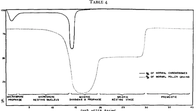
The relation between chromosome aberrations, microspore sterility, and the stage of nuclear development, is shown in table 4. The suscepti- bility of the chromosomes to X-ray treatment is greatest a t meiosis and presumably at meiotic prophase. Since all the chromosomes found in a
tetrad of resting microspores are already differentiated at late pachytene of meiosis, it appears that the meiotic chromosomes are much more sus- ceptible to X-ray breakage than the chromosomes in the resting nuclei of the microspores. I n view of the selected sample of microspores resulting from X-rayed meiotic cells, it seems probable that the chromosomes a t meiosis are at least ten times as susceptible as chromosomes at the resting stage in the microspore. The prophase stage of mitosis is more susceptible to X-rays than the resting stage, but a t prophase about half the breaks are chromatid breaks, while X-rayed resting nuclei show only chromosome breaks at metaphase and anaphase. However, the mitotic prophase stage is about twice as susceptible to X-ray treatment as the mitotic resting stage.
T H E E F F E C T O F TEMPERATURE ON CHROMOSOME SUSCEPTIBILITY TO X-RAYS
X-RAY INDUCED ABERRATIONS 505 Tradescantia microspores at various temperatures to determine if tem- perature at the time of radiation has any effect on frequency of chromo- some aberrations. Flower stalks of Tradescantia were placed in thermos bottles containing water at different temperatures. The cuttings were placed in the thermos bottles about half an hour before raying and kept in the bottles during irradiation and for one hour after raying. They were then placed in water a t room temperature and examined each day for about a week. Two series of observations were made, one irradiated with
a dose of IOO r at temperatures of 6" and 4ooC, respectively, and the other subjected to temperatures of 7 O , 25O, and 37°C and X-rayed with 2001.
The first series showed no significant difference in percentage of abnormali- ties at the two temperatures. Those irradiated at 6°C showed chromosome aberrations in 2 0 percent of the microspores, while those irradiated at
4ooC showed chromosome aberrations in 19 percent of the microspores. Similar results were obtained in the second series. The data are shown in table
5.
The microspores rayed at 37°C do show a higher average percent- age of cells with chromosome breaks, but in view of the great variability found on different days, the differences in aberrations a t different tem- peratures are not significant. Evidently there is no temperature coefficient for X-ray induced chromosome aberrations.TABLE 5
X-ray eJects at diJerent temperatures. Series 8. X200 r. Oct. 18, 1937. Tradescantia microspores.
MIC. DAYS AT 7°C AT 25'C AT 37°C COMBINED STE- AFTER
RAYING TOTAL % B R . TOTAL % B R .
148 59 103 26 63 21
57 23 78 1 2
142 18
56 30 76 46
132 65 '53 1 7
140 9 169 I O
170 I 7
47 I9
211 71
48 23.
TOTAL % B R .
1 1 1 43 132 1 1
66 8 63 I O
25 24 '43 14 I35 50
I37 90
TOTAL % B R .
391 62 388 18 269 12 289 1 2
273 16 332 1 7 239 40
424 73
RILITY % 25 25 26 I9 5' 53 60 20
Total 723 31 1070 32 812 36 260.5 33
The percentage of breaks based on number of division figures with breaks or fusions in one or more chromosomes.
The temperature experiments were conducted in October, and the microspore cycle at this time is about one week, as compared with about two weeks in January (cf. table 3). It will be noted that on a cell basis the abnormalities induced at meiosis are only slightly greater than those
506 KARL S A X
THE RELATION BETWEEN X-RAY DOSAGE AND
CHROMOSOME ABERRATIONS
I n both plants and animals the mutation rate induced by X-rays is
directly proportional to the dose. From this relationship it is concluded that single “hits” are responsible for the mutations. The accuracy with which chromosome aberrations can be analyzed in Tradescantia micro- spores makes possible a critical analysis of the relation between X-ray dosage and the percentage of chromosome aberrations.
I n the first series of experiments, flowering stalks of Tradescantia were subjected to X-ray doses of 150, 300, 600, and 1200 r. The only varying
factor in the treatment was the length of time necessary for giving the respective doses. The cytological observations were made on the third and fifth day after raying. A dicentric chromosome was classed as two breaks, as were the ring chromosomes. Since 8-90 percent of all visible aberra- tions are chromosome fusions accompanied by fragments, it makes little difference whether we class such aberrations as single or double breaks.
A
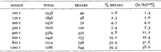
second series of buds was subjected to X-ray doses of 100, 200, 400,and 800 roentgens. The combined data from both series are shown in table 6. The log of the dosage plotted against the log of the percentage of aberrations gives a straight line, and leads to the derivation of an equa- tion for the relationship between dosage intensity and percentage of chromosome breaks.
YOB
= (Ir/80)’.~.TABLE 6
Relalion of breaks and X-ray dose.
DOSAGE TOTAL BREAKS % BREAKS (Ir/80)1.5%
100 r 2538 40 I .6 1.4
150 r 1896 48 2.5 2.6
200 r 1476 70 4.7 4.0
300 r 1626 I 2 0 7.4 7 . 3
400 r 3384 332 9 . 8 1 1 . 2
600 r 1446 275 19.0 20.4
b r 2214 796 35.9 31.6
1 2 0 0 r 1086 644 5 9 . 3 58.0
X-RAY INDUCED ABERRATIONS 507
The relation between dosage and chromosome breaks suggests that the double breaks are caused by independent X-ray hits. A number of investi- gators working with X-ray effects have devised methods for determining whether one, two, or more hits are necessary to produce a given effect. According to WYCKOFF and RIVERS (1930)~ if one hit is necessary to kill, the survival ratio is e-an, where a is the probability than an electron will
DESCRIPTION OF TABLE 7
The relation between dosage and chromosome aberration compared with theoretical curves based on equations for I hit (%B = I -eo*) and 2 hits [%B = I - e 4 * an)] reactions. Single breaks tend to occur in direct proportion to dosage.
eo
5 0
40
% B
30
2 0
10
hit the object and n is the number of electrons shot a t the object. If two hits are necessary to produce the effect, the survival ratio is ecan(I+an). Applying these formulae to our data, we have determined the theoretical curve for chromosome aberrations a t various dosages assuming that one hit is effective (%B = I -e-Qn), and that two hits are necessary [%B =
I
-
e-an (1+an)]. We have, in each case, taken an arbitrary value of a n508 KARL SAX
evidently caused by single hits, even though the chromosome may be split into two chromatids at the time of breakage.
DISCUSSION
Most of the earlier analyses of chromosome aberrations induced by X-rays were based on the assumption that a single hit was so localized in its effect that only a single chromatid could be broken a t a given locus. CARLSON (1937), working with irradiated somatic cells of Chortophaga, finds that an X-ray hit can break one or both of the two sister chromatids. KAUFMANN ( I 93 7) finds complex chromosome rearrangements following
X-radiation of Drosophila sperm and concludes that sister chromatids can be broken simultaneously a t the same locus by secondary effects of a single hit. This cytological work is in accord with the genetic results of
PATTERSON (1933) and MOORE (1934).
The fact that both chromosome and chromatid breaks occur at meta- phase and anaphase from
7
to 7 2 hours after raying the Tradescantiamicrospores, clearly indicates that a single hit can break one or both chromatids a t the same locus. Chromosome and chromatid breaks often occur in the same cell, and may occur in the same chromosome. It seems highly improbable that the time of splitting of the chromosome varies from
7
to 7 2 hours before the chromosomes reach the metaphase stage.The proportion of chromosome aberrations produced by different amounts of irradiation also indicates that some of the fusions may be caused by a single hit which breaks two adjacent chromosomes.
X-RAY INDUCED ABERRATIONS 509
chromosome breaks at the resting stage does not prove that the chromo- some is single a t this stage of nuclear development. The cytological studies of HUSKINS and HUNTER (193 5)
,
KAUFMANN (I 937)
,
CARLSON ( I 937),and MATHER (1937)~ all show that the chromosome is split early in the resting stage. The split may occur earlier, as NEBEL (1937) maintains, although the studies based on direct observation of the duality of the chromosome are not so critical.
Chromosome breaks and fusions have been attributed to two different mechanisms, fusions followed by breaks, and breaks followed by a fusion of broken ends. The behavior of irradiated Tradescantia chromosomes strongly supports the second hypothesis. While it is true that the earliest induced aberrations are chromosome fusions, these are seldom accom- panied by chromosome fragments and are of little significance in the production of permanent chromosome aberrations. The conclusions based on these temporary fusions (MARSHAK 1937) are not valid so far as per- manent X-ray effects are concerned (MARQUARDT 1937 and WHITE 1937). These primary fusions are induced also by heat (SAX 1937) and by age (BARBER 1938).
All breaks during the first 24 hours after irradiation of Tradescantia microspores are single chromatid and chromosome breaks. Irradiation of the resting nuclei also produces single breaks, although fusions are much more frequent a t this stage. These single chromatid and chromosome breaks can not be attributed to fusions followed by breaks. The evidence that most of the fusions between different chromosomes are dependent on two adjacent hits also indicates that the breaks occur first, followed by fusion of broken ends (cf. table 7).
The mutation rate is directly proportional to the dosage of X-rays, while the chromosome aberrations show a geometric increase with in- creased dosage. If mutation is associated with chromosome aberration, one might expect the dosage relationships of mutation and chromosome aberration to be similar. But more than 80 percent of the chromosome aberrations which can be detected involve fusions between different chromosomes, or between different arms of the same chromosome. These aberrations, as well as the simple deficiencies, will tend to be eliminated in successive cell generations, and most of the aberrations which survive will be reciprocal translocations, inversions, and small deficiencies. The small inversions and deficiencies can be induced by the effects of a single X-ray hit.
510 KARL SAX
mechanism of such changes. The chromosomes are in the form of relaxed coiled chromonemata during the resting stage. At early prophase, new minor spirals develop as the relic coils disappear. The gyres of the new minor spirals increase in size and decrease in number, so that a t metaphase there are 2 0 to 2 5 coils per chromosome in the Tradescantia microspore
(SAX and SAX 1936). A single X-ray hit can break two adjacent gyres, and the reunion of broken ends in new associations will produce small deficiencies and inversions. This mechanism is essentially the same as HUSTED (1937) finds occurring spontaneously in Pancratium. The dia- grams (textfig. I ) showing these structural changes are based on HUSTED’S
illustrations.
TEXTFIGURE I . Postulated mechanism for production of small deficiencies and inversions.
Breaks in two adjacent gyres of the coiled chromonema, followed by a criss-cross reunion, will produce a ring deletion which is either locked around the chromonema or is free. Breaks in two adjacent gyres followed by reunion of adjacent ends will lead to a small inversion.
The size of the deficiency or inversion will depend upon the diameter of
the chromonematic spiral. The diameter of the gyre will vary a t different periods in the mitotic cycle. At earliest prophase, two types of spirals are found,-large relic spirals and the new minor spirals of very small diameter. At metaphase, the deletion or inversion of a gyre would involve about 4 or 5 percent of the length of the chromonema in a single Trades- cantia chromosome. Fragments a t least this small are found occasionally in irradiated microspores (figs. 21 and 27). If a similar mechanism be
postulated for “molecular spirals” (DARLINGTON 1937)’ the aberrations could not be detected cytologically and might be considered as chemical changes in the gene.
X-RAY INDUCED ABERRATIONS 5 1 1
some aberrations (MARSHAK 1935). This differential susceptibility has been attributed to differences in p H (ZIRKLE 1936), to water content (GUSTAFSSON 1937), and to differences in amount of chromatin around the gene string (MARSHAK 1935). The differential susceptibility of meiotic and mitotic nuclei is difficult to reconcile with differences in p H involving the isoelectric point during each of the nuclear cycles. STADLER finds no differential mutation rate in X-rayed seeds which differ in water content, and GUSTAFSSON’S observations on chromosome fusions may be attributed to the initiation of prophase stages induced by the absorbed water. The chromatin around the gene string certainly could not serve as an insulation against X-ray hits, although it might reduce the flexibility of the gene string so that broken ends could not fuse in new associations. As GOOD-
SPEED has suggested, the cellular activity seems to play a part in X-ray
susceptibility. As applied to the chromosomes, it appears that the period of greatest sensitivity to irradiation is correlated with the greatest activity in the coiling mechanism, both minor coiling and relational coiling. At this time the chromosomes appear to be under torsional strain, and some of the breaks will be prevented from rejoining in the original position, and adjacent breaks in adjacent chromatids or chromosomes will join in new associations (CATCHESIDE 1936). The great susceptibility of meiotic prophase chromosomes can be attributed to relational coiling of both chromatids and chromosomes and the greatly increased length of the chromonemata which would provide greater opportunity for union of breaks in adjacent chromosomes. Gross chromosome aberrations would not be expected frequently in the metaphase chromosomes, owing to closely coiled spirals, or in the resting stage where the chromosomes are not under much torsional strain. The distribution of X-ray-induced mutations is not a t random in the chromosomes of Drosophila (GOWEN and GAY 1yj3), and chromosome breaks are not at random in Crepis (LEWITSKY and SIZOVA 1935), or in Tradescantia. The concentration of breaks in the proximal half of the chromosome arms may be associated with greater mechanical stress in that region. The differential susceptibility of different stages of nuclear development and of different chromosome loci indicates that permanent breaks and fusions are, in part, dependent upon secondary factors which are effective during irradiation.
5 1 2 KARL SAX
Assuming that the gene is spherical, GOWEN and GAY (1933) found the diameter of the sensitive volume to be 0.01~. MARSHAK (1937), working
with plants, estimates the diameter of the gene string to be .OOIP, while HASKINS and ENZMANN (1938) calculate the diameter of the sensitive volume as .014p. At best, these determinations are only rough approxi- mations, and are subject to other errors because it is assumed that every hit in the sensitive region of the gene string produces a chromosome break or mutation, regardless of the state of development of the nucleus. How- ever, the use of these measurements of sensitive volume is of some interest as applied to chromosome breaks in irradiated Tradescantia. Using MARSHAK’S calculation of the number of electrons which strike a given nuclear area per roentgen of dosage, we can estimate the theoretical number of X-ray hits striking the chromosomes of Tradescantia. The total length of the haploid gene string in the six chromosomes of Trades- cantia is approximately 4 8 0 ~ . If the diameter of the essential part of the gene string is . O O I ~ . as estimated by MARSHAK, we should expect
1.5
hitsper cell a t 150 r, but if the gene string has a diameter of .OI, as calculated
by GOWEN and GAY, we should expect
15
hits in the chromosomes of a single microspore. The actual number of breaks induced by 150 r a t the time of greatest sensitivity in meiosis is about 3; a t mitotic prophase, about0.7;
and a t the resting stage of the microspore nucleus, about 0 . 2 percell. If the maximum number of breaks is a measure of hits, the diameter of the actual gene string of Tradescantia chromosomes is between .OI and . O O I ~ . But if any part of the visible chromonema is hit by an electron,
the secondary effects could certainly spread far enough to cause breaks in the gene string, since a single “hit” can break two chromatids which are at least o . 1 ~ apart. Since the chromosomes are visible a t the stage of
greatest elongation at leptotene and early pachytene, we can take the diameter of a visible chromonema as about 0 . 1 ~ . Calculated on this basis,
the total number of hits in all chromosomes of a microspore would be
150 at a dosage of 150 r. The relatively great X-ray intensities used to induce mutations in plants and animals also indicate that few X-ray hits are effective in inducing mutations.
X-RAY INDUCED ABERRATIONS 513
tionship also suggests that most breaks are temporary. I n addition, we have the evidence from direct observation. Achromatic lesions are fre- quent, and these appear to be induced by partial breakage or by a reunion of broken ends. The evidence from pollen sterility shows that few of the X-ray hits produce haploid lethals. Evidently few of the hits on the gene string and its surrounding chromatin are effective in producing either permanent chromosome aberrations or mutations.
I n many respects the X-ray-induced breaks resemble the spontaneous breaks occurring a t the time of crossing over. They occur most frequently a t the time of differentiation of sister chromatids; broken ends usually reunite with other broken ends; the distribution of breaks which lead to aberrations is not a t random in the chromosome; and there is no evidence that the break is the direct cause of mutation.
The fact that there is no temperature coefficient for X-ray-induced chromosome aberrations or mutations indicates that the X-ray effect is immediate and is not due to delayed chemical reactions. There is no evidence for any delay in either the breaking of chromosomes or the pro- duction of mutations following irradiation.
It is well known that broken ends of chromosomes have a strong tend- ency to fuse with other broken ends, and that the normal ends of chromo- somes have characteristics not found a t interstitial loci. Permanent fusion of normal chromosome ends does not occur even in irradiated cells. I n some cases a break in a chromosome is followed by the fusion of the ends of adjacent broken chromatids. Breaks in inversion bridges at meiosis appear to produce a terminal fusion of sister chromatids at the break, so that a single bridge is formed a t the following mitotic division in the microspore (SAX 1937). I n the X-rayed microspores, such fusions occur when the chromosome is irradiated at late prophase when the chromatids are visibly differentiated; but no such fusion occurs if the chromosomes are irradiated during the resting stage. At this time, they usually react to irradiation as though there were only a single chromonema, and when the chromatids become differentiated and are analyzed at metaphase, there is no fusion of broken ends of adjacent sister chromatids (figs. 12
and 2 2 ) . This behavior indicates that broken ends of chromosomes induced
by X-rays may function as normal ends and separate freely without fusion and bridge formation. I t is not clear how these results can be reconciled with earlier observations.
A comparison of X-ray-induced mutations and induced chromosome aberrations shows great similarity of the reactions. Both show an immedi- ate effect of irradiation; there is no temperature coefficient for X-ray effects; there is differential susceptibility to irradiation; and there is
5 I4 KARL SAX
some. The discrepancy in dosage-response relations can be attributed to the elimination of most of the gross chromosome aberrations. The X-rays are not unique in their effects on mutation and chromosome aberrations, because both changes are produced simultaneously by age, or heat, or genetic factors (SAX 1937).
The similarity in chromosome aberration and mutation response to irradiation; the simultaneous production of both chromosome aberration and mutations by X-rays, heat, and age; and the fact that many of the X-ray hits produce neither chromosome aberrations nor mutations, sug- gests that mutations are produced by structural changes in the chromo- somes. Mutations frequently are associated with chromosome aberrations, and several geneticists have suggested that all mutations are caused by deletions or chromosome rearrangement ( GOLDSCHMIDT 1938). SOKOLOW
(1937) believes that all mutations are caused by position effects, some of
which involve only a few bands in the salivary chromosome of Drosophila.
DEMEREC (1937) finds that many of the X-ray-induced lethals in Dro- sophila are associated with chromosome deficiencies; but for spontaneous lethals and for visible mutations, no corresponding chromosome aberra- tions cbuld be detected in the salivary gland chromosomes. Moreover, many known translocations and inversions in both plants and animals are not associated with any phenotypic differentiation. If mutations are caused only by chromosome aberrations, many of the changes must be of a submicroscopic order, and the distinction between gene changes and position effect becomes arbitrary.
SUMMARY
An analysis of X-ray induced chromosome aberrations in Tradescantia microspores has shown that :-
I. The first recognizable aberrations are fusions of sister chromatids
with no acentric fragments. The more significant visible aberrations which appear later include terminal deletions of chromatids and chromosomes, chromatid and chromosome fusions accompanied by acentric fragments, and reciprocal translocations.
2. Chromosome rearrangements are caused by breaks followed by fusion
of broken ends.
3. The effects of a single X-ray “hit” can break one or both sister chromatids, and may break two adjacent chromosomes.
X-RAY INDUCED ABERRATIONS 5 1 5
5 . The chromosome is split early in the resting stage of the nucleus, and may be split earlier.
6. The percentage of gross chromosome aberrations increases geometri- cally with increased X-ray dosage, indicating that most of the chromosome fusions are dependent upon two independent adjacent hits. Most of these aberrations are eliminated in a few cell generations.
7.
There is no temperature coefficient for X-ray induced chromosome aberrations, indicating that the X-ray effect is not caused by a secondary chemical reaction.8. There is considerable differential susceptibility to irradiation at different periods in the meiotic and mitotic cycles. The greatest frequency of chromosome aberrations is associated with greatest chromosome activity ,-at meiotic and mi to tic prophase.
9. Chromosome breaks do not occur at random in the chromosome. IO. The relation between irradiation and chromosome aberration is sim- ilar to the relation between irradiation and mutation. Small inversions and deletions induced by a single “hit” can be attributed to breaks and fusions in adjacent coiled chromonemata.
This work was supported, in part, by a grant from the National Re- search Council Committee on Radiation. The writer is indebted also to Dr.
W.
J. CROZIER for the use of the X-ray equipment, to Dr.E. V.
ENZMANN for raying the Tradescantia buds, and to Margery Poole for technical assistance.LITERATURE CITED
BARBER, H. N., 1938 Delayed mitosis and chromatid fusion. Nature 141 : 80.
CARLSON, J. GORDON, 1937 Some effects of X-radiation on somatic chromosomes of Chortophaga
CATCHESIDE, D. G., 1936 Biological effects of irradiation. Sci. J. Roy. Coll. Sci. 6: 71-76. DARLINGTON, C. D., 1937 Recent advances in cytology. xvif671 pp. Philadelphia, P. Blakiston. DEMEREC, M., 1937 Relationship between various chromosomal changes in Drosophila melano-
DUGGAR, B. M., 1936 Biological effects of radiation. Vols. I and 2, pp. 1343, New York, McGraw- GOLDSCRMIDT, R., 1938 Physiological genetics. 375 pp., New York, McGraw-Hill.
GOWEN, J. W. AND GAY, E.H., 1933 Gene number, kind and size in Drosophila. Genetics 18:
GUSTAFSSON,
AKE,
1937 Thedifferent stability of chromosomes and the nature of mitosis. Heredi- HANSON, F. B., 1933 Radiation-genetics. Physiol. Rev. 13: 466-496.HASKINS, C. P. AND ENZMANN, E. V., 1938 A determination of the magnitude of the cell “sensitive volume” associated with the white-eye mutation in X-rayed Drosophila. 111. Proc. Nat. Acad. Sci. Wash. 24: 136-141.
HUSKINS, C. L. AND HUNTER, A. W. S., 1935 The effects of X-radiation bn chromosomes in the
microspores of Trillium erectum Linn. Proc. Roy. Soc. London, Ser. B. 117: 22-33.
HUSTED, L., 1937 Chromosome breakage and knot formation in Paris and Pancratium. J. Genet. 34: 329-338.
viridifasciata. Genet. Soc. Rec. (abstract) 6: 143.
gaster. Cytologia, Fujii Jub. Vol. 1125-1132. Hill.
1-31.
516 KARL SAX
KAUFMANN, B. P., 1937 Complex chromosomal rearrangements following X-radiation of sperm of LEWITSKY, G. AND SIZOVA, M., 1935 Further studies on regularities in chromosome transformations MARQ'KJARDT, H., 1937 Neuere haryologische Probleme und Ergebnisse. 2. Bot. 31: 572-593. MARSHAK, A., 1935 The effect of X-rays on chromosomes in diEerent stages of meiosis. J. Gen.
Physiol. 19: 179-198.
1937 The effect of X-rays on chromosomes in mitosis. Proc. Nat. Acad. Sci. Wash. 23: 362- 369.
MATHER, K., 1937 The experimental determination of the time of chromosome doubling. Proc. Roy. Soc. London 124: 97-106.
MOORE, W. B., 1934 A comparison of the frequencies of visible mutations produced by X-ray treatment in different developmental stages of Drosophila. Genetics 19: 209-222.
MULLER, H. J., 1932 Further studies on the nature and causes of gene mutation. Proc. Sixth Intern. Congress Genetics 213-255.
NEBEL, B. R., 1937 Chromosome structure. XII. Further radiation experiments with Tradescan- tia. Amer. J. Bot. 24: 365-372.
OIIVER, C. P., 1934 Radiation genetics. Quart. Rev. Biol. 9: 381-408.
PATTERSON, J. T., 1933 The mechanism of mosaic formation in Drosophila. Genetics 18: 32-52. RILEY, H. P., 1936 The effect of X-rays on the chromosomes of Tradescantia gigantea. Cytologia SAX, KARL, 1937 Chromosome behavicr and nuclear development in Tradescantia. Genetics 22 :
Drosophila melanogaster. Genet. Soc. Rec. (abstract) 6 : 154.
in Crepis capilleris induced by X-rays. C. R. Acad. Sci. U.S.S.R. 4: 70-71.
7: 131-142.
523-533.
1937 Effect of variations in temperature on nuclear and cell division in Tradescantia. Amer. J. Bot. 24: 218-225.
haben. Cytologia. Fujii Mem. Vol. 343-359. Nat. Acad. Sci. Wash. 22: 572-591.
SOKOLOW, N. N., 1937 Untersuchung von Chromosomenabschnitten, die eine Aberration erlitten STADLER, L. J., AND SPRAGUE, G. F., 1936 Genetic effects of ultra-violet radiation in maize. Proc. STUBBE, H., 1937 Der gegenwartige Stand der Strahlengenetik. Naturwiss. 30: 483-506. TIMOFEEFF-RESSOVSKY, N. W., 1937 Mutationsforschung in der Vererbungslehre. Dresden und WHITE, M. J. D., 1937 The effect of X-rays on the first meiotic division in three species of Orthop- WYCKOFF, R. W. G., AND RIVERS, T. M., 1930 The effect of cathode rays upon certain bacteria. ZIRKLE, R. E., 1936 Modification of radiosensitivity by means of readily penetrating acids and
Leipzig.
tera. Proc. Roy. Soc. London 124: 183-196. J. Exp. Med. 51: 921-32.

![DESCRIPTION OF based on equations for The relation between dosage and chromosome aberration compared with theoretical curves hit (%B = TABLE 7 -eo*) and hits [%B an)] reactions](https://thumb-us.123doks.com/thumbv2/123dok_us/1566319.1192512/14.477.78.398.216.455/description-equations-relation-chromosome-aberration-compared-theoretical-reactions.webp)
