Journal of Chemical and Pharmaceutical Research, 2017, 9(11):67-72
Research Article
CODEN(USA) : JCPRC5
ISSN : 0975-7384
67
In vitro
and
In silico
Studies of
Murraya koenigii (L)
against
Streptococcus
mutant
PS Shabnashmi
*and C Cynthia
Department of Biotechnology, Bishop Heber College, Tiruchirappalli, Tamil Nadu, India _____________________________________________________________________________
ABSTRACT
Medicinal plants have been used in traditional healthcare system in human history and are considered as a source of healthy human life. Different parts of the plants like roots, leaves, stem, bark, fruits and seeds have been used in fighting infection and strengthening the immune system. Murraya koenigii is a potential medicinal plant extremely valued for its characteristic aroma and bioactive compounds. In this study Murraya koenigii leaves extracts were screened for their In vitro antibacterial activity against streptococcus mutant by disc diffusion method in comparison with standard antibiotic streptomycin, penicillin, gentamycin and kanamycin. Streptococcus mutant commonly found in the human oral cavity, and is a major contributor of tooth decay. The ethanol, chloroform, methanol and hot water extracts of leafs of the plant were screened for phytochemical properties. Phytochemical screening study well showed the presence of carbohydrates, alkaloids, steroids, glycosides, protein, tannin, quinone, saponins and flavonoids in the leaf extracts of the plant. The hot water extract showed better antibacterial activity against streptococcus mutant. In silico study was carried out to screen the marker compounds from Murraya koenigii. Bismurrayafoline A and murrayazoin showed least binding energy with Streptococcus proteins.
Keywords: Murraya koenigii (L); Streptococcus mutant; Tooth decay; Antibacterial activity; Molecular docking
_____________________________________________________________________________
INTRODUCTION
68
carcinoma, hepg2 cells. Streptococcus mutants, a Gram-positive facultative anaerobic bacterium [6] known to produce lactic acid as part of its metabolism. It has been implicated most of all as the initiator of dental caries [7].
Streptococcus mutants has an abilityto bind to tooth surfaces in the presence of sucrose by the formation of water insoluble glucans, a polysaccharide that aids in binding the bacterium to the tooth [8]. Water insoluble glucan has also been found to lower the calcium and phosphate concentration of saliva, decreasing its ability to repair the tooth decay caused by bacterial lactic acid. The result of decay can greatly affect the overall health of the individual. In this present investigation was made to evaluate antibacterial activity of Murraya koenigii through in vitro and in silico approach.
MATERIALS AND METHODS Preparation of Plant Extract
The leaves of the plant Murraya koenigii (L) leaves were carefully removed and washed thoroughly 2-3 times with running water and with distilled water to remove dust particles. The leaves were air-dried in a shade under room temperature for seven days and then crushed into coarse powdery substance by using mortar and pestle. 100 gram of the powdered leaves were subjected to maceration in methanol (100 g/250 mL), ethanol (100 g/250 mL), chloroform (100 g/250 mL) and hot water (20 g/200 mL). The extracts were stored at 5°C for further experimental study.
Hot Water Extract
20 g of leaf powder boiled in 200 ml of distilled water with constant stirring for 30 minutes. Then cool at room temperature. Centrifuged at 5000 rpm for 15 minutes. The supernatant was again filtered using what-man no: 1 filter paper. The extract stored at 4°C.
Screening for Antibacterial Activity Assay
The antibacterial activity of extracts was determined by the disc diffusion method [9]. The extracts were performed against streptococcus mutant. The Petri plates were washed and placed in a hot air oven for sterilization. After sterilization, nutrient agar medium was poured into each sterile petri plates and allowed to solidify in a laminar air flow chamber. After solidification, using a sterile cotton swabs, fresh bacterial culture with known population count was spread over the plate by spread plate. The sterile disc was dissolved separately in methanol, ethanol, chloroform and hot water extracts and placed on seeded plates with the help of a sterile forceps. The plates were incubated at 37°C for 24-48 hrs. After the incubation, the plates were observed for formation of clear zone around the disc indicated the presence of antibacterial activity. The zone of inhibition was calculated by measuring the diameters of the zone around the disc.
Phytochemical Screening of the Leaves Extract
Qualitative screenings for the presence of various phytochemical compounds [10-13] were performed using the methanol, ethanol, chloroform and hot water extract.
Test for Steroids
Liebermann Burchard test:
Plant extracts was dissolved in a few drops of acetic anhydride. It was gently warmed and cooled under the tap water and a drop of concentrated sulphuric acid was added along the sides of the test tube. Appearance of green color indicates the presence of Steroids.
Test for Tannins Ferric Chloride test:
The test solution was treated with ferric chloride solution, a dark green or blue green coloration indicates the presence of Tannins.
Test for Alkaloids Mayers test:
Plant extracts was shaken with few drops of 2 N HCL. An aqueous layer formed which was decanted and one or two drops of Mayer’s reagent added. Formation of white precipitate indicates the presence of alkaloids.
69
Foam test:
Substance (extracts) shaken with water, foamy lather formation indicates the presence of saponins.
Test for Quinones
The plant extract was treated with concentrated HCl and observed for the formation of yellow precipitate it indicates the presence of Quinone.
Test for Amino Acids
2 ml of sample and 2 ml of ninhydrin reagent added and kept in water bath for 20 minutes. Appearance of purple color indicated the presence of amino acids the sample.
Test for Flavonoids Shinado’s test:
To the substance (extracts) in alcohol, few magnesium turnings and few drops of concentrated hydrochloric acid were added and boiled for five minutes. Red coloration shows the presence of flavonoids.
Molecular Docking
Molecular docking is a method to confirm the binding mode and interaction energy for the ligands with the target protein. Automated docking was performed using AutoDock 4.3.
Target Proteins
Crystal structures of target proteins of streptococcus mutant such as AlkD2 (PDB ID: 4X8Q)[14], SMU1763C (PDB ID: 4R0J) [15], dextran glucosidase (PDB ID: 4XB3) [16]was obtained from RCSB Protein data bank.
Natural Compounds from Murraya koenigii
The Structure of compounds of Murraya koenigii such as Bismurrayafoline A (PubChem CID: 375158),
Murrayacine (PubChem CID: 5319962),murrayazoin (PubChem CID: 21770913) were retrieved from PubChem databases.
RESULTS AND DISCUSSION
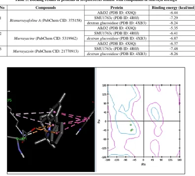
In this study evaluate the antibacterial activities of the crude extracts obtained from the leaves of Murraya koenigii using different solvents like methanol, ethanol, chloroform and hot water against streptococcus mutant. The antimicrobial efficacy of the extracts of Murraya koenigii leaves was quantitatively evaluated on the basis of inhibition zone in mm (Table 1) following the disc diffusion method. Methanol, ethanol, chloroform and hot water extracts have shown better activity than the standard drug. Hot water extract was more effective against streptococcus mutant. The preliminary phytochemical screening revealed the presence of steroids, tannins, alkaloids, saponins, quinones, amino acids, flavonoids in some of the extracts (Methanol, ethanol, chloroform and hot water) as shown in the Table 2. The methanol extract shows the presence of steroids, alkaloids, saponins, amino acids and flavonoids. The ethanol extract shows steroids, alkaloids, quinones, amino acids and flavonoids. The chloroform extract shows the presence of steroids and alkaloids while the hot water extract shows the presence of steroids, alkaloids, saponins, amino acids and flavonoids. All of these compounds have been shown to be potent antibacterial. Docking studies were performed for streptococcus mutant proteins with three marker compounds. The interaction of protein and ligands for the docked ligands with least binding energy was calculated. Bismurrayafoline A and
70
Table 1: Antibacterial activity of different extracts of leaves of Murraya koenigii against streptococcus mutant
S.No: Extract Zone of inhibition (mm)
1 Hot Water 19
2 Ethanol 12
3 Chloroform 14
4 Methanol 11
5 Streptomycin 7
6 Penicillin 2
7 Kanamycin 6
8 Gentamycin 8
Table 2: Showing qualitative phytochemical analyses of different extracts of leaves of Murraya koenigii
S.No Phytochemical compound Presence or absence
Methanol Ethanol Chloroform Hot water
1 Steroids + + + +
2 Tannins - - - -
3 Alkaloids + + + +
4 Saponins + - - +
[image:4.612.111.507.288.646.2]5 Quinones - + - -
Table 3: Docking results of proteins of streptococcus mutant with compounds of Murraya koenigii
S.No Compounds Protein Binding energy (kcal/mol)
1
Bismurrayafoline A (PubChem CID: 375158)
AlkD2 (PDB ID: 4X8Q) -6.44
SMU1763c (PDB ID: 4R0J) -7.29
dextran glucosidase (PDB ID: 4XB3) -8.24
AlkD2 (PDB ID: 4X8Q) -5.35
2
Murrayacine (PubChem CID: 5319962)
SMU1763c (PDB ID: 4R0J) -6.41
dextran glucosidase (PDB ID: 4XB3) -6.87
AlkD2 (PDB ID: 4X8Q) -6.37
3
Murrayazoin (PubChem CID: 21770913) SMU1763c (PDB ID: 4R0J) -7.48
dextran glucosidase (PDB ID: 4XB3) -8.26
71
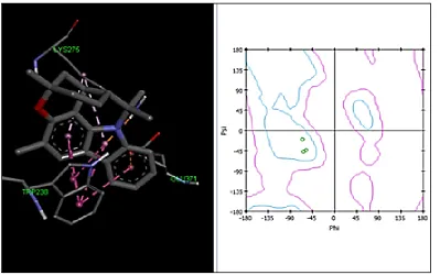
Figure 2: Murrayazoin interaction with dextran glucosidase
CONCLUSION
The present investigation revealed that the extracts from the leaves of Murraya Koenigii exhibited antimicrobial properties. Which explain the basis for its use in traditional medicines to treat tooth decay. Hot water extracts showed significant inhibitory activity against streptococcus mutant. The present study concludes that the Murraya koenigii may serve as a potential source of bioactive compounds in the prevention of tooth decay. The potential for developing antimicrobials from higher plants appears rewarding as it leads to the development of new drugs which is required today.
ACKNOWLEDGEMENTS
The authors are thankful to the Head, Biotech and Bio informatics department, Bishop Heber College, Tiruchirappalli, Tamilnadu, India.
REFERENCES
[1] PK Mukherjee; A Wahil. J Ethnopharmacol. 2006, 103, 25-35.
[2] MK Vinuthan; V Girish Kumar; JP Ravindra; NK Jayaprakash. Indian J Physiol Pharmacol. 2005, 48, 348-352.
[3] S Sathaye; DA Poornima; BM Vinam; BZ Vijay; DK Ramesh; K Harpreet; GR Roopali. Int J Pharm Bio Sci. 2012, 3(1), 430-438.
[4] NU Maheswari; N Cholarani. J Chem Pharm Res. 2013, 5(4), 120-123. [5] AB Sharangi; S Guha. Sci Int. 2013, 1(9), 312-317.
[6] X Li; MK Kristin; L Ronstadt; I Olsen. Clin Microbiol Rev. 2000, 13, 547-558. [7] L Simon. Undergraduate Res J. 2007.
[8] WJ Loesche. Microbiol Rev. 1986, 50(4), 353-380.
[9] AW Bauer; MM Kirby; JC Sherris; M Tuurck. American J Clin Pathol. 1966, 45, 493-496.
[10] SK Bhandary; SN Kumari; SB Vadisha; KP Sharmila; MP Bekal. Nitte University J Health Sci. 2012, 2(4), 34-38.
[11] S Deka; RK Sharma; M Lahkar. Pharm Innovation J. 2015, 4(2), 59-61.
72
[14] MA Elwood; S Rongxin; KA Lyle; E Brandt. PLOS ONE. 2015, 10(5).
[15] K Tan, X Xu, H Cui, S Liu, A Savchenko, A Joachimiak. The crystal structure of a functionally
uncharacterized protein SMU1736c from Sterptococcus mutans. Midwesr centre for structural genomics,
2014.