Single and multiphoton excited
fluorescence from serotonin complexed
with Bcyclodextrin
Botchway, SW, Bisby, RH, Dad, S and Parker, AW
http://dx.doi.org/10.1039/b508602g
Title Single and multiphoton excited fluorescence from serotonin complexed with Bcyclodextrin
Authors Botchway, SW, Bisby, RH, Dad, S and Parker, AW
Type Article
URL This version is available at: http://usir.salford.ac.uk/10092/
Published Date 2006
USIR is a digital collection of the research output of the University of Salford. Where copyright permits, full text material held in the repository is made freely available online and can be read, downloaded and copied for noncommercial private study or research purposes. Please check the manuscript for any further copyright restrictions.
Single- and multi-photon excited fluorescence from serotonin
complexed with
β
-cyclodextrin
Roger H. Bisbya*, Stanley W. Botchwayb, Shakeela Dada and Anthony W. Parkerb
a
Biosciences Research Institute, University of Salford, Salford M5 4WT, UK
b
Lasers for Science Facility, Rutherford Appleton Laboratory, Chilton, OX11 0QX,
UK
*Author for correspondence
Professor R H Bisby
Biosciences Research Institute
Peel Building
University of Salford
Salford M5 4WT, UK
Email:-
Summary
The fluorescence of serotonin on binding with β-cyclodextrin has been studied using
both steady-state and time-resolved methods. Steady state fluorescence intensity of
serotonin at 340 nm showed ~ 30% increase in intensity on binding with Ka ~ 60 dm3
mol-1 and the fluorescence lifetimes showed a corresponding increase. In contrast, the
characteristic green fluorescence (‘hyperluminescence’) of serotonin observed upon
multiphoton near-infrared excitation with sub-picosecond pulses was resolved into
two lifetime components assigned to free and bound serotonin. The results are of
interest in relation to selective imaging and detection of serotonin using the unusual
hyperluminescence emission and in respect to recent determinations of serotonin by
capillary electrophoresis in the presence of cyclodextrin. The results also suggest that
hyperluminescence occurs from multiphoton excitation of a single isolated serotonin
molecule.
Keywords:- serotonin, cyclodextrin, fluorescence lifetime, multiphoton, hyperluminescence
Introduction
Levels of the neurotransmitter serotonin have been linked to clinical depression and
suicide.1 This has consequently created considerable interest in the development of
analytical methods for determining serotonin levels in biological samples such as
cerebrospinal fluid.2 A recent report describes the use of capillary electrophoresis in
combination with hydroxypropyl-β-cyclodextrin for analyzing serotonin levels in
brain microdialysis.3 The importance of neurotransmitters has also stimulated research
to develop methods to locate them within living cells4 at physiological levels, in
particular, and non-linear spectroscopic methods linked to microscopy have much
potential in the field of neurobiology.5 The peak UV absorption of the
5-hydroxyindole chromophore in both 5-hydroxytryptophan and serotonin occurs at a
slightly longer wavelength than tryptophan. This has enabled the use of
5-hydroxytryptophan as a fluorescence probe to study conformation and mobility
within proteins.6 In RBL rat mast cells, confocal three-photon excitation using
ultrafast near infrared laser pulses has enabled imaging of serotonin distribution and
release by monitoring the UV fluorescence at 340 nm.7,8 We and others are presently
exploring the further possibility for selective intracellular imaging of serotonin by
taking advantage of the specific green emission (’hyperluminescence’) produced by
multiphoton excitation of serotonin and 5-hydroxytryptophan.9,10 However, the
identity of the molecular photoproduct responsible for hyperluminescence remains
elusive. The supra-linear dependence of hyperluminescence intensity on serotonin
concentration in phosphate buffer11 implies the possible involvement of a dimeric or
higher aggregate photoproduct, although rapid capillary electrophoresis experiments
indicate the photoproduct is of a similar size to serotonin itself.12 The present
experiments, in which a molecule of serotonin is isolated within a cyclodextrin cavity,
seek to resolve some aspects of this fundamental issue.
Cyclodextrins, especially β-cyclodextrin (cycloheptaamylose), have found varied
applications including drug solubilisation and delivery and in analytical chemistry
procedures.13 This wide range of applications comes from their ability to form
inclusion complexes and act as a host for various guest molecular species.
groups arranged around the external rim. These features provide both isolation of the
guest molecule from the surrounding environment as well as a means of improving
aqueous solubility of hydrophobic compounds. Cyclodextrins are able to modify the
behaviour of electronically excited states of organic molecules,14 for example in
permitting room temperature phosphorescence,15 enhancement of fluorescence
quantum yields16-19 and influencing the outcome of photochemical reactions.20 This
present work has sought to further the earlier work using capillary electrophoresis in
combination with hydroxypropyl-β-cyclodextrin 3 and quantify the nature of the
serotonin:β-cyclodextrin complex and characterise the photophysical properties of
both UV fluorescent and multiphoton hyperluminescence from serotonin.
Experimental
All chemicals including serotonin and β-cyclodextrin were purchased from
Sigma-Aldrich. Solutions were prepared in phosphate buffer (20 mmol dm-3, pH 7.0)
containing NaCl (0.15 mol dm-3).
Fluorescence spectra were measured using a Spex Fluoromax spectrofluorimeter
using the manufacturer’s correction curve to obtain corrected spectra. Fluorescence
lifetimes were measured using the time-correlated single photon counting technique
using as the excitation source a Spectra-Physics Ti:Sapphire laser (120 fs pulses, 80
MHz) pumped by a 20 W argon ion laser (Spectra Physics Ltd). For one-photon
excitation at 300 nm, the Ti:Sapphire laser output was shifted to 600 nm in a Mira
OPO (Coherent) and doubled to 300 nm using a type 1 barium borate (BBO),
delivering ~ 4mW to the sample. Fluorescence at 340 nm from a sample in a standard
1 cm quartz cuvette was selected by a monochromator (M500, IBH) and detected with
a Becker & Hickl GmbH photon-counting photomultiplier (Model PMH-100) linked
to a Becker and Hickel SPC700 multichannel analyser card. Fluorescence decays
were deconvoluted with the instrument response function using Edinburgh
Instruments software. Multiphoton excitation at 750 nm used the direct output of the
Ti:Sapphire laser (~60 mW at the sample). A drop of the sample on a cover slip was
placed on the stage of a fluorescence microscope (Nikon TE2000) and green emission
selected with a 505 nm interference filter (500IU25, Comar, UK). Analysis of
(
1 [ -CD])
CD] -[
max
0 β
β
A A
K F K F F
+ + =
Results and discussion
Ultraviolet fluorescence
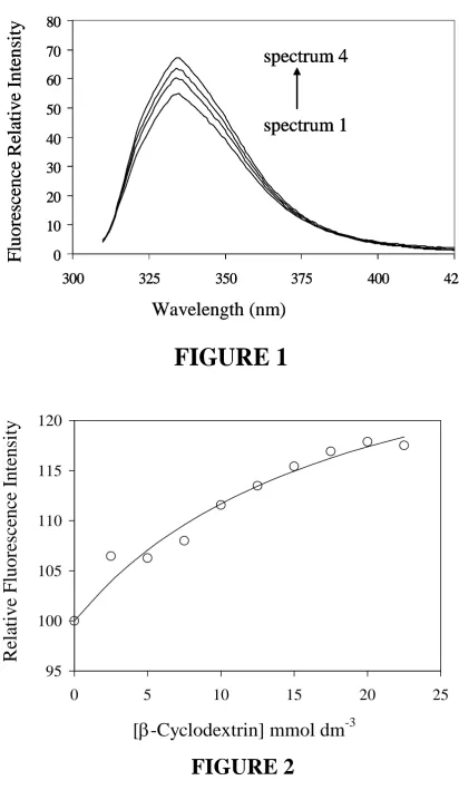
Serotonin fluoresces in aqueous solution at pH 7 with a maximum at 334 nm on
excitation at 300 nm. Addition of β-cyclodextrin to solutions of serotonin (5 µmol
dm-3) in solutions buffered with phosphate (20 mmol dm-3, pH 7.0) and NaCl (0.15
mol dm-3) resulted in a small enhancement of the fluorescence intensity (Figure 1).
Neither the excitation nor emission spectra showed any significant changes in shape
or peak wavelengths with the addition of β-cyclodextrin. This is consistent with the
observation that the fluorescence properties of 5-hydroxyindole are relatively
insensitive to solvent polarity.21
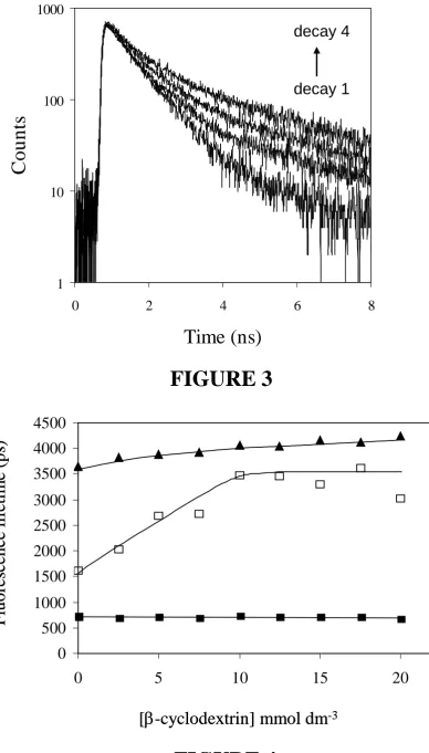
The binding parameters for the serotonin (S) - β-cyclodextrin (β-CD) complex were
evaluated on the basis of formation of a 1:1 complex as has previously been
established in similar cases:-16,17
S + β-CD [S- β-CD] ……. (1)
The association constant of the complex, KA, was found by direct non-linear least
squares fitting of plots of fluorescence intensity (F) versus β-cyclodextrin
concentration according to the binding equation (2), where F0 is the intensity in the
absence of β-cyclodextrin and Fmax is the maximal increase in intensity at infinite β-cyclodextrin concentration :-
…….(2)
Intensities were obtained by integration of the fluorescence spectra on excitation at
300 nm and are shown plotted against β-cyclodextrin concentration in Figure 2. The
best fit to equation (2) is shown by the line with KA = 53.2 ± 17.3 dm3 mol-1 and the
value of Fmax corresponding to 33.8 ± 6.1% increase in fluorescence intensity of the
fully complexed serotonin. (Note: The precision of the measurements of such a small
KA for serotonin: β-cyclodextrin binding is limited by the solubility of β-cyclodextrin
noteworthy that the value of KA for binding of serotonin by β-cyclodextrin obtained
here is less than those for tryptamine17 and tryptophan16 (160 ± 30 and 184 ± 18 dm3
mol-1 respectively) but the overall values for increases in fluorescence intensity on
binding are similar.
Time-resolved fluorescence decays of serotonin, excited at 300 nm and detected at
340 nm, were measured using the time-correlated single photon counting technique.
In the absence of cyclodextrin, the fluorescence decay was satisfactorily deconvoluted
(χ2 < 1.2) as a single exponential with a lifetime of 3.65 ns, in good agreement with a previous determination of 3.80 ns at pH 7.22 Addition of β-cyclodextrin resulted in an
increase in fluorescence lifetime (Figure 4), reaching a value of 4.24 ns in the
presence of 20 mmol dm-3β-cyclodextrin. The increase in fluorescence lifetime
(16%) closely matches the rise of steady state fluorescence intensity (18%) at 20
mmol dm-3β-cyclodextrin recorded above.
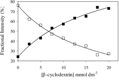
Time-resolved hyperluminescence
Multiphoton excitation of serotonin using an ultrafast near-infrared laser is known to
produce the green fluorescence at 505 nm (‘hyperluminescence’). This corresponds
to a 6-photon process, involving the initial formation of photochemical intermediate
in a four-photon step, followed by excitation of the intermediate by a further two
photons.9 For this work we used a focussed laser beam (750 nm, 130 fs pulses, 80
MHz, 56 mW at the sample) produced by a Ti:sapphire laser. The decay of
hyperluminescence from solutions of serotonin (1 mmol dm-3, pH 7.0) was
biexponential. Due to the inherently weaker emission in the hyperluminescence
experiments, a higher serotonin concentration (1 mmol dm-3) was used. However over
most of the range of cyclodextrin concentrations used (2.5 to 20 mmol dm-3), the
cyclodextrin is present in modest excess (≥ 5:1 molar ratio) and analysis of data using
equation (2) is justified. Representative time-resolved decays are shown in Figure 3
and analysis shows they contain a short lifetime (τ1) of 710 ± 17 ps that is found to be
independent of cyclodextrin concentration. However, the lifetime of a second
component (τ2) increased from 1.65 ns in the absence of β-cyclodextrin to a limiting
More complex analysis of the data was not attempted and the increase in the long
lifetime component to this limiting value is taken to indicate the eventual
predominance of the longer lifetime component due to serotonin to β-cyclodextrin.
The fractional intensity of the longer lifetime component (f2), defined by equation 3
where α1 and α2 are the pre-exponential factors from the fitting analysis,
increased with increasing β-cyclodextrin concentration(Figure 5). The variations of
the fractional intensities of the two lifetime components versus β-cyclodextrin
concentration are shown in Figure 5 with the solid lines representing best fits to the
equivalent of equation (2) with KA = 59.4 ± 11.2 and 53.8 ± 10.6 dm3 mol-1 for the
short and long component intensities respectively. These values are consistent with
the binding constant (53.2 ± 17.3 dm3 mol-1) obtained from the steady state
fluorescence experiments described above. Extrapolation of the data shows that at
saturating β-cyclodextrin concentrations only emission from bound serotonin is
observed with the percentage intensity of the lifetime for the bound form of 120 ±
11% and that for the free form of 15 ± 10%.
As similarly reported in a study of fluorescence lifetimes measured on the binding
2-amino-5,6-dimethylbenzimidazole to β-cyclodextrin,19 the hyperluminescence
decays from serotonin clearly contain exponential components attributable to bound
and free species. For the hyperluminescence the principal lifetimes of bound and free
serotonin are 3.4 ns and 0.71 ns respectively. It might have been anticipated that
differentiation of bound and free serotonin would be observed in the single
photon-excited ultraviolet (340 nm) fluorescence decays from serotonin. However since both
intensity and lifetimes show equivalent small increases of only 16-18% over the
available β-cyclodextrin concentration range, the two lifetimes may not have been
identified within the deconvolution process. Hyperluminescence from
5-hydroxytryptophan has been shown to have a similarly short lifetime in aqueous
solution of 0.91 ns.10 Unlike the fluorescence lifetime of the normal ultraviolet (340
nm) fluorescence of 5-hydroxytryptophan,21 hyperluminescence lifetimes were found )
3 ( ...
1 1 2 2
2 2
2 α τ α τ
τ α
+ =
to be very responsive to solvent,10 increasing up to 3.1 ns in 90% ethanol in water, a
value rather similar to the lifetime identified here with the hyperluminescent species
bound within the β-cyclodextrin cavity.
Hyperluminescence of molecules such as serotonin and 5-hydroxytryptophan offers
new opportunities in the detection11 and imaging23 of these compounds. The transient
species responsible for hyperluminescence remains unidentified, although new routes
to multiphoton excitation of serotonin have been explored recently.24 Capillary
electrophoresis experiments have been used to eliminate dimer or higher multimers of
serotonin as being responsible for the intermediate that produces the
hyperluminescence.12 In biological systems there is the possibility that serotonin may
be bound to proteins such as albumin, and that binding may affect attempts to
quantify or image serotonin using hyperluminescence. The results show that whilst
native ultraviolet fluorescence may be relatively unaffected by binding, the
hyperluminescence lifetime is more sensitive and might be used as an indicator of
such interactions.
In the present experiments there are two possibilities leading to hyperluminescence
from serotonin with the lifetime of 3.4 ns. This value is characteristic of the
fluorophore being located within an environment with a polarity lower than water.
Thus the first possibility is the photochemical conversion of serotonin to the
hyperluminescent intermediate arises from the 4-photon excitation (at 750 – 830 nm)
of a serotonin molecule already complexed within the β-cyclodextrin cavity. This
intermediate whilst still complexed may then be further excited by a further two
photons to generate the hyperluminescence. In this case the hyperluminescence
comes from an isolated single serotonin molecule. An alternative mechanism for the
hyperluminescence with 3.4 ns lifetime is that the initial intermediate is formed by the
four-photon process in solution. This may then associate with cyclodextrin during its
lifetime (>100 microseconds)9 before being excited to give hyperluminescence from
within the cyclodextrin cavity. In the latter case the similarity of the association
constants in the static fluorescence observed at 340 nm generated by UV excitation
and the time-resolved hyperluminescence experiments indicate similar sizes and
support the hypothesis that hyperluminescence occurs from a monomer species
derived from serotonin, and that it should be possible to observe hyperluminescence
from a single residue of 5-hydroxytryptophan within a protein.
Acknowledgements
The authors thank the Biological and Biotechnological Research Council (BBRSC)
for a grant in support of this work, CCLRC for access to the Central Laser Facility
References
1. A.S.Elhwuegi, Progress in Neuro-Psychopharmacology and Biological Psychiatry, 2004, 28, 435-451.
2. M.Asberg, Ann. N.Y. Acad. Sci., 1997, 836, 158-181.
3. N.Beturquia, F.Couderc, V.Sauvinet, C.Ordset, S.Parrot, C.Bayle, B.Renaus and L.Denoroy, Electrophoresis, 2005, 26, 1071-1079.
4. S.Okumoto, L.L.Looger, K.D.Michever, R.J.Reimer, S.J.,Smith and W.B.Frommer,
Proc.Natl.Acad.Sci.U.S.A., 2005, 102, 8740-8745.
5. J.Merz, Curr.Opin.Neurobiol., 2004, 14, 610-616
6. J.B.A.Ross, A.G.Szabo and C.W.V.Hogue, Methods Enzymol., 1997, 278, 151-190.
7. S.Maiti, J.B.Shear, R.M.Williams, W.R.Zipfel and W.W.Webb, Science, 1997, 275, 530-532.
8. R.M.Williams, J.B.Shear, W.R.Zipfel, S.Maiti and W.W.Webb, Biophys.J., 1999, 76, 1835-1846.
9. J.B.Shear, C.Xu and W.W.Webb, Photochem.Photobiol., 1997, 65, 931-936.
10. R.H.Bisby, M.Arvanitidis, S.W.Botchway, I.P.Clark, A.W.Parker and D.Tobin,
Photochem.Photobiol.Sci., 2003, 2, 157-162.
11. M.L.Gostkowski, J.Wei and J.B.Shear, Anal.Biochem., 1998, 260, 244-250.
12. M.J.Gordon, E.Okerberg, M.L.Gostkowski and J.B.Shear, J.Am.Chem.Soc., 2001,
123, 10780-10781.
13. J.Szejtli, Chem.Rev., 1998, 98, 1743-1753.
14. P.Bortolus and S.Monti, Adv.Photochem., 1996, 21, 1-119.
15. S.Scypinski, and L.J.C.Love, Anal. Chem.,1984,56, 331-336.
16. A.Orstan and J.B.A.Ross, J.Phys.Chem., 1987, 91, 2739-2745.
17. R.E.Galian, A.V.Veglia and R.H. de Rossi, Analyst, 1998, 123, 1587-1591.
18. C.Donze, E.Rizzarelli and G.Vecchio, Supramol.Chem., 1998, 10, 33-42.
19. M.A.El-Kemary and I.M.El-Mehasseb, Talanta, 2004, 62, 317-322.
21. K.Lotte, R.Plessow and A.Brockhinke, Photochem.Photobiol.Sci., 2004, 3, 348-359.
22. A.Chattopadhyay, R.Rukmini and S.Mukerjee, Biophys.J., 1996, 71, 1952-1960.
23. S.B.Botchway, K.Brindle and A.W.Parker, Biochem. J., 2005 in press (doi:10.1042/BJ20050648).
24. M.L.Gostkowski, R.Allen, M.L.Plenert, E.Okerberg, M.J.Gordon,and J.B.Shear,
Figure legends
FIGURE 1 Fluorescence spectra from serotonin (5 µmol dm-3) excited at 300 nm in solutions at pH 7 containing:- 0 (curve 1); 5 (curve 2); 10 (curve 3) and 20 (curve 4) mmol dm-3β-cyclodextrin.
FIGURE 2 Increase in fluorescence intensity from solutions of serotonin (5 µmol dm-3) at pH 7 on addition of β-cyclodextrin. The solid line indicates the fit with Ka= 53.2 dm3 mol-1, as described in the text.
FIGURE 3 Nanosecond time-resolved fluorescence decays at 505 nm following multiphoton excitation at 750 nm of serotonin (1 mmol dm-3) in
solutions (pH 7) containing β-cyclodextrin:- none (trace 1); 5 mmol dm-3 (trace 2); 10 mmol dm-3 (trace 3) and 20 mmol dm-3 (trace 4).
FIGURE 4 Fluorescence (hyperluminescence) lifetimes of the biexponential decay from serotonin (1 mmol dm-3) on multiphoton excitation at 750 nm and pH 7 with increasing β-cyclodextrin concentration. Emission was observed at 505 nm. Increasing the β-cyclodextrin concentration had no effect on the short lifetime component (■), whilst the lifetime of longer component increased (□). Also shown are the single exponential lifetimes of the UV fluorescence at 340 nm, excited at 300 nm (▲).
0
10
20
30
40
50
60
70
80
300
325
350
375
400
425
spectrum 4
spectrum 1
F
lu
o
res
cen
ce R
el
at
iv
e I
n
ten
si
ty
Wavelength (nm)
0
10
20
30
40
50
60
70
80
300
325
350
375
400
425
spectrum 4
spectrum 1
F
lu
o
res
cen
ce R
el
at
iv
e I
n
ten
si
ty
0
10
20
30
40
50
60
70
80
300
325
350
375
400
425
spectrum 4
spectrum 1
0
10
20
30
40
50
60
70
80
300
325
350
375
400
425
[image:13.595.84.497.48.758.2]spectrum 4
spectrum 1
spectrum 4
spectrum 1
F
lu
o
res
cen
ce R
el
at
iv
e I
n
ten
si
ty
Wavelength (nm)
FIGURE 1
95
100
105
110
115
120
0
5
10
15
20
25
[
β
-Cyclodextrin] mmol dm
-3[
β
-cyclodextrin] mmol dm
-3F
lu
o
res
cen
ce l
if
et
im
e (
ps
)
0
500
1000
1500
2000
2500
3000
3500
4000
4500
0
5
10
15
20
[
β
-cyclodextrin] mmol dm
-3F
lu
o
res
cen
ce l
if
et
im
e (
ps
)
0
500
1000
1500
2000
2500
3000
3500
4000
4500
0
5
10
15
20
[image:14.595.127.516.55.737.2]FIGURE 4
FIGURE 2
FIGURE 3
1 10 100 10000 2 4 6 8
Time (ns)
Cou
n
ts
decay 4
decay 1
1 10 100 10000 2 4 6 8
Time (ns)
Cou
n
ts
decay 4
decay 1
1 10 100 10000 2 4 6 8