CD4
+
Tregs and immune control
Zoltán Fehérvari, Shimon Sakaguchi
J Clin Invest. 2004;
114(9)
:1209-1217.
https://doi.org/10.1172/JCI23395
.
Recent years have seen Tregs become a popular subject of immunological research.
Abundant experimental data have now confirmed that naturally occurring CD25
+CD4
+Tregs
in particular play a key role in the maintenance of self tolerance, with their dysfunction
leading to severe or even fatal immunopathology. The sphere of influence of Tregs is now
known to extend well beyond just the maintenance of immunological tolerance and to
impinge on a host of clinically important areas from cancer to infectious diseases. The
identification of specific molecular markers in both human and murine immune systems has
enabled the unprecedented investigation of these cells and should prove key to ultimately
unlocking their clinical potential.
Review Series
Find the latest version:
CD4
+
Tregs and immune control
Zoltán Fehérvari1 and Shimon Sakaguchi1,2,3
1Department of Experimental Pathology, Institute for Frontier Medical Sciences, Kyoto University, Kyoto, Japan. 2Laboratory of Immunopathology,
Research Center for Allergy and Immunology, The Institute for Physical and Chemical Research (RIKEN), Yokohama, Japan.
3Core Research for Evolutional Science and Technology, Japan Science and Technology Agency, Kawaguchi, Japan.
Recent years have seen Tregs become a popular subject of immunological research. Abundant experimental data
have now confirmed that naturally occurring CD25
+CD4
+Tregs in particular play a key role in the maintenance of
self tolerance, with their dysfunction leading to severe or even fatal immunopathology. The sphere of influence of
Tregs is now known to extend well beyond just the maintenance of immunological tolerance and to impinge on
a
host of clinically important areas from cancer to infectious diseases. The identification of specific molecular
mark-ers in both human and murine immune systems has enabled the unprecedented investigation of these cells and
should prove key to ultimately unlocking their clinical potential.
Introduction
Naturally occurring CD25+CD4+ suppressor or Tregs cells play
an active part in establishing and maintaining immunological unresponsiveness to self constituents (i.e., immunological self tolerance) and negative control of various immune responses to non-self antigens (1). Although not a new idea for immunologists, the existence of Tregs as a definite cellular entity has been of great controversy until recently because of the paucity of reliable mark-ers for defining the cell, the ambiguity in the molecular basis of suppressive phenomena, the lack of ample evidence for their roles in immunological disease, and even the elusive nature of some suppressive phenomena themselves (2). Recent years, however, have witnessed increasing interest in Tregs in many fields of basic and clinical immunology. Among the several types of Tregs so far reported, naturally occurring CD25+CD4+ Tregs are the main
focus of current research, because accumulating evidence indi-cates that this population plays a crucial role in the maintenance of immunological self tolerance and negative control of patho-logical as well as physiopatho-logical immune responses. A prominent feature of CD25+CD4+ Tregs is that the majority, if not all, of
them are naturally produced by the normal thymus as a function-ally distinct and mature subpopulation of T cells and persist in the periphery with stable function, and that their generation is, at least in part, developmentally controlled (1). Congenital defi-ciency of this population, therefore, results in serious impair-ment of self tolerance and immunoregulation, leading to severe autoimmunity, immunopathology, and allergy in humans (3). On the other hand, their natural presence in the immune system as a phenotypically distinct population makes them a good target for designing ways to treat or prevent immunological diseases and to control pathological as well as physiological immune responses. In addition to this naturally arising “professional” Treg popula-tion, there are several other types of Tregs that can be induced from naive T cells by antigenic stimulation under specialized con-ditions in the periphery (4, 5). Although physiological roles for
these inducible or “adaptive” Tregs need to be fully established, they can still be exploited as a therapeutic tool (6). In this article, we shall review recent progress in our understanding of the roles of natural and adaptive CD4+ Tregs in immune tolerance and
neg-ative control of immune responses. We shall also touch briefly on their possible clinical applications.
Naturally occurring CD25+CD4+ Tregs in self tolerance
and their production by the normal thymus
Experimental evidence for the existence of Tregs with autoim-mune-inhibitory activity has been suggested in various animal models of autoimmune disease for many years (7, 8). Neonatal thymectomy, for example, leads to spontaneous development of autoimmune diseases including gastritis, thyroiditis, and oopho-ritis in selected strains of mice (7–9). Adult thymectomy and sub-sequent sublethal X-irradiation produced thyroiditis and type 1 diabetes (T1D) in selected strains of rats (10, 11). In NOD mice or Bio-Breeding (BB) rats, which spontaneously develop T1D and autoimmune thyroiditis, inoculation with CD4+ T cells from
his-tocompatible normal animals effectively prevented T1D (12, 13). On the other hand, characterization of effector T cells mediating these organ-specific autoimmune diseases firmly documented that CD4+ Th cells destroy the target organs/tissues by helping B
cells to form specific autoantibodies and by inducing cell-mediat-ed immune responses to the target self antigens. Collectively, these findings suggested that normal individuals harbor 2 functionally distinct populations of CD4+ T cells, one capable of mediating
autoimmune disease and the other capable of dominantly inhibit-ing it in the normal physiological state (8). To test this hypothesis directly, attempts were made from the mid-1980s onward to dis-sect these 2 CD4+ T cell populations by expression levels of
par-ticular cell surface molecules and to examine their potential cor-relation with autoimmune induction or inhibition. When CD4+
splenic T cell suspensions prepared from normal mice or rats were depleted of CD25+, RT6.1+, CD5high, or CD45RB/RClow cells and
the remaining CD4+ T cells transferred to syngeneic T
cell–defi-cient mice or rats, the recipients spontaneously developed various organ-specific autoimmune diseases (including T1D, thyroiditis, and gastritis) and systemic wasting disease in a few months; recon-stitution of the eliminated population inhibited the development of autoimmune disease (1, 8, 14). A similar transfer experiment also induced inflammatory bowel disease (IBD), which appeared to result from an excessive immune response of T cells to
com-Nonstandard abbreviations used: GITR, glucocorticoid-induced TNF receptor family–related gene; GITRL, GITR ligand; IBD, inflammatory bowel disease; IDO, indoleamine 2,3-dioxygenase; IPEX, immune dysregulation, polyendocrinopathy, enteropathy, X-linked syndrome; RAG, recombinase-activating gene; T1D, type 1 diabetes; Tr1, T regulatory cell type 1.
Conflict of interest: The authors have declared that no conflict of interest exists.
review series
mensal bacteria in the intestine (15). Currently CD25 is the most specific cell surface marker for such autoimmune- and IBD-pre-ventive CD4+ T cells, because CD25+CD4+ T cells, which constitute
5–10% of CD4+ T cells in normal naive mice, are included in the
CD5high or CD45RBlowCD4+ population, and furthermore their
depletion alone is sufficient to cause autoimmune disease/IBD, while their reconstitution is effectively able to inhibit autoimmune disease/IBD, in various models (1, 16). Additionally, the various immunological properties of natural Tregs, including their in vitro suppressive activity, are assigned to CD25+CD4+ T cells naturally
arising in the immune system (1, 8, 14). It should be noted, how-ever, that CD25 is not an absolute marker for naturally occurring Tregs, since it is also expressed at high levels on activated but oth-erwise conventional nonregulatory T cells. We shall discuss this caveat later in the article.
The normal thymus produces the majority, if not all, of CD25+CD4+ Tregs as a functionally mature T cell subpopulation,
which appears to constitute a distinct cellular lineage and to be contiguous with those found in the periphery (17) (Figure 1). As shown with the transfer of CD25–CD4+ spleen cells described
above, transfer of mature thymocyte suspensions depleted of CD25+ thymocytes produced various autoimmune diseases in
syngeneic T cell–deficient mice (17). This indicates that the normal thymus is continuously producing pathogenic self-reactive CD4+
T cells as well as functionally mature CD25+CD4+ Tregs capable of
controlling them. This centralized production of Tregs has been referred to as “the third function of the thymus” (18).
Accumulating evidence also indicates that thymic development of CD25+CD4+ Tregs requires unique interactions of their TCR
with self-peptide/MHC complexes expressed by thymic stromal cells (19). In TCR transgenic mice, for example, a large number of CD25+CD4+ T cells express endogenous TCR α chains paired with
transgenic β chains; recombinase-activating gene-2 (RAG-2)
defi-ciency, which blocks the gene rearrangement of the endogenous TCR α chain locus, abrogates the development of CD25+CD4+
Tregs in such TCR transgenic mice (17, 20). Furthermore, com-pared with thymic selection of other T cells, the development of CD25+CD4+ Tregs requires higher-avidity interactions of their
TCRs with self peptide/MHC or class II MHC itself expressed on the thymic stromal cells (especially cortical epithelial cells), yet the required avidity must not be so high as to lead to their deletion (19–23). Accessory molecules, such as CD28, B7, and CD40, expressed on developing thymocytes and thymic stromal cells also contribute to the thymic generation of CD25+CD4+ Tregs (24, 25).
The naturally occurring CD4+ Treg phenotype
Naturally occurring CD4+ Tregs constitutively express a variety of
cell surface molecules more commonly associated with activated/ memory cells, most significantly CD25, CD45RBlow, CD62L, CD103,
cytotoxic T lymphocyte antigen-4 (CTLA-4, or CD152), and gluco-corticoid-induced TNF receptor family–related gene (GITR) (15, 16, 26–30). Neuropilin-1, a molecule more usually associated with axon guidance, was very recently reported to be constitutively expressed by natural Tregs and, interestingly, is downregulated on conventional T cells following activation (31). Even though none of these markers is uniquely expressed by naturally occurring CD4+ Tregs, their level
of expression and constitutive nature have still made them useful as functional descriptors by enabling the consistent isolation and investigation of CD4+ T cells with regulatory properties. The
natu-rally occurring Treg surface phenotype indicates that they are in an antigen-primed state and are, at least superficially, similar to mem-ory-type T cells. Judging from the finding that CD25+CD4+ Tregs
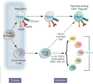
[image:3.585.47.347.85.356.2]require a high-avidity interaction with self peptide/MHC for their thymic development and become functional within the thymus, one could speculate that they are broad in antigen specificity yet more capable of recognizing self antigens than other T cells are.
Figure 1
Regulatory CD4+ cells can develop in a number of ways, although the mechanisms by which these occur and the relationship of the resulting cells to one anoth-er are contestable. Thymically genanoth-erated Treg cells, otherwise known as natural TR cells or CD25+CD4+ TR cells, develop intrathymically according to a special-ized combination of TCR and costimulatory signals. Extrathymically generated TR cells, e.g., Tr1 cells or Th3 cells, can be generated under a whole host of conditions. Whether a conventional naive CD4+ T cell can be converted in the periphery to a de facto Foxp3+
Although CD25 has so far proven to be the best surface marker for thymically produced CD4+ Tregs, it can be expressed on any T
cell following activation (1). In the human system, where there are relatively large numbers of activated T cells, this is especially prob-lematic. Currently, therefore, the best way to select natural human CD4+ Tregs is to sort the population that is very high in CD25
(32). The high constitutive expression of CD25 by Tregs begs the question of whether it is simply a convenient marker or a molecule essential for their function. Several lines of evidence indicate that CD25 is indispensable for the maintenance of natural CD25+CD4+
Tregs in the immune system. For example, it has been shown that mice deficient in IL-2, IL-2Rα (CD25), or IL-2Rβ (CD122) develop lethal inflammatory disease, termed IL-2 deficiency syndrome, which can be prevented by inoculation of normal CD25+CD4+ T
cells as long as a source of IL-2 is made available experimentally (33–35). Our own experiments indicated that neutralization of IL-2 selectively reduced numbers of CD25+CD4+ T cells in normal
mice and consequently produced organ-specific autoimmune dis-eases similar to those produced by depletion of natural Tregs (R. Setoguchi et al., manuscript submitted for publication). Collec-tively, these results suggest that IL-2 is essential for the develop-ment, maintenance, and function of CD25+CD4+ Tregs.
GITR and its role in CD25+CD4+ Treg function is an
interest-ing area. This molecule was identified as a constitutively expressed marker for naturally occurring Tregs, but, like most such candidate molecules involved in Treg identification, it is also upregulated on conventional activated CD4+ T cells (29, 30). An anti–mouse
GITR mAb (DTA-1) is able to block CD25+CD4+ Treg suppression
in vitro, and, furthermore, its injection leads to the induction of autoimmunity in vivo as well as enhancing the proliferation of CD25–CD4+ cells by transducing a costimulatory signal (29, 36).
Since DTA-1 is nondepleting, it was originally presumed to pri-marily transmit a suppression-blocking signal to the CD25+CD4+
Treg. However, some very recent data instead suggest that liga-tion of GITR on activated T cells, not Tregs, renders them resistant to suppression (37). The natural ligand for GITR (GITRL) has now also been cloned and its distribution elucidated (38). GITRL is expressed on APCs (DCs, macrophages, and B cells) but is downregulated following maturation. Therefore the relative dis-tribution patterns of GITR on activated T cells and Tregs, and of GITRL on APCs, suggest a complex dynamic of interaction, which is only just being elucidated.
Identification of an unambiguous surface marker for naturally occurring CD4+ Tregs remains something of a Holy Grail,
espe-cially where the isolation of human Tregs for clinical purposes is concerned. Efforts in this direction may well guide the progress of research in naturally occurring human Tregs.
FOXP3 as a master control gene for Treg development
A deeper understanding of the developmental processes of nat-ural Tregs, as suggested by the neonatal thymectomy model of autoimmune disease, evolved out of studies on the Scurfy mouse and the human disease IPEX (immune dysregulation, polyen-docrinopathy, enteropathy, X-linked syndrome). IPEX is an X-linked immunodeficiency syndrome associated with autoim-mune disease in multiple endocrine organs (such as T1D and thyroiditis), IBD, atopic dermatitis, and fatal infections (3). The Scurfy mouse strain exhibits a fatal X-linked lymphoprolifera-tion characterized by a multiorgan immunopathology very simi-lar to the human disease IPEX (39–41). The causative gene, Foxp3
(FOXP3 in humans), which underlies both syndromes, encodes a forkhead/winged-helix family transcriptional repressor called Scurfin (42–44). The striking similarities seen between mutations in Foxp3/FOXP3 and depletion of CD25+CD4+ Tregs led several
groups to investigate the relationship of this gene to Treg devel-opment and function. Experiments in mice indeed demonstrated
Foxp3 mRNA and Scurfin protein to be specifically expressed in CD25+CD4+ Tregs, and, in contrast to the cell surface markers
used to date, they were never observed in non-Tregs following conventional activation or differentiation into Th1 and Th2, nor in natural killer T cells (45–47).Subsequent studies, also in mice, have further demonstrated the existence of a small population of CD25–CD4+ T cells that are nevertheless still Foxp3+ and have
a regulatory function (ref. 1, and M. Ono et al., manuscript sub-mitted for publication). Scurfy mutant mice, or those with a tar-geted deletion of Foxp3, were unable to support the development of natural CD25+CD4+ Tregs, although they contained large
numbers of chronically activated CD25+ nonregulatory T cells
(45, 47). By contrast, the number of CD25+CD4+ Tregs increased
significantly in transgenic mice overexpressing Foxp3 (45). A final critical observation showed that retroviral transduction of
Foxp3 into Foxp3– nonregulatory CD25–CD4+ T cells bestowed on
them a fully functional Treg phenotype; e.g., cotransfer of Foxp3 -transduced T cells with CD25–CD4+ T cells prevented
autoim-mune disease and IBD in SCID mice (see above) (46, 47). Broadly speaking, an equivalent pattern of FOXP3 expression has now also been reported in human cells, with Treg-like prop-erties being similarly transferable by retroviral transduction (48–51). Already, however, some discrepancies are beginning to emerge between the behavior of human and that of mouse
FOXP3/Foxp3 expression. For instance, there is at least 1 example of FOXP3 being apparently induced following standard antibody-mediated activation of normal CD25– human T cells; this has
not been observed thus far in the murine model (50). Similarly, some instances of CD25– human T cell activation by DCs have
also resulted in FOXP3 upregulation (refs. 52, 53, and see below). The possibility remains, however, that the induction of FOXP3
expression in human CD25– cells may simply be a result of the
expansion of the human counterpart to the murine CD25–CD4+ Foxp3+ population described above, as these studies all isolated
their Tregs solely on the basis of CD25 (51).
Thus, Foxp3/FOXP3 appears to be a master control gene for the development and function of natural CD25+CD4+ Tregs. Given
that humans bear natural CD25+CD4+ Tregs with a phenotype
and function comparable to those found in rodents (32), it is most likely that in IPEX, disruption of the FOXP3 gene abrogates the development of thymic Tregs, leading to hyperactivation of T cells reactive with self antigens, commensal bacteria in the intestine, or innocuous environmental substances, and thus causing autoim-mune polyendocrinopathy, IBD, or allergy, respectively. This has several implications for self tolerance and autoimmune/inflam-matory disease in humans. First, this is so far the clearest example that an abnormality in naturally arising Tregs is a primary cause of human autoimmune disease, IBD, and allergy. Second, the development of natural Tregs is, at least in part, genetically and developmentally programmed. Third, hemizygous defects of the
review series
and FOXP3-normal ones as a genetic mosaic, yet they are never-theless completely normal (54). This observation demonstrates that even reduced numbers of Foxp3+ Tregs are able to dominantly
control pathogenic T cells, and, further, that even a partial resto-ration of Tregs could be sufficient to cure IPEX or, indeed, other autoimmune pathologies. Mechanistic data on Foxp3 are thus far lacking; it is therefore currently unclear how it exerts its effects at a molecular level. The molecular interactions of Foxp3, and indeed the signals triggering its expression, are now an intensely investi-gated area, and unraveling them may well prove critical to exploit-ing natural Tregs in a therapeutic settexploit-ing.
Functional characteristics of natural CD25+CD4+ Tregs
and their mechanisms of suppression
Without question the most remarkable feature of CD25+CD4+
Tregs is their ability to dampen immune responses. They appear capable of suppressing a wide variety of immune cells, encompass-ing those of both the innate (55–57) and the adaptive immune systems (58–60). This suppressive ability can be modeled in vitro by mixing of titrated numbers of highly purified CD25+CD4+
Tregs and responder cells, typically CD25–CD4+ T cells plus a T
cell stimulus. Under such conditions, the CD25+ population
sup-presses both the proliferation and, more fundamentally, the IL-2 production of the CD25– cells in a dose-dependent manner (58,
59, 61). CD25+CD4+ Tregs themselves require TCR stimulation,
and, it now seems, IL-2, to actually trigger their suppressive effects, but once this condition has been satisfied their ensuing suppres-sion can act non–antigen-specifically (58, 59, 61). Therefore, sup-pression is an active process and can be directed against bystander cells. Curiously, CD25+CD4+ Tregs themselves are anergic in vitro,
i.e., they do not proliferate or produce IL-2 in response to con-ventional T cell stimuli such as plate-or bead–bound anti-CD3, concanavlin A (ConA), or splenic APCs. This anergy can, however, be broken by a sufficiently potent stimulus, e.g., the addition of high-dose exogenous IL-2 or anti-CD28, or the use of mature DCs as APCs (27, 58, 59, 61–63). Some of these strong stimuli, particu-larly mature DCs, also perturb CD25+CD4+ Treg suppression both
in vitro and in vivo (63, 64). At least in vitro, anergy seems to be the default state of naturally occurring Tregs, since they revert back to it once potent stimulation is withdrawn (58). In vivo, however, CD25+CD4+ Treg anergy is not readily observed; instead they seem
to have a highly active rate of turnover (33, 65). It seems likely, then, that CD25+CD4+ Treg anergy is an in vitro phenomenon,
merely reflecting an exacting set of activation requirements gener-ally absent from cell culture.
Given that the ability to control immune responses is the cardinal feature of CD25+CD4+ Tregs, it is surprising that their mechanism(s)
of suppression remains elusive. Essentially, Treg suppression can be divided into those mechanisms mediated by relatively far-reaching soluble factors and those requiring intimate cell contact. In vivo experiments based chiefly on the IBD model mentioned previously have demonstrated the importance of the immunomodulatory cytokines IL-10 and TGF-β (66). By blocking IL-10 signaling in vivo with an anti–IL-10 receptor mAb, it was possible to abrogate the normal colitis-preventative action of CD45RBlow cells (66). Similarly,
CD45RBlow T cells from IL-10–/– mice lacked their otherwise intrinsic
ability to protect from colitis and, moreover, were even colitogenic themselves when transferred alone (66). The importance of IL-10 is further underscored by the observation that IL-10–/– mice
spon-taneously develop colitis (67, 68). Examination of the in vivo role
of TGF-β has generally painted a similar picture to that of IL-10, with Treg function being blocked by the presence of neutralizing anti–TGF-β mAbs (69). Some data also suggest that TGF-β may not necessarily act as a soluble factor but can also be found on the surface of activated CD25+CD4+ Tregs and may therefore act in a
membrane-proximal manner (70). Interestingly, virtually all TGF-β+
CD25+CD4+ Tregs also express thrombospondin, a factor capable
of converting normally latent TGF-β into its active form (71). There should be a note of caution regarding these in vitro studies on TGF-β, since a comprehensive analysis by a second group failed to demonstrate any role for it in vitro (72).
The confusion over a definitive CD25+CD4+ Treg suppression
mechanism is compounded when viewed in the context of the in vitro data, since here the overwhelming evidence highlights direct cell-cell interaction, and not cytokines, as being critical (58, 59, 73). Several lines of evidence lead to this conclusion: with the exception of the study on membrane-bound TGF-β alluded to above, both anti–IL-10 and anti–TGF-β fail to perturb CD25+CD4+ Treg
sup-pression (58, 59, 70, 72). Similarly, supernatants from suppressed cultures or activated CD25+CD4+ Tregs show no inherent
suppres-sive activity, nor can suppression be observed across a semiperme-able membrane (58, 59). Collectively, these in vitro observations therefore appear to obviate a role not just for IL-10 and TGF-β but for soluble factors in general.
The actual membrane events occurring during suppression that depends on CD25+CD4+ Treg contact have yet to be
clari-fied. The most simplistic models propose competition for APCs and specific MHC/peptide antigenic complexes. Additionally, the constitutive expression of the high-affinity IL-2 receptor could make naturally occurring Tregs into an effective IL-2 sink, depriv-ing potential autoreactive T cells of this essential growth factor (74). However, given the relative physiological scarcity of natu-rally occurring Tregs, it is perhaps unlikely that a simple competi-tive-adsorptive model alone could account for their suppressive action in vivo. Other models of CD25+CD4+ Treg suppression
propose a more proactive and antagonistic form of suppression that relies on the expression of specific “inhibitory” molecules. The identity and indeed even the very existence of such an inhibi-tory molecule are uncertain, but 1 potential molecule could be Treg–expressed CTLA-4. Aside from its well-established high affinity for the costimulatory molecules B7.1 and B7.2 (CD80 and CD86, respectively), CTLA-4 has also recently been shown to trigger the induction of the enzyme indoleamine 2,3-dioxygen-ase (IDO) when interacting with its ligands on DCs (75–78). IDO catalyzes the conversion of tryptophan to kynurenine and other metabolites, which have potent immunosuppressive effects in the local environment of the DC. In this way, CD25+CD4+ Tregs may
exert their suppression by proxy through their action on APCs. Another APC-centric mode of suppression could be via the per-turbation of antigen-presenting capacity. In support of this con-cept, one report has demonstrated that purified CD25+CD4+ Tregs
are able to downregulate the expression of both CD80 and CD86 on DCs, converting them into inefficient APCs (57). At any rate, CD25+CD4+ Tregs need not act exclusively via the APC, since they
are quite capable of suppressing in the context of “APC-free” sys-tems such as plate- or bead–bound antibodies or MHC/peptide tetramers (58, 79). At least in vitro, direct suppression of the target cell is therefore still also possible.
study suggested that engagement of CD80, and to a lesser extent CD86, on the responder T cell and not the APC was responsible for the transmission of a negative signal, and therefore these were the molecular targets through which Tregs exert their function (80). In support of this, the authors demonstrated that B7–/– responder
cells were resistant to suppression in vitro and induced a fatal wasting disease refractory to cotransferred CD25+CD4+ Tregs
(80). Again the obvious candidate Treg molecule for this inhibi-tory interaction would be CTLA-4, although this fails to explain the paradoxically intact suppression mediated by CTLA-4–/–
CD25+CD4+ Tregs (27). The presence of B7 on conventional T cells
has been known for several years, and it would be interesting, then, if this hitherto puzzling expression pattern were shown to play a role in Treg–mediated suppression (81, 82). While the identifica-tion of a membrane-bound CD25+CD4+ Treg–specific inhibitory
molecule remains inconclusive, some very recent work has sug-gested that the CD4-related molecule LAG-3 may be important (CD223), though this awaits independent confirmation (83, 84). Proving a negative hypothesis is always a difficult task, but it may yet be shown that there are no truly unique Treg–associated mol-ecules responsible for inhibition. Rather, the specialized functions of Tregs could simply be the product of known molecules acting semi-redundantly, which together generate a suppressive pheno-type. An integrated summary of CD25+CD4+ Treg suppressive
mechanisms is shown in Figure 2.
Given the relative physiological scarcity of CD25+CD4+ Tregs, it
seems likely that in vivo they would use mechanisms to amplify their suppressive action, and ones that are not normally fully appreciated under in vitro analysis. This could occur either by the modification
of APCs as outlined above or by the “infectious” spreading of toler-ance to conventional T cells. In accordtoler-ance with this, some recent work has demonstrated that human CD25+CD4+ Tregs can confer
a suppressive phenotype to conventional CD4+ T cells in a
contact-dependent manner (52). These newly generated regulatory-like cells then suppress by means of IL-10 or TGF-β. This would constitute a mechanism of not only spreading a suppressive phenotype but also making it more efficient on a per-cell basis by engaging the action of soluble mediators. Most satisfyingly, this scenario could also finally reconcile some of the disparities observed between the in vitro and the in vivo mechanisms of CD25+CD4+ Treg suppression.
Extrathymic generation of CD4+ Tregs
The possibility of extrathymic CD25+CD4+ Treg generation is
currently contentious. An interesting recent study in this area readdressed so-called “low-zone” tolerance (85). This phenom-enon was first observed decades ago and was described as the antigen-specific tolerance resulting from subimmunogenic doses of antigen given i.v. (86). Using an osmotic pump to deliver min-ute controlled quantities of antigenic peptide to TCR trans-genic mice, the authors of this updated study demonstrated the appearance of CD25+CD4+ Tregs measurable by their function,
surface phenotype, and Foxp3 expression. Importantly, the study showed that Treg development could still occur in thymecto-mized TCR transgenic mice on a RAG knockout background, which are normally wholly lacking in CD25+CD4+ Tregs (17, 20,
46, 85). It therefore seems possible that CD25+CD4+ Treg
[image:6.585.49.342.82.403.2]devel-opment, as measured by Foxp3 and suppressive function, can occur in conventional T cells under specific in vivo activation
Figure 2
review series
conditions, therefore demonstrating a potentially clinically sig-nificant developmental plasticity.
The involvement of TGF-β in many facets of Treg behavior is well known (87), but a recent report has also suggested that its exog-enous addition results in development of Foxp3+ Tregs, from
con-ventional and even from RAG knockout CD4+ T cells (refs. 87, 88).
However, it remains to be seen whether generation of CD25+CD4+
Tregs by the mechanisms described above can occur outside of the
relatively artificial confines of a TCR transgenic system, and indeed whether the initial cell populations contain only “truly” naive T cells and no potential Foxp3+CD25+CD4+ Treg precursors.
In the original demonstration of extrathymically generated regu-latory cells, the cells were termed T reguregu-latory cell type 1 (Tr1) or Th3 cells (89, 90). It is likely that these cells form a cell type distinct from their thymically generated CD25+CD4+ Treg counterparts
that have been elaborated on above. Tr1 and related cells have been generated using a variety of approaches, typically involving T cell activation in the presence of immunomodulating cytokines or repetitive stimulation with nonprofessional APCs. Tr1 cells were initially generated by chronic stimulation of normal nonregula-tory T cells in the presence of IL-10 (89). Such cells secrete a pat-tern of cytokines distinct from that of the more usual Th1 or Th2 profile and are characterized by high levels of IL-10 and generally low levels of TGF-β and IL-5 (89). Moreover, Tr1 cells are function-ally suppressive in vivo and able to prevent the development of Th1 autoimmune diseases such as colitis (89, 91). Th3 cells, on the other hand, were cloned from the mesenteric lymph nodes of mice orally tolerized with myelin basic protein (90). The majority of such cells produce TGF-β and varying levels of Th2 cytokines and suppress the induction of experimental autoimmune encephalitis (90). In vitro treatment of human and mouse T cells with a com-bination of the immunosuppressants vitamin D3 and
dexametha-sone has also resulted in the generation of Foxp3– regulatory cells,
but with properties somewhat distinct from those reported for Tr1 or Th3 cells (92–94). Finally, there is also a study suggesting that signaling through the complement receptor (CD46) concomitant to more conventional TCR activation can trigger the peripheral induction of human CD4+ regulatory cells (95).
Much attention has also focused on the influence that DCs may have on the extrathymic development of regulatory cells. Stimula-tion with immature DCs (i.e., low levels of costimulatory molecules) and stimulation with DCs modified by pretreatment with IL-10 or TGF-β have both been shown to result in the induction of anergic cells with suppressive capabilities in vitro and in vivo (96–98). Cur-rent models of DC-based tolerance state that T cell antigen recogni-tion on immature DCs results in tolerizarecogni-tion whereas mature DCs elicit effector responses (99). A system structured in this way would
be effective at maintaining self tolerance in the physiological steady state, i.e., in the absence of inflammatory “danger signals,” yet would support productive immune responses following DC matu-ration triggered by the presence of microbes. However, there would always be the potential danger that DCs matured during “sterile” inflammation, e.g., following mechanical injury, could elicit immu-nity to autoantigens. Similarly, self tolerance could theoretically also be broken by autoantigens presented on DCs matured during a contemporaneous microbial infection. It seems, though, that the immune system may have yet another level of control to protect against just the kind of scenarios outlined above. In support of this, it was recently reported that the response of conventional human CD4+ T cells to autologous peptides presented by mature, but not
by immature, DCs results in the generation of regulatory-like T cells (53). If confirmed, this ability of the immune system to so dra-matically alter the outcome of a response depending on the antigen being recognized is rather remarkable, especially given the appar-ently matured DC phenotype. Possibly the net response is attribut-able to the nature or source of ancillary signals, e.g., which toll-like receptors (TLRs) are being engaged, alone or in combination.
Extrathymically generated regulatory cells represent a hetero-geneous assemblage whose ontogenic relationship to naturally occurring Tregs is still being determined. The only really clear point of convergence between the 2 broad families of regulatory cells is that they share a suppressive capability. One interpretation would suggest that peripherally generated regulatory cells merely represent a specialized activation state of conventional CD4+ cells
(i.e., “adaptive regulatory cells”) whereas CD25+CD4+ Tregs are
a de facto lineage by virtue of their distinctive Foxp3 expression, although the most recent data stemming from the use of TGF-β or low-zone tolerance induction protocols are perhaps blurring even this distinction (85, 88). The use of Foxp3/FOXP3 to disentangle this conundrum has been only partially successful. As far as mice are concerned, Foxp3 seems, by and large, to be a stable marker expressed only in naturally occurring CD25+CD4+ Tregs and thus
far is not in most models of extrathymically generated regulatory cells (but see refs. 85, 88). In contrast, human FOXP3 expression appears far less stringent, with some reports already demonstrating upregulation in extrathymically generated regulatory cells follow-ing even basic activation (52, 53). Whether the apparent variability in human FOXP3 and, to a much lesser extent, mouse Foxp3 expres-sion undermines its importance as an unambiguous marker for naturally occurring Tregs remains to be seen.
Conclusion and clinical perspective
Abundant evidence now strongly supports the once controversial existence of Tregs as key controllers of self tolerance. It also now
Table 1
Potential clinical applications of CD25+CD4+ Tregs
Target condition Potential therapeutic approach
Enhancement of CD25+CD4+ Treg function Organ transplantation, Transfer of Tregs or enhancement of their function allows specific
autoimmune disease, suppression of immune responses; e.g., ex vivo gene transduction allergy of Foxp3; ex vivo generation of regulatory cells using
cytokines, pharmacological agents, or modified DCs Reduction of CD25+CD4+ Treg function Cancer, infectious Removal of Tregs or blocking of their function boosts immune
seems that their roles can be expanded to many areas of immunol-ogy, in fact, potentially, to any scenario where the suppression and/ or tuning of an immune response is required. A strategic manipu-lation of Tregs, either naturally occurring or extrathymically gen-erated, to dampen or enhance their functions as appropriate may prove to have great clinical benefit (Table 1). Already manipula-tion of CD25+CD4+ Tregs in various animal models has provided
encouraging results for both enhancement of tumor immunity and maintenance of allograft tolerance (100–104). In the case of organ transplantation in particular, CD25+CD4+ Tregs seem
to offer a flexible and adaptive form of immunological control apparently not achievable with standard small-molecule immu-nosuppression (see for example refs. 102, 104, 105). These practi-cal applications will be expanded upon in other Reviews in this series. Informed by the murine studies, recent experiments are also increasingly demonstrating the significant roles CD25+CD4+ Tregs
can play in human pathologies as varied as rheumatoid arthritis, multiple sclerosis, HIV infection, and allergy (106–112). Recent advances in our understanding of CD25+CD4+ Treg development
and important functional markers such as the association with
Foxp3/FOXP3 have permitted the accurate isolation and manipula-tion of these cells in mice and, importantly, their human counter-parts. Understanding the events both upstream and downstream
of Foxp3/FOXP3 may enable us to “tailor-make” large numbers of CD25+CD4+ Tregs to specifically suppress immune responses in
autoimmunity and allergy or to antagonize them where a boost of immunity is required, e.g., in microbial and antitumor responses. The potential clinical focus, though, need not be solely on thy-mically produced CD25+CD4+ Tregs, since peripherally generated
regulatory cells such as Tr1, with their potent cytokine-mediated suppressive capacity, may also hold great therapeutic promise.
Acknowledgments
We apologize to those researchers whose work, because of space restrictions, has not been cited in this review. We thank our col-leagues who have allowed the prepublication mention of their work and engaging discussion. Z. Fehérvari is supported by the Japan Society for the Promotion of Science and S. Sakaguchi by grants-in-aid from the Ministry of Education, Culture, Sports, Science and Technology and the Ministry of Human Welfare of Japan.
Address correspondence to: Zoltán Fehérvari, Department of Experimental Pathology, Institute for Frontier Medical Scienc-es, Kyoto University, Shogo-in 53, Sakyo-ku, Kyoto 606-8507, Japan. Phone: 81-75-751-3888; Fax: 81-75-751-3820; E-mail: [email protected].
1. Sakaguchi, S. 2004. Naturally arising CD4+ regu-latory t cells for immunologic self-tolerance and negative control of immune responses. Annu. Rev. Immunol.22:531–562.
2. Bloom, B.R., Salgame, P., and Diamond, B. 1992. Revisiting and revising suppressor T cells. Immunol. Today.13:131–136.
3. Gambineri, E., Torgerson, T.R., and Ochs, H.D. 2003. Immune dysregulation, polyendocrinopa-thy, enteropapolyendocrinopa-thy, and X-linked inheritance (IPEX), a syndrome of systemic autoimmunity caused by mutations of FOXP3, a critical regulator of T-cell homeostasis. Curr. Opin. Rheumatol.15:430–435. 4. Roncarolo, M.G., Bacchetta, R., Bordignon, C.,
Narula, S., and Levings, M.K. 2001. Type 1 T regu-latory cells. Immunol. Rev.182:68–79.
5. Weiner, H.L. 2001. Induction and mechanism of action of transforming growth factor-beta-secreting Th3 regulatory cells. Immunol. Rev.182:207–214. 6. Bluestone, J.A., and Abbas, A.K. 2003. Natural
ver-sus adaptive regulatory T cells. Nat. Rev. Immunol. 3:253–257.
7. Sakaguchi, S., Takahashi, T., and Nishizuka, Y. 1982. Study on cellular events in post-thymectomy autoimmune oophoritis in mice. II. Requirement of Lyt-1 cells in normal female mice for the preven-tion of oophoritis. J. Exp. Med.156:1577–1586. 8. Sakaguchi, S., Fukuma, K., Kuribayashi, K., and
Masuda, T. 1985. Organ-specific autoimmune diseases induced in mice by elimination of T cell subset. I. Evidence for the active participation of T cells in natural self-tolerance; deficit of a T cell subset as a possible cause of autoimmune disease.
J. Exp. Med.161:72–87.
9. Asano, M., Toda, M., Sakaguchi, N., and Sakaguchi, S. 1996. Autoimmune disease as a consequence of developmental abnormality of a T cell subpopula-tion. J. Exp. Med.184:387–396.
10. Penhale, W.J., Farmer, A., McKenna, R.P., and Irvine, W.J. 1973. Spontaneous thyroiditis in thy-mectomized and irradiated Wistar rats. Clin. Exp. Immunol.15:225–236.
11. Fowell, D., and Mason, D. 1993. Evidence that the T cell repertoire of normal rats contains cells with the potential to cause diabetes. Characterization of the CD4+ T cell subset that inhibits this autoim-mune potential. J. Exp. Med.177:627–636.
12. Boitard, C., Yasunami, R., Dardenne, M., and Bach, J.F. 1989. T cell-mediated inhibition of the transfer of autoimmune diabetes in NOD mice. J. Exp. Med. 169:1669–1680.
13. Greiner, D.L., et al. 1987. Depletion of RT6.1+ T lymphocytes induces diabetes in resistant bio-breeding/Worcester (BB/W) rats. J. Exp. Med. 166:461–475.
14. Powrie, F., and Mason, D. 1990. OX-22high CD4+ T cells induce wasting disease with multiple organ pathology: prevention by the OX-22low subset.
J. Exp. Med.172:1701–1708.
15. Powrie, F., Leach, M.W., Mauze, S., Caddle, L.B., and Coffman, R.L. 1993. Phenotypically distinct subsets of CD4+ T cells induce or protect from chronic intestinal inflammation in C. B-17 scid mice. Int. Immunol.5:1461–1471.
16. Sakaguchi, S., Sakaguchi, N., Asano, M., Itoh, M., and Toda, M. 1995. Immunologic self-tolerance maintained by activated T cells expressing IL-2 receptor alpha-chains (CD25). Breakdown of a single mechanism of self-tolerance causes various autoimmune diseases. J. Immunol.155:1151–1164. 17. Itoh, M., et al. 1999. Thymus and autoimmunity:
production of CD25+CD4+ naturally anergic and suppressive T cells as a key function of the thy-mus in maintaining immunologic self-tolerance.
J. Immunol.162:5317–5326.
18. Seddon, B., and Mason, D. 2000. The third func-tion of the thymus. Immunol. Today.21:95–99. 19. Jordan, M.S., et al. 2001. Thymic selection of
CD4+CD25+ regulatory T cells induced by an ago-nist self-peptide. Nat. Immunol.2:301–306. 20. Kawahata, K., et al. 2002. Generation of
CD4(+)CD25(+) regulatory T cells from autoreactive T cells simultaneously with their negative selection in the thymus and from nonau-toreactive T cells by endogenous TCR expression.
J. Immunol.168:4399–4405.
21. Stephens, G.L., and Ignatowicz, L. 2003. Decreas-ing the threshold for thymocyte activation biases CD4+ T cells toward a regulatory (CD4+CD25+) lineage. Eur. J. Immunol.33:1282–1291.
22. Sakaguchi, S., et al. 2003. Thymic generation and selection of CD25+CD4+ regulatory T cells: impli-cations of their broad repertoire and high reac-tivity for the maintenance of immunological
self-tolerance. Novartis Found. Symp.252:6–16. 23. Hsieh, C.S., et al. 2004. Recognition of the
periph-eral self by naturally arising CD25(+) CD4(+) T cell receptors. Immunity.21:267–277.
24. Salomon, B., et al. 2000. B7/CD28 costimulation is essential for the homeostasis of the CD4+CD25+ immunoregulatory T cells that control autoim-mune diabetes. Immunity.12:431–440.
25. Kumanogoh, A., et al. 2001. Increased T cell auto-reactivity in the absence of CD40-CD40 ligand interactions: a role of CD40 in regulatory T cell development. J. Immunol.166:353–360.
26. Lehmann, J., et al. 2002. Expression of the integrin alpha Ebeta 7 identifies unique subsets of CD25+ as well as CD25- regulatory T cells. Proc. Natl. Acad. Sci. U. S. A.99:13031–13036.
27. Takahashi, T., et al. 2000. Immunologic self-toler-ance maintained by CD25(+)CD4(+) regulatory T cells constitutively expressing cytotoxic T lympho-cyte-associated antigen 4. J. Exp. Med.192:303–310. 28. Read, S., Malmstrom, V., and Powrie, F. 2000. Cytotoxic T lymphocyte-associated antigen 4 plays an essential role in the function of CD25(+)CD4(+) regulatory cells that control intestinal inflamma-tion. J. Exp. Med.192:295–302.
29. Shimizu, J., Yamazaki, S., Takahashi, T., Ishi-da, Y., and Sakaguchi, S. 2002. Stimulation of CD25(+)CD4(+) regulatory T cells through GITreg breaks immunological self-tolerance. Nat. Immunol. 3:135–142.
30. McHugh, R.S., et al. 2002. CD4(+)CD25(+) immu-noregulatory T cells: gene expression analysis reveals a functional role for the glucocorticoid-induced TNF receptor. Immunity.16:311–323. 31. Bruder, D., et al. 2004. Neuropilin-1: a surface
marker of regulatory T cells. Eur. J. Immunol. 34:623–630.
32. Baecher-Allan, C., Brown, J.A., Freeman, G.J., and Hafler, D.A. 2001. CD4+CD25high regula-tory cells in human peripheral blood. J. Immunol. 167:1245–1253.
33. Almeida, A.R., Legrand, N., Papiernik, M., and Frei-tas, A.A. 2002. Homeostasis of peripheral CD4+ T cells: IL-2R alpha and IL-2 shape a population of regulatory cells that controls CD4+ T cell numbers.
J. Immunol.169:4850–4860.
review series
L. 2002. CD4 regulatory T cells prevent lethal autoimmunity in IL-2Rbeta-deficient mice. Impli-cations for the nonredundant function of IL-2.
Immunity.17:167–178.
35. Papiernik, M., de Moraes, M.L., Pontoux, C., Vas-seur, F., and Penit, C. 1998. Regulatory CD4 T cells: expression of IL-2R alpha chain, resistance to clonal deletion and IL-2 dependency. Int. Immunol. 10:371–378.
36. Kanamaru, F., et al. 2004. Costimulation via glu-cocorticoid-induced TNF receptor in both con-ventional and CD25+ regulatory CD4+ T cells.
J. Immunol.172:7306–7314.
37. Stephens, G.L. et al. 2004. Engagement of glucocorticoid-induced TNFR family-related receptor on effector T cells by its ligand mediates resistance to suppression by CD4+CD25+ T cells.
J. Immunol. 173:5008–5020.
38. Tone, M., et al. 2003. Mouse glucocorticoid-induced tumor necrosis factor receptor ligand is costimulatory for T cells. Proc. Natl. Acad. Sci. U. S. A.
100:15059–15064.
39. Blair, P.J., et al. 1994. CD4+CD8- T cells are the effector cells in disease pathogenesis in the scurfy (sf) mouse. J. Immunol.153:3764–3774.
40. Godfrey, V.L., Wilkinson, J.E., Rinchik, E.M., and Russell, L.B. 1991. Fatal lymphoreticular disease in the scurfy (sf) mouse requires T cells that mature in a sf thymic environment: potential model for thymic education. Proc. Natl. Acad. Sci. U. S. A. 88:5528–5532.
41. Lyon, M.F., Peters, J., Glenister, P.H., Ball, S., and Wright, E. 1990. The scurfy mouse mutant has pre-viously unrecognized hematological abnormalities and resembles Wiskott-Aldrich syndrome. Proc. Natl. Acad. Sci. U. S. A.87:2433–2437.
42. Brunkow, M.E., et al. 2001. Disruption of a new forkhead/winged-helix protein, scurfin, results in the fatal lymphoproliferative disorder of the scurfy mouse. Nat. Genet.27:68–73.
43. Bennett, C.L., et al. 2001. The immune dysregula-tion, polyendocrinopathy, enteropathy, X-linked syndrome (IPEX) is caused by mutations of FOXP3.
Nat. Genet.27:20–21.
44. Schubert, L.A., Jeffery, E., Zhang, Y., Ramsdell, F., and Ziegler, S.F. 2001. Scurfin (FOXP3) acts as a repressor of transcription and regulates T cell acti-vation. J. Biol. Chem.276:37672–37679.
45. Khattri, R., Cox, T., Yasayko, S.A., and Ramsdell, F. 2003. An essential role for Scurfin in CD4+CD25+ T regulatory cells. Nat. Immunol.4:337–342. 46. Hori, S., Nomura, T., and Sakaguchi, S. 2003.
Con-trol of regulatory T cell development by the tran-scription factor Foxp3. Science.299:1057–1061. 47. Fontenot, J.D., Gavin, M.A., and Rudensky, A.Y.
2003. Foxp3 programs the development and function of CD4+CD25+ regulatory T cells. Nat. Immunol.4:330–336.
48. Oswald-Richter, K., et al. 2004. HIV infection of naturally occurring and genetically reprogrammed human regulatory T-cells. PLoS Biol. 2:E198. 49. Weiss, L., et al. 2004. Human immunodeficiency
virus-driven expansion of CD4+CD25+ regula-tory T cells which suppress HIV-specific CD4 T-cell responses in HIV-infected patients. Blood.
doi:10.1182/blood-2004-01-0365.
50. Walker, M.R., et al. 2003. Induction of FoxP3 and acquisition of T regulatory activity by stimu-lated human CD4+CD25– T cells. J. Clin. Invest.
112:1437–1443. doi:10.1172/JCI200319441. 51. Yagi, H., et al. 2004. Crucial role of FOXP3 in the
development and function of human CD25+CD4+ regulatory T cells. Int. Immunol. In press. 52. Stassen, M., et al. 2004. Human CD25+ regulatory
T cells: two subsets defined by the integrins alpha 4 beta 7 or alpha 4 beta 1 confer distinct suppres-sive properties upon CD4+ T helper cells. Eur. J. Immunol.34:1303–1311.
53. Verhasselt, V., et al. 2004. Induction of FOXP3-expressing regulatory CD4pos T cells by human mature autologous dendritic cells. Eur. J. Immunol. 34:762–772.
54. Tommasini, A., et al. 2002. X-chromosome inacti-vation analysis in a female carrier of FOXP3 muta-tion. Clin. Exp. Immunol.130:127–130.
55. Maloy, K.J., et al. 2003. CD4+CD25+ T(R) cells sup-press innate immune pathology through cytokine-dependent mechanisms. J. Exp. Med.197:111–119. 56. Serra, P., et al. 2003. CD40 ligation releases
imma-ture dendritic cells from the control of regulatory CD4+CD25+ T cells. Immunity.19:877–889. 57. Cederbom, L., Hall, H., and Ivars, F. 2000.
CD4+CD25+ regulatory T cells down-regulate co-stimulatory molecules on antigen-presenting cells.
Eur. J. Immunol.30:1538–1543.
58. Takahashi, T., et al. 1998. Immunologic self-toler-ance maintained by CD25+CD4+ naturally anergic and suppressive T cells: induction of autoimmune disease by breaking their anergic/suppressive state.
Int. Immunol.10:1969–1980.
59. Thornton, A.M., and Shevach, E.M. 1998. CD4+CD25+ immunoregulatory T cells suppress polyclonal T cell activation in vitro by inhibiting interleukin 2 production. J. Exp. Med.188:287–296. 60. Janssens, W., Carlier, V., Wu, B., VanderElst, L., Jacquemin, M.G., and Saint-Remy, J.M. 2003. CD4+CD25+ T cells lyse antigen-presenting B cells by Fas-Fas ligand interaction in an epitope-specific manner. J. Immunol.171:4604–4612.
61. Thornton, A.M., Donovan, E.E., Piccirillo, C.A., and Shevach, E.M. 2004. Cutting edge: IL-2 is critically required for the in vitro activation of CD4+CD25+ T cell suppressor function.
J. Immunol.172:6519–6523.
62. Thornton, A.M., Piccirillo, C.A., and Shevach, E.M. 2004. Activation requirements for the induction of CD4+CD25+ T cell suppressor function. Eur. J. Immunol.34:366–376.
63. Yamazaki, S., et al. 2003. Direct expansion of functional CD25+ CD4+ regulatory T cells by antigen-processing dendritic cells. J. Exp. Med. 198:235–247.
64. Pasare, C., and Medzhitov, R. 2003. Toll path-way-dependent blockade of CD4+CD25+ T cell-mediated suppression by dendritic cells. Science. 299:1033–1036.
65. Gavin, M.A., Clarke, S.R., Negrou, E., Gallegos, A., and Rudensky, A. 2002. Homeostasis and anergy of CD4(+)CD25(+) suppressor T cells in vivo. Nat. Immunol.3:33–41.
66. Asseman, C., Mauze, S., Leach, M.W., Coffman, R.L., and Powrie, F. 1999. An essential role for interleukin 10 in the function of regulatory T cells that inhibit intestinal inflammation. J. Exp. Med. 190:995–1004.
67. Suri-Payer, E., and Cantor, H. 2001. Differential cytokine requirements for regulation of autoim-mune gastritis and colitis by CD4(+)CD25(+) T cells. J. Autoimmun.16:115–123.
68. Berg, D.J., et al. 1996. Enterocolitis and colon can-cer in interleukin-10-deficient mice are associated with aberrant cytokine production and CD4(+) TH1-like responses. J. Clin. Invest.98:1010–1020. 69. Powrie, F., Carlino, J., Leach, M.W., Mauze, S.,
and Coffman, R.L. 1996. A critical role for trans-forming growth factor-beta but not interleukin 4 in the suppression of T helper type 1-mediated colitis by CD45RB(low) CD4+ T cells. J. Exp. Med. 183:2669–2674.
70. Nakamura, K., Kitani, A., and Strober, W. 2001. Cell contact-dependent immunosuppression by CD4(+)CD25(+) regulatory T cells is mediated by cell surface-bound transforming growth factor beta. J. Exp. Med.194:629–644.
71. Oida, T., et al. 2003. CD4+CD25- T cells that express latency-associated peptide on the surface
suppress CD4+CD45RBhigh-induced colitis by a TGF-beta-dependent mechanism. J. Immunol. 170:2516–2522.
72. Piccirillo, C.A., et al. 2002. CD4(+)CD25(+) regu-latory T cells can mediate suppressor function in the absence of transforming growth factor beta1 production and responsiveness. J. Exp. Med. 196:237–246.
73. Zhang, X., Izikson, L., Liu, L., and Weiner, H.L. 2001. Activation of CD25(+)CD4(+) regulatory T cells by oral antigen administration. J. Immunol. 167:4245–4253.
74. De La Rosa, M., Rutz, S., Dorninger, H., and Scheffold, A. 2004. Interleukin-2 is essential for CD4(+)CD25(+) regulatory T cell function. Eur. J. Immunol.34:2480–2488.
75. Grohmann, U., et al. 2002. CTLA-4-Ig regulates tryptophan catabolism in vivo. Nat. Immunol. 3:1097–1101.
76. Fallarino, F., et al. 2003. Modulation of tryptophan catabolism by regulatory T cells. Nat. Immunol. 4:1206–1212.
77. Munn, D.H., Sharma, M.D., and Mellor, A.L. 2004. Ligation of B7-1/B7-2 by human CD4(+) T cells triggers indoleamine 2,3-dioxygenase activity in dendritic cells. J. Immunol.172:4100–4110. 78. Collins, A.V., et al. 2002. The interaction
proper-ties of costimulatory molecules revisited. Immunity. 17:201–210.
79. Piccirillo, C.A., and Shevach, E.M. 2001. Cut-ting edge: control of CD8+ T cell activation by CD4+CD25+ immunoregulatory cells. J. Immunol. 167:1137–1140.
80. Paust, S., Lu, L., McCarty, N., and Cantor, H. 2004. Engagement of B7 on effector T cells by regulatory T cells prevents autoimmune disease. Proc. Natl. Acad. Sci. U. S. A.101:10398–10403.
81. Greenfield, E.A., et al. 1997. B7.2 expressed by T cells does not induce CD28-mediated costimulatory activity but retains CTLA4 binding: implications for induction of antitumor immunity to T cell tumors. J. Immunol.158:2025–2034.
82. Prabhu Das, M.R., et al. 1995. Reciprocal expres-sion of co-stimulatory molecules, B7-1 and B7-2, on murine T cells following activation. Eur. J. Immunol.25:207–211.
83. Workman, C.J., and Vignali, D.A. 2003. The CD4-related molecule, LAG-3 (CD223), regulates the expansion of activated T cells. Eur. J. Immunol. 33:970–979.
84. Huang, C.-T., et al. Role of LAG-3 in regulatory T cells. Immunity. In press.
85. Apostolou, I., and Von Boehmer, H. 2004. In vivo instruction of suppressor commitment in naive T cells. J. Exp. Med.199:1401–1408.
86. Mitchison, N.A. 1964. Induction of immunological paralysis in two zones of dosage. Proc. R. Soc. Lond. B Biol. Sci.161:275–292.
87. Gorelik, L., and Flavell, R.A. 2002. Transform-ing growth factor-beta in T-cell biology. Nat. Rev. Immunol.2:46–53.
88. Chen, W., et al. 2003. Conversion of peripheral CD4+CD25- naive T cells to CD4+CD25+ regula-tory T cells by TGF-beta induction of transcription factor Foxp3. J. Exp. Med.198:1875–1886. 89. Groux, H., et al. 1997. A CD4+ T-cell subset inhibits
antigen-specific T-cell responses and prevents coli-tis. Nature.389:737–742.
90. Chen, Y., Kuchroo, V.K., Inobe, J., Hafler, D.A., and Weiner, H.L. 1994. Regulatory T cell clones induced by oral tolerance: suppression of autoim-mune encephalomyelitis. Science.265:1237–1240. 91. Groux, H. 2003. Type 1 T-regulatory cells: their role
in the control of immune responses. Transplanta-tion.75(Suppl. 9):8S–12S.
inhibited by T helper type 1 (Th1)- and Th2-induc-ing cytokines. J. Exp. Med.195:603–616. 93. Vieira, P.L., et al. 2004. IL-10-secreting
regula-tory T cells do not express Foxp3 but have com-parable regulatory function to naturally occur-ring CD4+CD25+ regulatory T cells. J. Immunol. 172:5986–5993.
94. Gregori, S., et al. 2001. Regulatory T cells induced by 1 alpha,25-dihydroxyvitamin D3 and mycophe-nolate mofetil treatment mediate transplantation tolerance. J. Immunol.167:1945–1953.
95. Kemper, C., et al. 2003. Activation of human CD4+ cells with CD3 and CD46 induces a T-regulatory cell 1 phenotype. Nature.421:388–392.
96. Sato, K., Yamashita, N., Baba, M., and Matsuyama, T. 2003. Regulatory dendritic cells protect mice from murine acute graft-versus-host disease and leukemia relapse. Immunity.18:367–379. 97. Sato, K., Yamashita, N., Baba, M., and Matsuyama,
T. 2003. Modified myeloid dendritic cells act as regulatory dendritic cells to induce anergic and regulatory T cells. Blood.101:3581–3589. 98. Jonuleit, H., Schmitt, E., Schuler, G., Knop, J., and
Enk, A.H. 2000. Induction of interleukin 10-pro-ducing, nonproliferating CD4(+) T cells with regu-latory properties by repetitive stimulation with allogeneic immature human dendritic cells. J. Exp. Med.192:1213–1222.
99. Steinman, R.M., Hawiger, D., and Nussenzweig, M.C. 2003. Tolerogenic dendritic cells. Annu. Rev. Immunol.21:685–711.
100. Shimizu, J., Yamazaki, S., and Sakaguchi, S. 1999. Induction of tumor immunity by remov-ing CD25+CD4+ T cells: a common basis between tumor immunity and autoimmunity. J. Immunol. 163:5211–5218.
101. Sutmuller, R.P., et al. 2001. Synergism of cytotoxic T lymphocyte-associated antigen 4 blockade and depletion of CD25(+) regulatory T cells in antitu-mor therapy reveals alternative pathways for sup-pression of autoreactive cytotoxic T lymphocyte responses. J. Exp. Med.194:823–832.
102. Edinger, M., et al. 2003. CD4+CD25+ regulatory T cells preserve graft-versus-tumor activity while inhibiting graft-versus-host disease after bone mar-row transplantation. Nat. Med.9:1144–1150. 103. Trenado, A., et al. 2003. Recipient-type specific
CD4+CD25+ regulatory T cells favor immune
reconstitution and control graft-versus-host disease while maintaining graft-versus-leuke-mia. J. Clin. Invest.112:1688–1696. doi:10.1172/ JCI200317702.
104. Nishimura, E., Sakihama, T., Setoguchi, R., Tanaka, K., and Sakaguchi, S. 2004. Induction of antigen-specific immunologic tolerance by in vivo and in vitro antigen-specific expansion of naturally aris-ing Foxp3+CD25+CD4+ regulatory T cells. Int. Immunol.16:1189–1201.
105. Hoffmann, P., Ermann, J., Edinger, M., Fath-man, C.G., and Strober, S. 2002. Donor-type CD4(+)CD25(+) regulatory T cells suppress lethal acute graft-versus-host disease after allogeneic
bone marrow transplantation. J. Exp. Med. 196:389–399.
106. Baecher-Allan, C., and Hafler, D.A. 2004. Suppressor T cells in human diseases. J. Exp. Med.200:273–276. 107. Viglietta, V., Baecher-Allan, C., Weiner, H.L., and
Hafler, D.A. 2004. Loss of functional suppression by CD4+CD25+ regulatory T cells in patients with multiple sclerosis. J. Exp. Med.199:971–979. 108. Karlsson, M.R., Rugtveit, J., and Brandtzaeg, P.
2004. Allergen-responsive CD4+CD25+ regulatory T cells in children who have outgrown cow’s milk allergy. J. Exp. Med.199:1679–1688.
109. Ehrenstein, M.R., et al. 2004. Compromised func-tion of regulatory T cells in rheumatoid arthritis and reversal by anti-TNFα therapy. J. Exp. Med. 200:277–285.
110. Kinter, A.L., et al. 2004. CD25+CD4+ regulatory T cells from the peripheral blood of asymptomatic HIV-infected individuals regulate CD4+ and CD8+ HIV-specific T cell immune responses in vitro and are associated with favorable clinical markers of disease status. J. Exp. Med.200:331–343. 111. Cao, D., et al. 2003. Isolation and functional
char-acterization of regulatory CD25brightCD4+ T cells from the target organ of patients with rheumatoid arthritis. Eur. J. Immunol.33:215–223.