Nitric oxide decreases stability of mRNAs
encoding soluble guanylate cyclase subunits in
rat pulmonary artery smooth muscle cells.
G Filippov, … , D B Bloch, K D Bloch
J Clin Invest.
1997;
100(4)
:942-948.
https://doi.org/10.1172/JCI119610
.
Nitric oxide stimulates soluble guanylate cyclase (sGC) to convert GTP to the intracellular
second messenger cGMP. In rat pulmonary artery smooth muscle cells, sGC is an obligate
heterodimer composed of alpha1 and beta1 subunits. We investigated the effect of NO
donor compounds on sGC subunit gene expression in rat pulmonary artery smooth muscle
cells. Sodium nitroprusside and S-nitroso-glutathione decreased sGC subunit mRNA and
protein levels, as well as sGC enzyme activity. 1H-[1,2,4]oxadiazolo[4,3-a]quinoxalin-1-one,
an sGC inhibitor, blocked the effect of sodium nitroprusside on sGC subunit gene
expression, whereas 8-bromo cGMP decreased subunit mRNA levels, demonstrating that
NO-mediated decrease in sGC subunit mRNA levels is cGMP-dependent. sGC subunit
mRNA levels decreased more rapidly in rat pulmonary artery smooth muscle cells exposed
to NO than in cells exposed to actinomycin D, suggesting that NO decreases sGC subunit
mRNA stability. Actinomycin D and cycloheximide blocked the ability of NO to decrease
sGC subunit mRNA levels. These results demonstrate that NO decreases sGC subunit
mRNA stability via a transcription- and translation-dependent mechanism.
Research Article
Find the latest version:
J. Clin. Invest.
© The American Society for Clinical Investigation, Inc. 0021-9738/97/08/0942/07 $2.00
Volume 100, Number 4, August 1997, 942–948 http://www.jci.org
Nitric Oxide Decreases Stability of mRNAs Encoding Soluble Guanylate Cyclase
Subunits in Rat Pulmonary Artery Smooth Muscle Cells
Galina Filippov,*‡ Donald B. Bloch,*‡ and Kenneth D. Bloch*‡
*Cardiovascular Research Center and the Arthritis Unit of the General Medical Services, Massachusetts General Hospital; and ‡Department of Medicine, Harvard Medical School, Charlestown, Massachusetts 02129
Abstract
Nitric oxide stimulates soluble guanylate cyclase (sGC) to convert GTP to the intracellular second messenger cGMP. In rat pulmonary artery smooth muscle cells, sGC is an obli-gate heterodimer composed of a1 and b1 subunits. We in-vestigated the effect of NO donor compounds on sGC sub-unit gene expression in rat pulmonary artery smooth muscle cells. Sodium nitroprusside and S-nitroso-glutathione de-creased sGC subunit mRNA and protein levels, as well as sGC enzyme activity. 1H-[1,2,4]oxadiazolo[4,3-a]quinoxa-lin-1-one, an sGC inhibitor, blocked the effect of sodium ni-troprusside on sGC subunit gene expression, whereas 8-bromo cGMP decreased subunit mRNA levels, demonstrating that NO-mediated decrease in sGC subunit mRNA levels is cGMP-dependent. sGC subunit mRNA levels decreased more rapidly in rat pulmonary artery smooth muscle cells exposed to NO than in cells exposed to actinomycin D, sug-gesting that NO decreases sGC subunit mRNA stability. Ac-tinomycin D and cycloheximide blocked the ability of NO to decrease sGC subunit mRNA levels. These results demon-strate that NO decreases sGC subunit mRNA stability via a transcription- and translation-dependent mechanism. (J. Clin. Invest. 1997. 100:942–948.) Key words: cGMP •
NO-donor compound • actinomycin D • cycloheximide •
re-ceptor
Introduction
Nitric oxide, the active moiety of endothelium-derived relax-ing factor, has a critical role in the regulation of vascular tone and patency (reviewed in reference 1). NO regulates vascular cell proliferation, migration, and apoptosis. In addition, NO in-hibits platelet and leukocyte activation. NO also participates in nonvascular functions including neurotransmission and the re-sponse to infection. Excessive NO production contributes to cellular injury and the hemodynamic sequelae of sepsis (2). Extensive research has been directed toward identifying the mechanisms regulating the production of NO. However,
mech-anisms regulating responsiveness to NO are less well charac-terized.
NO acts in part by stimulating soluble guanylate cyclase (sGC),1 which converts GTP to cyclic GMP (cGMP). cGMP is an intracellular second messenger that activates cGMP-depen-dent protein kinase, as well as other cGMP receptor proteins (3), resulting in diverse effects including relaxation of vascular smooth muscle. sGC is a heterodimer composed of a and b
subunits with two isoforms of each subunit encoded in the rat genome: a1, a2, b1, and b2 (4). a1 and b1 subunits are the pre-dominant isoforms in the lung and vascular smooth muscle cells (reference 5 and our unpublished data).
Increasing evidence suggests that NO modulates gene ex-pression. NO regulates gene transcription via both cGMP-dependent and -incGMP-dependent mechanisms. Peunova and Eni-kolopov (6) found, in rat pheochromocytoma PC12 cells, that the NO-mediated increase in c-fos gene expression was cal-cium- and cGMP-dependent and appeared to involve protein kinase A activation of the CREB transcription factor. Pilz et al. (7) observed, in rodent fibroblast and epithelial cell lines, that NO and a membrane-permeable cGMP analogue acti-vated gene transcription via cis-acting nucleotide sequences containing a phorbol ester–response element. In contrast, Kourembanas et al. (8) found in endothelial cells that NO-medi-ated suppression of endothelin 1 and platelet-derived growth factor B chain gene transcription was cGMP independent. Similarly, DeCatarina et al. (9) and Shin et al. (10) reported that NO, but not cGMP analogues, inhibited transcription of genes encoding vascular cell adhesion molecule-1 and intercel-lular adhesion molecule-1 in endothelial (9) and smooth mus-cle (10) cells, possibly by stabilizing the numus-clear factor–kB in-hibitor, IkBa (11). NO can also modulate gene expression by altering mRNA stability. Pantopoulos and Hentze observed that NO stabilized transferrin receptor mRNA by activating binding of the iron-regulatory protein to iron-response ele-ments located in the 39 untranslated region of the mRNA (12). To investigate the effect of chronic exposure to NO on NO/ cGMP signal transduction, sGC subunit mRNA and protein levels and sGC enzyme activity were measured in cultured rat pulmonary artery smooth muscle cells (RPaSMC) exposed to chemical compounds that generate NO. Evidence is presented that chronic exposure to NO decreases sGC enzyme activity by decreasing the stability of the mRNAs encoding its subunits and that destabilization of sGC subunit mRNAs is dependent on gene transcription and protein synthesis.
Address correspondence to Kenneth D. Bloch, M.D., Cardiovascular Research Center, CNY 4, Massachusetts General Hospital, 149 13th Street, Charlestown, MA 02129. Phone: 617-724-9540; FAX: 617-726-5806; E-mail: [email protected]
Received for publication 19 June 1996 and accepted in revised form 22 May 1997.
Methods
These investigations were approved by the Subcommittee for Re-search Animal Studies at the Massachusetts General Hospital.
Reagents. Sodium nitroprusside (SNP) and cycloheximide were purchased from Sigma Chemical Co. (St. Louis, MO). S -nitroso-glu-tathione (GSNO) and spermine NONOate were obtained from Alexis Corp. (San Diego, CA). Oxidized and reduced glutathione,
S-nitroso-acetylpenicillamine (SNAP), 1H-[1,2,4]oxadiazolo[4,3-a] quinoxalin-1-one (ODQ), and actinomycin D were obtained from Calbiochem-Novabiochem Corp. (La Jolla, CA).
Cultured cells. Cultures of primary RPaSMC were prepared from explants of endothelium- and adventitia-stripped pulmonary arteries of adult Sprague-Dawley rats, as described by Yu et al. (13). RPaSMC were maintained in RPMI supplemented with 10% NuSe-rum (Collaborative Biomedical Products, Bedford, MA), penicillin, and streptomycin. Cells were used between passages 5 and 10.
RNA blot hybridization. RNA was extracted from RPaSMC us-ing the guanidine isothiocyanate–cesium chloride method, as de-scribed previously (14). 10 mg of total cellular RNA were fractionated in 1.3% agarose-formaldehyde gels containing ethidium bromide. RNA was transferred to MAGNA CHARGE membranes (Micron Separations Inc., Westboro, MA) and cross-linked by exposure to ul-traviolet light. Membranes were hybridized with rat sGC a1 and b1 subunit cDNA probes, as described previously (14) (cDNAs encod-ing sGC a1 and b1 subunits were generously provided by M. Nakane, Abbott Laboratories, Inc., Abbott Park, IL) (5). For some experi-ments, RNA blots were also hybridized with radiolabeled probes de-rived from a cDNA encoding the human glyceraldehyde-3-phosphate dehydrogenase (GAPDH; kindly provided by M. Alexander-Bridges, Howard Hughes Medical Institute, Massachusetts General Hospital) (15) and a cDNA encoding rat c-jun (kindly provided by T. Curran, Roche Institute of Molecular Biology, Nutley, NJ) (16). Membranes were also hybridized with a 15-fold molar excess of a 32P-labeled
oli-gonucleotide complementary to rat 18S RNA (17). Because riboso-mal RNA represents the vast majority of cellular RNA and 18S RNA is a fixed proportion of ribosomal RNA, hybridization of this oligo-nucleotide was used as a measure for each sample of the quantity of total RNA loaded on gels.
Measuring sGC subunit protein levels in RPaSMC.RPaSMC were homogenized in buffer containing 50 mM Tris-HCl, pH 7.6, 1 mM EDTA, 1 mM DTT, and 2 mM PMSF. Extracts were centrifuged at 100,000 g for 1 h at 48C. Supernatants (30 mg) of centrifuged cell ex-tracts were fractionated using 8% SDS-PAGE and transferred to ni-trocellulose filters (Micron Separations Inc.). Filters were blocked at room temperature for 1 h in phosphate-buffered saline, pH 7.4, con-taining 5% nonfat dry milk (PBS-milk), and then incubated at room temperature for 1 h with an immunoaffinity-purified polyclonal anti-serum directed against the sGC b1 subunit (prepared as described be-low). Bound antibody was detected by incubation of the filters with horseradish peroxidase protein A (Boehringer Mannheim Biochemi-cals, Indianapolis, IN) and visualized using chemiluminescence (En-hanced Chemiluminescence Kit; Amersham Life Sciences, Arlington Heights, IL) and exposure to x-ray film. Selected filters were also in-cubated with a monoclonal antiserum directed against the sGC a1 subunit (18) (kindly provided by M. Nakane), diluted 1:500 in PBS-milk. Bound antibodies were detected by incubation of filters with horseradish peroxidase–labeled goat anti–mouse immunoglobulin (Amersham Life Sciences) and were visualized using chemilumines-cence. Films were scanned using a Color Image Scanner (Seiko Ep-son Corp., Tokyo, Japan) and the National Institutes of Health Image 1.44 software. sGC subunit concentrations were estimated by measur-ing the absorbance on x-ray films correspondmeasur-ing to each sGC subunit. To prepare an antiserum directed against the sGC b1 subunit, a Pst1 restriction fragment of the b1 subunit cDNA was ligated into the prokaryotic expression plasmid pMAL-c2 (New England Biolabs Inc., Beverly, MA) and the plasmid was used to transform Escheri-chia coli. A fusion protein containing maltose-binding protein and
amino acids 67–377 of the rat sGC b1 subunit was purified by column chromatography. Purified fusion protein emulsified in complete Freund’s adjuvant was injected intradermally into rabbits. At monthly intervals, animals were injected intramuscularly with fusion protein emulsified in incomplete Freund’s adjuvant. Anti–b1 subunit antibodies were immunoaffinity-purified from rabbit sera using a glu-tathione-S-transferase (GST)–b1 subunit fusion protein (19). To pre-pare this fusion protein, the Pst1 restriction fragment of the b1 subunit cDNA was ligated into the prokaryotic expression vector pGEX-KG (20), and the plasmid was used to transform E. coli. Bac-terial extract containing the GST-b1 subunit fusion protein was frac-tionated using SDS-PAGE and transferred to a nitrocellulose mem-brane as described above. The memmem-brane was blocked in PBS containing 5% nonfat dry milk at 508C for 1 h and was then incubated with serum from rabbits immunized with the maltose-binding pro-tein–b1 subunit fusion protein (diluted 1:10 in blocking solution) for 1 h at room temperature. Bound antibodies were eluted by incuba-tion in 100 mM glycine-HCl, pH 2.5, for 10 min at room temperature. The antibody solution was neutralized by the addition of 1 M Tris buffer, pH 8.8. Affinity-purified antibodies were concentrated using an Amicon concentrator (W.R. Grace and Co., Beverly, MA).
Soluble guanylate cyclase enzyme activity.sGC enzyme activity was measured as described by Mittal (21). RPaSMC supernatants (30 mg) were incubated for 10 min at 378C in a reaction mixture containing 50 mM Tris-HCl, pH 7.5, 4 mM MgCl2, 0.5 mM
1-methyl-3-isobutyl-xanthine, 7.5 mM creatine phosphate, 0.2 mg/ml creatine phosphoki-nase, and 1 mM GTP with and without 1 mM sodium nitroprusside. The reaction was terminated by addition of 0.9 ml 0.05 N HCl and boiling for 3 min. cGMP in the reaction mixture was measured using a commercial RIA (Biomedical Technologies, Inc., Stoughton, MA). sGC enzyme activity is expressed as picomoles of cGMP produced per minute per milligram protein in the cell supernatant (mean6SD). sGC enzyme activities were compared by a factorial model of analysis of variance. When significant differences were detected, Scheffe’s analysis was used post hoc to compare groups. Significance was de-clared if P , 0.05.
Results
RPaSMC incubated in the presence or absence of an NO do-nor compound and was , 1% for both.
To determine whether NO-mediated changes in sGC sub-unit gene expression were correlated with changes in subsub-unit protein levels, sGC b1 subunit levels were measured in RPaSMC exposed to 1 mM SNP for 4, 8, and 24 h (Fig. 3, top). Levels of the sGC b1 subunit progressively decreased in RPaSMC exposed to SNP with a half-life between 4 and 8 h. In cells exposed to SNP for 24 h, sGC subunit levels were , 10% of that in control cells. The effect of SNP on sGC b1 subunit levels was dose dependent: incubation of RPaSMC with as lit-tle as 10 mM SNP decreased b1 subunit concentrations (Fig. 3,
bottomleft). Incubation of RPaSMC with 100 mM GSNO also decreased sGC b1 subunit levels (Fig. 3, bottom right). Using a monoclonal antibody directed against the rat sGC a1 subunit,
a1 subunit levels were found to decrease in cells exposed to SNP in parallel with b1 subunit levels (Fig. 3, top). These re-sults demonstrate that chronic exposure of RPaSMC to NO donor compounds decreases sGC subunit mRNA and protein levels.
[image:4.612.60.297.56.227.2]Exposure of rat pulmonary artery smooth muscle cells to NO donor compounds decreases sGC enzyme activity.To de-termine the effect of chronic NO exposure on sGC function, RPaSMC were exposed to 1 mM SNP for 4, 8, and 24 h, and
Figure 1. NO decreases sGC subunit mRNA levels in rat pulmonary artery smooth muscle cells. RNA extracted from RPaSMC exposed to 1026 to 1023 M SNP for 4 h, from RPaSMC exposed to 1 mM SNP
[image:4.612.317.556.62.210.2]for 1, 2, 4, 8, 16, and 24 h, and from untreated RPaSMC (C) was frac-tionated on formaldehyde-agarose gels, transferred to nylon mem-branes, and hybridized with sGC a1 and b1 subunit cDNA probes and with an oligonucleotide complementary to 18S ribosomal RNA. (The band below the a1 subunit mRNA represents residual signal from the earlier hybridization with the b1 subunit cDNA probe.) These results demonstrate that the NO-mediated decrease in sGC subunit mRNA levels is exposure duration and dose dependent.
Figure 2. NO generation by NO donor com-pounds decreases sGC subunit mRNA levels. RPaSMC were incubated for 4 h in the ab-sence (C) and presence of 1023 M SNP, 1023 M
sodium cyanide (CN), 1025 M SNAP, 1026 to
1024 M S-nitroso-glutathione (GSNO), and
1024 M oxidized glutathione (GS). RNA blots
[image:4.612.61.386.540.742.2]were prepared and hybridized with sGC a1 and b1 subunit cDNA probes and with an oli-gonucleotide complementary to 18S ribosomal RNA. These results suggest that sGC subunit mRNA levels decrease in response to the gen-eration of NO from NO donor compounds.
Figure 3. NO decreases sGC subunit protein levels in rat pulmonary artery smooth muscle cells. sGC a1 and b1 subunit levels were mea-sured in RPaSMC exposed to 1 mM SNP for 0, 4, 8, and 24 h (top). sGC b1 subunit levels were also measured in RPaSMC incubated for 24 h in the absence (C) or presence of 1025 to 1023 M SNP (lower left)
and 1024 M GSNO (lower right). Proteins in the soluble fraction of
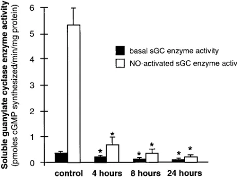
sGC enzyme activity was measured in the soluble fraction of cell extracts (Fig. 4). In extracts prepared from untreated RPaSMC, addition of SNP to the reaction mixture increased sGC enzyme activity . 10-fold (from 0.3860.07 to 5.460.58 pmol cGMP synthesized per min/mg protein). In extracts pre-pared from cells exposed to SNP for 4 h, basal and NO-stimu-lated sGC enzyme activity decreased by 47 and 87%, respec-tively (0.2060.05 and 0.6860.26; P , 0.01 for both). Similar decreases in basal and NO-stimulated sGC enzyme activity were observed in RPaSMC exposed for 4 h to 100 mM GSNO (data not shown). NO-activated sGC enzyme activity in RPaSMC exposed to SNP for 24 h was , 5% of that in control cells. These results demonstrate that chronic exposure to NO decreases the capacity of RPaSMC to synthesize cGMP.
Regulation of soluble guanylate cyclase subunit gene ex-pression by NO is mediated by a cGMP-dependent mecha-nism. To investigate the mechanisms responsible for the effect of NO on sGC subunit gene expression, RPaSMC were treated with 3 mM ODQ, an inhibitor of sGC, before exposure to SNP. Pretreatment with ODQ nearly completely blocked the ability of SNP to decrease sGC subunit gene expression (Fig. 5). To further investigate whether the sGC enzyme prod-uct cGMP could modulate sGC subunit gene expression, RPaSMC were incubated with the membrane-permeable cGMP analogue, 8-bromo cGMP. Exposure of RPaSMC to 1 mM 8-bromo cGMP for 4 h decreased sGC a1 and b1 subunit mRNA levels (Fig. 5). These results suggest that the effect of NO on sGC subunit gene expression is mediated by a cGMP-dependent mechanism.
Nitric oxide decreases soluble guanylate cyclase subunit mRNA stability. Exposure of RPaSMC to NO decreased lev-els of mRNAs encoding sGC a1 and b1 subunits. To
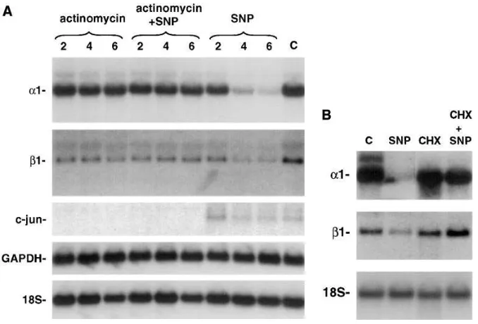
deter-mine if altered mRNA transcription or stability contributed to the NO-mediated decrease in sGC subunit mRNA levels, the effect of actinomycin D, an RNA polymerase inhibitor, on sGC subunit gene expression was measured in RPaSMC incu-bated in the presence or absence of SNP. In RPaSMC exposed to 10 mM actinomycin D for up to 6 h, the levels of sGC a1 and
b1 subunit mRNAs were unchanged (Fig. 6 A). In RPaSMC incubated in the presence of SNP, the levels of sGC a1 and b1 subunit mRNAs decreased markedly within 4 h. Incubation of RPaSMC with actinomycin D blocked the ability of SNP to de-crease sGC subunit mRNA levels. An mRNA with a short half-life, c-jun, was detected in control and SNP-treated RPaSMC, but not in actinomycin D–treated cells, demonstrat-ing that the concentration of actinomycin D used was sufficient to block gene transcription in RPaSMC. Levels of the mRNA encoding glyceraldehyde-3-phosphate dehydrogenase, an mRNA with a long half-life, were not altered by exposure of RPaSMC for up to 6 h to actinomycin D, SNP, or the combination (Fig. 6
A). These results demonstrated that SNP did not decrease levels of all mRNAs. Incubation of RPaSMC with the protein synthesis inhibitor cycloheximide (100 mM) also prevented the NO-induced decrease in sGC subunit mRNA levels (Fig. 6 B). In additional studies, actinomycin D and cycloheximide were found to block the effect of GSNO on sGC subunit gene expression, and sGC subunit mRNA levels decreased more rapidly in GSNO-treated RPaSMC than in actinomycin D–treated cells (data not shown). These results suggest that NO decreases the stability of sGC subunit mRNAs selectively via a mechanism that is both transcription and translation de-pendent.
Discussion
[image:5.612.315.554.57.241.2]Exposure of rat pulmonary artery smooth muscle cells to NO donor compounds decreased expression of genes encoding both the a1 and b1 subunits of the heterodimeric sGC enzyme. Diminished sGC subunit gene expression was accompanied by
[image:5.612.57.288.60.235.2]Figure 4. sGC enzyme activity is decreased in rat pulmonary artery smooth muscle cells chronically exposed to NO. The soluble fraction of extracts derived from untreated RPaSMC (control) and RPaSMC exposed to 1 mM SNP for 4, 8, and 24 h (three separate experiments for each time point) was assayed for sGC enzyme activity in the ab-sence (closed boxes) and presence (open boxes) of 1 mM SNP. sGC enzyme activity is expressed as picomoles of cGMP produced per minute per milligram of cell extract supernatant (6SD). *Significant differences from control values (P , 0.01). These results show that basal and NO-activated sGC enzyme activity is reduced in RPaSMC chronically exposed to NO.
decreased subunit protein levels and decreased basal and NO-activated sGC enzyme activity. The concentrations of NO do-nor compounds used in this study are comparable with those required to modulate expression of genes encoding other pro-teins, including endothelin 1 (8), macrophage-colony stimulat-ing factor (22), and the endothelin A receptor (23). Experi-ments using NO synthase inhibitors demonstrated that levels of NO sufficient to modulate expression of these genes are at-tainable in endothelial cells (8, 22) and in cytokine-treated smooth muscle cells (23). Moreover, using an NO-selective electrode, Ichimori et al. observed that concentrations of NO generated by 10 mM SNAP in biological fluids are similar to those produced by endothelial cells stimulated with 1 mM ace-tylcholine (24). Taken together, these observations suggest that concentrations of NO sufficient to alter sGC subunit mRNA levels are attainable in vivo.
Exposure of RPaSMC to a membrane-permeable cGMP analogue, 8-bromo cGMP, decreased sGC subunit gene ex-pression, whereas an inhibitor of sGC enzyme activity, ODQ, attenuated the ability of NO to alter sGC subunit mRNA lev-els. These results suggest that the effect of NO on sGC subunit gene expression is cGMP dependent. Other investigators have also suggested that cGMP regulates sGC subunit gene expres-sion. Ujiie et al. (25) observed that incubation of cultured rat medullary interstitial cells with agents that increased intracel-lular cGMP concentrations decreased sGC enzyme activity and that exposure of these cells to 0.3 mM SNP modestly de-creased sGC a1 and b1 subunit mRNA levels. Similarly, Papa-petropoulos et al. (26) observed that incubation of aortic smooth muscle cells with Zaprinast, an inhibitor of cGMP deg-radation by type 5 phosphodiesterase, decreased sGC subunit gene expression. Increased cGMP levels may alter sGC sub-unit gene expression via activation of cGMP-dependent pro-tein kinase. Alternatively, cGMP may inhibit phosphodi-esterase activity (27), leading to increased cAMP levels that in
turn may decrease sGC subunit mRNA levels, as described previously in other cell types (28–30).
Nitric oxide appears to decrease sGC subunit mRNA sta-bility via a transcription-dependent mechanism. This conclu-sion is based on the observations that sGC subunit mRNA lev-els decrease more rapidly in RPaSMC exposed to NO than in cells exposed to actinomycin D and that pretreatment of RPaSMC with actinomycin D blocked the ability of NO to al-ter sGC subunit mRNA levels. The observations that levels of c-jun and GAPDH mRNAs did not change markedly in RPaSMC exposed to SNP are consistent with NO mediating a selective effect on sGC subunit mRNAs. Incubation of RPaSMC with the protein synthesis inhibitor cycloheximide also blocked the ability of NO to decrease sGC subunit mRNA levels, supporting the hypothesis that NO induces syn-thesis of an mRNA destabilizing protein. However, it should be noted that, for some mRNAs, low levels of mRNA transla-tion are required for the degradatransla-tion of the molecule (31). We recently reported that incubation of PC12 cells with nerve growth factor decreases sGC subunit mRNA levels via a simi-lar transcription- and translation-dependent mechanism (30). These observations suggest that shared regulatory mechanisms may control sGC subunit gene expression in these two cell types.
[image:6.612.57.396.102.332.2]NO-induced modulation of mRNA stability has been re-ported previously. Pantopoulos and Hentze (12) observed that NO stabilized the mRNA encoding the transferrin receptor by activating binding of an iron-regulatory protein (IRP) to se-quences (iron-response elements, IREs) in the 39 untranslated region of the mRNA. It is unlikely that the IRP/IRE system (reviewed in reference 32) is responsible for the effect of NO on sGC subunit mRNA stability because IRP/IRE modulation of mRNA stability does not require gene transcription and consensus IRE sequences are absent from the reported se-quences of the mRNAs encoding a1 and b1 subunits (5). The
NO-mediated transcription/translation-dependent destabiliza-tion of sGC subunit mRNAs appears to represent a novel mechanism by which NO modulates gene expression.
Chronic exposure of RPaSMC to SNP decreased sGC en-zyme activity. However, whereas NO-activated sGC enen-zyme activity decreased 87% within 4 h, more than4 h were re-quired to decrease sGC subunit protein levels by 50%. These results suggest that chronic exposure to NO donor compounds decreases the specific activity of the sGC enzyme, as well as the levels of its subunits. Mechanisms regulating the specific activity of the sGC enzyme have been extensively investigated (reviewed in reference 27). Other investigators have observed that chronic exposure of cultured vascular smooth muscle cells decreased sGC enzyme activity (26, 33). Papapetropoulos et al. (26) observed that pretreatment with reducing agents such as ascorbic acid restored NO-stimulated cGMP synthesis in RPaSMC chronically exposed to SNP and suggested that chronic exposure to NO may impair sGC enzyme activity by altering the redox state of the enzyme.
The decrease in sGC enzyme activity associated with chronic exposure to NO may have important clinical implica-tions. Downregulation of sGC function may have a protective effect under conditions associated with elevated NO levels. For example, cells respond to viral, bacterial, or parasitic infec-tion by producing high concentrainfec-tions of NO (1). It is possible that NO-mediated downregulation of sGC function represents a homeostatic mechanism preventing excessive activation of cGMP-dependent protein kinase and other cGMP-dependent mechanisms and, thereby, attenuating NO-mediated host cell toxicity or hemodynamic compromise. Chronic administration of NO donor compounds, such as nitroglycerin, impairs the ability of vascular smooth muscle to respond to additional NO. Clinically, this phenomenon, referred to as nitrate tolerance, is important for patients with coronary artery disease to whom nitroglycerin is chronically administered. The etiology of ni-trate tolerance is multifactorial (34) with decreased sGC en-zyme activity having an important role (35, 36). Although the concentrations of NO donor compounds required to elicit ni-trate tolerance are typically greater in cultured cells than in vivo, the observations presented here suggest that decreased sGC subunit mRNA levels may contribute to impaired NO re-sponsiveness in nitrate tolerance.
In summary, chronic exposure of rat pulmonary artery smooth muscle cells to NO donor compounds decreased sGC subunit mRNA and protein levels and decreased sGC enzyme activity. The NO-induced decrease in the levels of mRNAs en-coding both sGC subunits was cGMP, actinomycin D, and cy-cloheximide dependent. These results suggest that NO induces synthesis of a factor (probably a protein) that selectively desta-bilizes sGC subunit mRNAs. NO-induced modulation of sGC subunit mRNA levels illustrates a novel mechanism by which NO regulates gene expression.
Acknowledgments
The authors thank Dr. M. Nakane for providing the sGC subunit cDNAs and the monoclonal antibody directed against the sGC a1 subunit, Dr. M. Alexander-Bridges for providing the human glyceral-dehyde-3-phosphate dehydrogenase cDNA probe, and Dr. T. Curran for providing the c-jun cDNA probe. The authors also thank Drs. H. Liu, M. Takata, and K.J. Bloch for their review of this manuscript.
This work was supported by National Institutes of Health grants
HL-55377 (K.D. Bloch), AR-01866 (D.B. Bloch), and DK-51179 (D.B. Bloch), by an Arthritis Investigator Award from the Arthritis Foundation (D.B. Bloch), and by a grant to the Cardiovascular Re-search Center from Bristol Myers Squibb Pharmaceuticals. K.D. Bloch is an Established Investigator of the American Heart Associa-tion.
References
1. Schmidt, H.H.H.W., and U. Walter. 1994. NO at work. Cell. 78:919–925. 2. Cobb, J.P., and R.L. Danner. 1996. Nitric oxide and septic shock. JAMA (J. Am. Med. Assoc.). 275:1192–1196.
3. Lincoln, T.M., and T.L. Cornwell. 1993. Intracellular cyclic GMP recep-tor proteins. FASEB J. 7:328–338.
4. Wong, S.K.-F., and D.L. Garbers. 1992. Receptor guanylyl cyclases. J. Clin. Invest. 90:299–305.
5. Nakane, M., K. Arai, S. Saheki, T. Kuno, W. Buechler, and F. Murad. 1990. Molecular cloning and expression of cDNAs coding for soluble guanylate cyclase from rat lung. J. Biol. Chem. 265:16841–16845.
6. Peunova, N., and G. Enikolopov. 1993. Amplification of calcium-induced gene transcription by nitric oxide in neuronal cells. Nature (Lond.). 364:450– 453.
7. Pilz, R.B., M. Suhasini, S. Idress, J.L. Meinkoth, and G.R. Boss. 1995. Ni-tric oxide and cGMP analogs activate transcription from AP-1-responsive pro-moters in mammalian cells. FASEB J. 9:552–558.
8. Kourembanas, S., L.P. McQuillan, G.K. Leung, and D.V. Faller. 1993. Nitric oxide regulates the expression of vasoconstrictors and growth factors by vascular endothelium under both normoxia and hypoxia. J. Clin. Invest. 92:99– 104.
9. DeCatarina, R., P. Libby, H.-B. Peng, V.J. Thannickai, T.B. Rajava-shisth, M.A. Gimbrone, W.S. Shin, and J.K. Liao. 1995. Nitric oxide decreases cytokine-induced endothelial activation. J. Clin. Invest. 96:60–68.
10. Shin, W.S., Y.-H. Hong, H.-B. Peng, R. DeCatarina, P. Libby, and J.K. Liao. 1996. Nitric oxide attenuates vascular smooth muscle cell activation by in-terferon-g. J. Biol. Chem. 271:11317–11324.
11. Peng, H.-B., P. Libby, and J.K. Liao. 1995. Induction and stabilization of IkBa by nitric oxide mediates inhibition of NF-kB. J. Biol. Chem. 270:14214– 14219.
12. Pantopoulos, K., and M.W. Hentze. 1995. Nitric oxide signaling to iron-regulatory protein: direct control of ferritin mRNA translation and transferrin receptor mRNA stability in transfected fibroblasts. Proc. Natl. Acad. Sci. USA. 92:1267–1271.
13. Yu, F.-S., S.-L. Lee, J.J. Lanzillo, and B.L. Fanburg. 1989. Endothelial cell inhibition of hypoxia-induced stimulation of serotonin uptake by vascular smooth muscle cells. Am. Rev. Respir. Dis. 138:1144–1148.
14. Bloch, K.D., G. Filippov, L.S. Sanchez, M. Nakane, and S.M. de la Monte. 1997. Expression of soluble guanylate cyclase, a nitric oxide receptor, is regulated during pulmonary development. Am. J. Physiol. 272:L400–L406.
15. Ercolani, L., B. Florence, M. Denaro, and M. Alexander. 1988. Isolation and complete sequence of a functional human glyceraldehy3-phosphate de-hydrogenase gene. J. Biol. Chem. 263:15335–15341.
16. Abate, C., D. Luk, and T. Curran. 1991. Transcriptional regulation by Fos and Jun in vitro: interaction among multiple activator and regulatory do-mains. Mol. Cell. Biol. 11:3624–3632.
17. Lee, R.T., K.D. Bloch, J.M. Pfeffer, M.A. Pfeffer, E.J. Neer, and C.E. Seidman. 1988. Atrial natriuretic factor gene expression in ventricles of rats with spontaneous biventricular hypertrophy. J. Clin. Invest. 81:431–434.
18. Kamisaki, Y., S. Saheki, M. Nakane, J.A. Palmieri, T. Kuno, B.Y. Chang, S.A. Waldman, and F. Murad. 1986. Soluble guanylate cyclase from rat lung exists as a heterodimer. J. Biol. Chem. 261:7236–7241.
19. Olmsted, J.B. 1981. Affinity purification of antibodies from diazotized paper blots of heterogeneous protein samples. J. Biol. Chem. 256:11955–11957. 20. Guan, K.-L., and J.E. Dixon. 1991. Eukaryotic proteins expressed in Es-cherichia coli: an improved thrombin cleavage and purification procedure of fu-sion proteins with glutathione-S-transferase. Anal. Biochem. 192:262–267.
21. Mittal, C.K. 1986. Determination of adenylate cyclase and guanylate cy-clase activities in cells and immune system. Methods Enzymol. 132:422–428.
22. Peng, H.-B., T.B. Rajavashisth, P. Libby, and J.K. Liao. 1995. Nitric ox-ide inhibits macrophage-colony stimulating factor gene transcription in vascular endothelial cells. J. Biol. Chem. 270:17050–17055.
23. Redmond, E.M., P.A. Cahill, R. Hodges, S. Zhang, and J.V. Sitzmann. 1996. Regulation of endothelin receptors by nitric oxide in cultured rat vascular smooth muscle cells. J. Cell. Physiol. 166:469–479.
24. Ichimori, K., H. Ishida, F. Fukahori, H. Nakazawa, and E. Murakami. 1994. Practical nitric oxide measurement employing a nitric oxide–selective electrode. Rev. Sci. Instrum. 65:2714–2718.
26. Papapetropoulos, A., C.Y. Go, F. Murad, and J.D. Catravas. 1996. Mechanisms of tolerance to sodium nitroprusside in rat cultures aortic smooth muscle cells. Br. J. Pharmacol. 117:147–155.
27. Hobbs, A.J., and L.J. Ignarro. 1996. The nitric oxide-cGMP signal trans-duction system. In Nitric Oxide and the Lung. W.M. Zapol and K.D. Bloch, ed-itors. Lung Biology in Health and Disease. Marcel Dekker Inc., New York. pp. 1–57.
28. Shimouchi, A., S.P. Janssens, D.B. Bloch, W.M. Zapol, and K.D. Bloch. 1993. Cyclic AMP regulates soluble guanylate cyclase b1 subunit gene expres-sion in RFL-6 rat fetal lung fibroblasts. Am. J. Physiol. 265:L456–L461.
29. Papapetropoulos, A., N. Marczin, G. Mora, A. Milici, F. Murad, and J.D. Catravas. 1995. Regulation of vascular smooth muscle soluble guanylate cyclase activity, mRNA, and protein levels by cAMP-elevating agents. Hyper-tension. 26:696–704.
30. Liu, H., T. Force, and K.D. Bloch. 1997. Nerve growth factor decreases soluble guanylate cyclase in rat pheochromocytoma PC12 cells. J. Biol. Chem.
272:6038–6043.
31. Sachs, A.B. 1993. Messenger RNA degradation in eukaryotes. Cell. 74: 413–421.
32. Klausner, R.D., T.A. Rouault, and J.B. Harford. 1993. Regulating the fate of mRNA: the control of cellular iron metabolism. Cell. 72:19–28.
33. Zhang, L.M., M.R. Castresana, S. Stefansson, and W.H. Newman. 1993. Tolerance to sodium nitroprusside: studies in cultured porcine vascular smooth muscle cells. Anesthesiology. 79:1094–1103.
34. Munzel, T., S. Kurz, T. Heitzer, and D.G. Harrison. 1996. New insights into mechanisms underlying nitrate tolerance. Am. J. Cardiol. 77:24–30.
35. Axelsson, K.L., and R.G. Andersson. 1983. Tolerance towards nitro-glycerin, induced in vivo, is correlated to a reduced cGMP response and an al-teration in cGMP turnover. Eur. J. Pharmacol. 88:71–79.