Regulation of transforming growth factor-beta 1
expression by the hepatitis B virus (HBV) X
transactivator. Role in HBV pathogenesis.
Y D Yoo, … , G Jay, S J Kim
J Clin Invest. 1996;
97(2)
:388-395.
https://doi.org/10.1172/JCI118427
.
TGF-beta 1 has been implicated in the pathogenesis of liver disease. The high frequency of
detection of the hepatitis B virus X (HBx) antigen in liver cells from patients with chronic
hepatitis, cirrhosis, and liver cancer suggested that expression of HBx and TGF-beta 1 may
be associated. To test this possibility, we examined the expression of TGF-beta 1 in the
liver of transgenic mice expressing the HBx gene. We show that the patterns of expression
of TGF-beta 1 and Hbx protein are similar in these mice and that HBx activates transcription
of the TGF-beta 1 gene in transfected hepatoma cells. The cis-acting element within the
TGF-beta 1 gene that is responsive to regulation by Hbx is the binding site for the Egr family
of transcription factors. We further show that the Egr-1 protein associates with the HBx
protein, allowing HBx to participate in the transcriptional regulation of immediate-early
genes. Our results suggest that expression of Hbx might induce expression of TGF-beta 1 in
the early stages of infection and raise the possibility that TGF-beta 1 may play a role in
hepatitis B virus pathogenesis.
Research Article
Find the latest version:
The Journal of Clinical Investigation Volume 97, Number 2, January 1996, 388–395
Regulation of Transforming Growth Factor-
b
1 Expression by the Hepatitis B Virus
(HBV) X Transactivator
Role in HBV Pathogenesis
Young Do Yoo,* Hiroyuki Ueda,‡ Keunchil Park,* Kathy C. Flanders,* Young Ik Lee,§ Gilbert Jay,‡ and Seong-Jin Kim*
*Laboratory of Chemoprevention, National Cancer Institute, Bethesda, Maryland 20892; ‡Department of Virology, Jerome H. Holland
Laboratory, Rockville, Maryland 20855; §Laboratory of Molecular Genetics, Genetic Engineering Research Institute, Korea Institute of
Science and Technology, Daejon 305-606, Korea
Abstract
TGF-b1 has been implicated in the pathogenesis of liver disease. The high frequency of detection of the hepatitis B virus X (HBx) antigen in liver cells from patients with chronic hepatitis, cirrhosis, and liver cancer suggested that expression of HBx and TGF-b1 may be associated. To test this possibility, we examined the expression of TGF-b1 in the liver of transgenic mice expressing the HBx gene. We show that the patterns of expression of TGF-b1 and Hbx protein are similar in these mice and that HBx activates transcription of the TGF-b1 gene in transfected hepatoma cells. The cis-acting element within the TGF-b1 gene that is responsive to regulation by Hbx is the binding site for the Egr family of transcription factors. We further show that the Egr-1 protein associates with the HBx protein, allowing HBx to participate in the transcriptional regulation of im-mediate–early genes. Our results suggest that expression of Hbx might induce expression of TGF-b1 in the early stages of infection and raise the possibility that TGF-b1 may play a role in hepatitis B virus pathogenesis. (J. Clin. Invest. 1996. 97:388–395.) Key words: hepatitis B virus • TGF-b1 • transcription • immunohistochemistry • hepatocellular car-cinoma
Introduction
Hepatitis B virus (HBV)1 causes acute and chronic liver cell
in-jury and inflammation and is strongly associated with liver can-cer (1). Its genome contains four recognized open reading frames, three of which code for known virion proteins (2). The fourth, called the HBV X gene (HBx), is conserved among all mammalian hepadnaviruses and has been shown to be ex-pressed both during viral infection (3–5) and in HBV-associ-ated hepatocellular carcinoma (6). Recently, it has been shown
that the woodchuck hepatitis virus X gene is important for the establishment of viral infection in woodchucks, suggesting that the HBx gene plays an important role in HBV replication in humans (7).
It is known that HBx trans-activates the expression of many viral and cellular transcriptional promoters (8–13). It ac-tivates transcription through certain cis-acting sequences, in-cluding transcription activator proteins AP-1 and AP-2 (14). HBx also forms protein–protein complexes with cellular tran-scription factors such as cyclic AMP-responsive element-binding protein (CREB) and activating transcription factor (ATF-2) and modifies their ability to bind transcriptional en-hancers (15). Recently, it has been reported that HBx can acti-vate protein kinase C, a key component of cellular signal trans-duction (16). Moreover, we have demonstrated that expression of the HBx gene alone is sufficient for the develop-ment of liver cancer in transgenic mice (17, 18).
Cytokines affect many functions in the liver, including amino acid, protein, lipid, mineral, and carbohydrate metabo-lism. In liver disease, cytokines are involved in the onset of in-trahepatic immune responses, in liver regeneration, and in the fibrotic and cirrhotic transformation of the liver after chronic chemical injury or viral infection (19). To understand the mechanisms by which HBx induces changes in the liver, we ex-amined the expression of TGF-b1, a cytokine that inhibits hepatocyte proliferation during liver regeneration (20–22) and stimulates the production of extracellular matrix proteins by hepatocytes during liver cirrhosis (23, 24). Recent results also suggest that TGF-b1 may play a role in the pathogenesis of fi-brosis in chronic hepatitis and cirrhosis (25) and in the devel-opment of hepatocellular carcinoma (26).
In this study, we examined the expression of TGF-b1 in the livers of transgenic animals harboring the HBx gene under its own control elements (17, 18). TGF-b1 expression correlated well with the expression of the HBx protein in the early focal lesions of altered hepatocytes, suggesting that HBx acted ei-ther directly or indirectly to increase TGF-b1 expression. We also showed that HBx trans-activates the TGF-b1 promoter through the Egr-1 binding sites. Additionally, TGF-b1 expres-sion was sustained in the adenoma and carcinoma leexpres-sions, sug-gesting that it might play a role in the progressive stages of hepatocellular carcinoma.
Methods
Immunohistochemistry. The liver tissues were fixed in 10% formalin and embedded in paraffin. 5-mm sections were immunostained with either anti-HBx antiserum or anti–TGF-b1 antibodies, visualized by the avidin–biotin complex method (Vector Laboratories, Inc., Burlin-game, CA), and counterstained with hematoxylin. TGF-b1 antibodies used for immunohistochemistry were raised in rabbits to the NH2
-ter-Address correspondence to Seong-Jin Kim, Building 41, Room B1106, National Cancer Institute, National Institutes of Health, Bethesda, MD 20892-5055. Phone: 301-496-5391; FAX: 301-496-8395. Keunchil Park’s present address is Division of Hemato-Oncology, Department of Internal Medicine, SamSung Medical Center, Seoul, Korea.
Received for publication 8 May 1995 and accepted in revised form 6 October 1995.
minal 1–30 amino acids of mature TGF-b1 (anti-CC and anti-LC) (27, 28). Anti-CC stained extracellular matrix associated TGF-b1, whereas anti-LC stained intracellular TGF-b1.
RNA isolation and blotting. Total RNAs from mouse tissues were extracted by acid guanidinium thiocyanate–phenol chloroform extraction. Total RNAs (10 mg each) were separated by electrophore-sis through 1% agarose–formaldehyde gels and transferred to nitro-cellulose membranes. Prehybridization, hybridization, and washing of the membrane were as described previously (29).
Cell culture, DNA transfection, and chloramphenicol acetyltrans-ferase (CAT) assays. HepG2 cells were grown in Dulbecco’s minimal essential medium supplemented with 10% FBS. For transient expres-sion assays, cells were plated at 1.2 3 106 per 10-cm dish and cultured
for 24 h before transfection by the calcium phosphate coprecipitation method. Cells were harvested 48 h after addition of DNA, and ex-tracts were assayed for CAT activity. All transfections were repeated at least three times. For normalization of transfection efficiencies in HepG2 cells, a growth hormone expression plasmid (pSVGH) was in-cluded in the cotransfections. The level of growth hormone expres-sion was determined using a growth hormone detection kit (Nichols Institute, San Juan, Capistrano, CA).
Mobility shift assays. Bacterially expressed Egr-1 protein was
kindly provided by Dr. V. Sukhatme (30). The double-stranded oligo-nucleotides were labeled by Klenow enzyme and gel purified. After bacterially expressed Egr-1 protein was incubated for 25 min at room temperature in binding buffer (20 mM Hepes, pH 7.5, 50 mM KCl, 1 mM dithiothreitol, 1 mM EDTA, 5% glycerol, 2 mg of double-stranded poly[dI-dC] nonspecific competitor, z 0.2 ng of 32P-labeled
probe), the reaction products were loaded onto a 5% polyacrylamide gel (39:1, acrylamide/bisacrylamide) and electrophoresed in 0.5 3
TBE (50 mM Tris, 50 mM boric acid, 1 mM EDTA) for 2.5–3.5 h at 8 V/cm. Gels were dried and autoradiographed.
[image:3.612.59.436.307.738.2]Plasmids. Human TGF-b1 promoter/CAT plasmids (29), GAL4-ATF-2 (31), GAL4-CREB (32), and G5BCAT reporter constructs (33) have been previously described. All GAL4-Egr-1 fusion plas-mids were constructed by inserting the appropriate Egr-1 DNA frag-ment in-frame to the GAL(1–147) sequence in the vector pSG424 (34). Egr-1 DNA fragments were produced by polymerase chain re-action. The 59-oligonucleotide used in all amplications contained an EcoRI site and the 39-oligonucleotide contained an XbaI site. Using these oligonucleotides, fragments were amplified according to the standard protocol of the GeneAmp kit (Perkin-Elmer Corp., Nor-walk, CT). The junctions of all GAL4 fusion plasmids were con-firmed by DNA sequencing. The HBx expression plasmids (pMAM
ND1-4 and CD1-4) used for the expression of native and mutant HBx proteins in eukaryotic cells were constructed by cloning the HBx-ORF insert into the SalI site of pMAM-neo (Promega, Madison, WI).
Glutathione S-transferase (GST)-HBx and GST-Egr-1 fusion proteins. Various GST-HBx and eight GST-Egr-1 fusion proteins ex-pressed in Escherichia coli were partially purified by adsorption to glutathione-Sepharose beads (Pharmacia Biotech Inc., Piscataway, NJ) in the presence of the detergent N-laurylsarcosine and Triton X-100 as has been described previously (35).
Egr-1 proteins and HBx protein generated by in vitro transcription and translation. Plasmids containing the Egr-1 (1–533), Egr-1 (32– 533), Egr-1 (147–533), Egr-1 (249–533), and Egr-1 (1–364) were con-structed by inserting appropriate PCR-generated fragments of Egr-1 at in-frame EcoRI/BamHI restriction sites in pGEM4 (Promega). For the synthesis of [35S]methionine-labeled HBx protein by in vitro
tran-scription and translation, pTM1/HBx plasmid was used as template for RNA synthesis by T7 RNA polymerase followed by translation in rabbit reticulocyte extracts (Promega).
Results
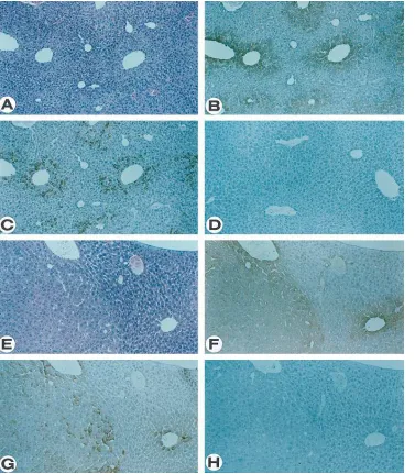
Expression of TGF-b1 in transgenic mice harboring the HBx gene. At 4 mo of age, the HBx-transgenic mice exhibited mul-tifocal areas of altered hepatocytes that were made up of cells with a poorly stained cytoplasm (Fig. 1 A) and were not de-tected in nontransgenic littermates (17, 18). Expression of the HBx protein as seen by immunohistochemical staining of a se-rial liver section using an anti-HBx serum correlated precisely
with the altered foci (Fig. 1 B), consistent with its role in the underlying histopathological change. The restricted expression of HBx in only a subset of cells may reflect the need for spe-cific transcription factors required for the activation of the vi-ral regulatory elements and present only in cells at a specific differentiation state.
Expression of TGF-b1 was assessed by immunohistochemi-cal staining using isotype-specific peptide antibodies. While TGF-b1 was not detected in the liver of normal mice (Fig. 1
D), it was found in the livers of the transgenic mice (Fig. 1 C). Interestingly, the cells that expressed TGF-b1 were located ex-clusively within the altered foci made up of hepatocytes (17) where HBx was also highly expressed (compare Fig. 1, B and
C), supporting the possibility that TGF-b1 expression could be activated by HBx. Whereas every cell within an altered focus expressed HBx, only a subset of them also expressed TGF-b1. This latter observation suggests that HBx expression, while necessary, is insufficient to induce TGF-b1 and argues for epi-genetic rather than epi-genetic events underlying TGF-b1 activa-tion by HBx.
At z 8–12 mo of age, tumor nodules begin to appear in the
livers of the transgenic mice. While many of these tumors had benign characteristics and were diagnosed as adenomas (Fig. 1
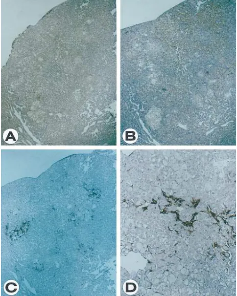
E), others appeared malignant and resembled hepatocellular carcinoma (Fig. 2). HBx protein was highly and uniformly ex-pressed in both adenoma and carcinoma lesions (Figs. 1 F and 2 A, respectively). Similarly, TGF-b1 expression was also de-tected in the tumors. For adenomas, the accumulation of intra-cellular TGF-b1 appeared to differ between individual cells as has been observed for cells in the altered foci, suggesting that, while its expression is dependent on the HBx protein, other factors may also be involved (Fig. 1 G). For carcinomas, the cells that expressed TGF-b1 were localized to discrete regions (Fig. 2 B). Interestingly, extracellular TGF-b1 is detected only in malignant tumors (Fig. 2 C and D) and not in benign lesions (Fig. 1 H). However, we cannot exclude the possibility that this represents a relative rather than an absolute difference in the level of accumulation between the two stages of the disease process.
Northern blot hybridization analysis of RNA extracted from a control mouse liver and from a transgenic liver with fo-cal lesions, adenomas, or carcinomas suggested that the in-crease in TGF-b1 expression in tumors may be regulated at the transcriptional level. The level of TGF-b1 mRNA was un-detectable in the control liver, but expressed at high levels in the tumor (Fig. 3). Taken together, our results demonstrate a
[image:4.612.56.298.365.668.2]Figure 2. Malignant tumor in the liver of an HBx-transgenic mouse. Serial sections of hepatocellular carcinoma from a 22-mo-old trans-genic mouse were immunostained with anti-HBx serum (A) and anti– TGF-b1 antibodies staining for intracellular (B) or extracellular TGF-b1 (C). Higher magnification of immunostaining for extracellu-lar TGF-b1 is also shown (D).
Figure 3. Northern blot analysis of TGF-b
strong association between expression of HBx and TGF-b1 in vivo and suggest the possibility that TGF-b1 might play a part in early stages of development of hepatocellular carcinoma in-duced by HBV infection.
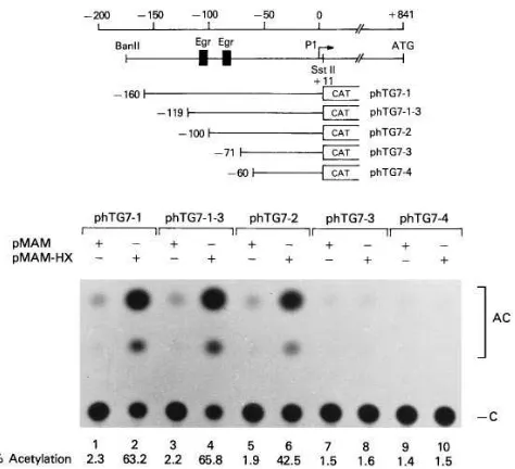
Trans-activation of the TGF-b1 promoter by HBx is medi-ated through the Egr-1 binding sites. To investigate whether increased expression of TGF-b1 in liver from the HBx-trans-genic mice was transcriptional, we examined the promoter ac-tivities of the TGF-b1 gene. The structures of the TGF-b 1-CAT chimeric plasmids used in this study have been described previously (29, 36). The chimeric plasmids were cotransfected into the hepatocarcinoma cell line, HepG2, with the control expression plasmid (pMAM) or with this same plasmid engi-neered to express HBx (pMAM-HX). Expression of the
TGF-b1-CAT reporter gene (phTG7-1) was 27-fold higher with pMAM-HX than with pMAM (Fig. 4, compare lanes 1 and 2).
We next sought to identify the specific cis-acting element that mediates responsiveness to HBx by testing a series of de-letion constructs of the TGF-b1 promoter linked to CAT. The induction dropped almost to the basal level when the deletion reached 271 (Fig. 4, compare lanes 7 and 8).
The TGF-b1 promoter contains two Egr-1 binding sites at positions 2119 to 2111 (59-CGCCCCCGC-39) and 282 to
274 (59-CCGGGGGCG-39) (Figs. 4 and 5). To determine if these sites are specifically involved in HBx-mediated transcrip-tional regulation, we generated chimeric constructs containing sequences between 2125 and 298 and between 293 and 263 ligated to the adenovirus E4D-38 promoter-CAT vector (37). Three-base substitution mutants of the Egr-1 binding sites were also generated and tested for HBx trans-activation (Fig. 5, A and B). No increase in CAT activity was observed when the control plasmid pE4D-38 was cotransfected with pMAX-HX, whereas pE4D-38(2125/298) and pE4D-38(293/263) that contain the separate Egr-1 sites both showed an increase in CAT activity under the same conditions (Fig. 5, A and B, re-spectively). Mutant constructs, pE4D-38(2125/298mt) and pE4D-38(293/263mt), were not activated by HBx (Fig. 5 A, lanes 5 and 6, and Fig. 5 B, lanes 3 and 4). We also demon-strated that bacterially expressed Egr-1 binds to these two Egr-1 binding sites (29, data not shown).
[image:5.612.61.298.56.272.2]A GAL4-Egr-1 fusion protein can mediate transcription ac-tivation by HBx. To confirm that the Egr-1 protein was directly involved in HBx-mediated transcriptional activation of TGF-b1, we designed a protein fusion experiment. Plasmids expressing various GAL4 fusion proteins were cotransfected with a CAT reporter construct (G5E1bCAT), which contained five GAL4 binding sites upstream of the AdE1b TATA box (33). To these transfection mixtures we added either the HBx expres-sion plasmid (pMAM-HX) or the control expresexpres-sion vector (pMAM). As expected, HBx did not stimulate transcription on cotransfection of the minimal GAL4 DNA-binding domain (Fig. 6, lanes 1 and 2). On cotransfection of GAL4-Egr-1, how-ever, transcription was greatly stimulated by HBx (Fig. 6, lanes 7 and 8). Even though it has been shown that the HBx protein forms protein–protein complexes with both CREB and ATF-2 (15), transcriptional stimulation was observed with GAL4-ATF-2 (Fig. 6, lanes 9 and 10) but not with GAL4-CREB (Fig. 6, lanes 5 and 6). Transcriptional stimulation was also not ob-served on cotransfection of GAL4-ATF-1 (Fig. 6, lanes 3 and 4) or GAL4-VP1, an activator carrying an acidic activating re-gion (Fig. 6, lanes 11 and 12). These results indicate that Egr-1
Figure 4. Identification of the HBx-responsive element in the
TGF-b1 promoter. An expression plasmid pMAM (lanes 1, 3, 5, 7, and 9) or the same vector expressing the HBx cDNA (pMAM-HX, lanes 2,
4, 6, 8, and 10) was cotransfected with reporter plasmids containing upstream elements of the human TGF-b1 gene. Representative ex-periments to determine CAT activity in extracts of transiently trans-fected HepG2 cells are shown. The fusion genes that were transtrans-fected are represented schematically at the top of the figure.
[image:5.612.60.374.592.738.2]can specifically support HBx-mediated transcriptional activa-tion.
Identification of the domain of HBx responsible for its trans-activation function. The amino acid sequence of the HBx protein reveals relatively few structural motifs that might be involved in trans-activation. Several reports indicate that the COOH terminus may act as an acidic activator (38). We next examined the effect of HBx deletion constructs on GAL4-Egr-1 transcription to identify the active region of the HBx protein.
A series of NH2- or COOH-terminal deletion constructs was generated as shown in Fig. 7. The ND-4 construct, in which 50 amino acids were deleted from the NH2 terminus of the HBx protein, was still able to activate GAL4-Egr-1 tran-scription, whereas removal of up to 20 amino acids from the COOH terminus abolished the ability of the protein to acti-vate transcription (Fig. 7). It had previously been demon-strated that the block of amino acids from position 132 to 139, containing the highly conserved sequence FVLGGCRH, is es-sential for maintaining the trans-activation function (39). Our results also suggest that the COOH terminus is required for ac-tivation of the TGF-b1 promoter.
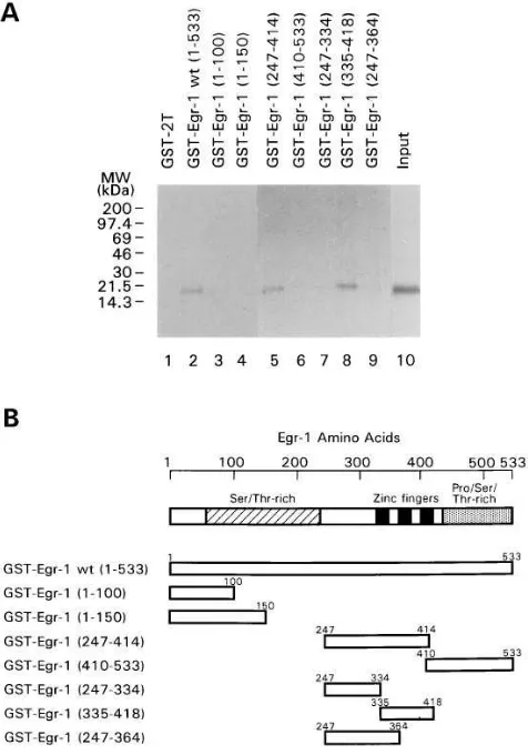
The HBx protein binds to Egr-1 in vitro.We next investi-gated the ability of HBx to interact directly with Egr-1 in vitro. Fig. 8 shows the results of the GST affinity chromatography experiment. An intact 17-kD [35S]methionine-labeled HBx
[image:6.612.60.368.90.229.2]protein prepared by in vitro translation was found to bind the GST-Egr-1 protein by GST affinity chromatography (Fig. 8 A, lane 2). The binding of HBx was not detected with agarose beads containing GST alone (data not shown). To identify
Figure 6. Stimulation by HBx of Egr-1–medi-ated transcription. An expression plasmid for HBx (pMAM-HX, lanes 2, 4, 6, 8, 10, and 12) or the parent vector (pMAM, lanes 1, 3, 5, 7, 9, and
[image:6.612.316.554.296.636.2]11) was cotransfected with a GAL4 reporter plasmid G5BCAT and expression vector for GAL4 binding domain (1–147) (lanes 1 and 2), GAL4-ATF-1 (lanes 3 and 4), GAL4-CREB (lanes 5 and 6), GAL4-Egr-1 (lanes 7 and 8), GAL4-ATF-2 (lanes 9 and 10), or the acidic acti-vator GAL4-VP1 (lanes 11 and 12). The re-porter construct G5BCAT contains 5 GAL4 binding sites upstream of the Ad E1b TATA box and a CAT reporter gene. GAL4-VP1 con-tains VP16 activator sequences between amino acids 411 and 454 fused to the GAL4 binding do-main, GAL4(1–147).
Figure 7. Identification of the domain of the HBx protein for its transactivation function. A schematic representation of the HBx pro-tein and its derivatives together with the fold induction are presented.
Figure 8. HBx protein interacts with the zinc finger domain of Egr-1 in a GST affinity chromatography. (A) HBx protein was synthesized and labeled with [35S]methionine in vitro. The bacterially expressed
GST-Egr-1 deletion chimeras were incubated with the amount of ra-diolabeled input protein shown in lane 10. After extensive washing, the coprecipitated radiolabeled proteins were resolved in a 4–20% gradient SDS polyacrylamide gel. In lane 1, the GST protein was used as a negative control. (B) Seven segments representing GST-Egr-1 deletion chimeras used in the GST affinity chromatography experi-ment were presented diagrammatically.
[image:6.612.59.294.578.703.2](247–364), were constructed using internal PCR primers (Fig. 8 B). All proteins from these chimeras were produced from IPTG-induced E. coli bacterial hosts and analyzed by SDS-PAGE (data not shown). The GST affinity assay was used to show that HBx protein can bind to the GST-Egr-1. The results revealed that HBx could bind to intact Egr-1 (1–533), Egr-1 (247–414), and Egr-1 (335–418), all containing the three zinc finger domains (Fig. 8 A, lanes 2, 5, and 8), but not to Egr-1 (1–100), Egr-1 (1–150), Egr-1 (410–533), Egr-1 (247–334), and Egr-1 (247–364) (Fig. 8 A, lanes 3, 4, 6, 7, and 9).
The design of the GST bead affinity binding assay was re-versed by using [35S]methionine-labeled in vitro–translated
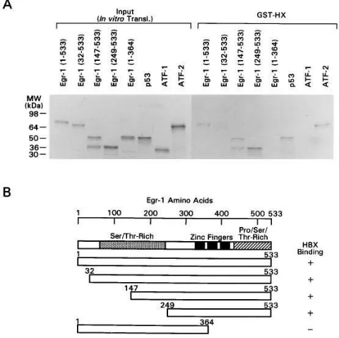
forms of Egr-1 to identify which segment of Egr-1 was in-volved in binding to HBx protein. In Fig. 9, a Sepharose bead– bound GST fusion protein containing the entire HBx protein was used. The results revealed that intact Egr-1 (1–533), Egr-1 (32–533), Egr-1 (147–533), and Egr-1 (249–533) could bind to the HBx protein. However, the NH2-terminal segment of Egr-1 (1–364), which does not contain the zinc finger domain, did not show any binding to HBx. [35S]Methionine-labeled in vitro–
synthesized preparations of the intact human p53 and the ATF-2 protein served as positive controls, and the ATF-1 pro-tein served as a negative control for the binding specificity in this assay (15, 40).
Discussion
It has been shown that HBx is capable of trans-activating a va-riety of viral (9, 12, 41) and cellular promoters (13, 16, 41). Re-cently, we also demonstrated that HBx is directly involved in
the development of liver cancer by generating transgenic mice harboring the entire HBx gene (17). Using these same trans-genic mice, we have now been able to show a strong associa-tion of expression of HBx and TGF-b1, which is known to play an important role in chronic hepatitis and liver cirrhosis (23– 25) in vivo. Moreover, in vitro experiments demonstrate that HBx can directly induce the expression of TGF-b1. We dem-onstrated that TGF-b1 localized immunochemically to the HBx-expressing cells and that sequences homologous to a pre-viously defined Egr-1–responsive element (30) mediate the regulation of TGF-b1 promoter activity by the HBx protein. These results suggest that TGF-b1 may be one of the media-tors of HBV pathogenesis.
Egr-1, also known as TIS-8 (42), Zif268 (43–45), Krox 24 (46), and NGFI-A (47), is a recently characterized transcrip-tion factor that is responsive to cell division signals (48). It con-tains three zinc fingers, which bind to the target sequence GCGGGGGCG (48, 49), also recognized by the zinc finger transcription factor WT1 (50). Egr-1 is rapidly induced in re-sponse to a variety of stimuli (48). Like the protooncogenes c-fos and c-jun, Egr-1 is induced within 30 min by extracellular growth signals in the absence of protein synthesis and when cells of certain lineages are cued to differentiate. Promoters of many genes including insulin-like growth factor II (51) contain Egr-1 binding sites. These observations suggest that regulation of Egr-1 function may lead to changes of cell physiology.
Mutational analysis of HBx has demonstrated that multiple structural motifs are separately involved in the activation of different promoters (38). The COOH terminus contains the moderately acidic alpha helix. Removal of up to 12 amino ac-ids from the COOH terminus did not change its trans -activa-tion func-activa-tion in vitro (8, 39, 52–54), whereas dele-activa-tion of amino acids between positions 132 and 139, containing the highly conserved sequence FVLGGCRH, resulted in the loss of ac-tivity (39). Interestingly, the HBx protein extends nine amino acids beyond that of the woodchuck hepatitis virus at the COOH terminus (7). It appears that the amino acids between positions 126 and 136 of woodchuck hepatitis virus are impor-tant for virus replication in the natural host, suggesting that this region is crucial to the function of the protein (7). In this study, we have also demonstrated that deletion of amino acids from positions 136 to 153 of the HBx protein abolished its
trans-activating function.
It is known that the HBx protein does not bind directly to DNA but instead forms protein–protein complexes with cellu-lar transcription factors like CREB and ATF-2, and alters their binding specificities (15). We have demonstrated that HBx stimulates transcription conferred by GAL4-ATF-2 but not by GAL4-CREB. Since ATF-2 lacks a constitutive activat-ing region (31), a cellular factor like the retinoblastoma gene product (55) or a viral protein such as the adenovirus E1a may bind to ATF-2 and supply the transcriptional activating func-tion. The fact that HBx is not able to induce transcription con-ferred by GAL4-CREB indicates that HBx only alters the DNA binding specificity of CREB and that the activation po-tential of CREB possibly requires phosphorylation (56, 57).
[image:7.612.57.297.404.643.2]In the present study, we have demonstrated that HBx forms a complex with the Egr-1 protein. Because phosphoryla-tion events modulate the activity of a number of transcripphosphoryla-tion factors, this could impart another level of regulation on Egr-1 activity. It is of interest to note that Natoli et al. (58) have sug-gested that the HBx protein might induce posttranslational
Figure 9. HBx protein interacts with the zinc finger domain of Egr-1. (A) Five kinds of Egr-1 deletion mutants that were synthesized in vitro and labeled with [35S]methionine were coprecipitated with the
GST-HBx fusion protein. The [35S]methionine-labeled Egr-1 deletion
mutants were subjected to the GST affinity chromatography. The [35S]methionine-labeled p53 and ATF-2 were used as positive
con-trols, and the [35S]methionine-labeled ATF-1 was used as a negative
modifications that render the AP1 complex more efficient in its DNA-binding ability. Phosphorylation and dephosphoryla-tion of c-Fos and c-Jun have been described and shown to have a critical role in the regulation of AP1 function (59, 60).
TGF-b1 is an important cytokine in the pathophysiology of liver fibrosis, stimulating the production of extracellular matrix. In hepatic fibrosis, a marked increase is seen in the hepatic extracellular matrix proteins, including collagens, glycoproteins, and glycosaminoglycans. In both experimental models of hepatic fibrosis and in patients with liver cirrhosis, increased expression of type I collagen genes is seen (61). The TGF-bs also stimulate type I collagen gene expression in pri-mary cultures of hepatocytes, Ito cells, and fibroblasts (23). Castilla et al. (25) have shown that the level of TGF-b1 mRNA in liver biopsy specimens correlated positively with hepatic fi-brosis in a large group of patients with chronic viral hepatitis, suggesting that TGF-b1 may play a role in the pathogenesis of hepatic fibrosis.
TGF-b1 most often acts as a negative growth regulator (62). This effect must be overcome before tumor progression can occur (63). Evidence from animal models as well as human tumors indicates that malignant tumors including human hepa-tocellular carcinoma secrete high levels of TGF-b1 (26, 64–66). Increased secretion of TGF-b1 by cells that have lost respon-siveness to its growth inhibitory activities is thought to facili-tate tumor progression by indirect means such as suppression of immune surveillance and stimulation of tumor stroma. In this study, we have demonstrated that the level of TGF-b1 mRNA is significantly increased in livers from the HBx -trans-genic mice and that expression of TGF-b1 correlates well with that of the HBx protein.
A recent study has shown that 84% of patients with pri-mary hepatocellular carcinoma who are HBV carriers were HBx positive in their tumor cells (67). Evidence that expres-sion of TGF-b1 correlates well with that of the HBx protein in transgenic mice and that TGF-b1 expression is also elevated in hepatic cirrhosis in patients with chronic viral hepatitis sug-gests that further work should be done to define the specific role of TGF-b1 in the pathogenesis of HBV-associated liver diseases, including primary hepatocellular carcinoma.
Acknowledgments
We thank V. Sukhatme for the Egr-1 cDNA and the Egr-1 polyclonal antibody, M. Thompson for the CREB, M. Green for GAL4-ATF-1 and ATF-2, R. Allison and L. Mullen for oligonucleotide syn-thesis, and Y. Kim for the generation of HBx expression constructs. We also thank A. Roberts, M. Sporn, L. Wakefield, and E. Tabor for discussions and the critical reading of the manuscript.
This study was supported in part by National Institutes of Health grant CA-51886 to G. Jay.
References
1. Beasley, R. P., C. C. Lin, L. Y. Hwang, and C. S. Chien. 1981. Hepatocel-lular carcinoma and hepatitis B virus. Lancet. ii:1129–1133.
2. Tiollais, P., C. Pourcel, and A. Dejean. 1985. The hepatitis B virus.
Na-ture (Lond.). 317:489–495.
3. Kay, A., E. Mandart, C. Trepo, and F. Galibert. 1985. The HBV HBx gene expressed in E. coli is recognised by sera from hepatitis patients. EMBO
(Eur. Mol. Biol. Organ.) J. 4:1287–1292.
4. Moriarty, A. M., H. Alexander, R. A. Lerner, and G. B. Thornton. 1985. Antibodies to peptides detect new hepatitis B antigen: serological correlation with hepatocellular carcinoma. Science (Wash. DC). 227:429–432.
5. Siddiqui, A., S. Jameel, and J. Mapoles. 1987. Expression of the hepatitis
B virus X gene in mammalian cells. Proc. Natl. Acad. Sci. USA. 84:2523–2517. 6. Wang, W., W. T. London, L. Lega, and M. A. Feitelson. 1991. HBxAg in the liver from carrier patients with chronic hepatitis and cirrhosis. Hepatology.
14:29–37.
7. Chen, H.-S., S. Kaneko, R. Girones, R. W. Anderson, W. E. Horn-buckle, B. C. Tennant, P. J. Cote, J. L. Gerin, R. H. Purcell, and R. H. Miller. 1993. The woodchuck hepatitis virus x gene is important for establishment of vi-rus infection in woodchucks. J. Virol. 67:1218–1226.
8. Levrero, M., C. Balsano, G. Natoli, M. L. Avantaggiati, and E. Elfassi. 1990. Hepatitis B virus X protein transactivates the long terminal repeats of hu-man immunodeficiency virus type 1 and 2. J. Virol. 64:3082–3086.
9. Seto, E., D.-X. Zhou, B. M. Peterlin, and T. S. B. Yen. 1989. Trans-acti-vation by the hepatitis B virus X protein shows cell-type specificity. Virology.
173:764–766.
10. Siddiqui, A., R. Gaynor, A. Srinivasan, J. Mapoles, and R. W. Farr. 1989. Trans-activation of viral enhancers including long terminal repeat of the
human immunodeficiency virus by the hepatitis B virus X protein. Virology.
169:479–494.
11. Twu, J. S., and R. H. Schloemer. 1987. Transcriptional trans-activating function of hepatitis B virus. J. Virol. 61:3448–3453.
12. Twu, J. S., and W. S. Robinson. 1989. Hepatitis B virus X gene can transactivate heterologous viral sequence. Proc. Natl. Acad. Sci. USA. 86:2046– 2050.
13. Twu, J. S., M. Y. Lai, D. S. Chen, and W. S. Robinson. 1993. Activation of the proto-oncogene c-Jun by the X protein of hepatitis B virus. Virology. 192: 346–350.
14. Seto, E., P. J. Mitchell, and T. S. B. Yen. 1990. Transactivation by the hepatitis B virus X protein depends on AP-2 and other transcription factors.
Nature (Lond.). 344:72–74.
15. Maguire, H. F., J. P. Hoeffler, and A. Siddiqui. 1991. HBV X protein al-ters the DNA binding specificity of CREB and ATF-2 by protein-protein inter-actions. Science (Wash. DC). 252:842–844.
16. Kekulé, A. S., U. Lauer, L. Weiss, B. Luber, and P. H. Hofschneider. 1993. Hepatitis B virus transactivator HBx uses a tumor promoter signalling pathway. Nature (Lond.). 361:742–746.
17. Kim, C.-M., K. Koike, I. Saito, T. Miyamura, and G. Jay. 1991. HBx
gene of hepatitis B virus induces liver cancer in transgenic mice. Nature
(Lond.). 351:317–320.
18. Ueda, H., S. J. Ullich, J. D. Gangemi, C. A. Kappel, L. Ngo, M. A. Fei-telson, and G. Jay. 1995. Functional inactivation but not structural mutation of p53 causes liver cancer. Nat. Genet. 9:41–47.
19. Andus, T., J. Bauer, and W. Gerok. 1991. Effects of cytokines on the liver. Hepatology. 13:364–375.
20. Braun, L., J. E. Mead, M. Panzica, R. Mikumo, G. I. Bell, and N.
Fausto. 1988. Transforming growth factor b mRNA increases during liver
re-generation: a possible paracrine mechanism of growth regulation. Proc. Natl.
Acad. Sci. USA. 85:1539–1543.
21. Fausto, N., and J. E. Mead. 1989. Regulation of liver growth: protoon-cogenes and transforming growth factors. Lab. Invest. 60:4–13.
22. Nakamura, T., Y. Tomita, R. Hirai, K. Yamaoka, K. Kaji, and A. Ichi-hara. 1985. Inhibitory effect of transforming growth factor-b on DNA synthesis
of adult rat hepatocytes in primary culture. Biochem. Biophys. Res. Commun.
133:1042–1050.
23. Czaja, M. J., F. R. Weiner, K. C. Flanders, M. A. Giambrone, R. Wind, L. Biempica, and M. A. Zern. 1989. In vitro and in vivo association of trans-forming growth factor-b1 with hepatic fibrosis. J. Cell Biol. 108:2477–2482.
24. Nakatsukasa, H., P. Nagy, R. P. Evarts, L. C. Hsia, E. Marsden, and S. S. Thorgeirsson. 1990. Cellular distribution of transforming growth factor-b1 and procollagen types I, III, and IV transcripts in carbon tetrachloride-induced rat liver fibrosis. J. Clin. Invest. 85:1833–1843.
25. Castilla, A., J. Prieto, and N. Fausto. 1991. Transforming growth factor
b1 and a in chronic liver disease: effects of interferon-a therapy. N. Eng. J. Med. 324:933–940.
26. Ito, N., S. Kawata, S. Tamura, K. Takaishi, Y. Shirai, S. Kiso, I. Yabuu-chi, Y. Matsudo, M. Nishioka, and S. Tarui. 1991. Elevated levels of
transform-ing growth factor-b messenger RNA and its polypeptide in human
hepatocellu-lar carcinoma. Cancer Res. 51:4080–4083.
27. Heine, U. I., K. C. Flanders, A. B. Roberts, E. F. Munoz, and M. B.
Sporn. 1987. Role of transforming growth factor b in the development of the
mouse embryo. J. Cell Biol. 105:2861–2876.
28. Flanders, K. C., N. L. Thompson, D. S. Cissel, E. Van Obberghen-Schilling, C. C. Baker, M. E. Kass, R. Ellingsworth, A. B. Roberts, and M. B.
Sporn. 1989. Transforming growth factor-b1: histochemical localization with
antibodies to different epitopes. J. Cell Biol. 108:653–660.
29. Kim, S.-J., K. Park, B. B. Rudkin, B. R. Dey, M. B. Sporn, and A. B. Roberts. 1994. Nerve growth factor induces transcription of transforming growth factor-b1 through a specific promoter element in PC12 cells. J. Biol.
Chem. 269:3739–3744.
31. Liu, F., and M. R. Green. 1990. A specific member of the ATF tran-scription factor family can mediate trantran-scription activation by the adenovirus E1a protein. Cell. 61:1217–1224.
32. Sheng, M., M. A. Thompson, and M. E. Greenberg. 1991. CREB: A
Ca21-regulated transcription factor phosphorylated by calmodulin-dependent
kinases. Science (Wash. DC). 252:1427–1430.
33. Lillie, J. W., and M. R. Green. 1989. Transcription activation by the ad-enovirus E1A protein. Nature (Lond.). 338:39–44.
34. Sadowski, I., J. Ma, S. Triszenberg, and M. Ptashne. 1988. GAL4-VP16 is an unusually potent transcriptional activator. Nature (Lond.). 335:563–564.
35. Frankel, S., R. Sohn, and L. Leinwand. 1991. The use of sarkosyl in gen-erating soluble protein after bacterial expression. Proc. Natl. Acad. Sci. USA.
83:1192–1196.
36. Kim, S.-J., A. Glick, M. B. Sporn, and A. B. Roberts. 1989.
Character-ization of the promoter region of the human transforming growth factor-b1
gene. J. Biol. Chem. 264:402–408.
37. Gilardi, P., and M. Perricaudet. 1984. The E4 transcriptional unit of
Ad2: far upstream sequences are required for its transactivation by E1A.
Nu-cleic Acids Res. 12:7877–7888.
38. Kwee, L., R. Lucito, B. Aufiero, and R. J. Schneider. 1992. Alternate translation initiation on hepatitis B virus X mRNA produces multiple polypep-tides that differentially transactivate class II and III promoters. J. Virol. 66: 4382–4389.
39. Arii, M., S. Takada, and K. Koike. 1992. Identification of three essen-tial regions of hepatitis B virus X protein for transactivation function.
Onco-gene. 7:397–403.
40. Wang, X. W., K. Forrester, H. Yeh, M. A. Feitelson, J.-R. Gu, and C. C. Harris. 1994. Hepatitis B virus X protein inhibits p53 sequence-specific DNA binding, transcriptional activity, and association with transcription factor
ERCC3. Proc. Natl. Acad. Sci. USA. 91:2230–2234.
41. Colgrove, R., S. Gwynn, and D. Ganem. 1989. Transcriptional activa-tion of homologous and heterologous genes by the hepatitis B virus gene prod-uct in cells permissive for viral replication. J. Virol. 63:4019–4026.
42. Lim, R. W., B. C. Varnum, and H. R. Herschman. 1987. Cloning of tet-radecanoyl phorbol ester-induced “primary response” sequences and their ex-pression in density-arrested Swiss 3T3 cells and a TPA nonproliferative variant.
Oncogene. 1:263–270.
43. Lau, L. F., and D. Nathans. 1987. Expression of a set of growth-related
immediate early genes in BALB/c3T3 cells: coordinate regulation with c-fos
and c-myc. Proc. Natl. Acad. Sci. USA. 84:1182–1186.
44. Christy, B. A., L. F. Lau, and D. Nathans. 1988. A gene activated in mouse 3T3 cells by serum growth factors encodes a protein with zinc finger se-quences. Proc. Natl. Acad. Sci. USA. 85:7857–7861.
45. Christy, B. A., and D. Nathans. 1989. DNA binding site of the growth factor-inducible protein Zif268. Proc. Natl. Acd. Sci. USA. 86:8737–8741.
46. Lemaire, P., O. Relevant, R. Bravo, and P. Charnay. 1988. Two mouse genes encoding potential transcription factors with identical DNA-binding do-mains are activated by growth factors in cultured cells. Proc. Natl. Acad. Sci.
USA. 85:4691–4695.
47. Milbrandt, J. 1987. A nerve growth factor-induced gene encodes a
possible transcriptional regulatory factor. Science (Wash. DC). 238:797–
799.
48. Sukhatme, V. P. 1990. Early transcriptional events in cell growth: the Egr family. J. Am. Soc. Nephrol. 1:859–866.
49. Madden, S. L., D. M. Cook, J. F. Morris, A. Gashler, V. P. Sukhatme, and F. J. Rauscher III. 1991. Transcriptional repression mediated by the WT1
Wilm’s tumor gene product. Science (Wash. DC). 253:1550–1553.
50. Rauscher III, F. J., J. F. Morris, T. J. Tournay, D. M. Cook, and T.
Cur-ran. 1990. Binding of the Wilm’s tumor locus zinc finger protein to the egr-1 consensus sequence. Science (Wash. DC). 250:1258–1262.
51. Drummond, I. A., S. L. Madden, P. Rohwer-Nutter, G. I. Bell, V. P. Sukhatme, and F. J. Rauscher III. 1992. Repression of the insulin-like growth
factor II gene by the Wilms tumor suppressor WT1. Science (Wash. DC). 257:
674–677.
52. Takada, S., and K. Koike. 1990. Trans-activation function of a 39 trun-cated X gene-cell fusion product from integrated hepatitis B virus DNA in chronic hepatitis tissues. Proc. Natl. Acad. Sci. USA. 87:5628–5632.
53. Unger, T., and Y. Shaul. 1990. The X-protein of the hepatitis B virus
acts as a transcription factor when targeted to its responsive element. EMBO
(Eur. Mol. Biol. Organ.) J. 9:1889–1895.
54. Ritter, S. E., T. M. Whitten, A. T. Quets, and R. H. Schloemer. 1991. An internal domain of the hepatitis B virus antigen is necessary for transactivat-ing activity. Virology. 182:841–845.
55. Kim, S.-J., S. Wagner, F. Liu, M. A. O’Reilly, P. D. Robbins, and M. R. Green. 1992. Retinoblastoma gene product activates expression of the human TGF-b2 gene through transcription factor ATF-2. Nature (Lond.). 358:331–334. 56. Yamamoto, K. K., G. A. Gonzalez, W. H. Briggs III, and M. R. Mont-miny. 1988. Phosphorylation-induced binding and transcriptional efficacy of nu-clear factor CREB. Nature (Lond.). 334:494–498.
57. Gonzalez, G. A., and M. R. Montiminy. 1989. Cyclic AMP stimulates somatostatin gene transcription by phosphorylation of CREB at serine 133.
Cell. 59:675–680.
58. Natori, G., M. L. Avantaggiati, P. Chirillo, A. Costanzo, M. Artini, C. Balsano, and M. Levrero. 1994. Induction of the DNA-binding activity of c-Jun/ c-Fos heterodimers by the hepatitis B virus transactivator pX. Mol. Cell. Biol.
14:989–998.
59. Boyle, W. J., T. Smeal, L. H. K. Defize, P. Angel, J. R. Woodgett, M. Karin, and T. Hunter. 1991. Activation of protein kinase C decreases phos-phorylation of c-Jun at serines that negatively regulate its DNA-binding activ-ity. Cell. 64:573–584.
60. Tratner, J., R. Ofir, and I. M. Verma. 1992. Alteration of a cyclic AMP-dependent protein kinase phosphorylation site in the c-Fos protein augments its transforming potential. Mol. Cell. Biol. 12:998–1006.
61. Panduro, A., F. Shalaby, L. Biempica, and D. A. Shafritz. 1988. Changes in albumin, alpha-fetoprotein and collagen gene transcription in CCl4
-induced hepatic fibrosis. Hepatology. 8:259–266.
62. Robert, A. B., and M. B. Sporn. 1990. The transforming growth
factor-bs. Peptide growth factors and their receptors, part I. M. B. Sporn and A. B. Roberts, editors. Springer-Verlag, Berlin. 419–472.
63. Sporn, M. B., and A. B. Roberts. 1985. Autocrine growth factors and cancer. Nature (Lond.). 313:747–749.
64. Derynck, R., D. V. Goeddel, A. Ullrich, J. V. Gutterman, R. D. Will-iams, T. S. Bringman, and W. H. Berger. 1987. Synthesis of messenger RNAs for transforming growth factors a and b and the epidermal growth factor recep-tor by human tumors. Cancer Res. 47:707–712.
65. Kim, S.-J., J. H. Kehrl, J. Burton, C. L. Tendler, K.-T. Jeang, D. Danielpour, C. Thevenin, K. Y. Kim, M. B. Sporn, and A. B. Roberts. 1990. Transactivation of the transforming growth factor b1 (TGF-b1) gene by human T lymphotrophic virus type 1 tax: a potential mechanism for increased produc-tion of TGF-b1 in adult T cell leukemia. J. Exp. Med. 172:121–129.
66. Unsal, H., Y. Cengiz, C. Marcais, M. Kew, M. Volkmann, H. Zentgraf, K. J. Isselbacher, and M. Ozturk. 1994. Genetic heterogeneity of hepatocellular carcinoma. Proc. Natl. Acad. Sci. USA. 91:822–826.