RESEARCH ARTICLE
Transcriptomic analysis of mouse limb tendon cells during
development
Emmanuelle Havis1,2,3,*, Marie-Ange Bonnin1,2,3,*, Isabel Olivera-Martinez1,2,*, Nicolas Nazaret4,
Mathilde Ruggiu1,2, Jennifer Weibel5, Charles Durand1,2, Marie-Justine Guerquin1,2, Christelle Bonod-Bidaud6, Florence Ruggiero6, Ronen Schweitzer5and Delphine Duprez1,2,3,‡
ABSTRACT
The molecular signals driving tendon development are not fully identified. We have undertaken a transcriptome analysis of mouse limb tendon cells that were isolated at different stages of development based on scleraxis (Scx) expression. Microarray comparisons allowed us to establish a list of genes regulated in tendon cells during mouse limb development. Bioinformatics analysis of the tendon transcriptome showed that the two most strongly modified signalling pathways were TGF-βand MAPK. TGF-β/SMAD2/3 gain- and loss-of-function experiments in mouse limb explants and mesenchymal stem cells showed that TGF-βsignalling was sufficient and required via SMAD2/3 to drive mouse mesodermal stem cells towards the tendon lineageex vivoandin vitro. TGF-βwas also sufficient for tendon gene expression in late limb explants during tendon differentiation. FGF does not have a tenogenic effect and the inhibition of the ERK MAPK signalling pathway was sufficient to activate Scx in mouse limb mesodermal progenitors and mesenchymal stem cells.
KEY WORDS: Transcriptome, Limb, Tendon, Mouse, Scleraxis, TGF-β, SMAD2/3, ERK
INTRODUCTION
Tendons transmit forces generated from muscle to bone in order to facilitate movement. Tendons are mainly composed of type I collagen fibres organised parallel to the axis of the tendon, which provide the tensile strength of tendons. One of the difficulties in studying tendon development is that the main molecular component, type I collagen, is not specific to tendons. The discovery of the basic helix-loop-helix transcription factor scleraxis (Scx) as a specific tendon and ligament marker was an important step in the study of tendon development (Schweitzer et al., 2001). The loss ofScxactivity in mice leads to a defect in the differentiation of the force-transmitting and intermuscular tendons, while not affecting the tendons anchoring muscles to the skeleton (Murchison et al., 2007). The type II transmembrane glycoprotein tenomodulin (Tnmd) is considered a marker of differentiated tenocytes. SCX has been shown to be required and sufficient for Tnmd expression (Docheva et al., 2005; Shukunami et al., 2006;
Murchison et al., 2007). Two other DNA-binding proteins, namely the zinc-finger protein early growth response 1 (EGR1) and the homeodomain protein mohawk (MKX), have been shown to be involved in tendon formation (Ito et al., 2010; Liu et al., 2010; Lejard et al., 2011; Guerquin et al., 2013). However, these two transcription factors, although important forCol1a1transcription in tendons, are not specific to tendons.
Experiments in embryology and genetic analyses have shown that limb tendon formation relies on muscle. In the absence of muscles, stylopod (arm) and zeugopod (forearm) tendon development is initiated, but is later arrested, suggesting the requirement of signals from muscles and/or of mechanical forces to complete tendon development (Kardon, 1998; Schweitzer et al., 2001; Edom-Vovard et al., 2002; Bonnin et al., 2005). By contrast, distal (autopod) tendons form independently of muscle (Kardon, 1998; Huang et al., 2013). TGF-βand FGF are the main signalling pathways identified as being involved in stylopod and zeugopod tendon development during the muscle-dependent phase of limb tendon development (Tozer and Duprez, 2005; Schweitzer et al., 2010). However, the TGF-βand FGF signalling pathways have been shown to be involved in limb tendon development in mouse and chick embryos, respectively (Edom-Vovard et al., 2002; Pryce et al., 2009). FGF is also required and sufficient for mouse and chick axial tendon formation (Brent et al., 2003, 2005; Smith et al., 2005).
In order to identify novel tendon markers and to determine which signalling pathways are involved during tendon development, we undertook a transcriptome analysis of mouse limb tendon cells at different stages of development. We established a list of novel tendon markers. Bioinformatics analysis of the transcriptome identified the TGF-β and MAPK pathways as those most substantially modified in limb tendon cells during development. Bioinformatics data combined with TGF-βand FGF gain- and loss-of-function in mouse limb explants and mesenchymal stem cells showed that the TGF-β/SMAD2/3 and ERK MAPK signalling pathways control the commitment of progenitor cells to enter the tendon lineage.
RESULTS
Isolation of tendon cells from mouse limbs at different stages of development
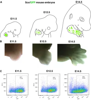
In order to isolate tendon cells, we took advantage of the Scx-GFP mouse line (Pryce et al., 2007), so thatScx-positive cells could be isolated by flow cytometry based on GFP fluorescence. We chose the E11.5 stage to select limb tendon progenitors and the E14.5 stage to target limb tendon differentiated cells, when tendons are well individualised. We also chose an intermediate time point at E12.5, as the transitory time point between the muscle-independent and -dependent phases of tendon formation. In the absence of muscles, Scxis normally expressed in E11.5 limbs, defining the
Received 3 February 2014; Accepted 17 July 2014
1
CNRS UMR 7622, IBPS-Developmental Biology Laboratory, Paris F-75005,
France.2Sorbonne Universités, UPMC Univ Paris 06, IBPS-Developmental Biology
Laboratory, Paris F-75005, France.3Inserm U1156, Paris F-75005, France.
4
ProfileXpert, SFR Lyon-Est, UMS 3453 CNRS/US7 INSERM, Lyon F-69008, France.
5
Research Division, Shriners Hospital for Children, Portland, OR 97239, USA.
6
Institut de Génomique Fonctionnelle de Lyon, UniversitéLyon 1, CNRS UMR5242,
Ecole Normale Supérieure de Lyon, Lyon F-69007, France.
*These authors contributed equally to this work
‡
Author for correspondence ([email protected])
DEVEL
O
muscle-independent phase, and is then lost in E14.5 muscleless limbs at the level of the forearm and arm (Schweitzer et al., 2001; Bonnin et al., 2005). In E12.5 muscleless limbs,Scxexpression is still present in ventral limb regions but starts to be downregulated in dorsal limb regions, at the level of the forearm (Bonnin et al., 2005; Pryce et al., 2009). We thus consider E12.5 as the transient time point between the muscle-independent and -dependent phases of forearm and arm tendon formation. Forelimbs were dissected from Scx-GFP embryos at E11.5, E12.5 and E14.5 (Fig. 1A,B). Scx-GFP+cells were then separated by flow cytometry (Fig. 1C);
50, 30 and 20 embryos were needed for the E11.5, E12.5 and E14.5 stages, respectively. The dissection and cytometry steps were performed three times for each time point in order to allow triplicate Affymetrix analyses.
Microarray analyses
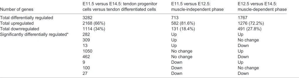
We performed three array comparisons: (1) E11.5 versus E12.5, corresponding to the muscle-independent phase of tendon formation; (2) E12.5 versus E14.5, corresponding to the muscle-dependent phase of arm and forearm tendon formation; and (3) E11.5 versus E14.5, corresponding to tendon progenitor cells versus tendon differentiated cells (Table 1). A total of 3282 genes (more than 10% of all transcripts in the genome array) were differentially regulated in limb tendon cells during development, between E11.5 and E14.5 (Table 1). A greater number of genes were differentially regulated during the muscle-dependent phase as compared with the muscle-independent phase (1767 versus 713; Table 1).
In total, 4888 regulated transcripts ( probe sets) can be hierarchically clustered (supplementary material Fig. S1A). We then asked whether differentially regulated genes in tendon cells represented specific Gene Ontology (GO) categories, which would
highlight differential biological activities during development (Table 2; supplementary material Fig. S1B). GO terms related to extracellular structure organisation, cell adhesion or response to wounding were highly represented in differentiated tendon cells at E14.5, whereas GO terms related to cell cycle were highly represented in tendon progenitor cells at E11.5 (Table 2; supplementary material Fig. S1B). In addition, very high enrichment scores and significant P-values were observed for the GO terms ‘extracellular matrix’, ‘cell adhesion’and ‘collagen’in tendon cells during limb development (Table 2), consistent with the massive increase of matrix synthesis during tendon development.
Genes displaying enriched expression in tendon cells during development
Few tendon-specific markers have been identified. In addition to the ScxandTnmdtendon markers, a series of matrix proteins has been described as being expressed or/and associated with tendon development, which we previously attempted to list (Edom-Vovard and Duprez, 2004). The mRNA relative expression of the main tendon collagen,Col1a1, and that of the tendon-associated collagens Col3a1,Col5a1,Col6a1, Col12a1and Col14a1was enhanced in tendon cells between E11.5 and E14.5 (Lejard et al., 2011). This is consistent with the high enrichment score of the‘collagen’biological process during tendon development (Table 2).
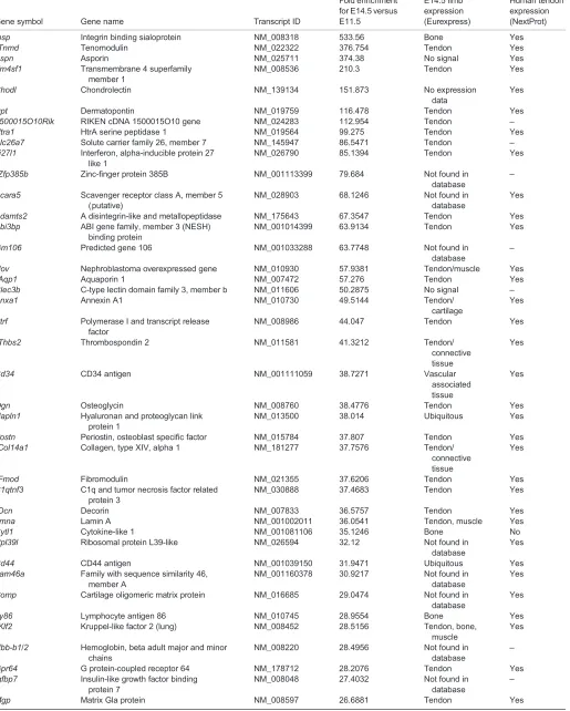
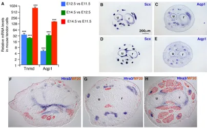
[image:2.612.49.347.409.744.2]One objective was to establish a list of tendon markers during development (supplementary material Table S1). We ordered the top 100 upregulated genes in E14.5 differentiated tendon cells versus E11.5 tendon progenitor cells, from high to low fold change (Table 3). We also analysed the expression of the ordered genes using Eurexpress, a transcriptome altas database for the mouse embryo. We found that the majority of the top 100 upregulated
Fig. 1. Strategy of tendon cell purification from forelimbs at different stages of mouse development.(A) Forelimbs were dissected from E11.5, E12.5 and E14.5 Scx-GFP mouse embryos. Dashed lines indicate the levels of dissection for forelimbs. (B) Representative images of dissected forelimbs at the different stages. (C) GFP+cells were isolated by flow cytometry. The boxed regions were used for transcriptome analysis.
DEVEL
O
genes were expressed in tendons (Table 3). The known tendon differentiation marker Tnmd was the second most differentially expressed gene on the list, displaying a 376-fold change between E14.5 and E11.5 (Table 3). In order to validate this list of tendon genes, we chose candidates not previously known to be related to tendons, starting with aquaporin 1 (Aqp1), a water channel protein, for which the fold change in expression levels was 57.2 between E11.5 and E14.5 in the array (Table 3). The dramatic increase of Aqp1mRNA levels was confirmed by RT-q-PCR (Fig. 2A).In situ hybridisation experiments showed thatAqp1is expressed in mouse forelimb tendons, similar toScx expression (Fig. 2B-E). We also chose HtrA serine peptidase 3 (Htra3) from the array and demonstrated its expression in E14.5 mouse limb tendons by in situhybridisation (Fig. 2F-H).Aqp1andHtra3, which were not previously known to be tendon related, displayed tendon-specific expression in mouse limbs (Fig. 2B-H). It should be noted thatScx did not appear in the array as a significantly upregulated gene, suggesting thatScxexpression levels did not change between E11.5 and E14.5, consistent with the little variation in Scx mRNA expression levels in mouse limbs (supplementary material Fig. S4A). We believe that this list of genes (Table 3; supplementary material Table S1) enriched in E14.5 tendon cells constitutes an important inventory of tendon markers.
TGF-βis the main signalling pathway upregulated in limb tendon cells during development
We next aimed to identify signalling pathways modified in limb tendon cells during development. We first used Genomatix software, which established gene associations with over 400 canonical pathways. In our tendon cell array, TGF-βwas the top pathway, displaying the highest number of upregulated genes in the three types of comparisons (Table 4): 59 and 102 genes of the TGF-βpathway (comprising 640
components) were significantly upregulated between E11.5 and E12.5 and between E12.5 and E14.5, respectively (Table 4). Consistent with our analysis using the Database for Annotation, Visualization and Integrated Discovery (DAVID) (Table 2), components of the cell cycle were significantly downregulated during tendon cell differentiation between E12.5 and E14.5 (Table 4).
In order to confirm the modification of TGF-β components in tendon cells, we analysed the variation of signal transduction KEGG pathways, which defines pathways from a more conventional point of view than Genomatix. Consistent with the Genomatix analysis (Table 4), TGF-β (KEGG N°4350) was the signalling pathway (among those of the signal transduction group) that displayed the highestP-value in terms of being modified between the three types of comparisons (Table 5). The components of the KEGG TGF-β pathway that display significant upregulation or downregulation in expression between the three time points of comparison are listed in supplementary material Table S2 and illustrated in supplementary material Fig. S2. The mRNA expression levels of TGF-βligands, receptors and extracellular components that were significantly differentially regulated in the arrays (supplementary material Table S2) were confirmed by RT-q-PCR analyses (Fig. 3A). Notably, all the genes encoding extracellular components [TGF-β, THBS, LTBP, decorin (DCN), TGFβi] of the ‘classical’ TGF-β pathway displayed significant upregulation in differentiated tendon cells versus progenitor tendon cells (supplementary material Table S2 and Fig. S2). Two of these extracellular components, namely the thrombospondin genesThbs2andThbs4, were expressed in mouse limb tendons (Fig. 3B-M) and are among the 100 top differentially expressed genes (Table 3).
[image:3.612.48.558.70.203.2]Based on bioinformatics analyses, RT-q-PCR and in situ hybridisation data, we concluded that the TGF-β pathway is the most active pathway in tendon cells during mouse limb development.
Table 1. Global reorganisation of the transcriptome of limb tendon cells during mouse development
Number of genes
E11.5 versus E14.5: tendon progenitor cells versus tendon differentiated cells
E11.5 versus E12.5: muscle-independent phase
E12.5 versus E14.5: muscle-dependent phase
Total differentially regulated 3282 713 1767
Total upregulated 2168 (66%) 582 (81.6%) 1276 (72.2%)
Total downregulated 1114 (34%) 131 (18.4%) 491 (27.8%)
Significantly differentially regulated* 282 Up Up
309 Up No change
13 Up Down
1050 No change Up
462 No change Down
9 Down Up
100 Down No change
27 Down Down
[image:3.612.47.572.618.728.2]*Shown are the number of genes that exhibit combinations of significant upregulation or downregulation, or no change, in E11.5 versus E14.5 compared with E11.5 versus E12.5 and with E12.5 versus E14.5.
Table 2. GO analysis in mouse limb tendon cells during development
E11.5 versus E12.5 E12.5 versus E14.5
Cluster High at E11.5 High at E12.5 High at E12.5 High at E14.5
Transcription factor activity (GOTERM-MF-FAT) 4 (1.3×10⁻⁸) 15.67 (3.4×10⁻⁸) Pattern specification process (GOTERM-BP-FAT) 4 (3.8×10⁻⁸)
Limb development (GOTERM-BP-FAT) 2.41 (1.1×10⁻³) 7.14 (7.3×10⁻⁹) Negative regulation of cell differentiation (GOTERM-BP-FAT) 2.66 (2.5×10⁻⁵)
Cell cycle (GOTERM-BP-FAT) 17.77 (2.9×10⁻²²)
Extracellular matrix (GOTERM-CC-FAT) 27.68 (6.9×10⁻²⁹) 38.55 (1.6×10–42)
Cell adhesion (GOTERM-BP-FAT) 13.5 (3.8×10⁻¹⁷) 26.58 (1.4×10⁻²⁸)
Collagen (SP-PIR-Keywords) 9.56 (2.0×10⁻¹¹) 12.64 (1.9×10⁻¹⁴)
Shown are enrichment scores with theP-value in parentheses. The DAVID bioinformatics resource 6.7 was used for this analysis.
DEVEL
O
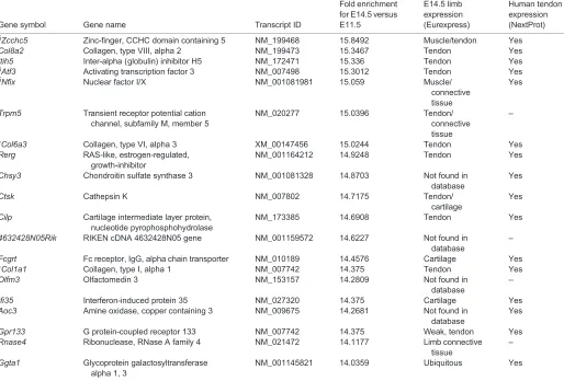
Table 3. Top 100 genes enriched in mouse limb tendon cells at E14.5 versus E11.5
Gene symbol Gene name Transcript ID
Fold enrichment for E14.5 versus E11.5
E14.5 limb expression (Eurexpress)
Human tendon expression (NextProt)
Ibsp Integrin binding sialoprotein NM_008318 533.56 Bone Yes
*Tnmd Tenomodulin NM_022322 376.754 Tendon Yes
Aspn Asporin NM_025711 374.38 No signal Yes
Tm4sf1 Transmembrane 4 superfamily member 1
NM_008536 210.3 Tendon Yes
Chodl Chondrolectin NM_139134 151.873 No expression
data
Yes
Dpt Dermatopontin NM_019759 116.478 Tendon Yes
1500015O10Rik RIKEN cDNA 1500015O10 gene NM_024283 112.954 Tendon –
Htra1 HtrA serine peptidase 1 NM_019564 99.275 Tendon Yes
Slc26a7 Solute carrier family 26, member 7 NM_145947 86.5471 Tendon –
Ifi27l1 Interferon, alpha-inducible protein 27 like 1
NM_026790 85.1394 Tendon Yes
§Zfp385b Zinc-finger protein 385B NM_001113399 79.684 Not found in
database
–
Scara5 Scavenger receptor class A, member 5 (putative)
NM_028903 68.1246 Not found in
database
Yes
Adamts2 A disintegrin-like and metallopeptidase NM_175643 67.3547 Tendon Yes Abi3bp ABI gene family, member 3 (NESH)
binding protein
NM_001014399 63.9134 Tendon Yes
Gm106 Predicted gene 106 NM_001033288 63.7748 Not found in
database
–
Nov Nephroblastoma overexpressed gene NM_010930 57.9381 Tendon/muscle Yes
‡Aqp1 Aquaporin 1 NM_007472 57.276 Tendon Yes
Clec3b C-type lectin domain family 3, member b NM_011606 50.2875 No signal –
Anxa1 Annexin A1 NM_010730 49.5144 Tendon/
cartilage
Yes
Ptrf Polymerase I and transcript release factor
NM_008986 44.047 Tendon Yes
‡Thbs2 Thrombospondin 2 NM_011581 41.3212 Tendon/
connective tissue
Yes
Cd34 CD34 antigen NM_001111059 38.7271 Vascular
associated tissue
Yes
Ogn Osteoglycin NM_008760 38.4776 Tendon Yes
Hapln1 Hyaluronan and proteoglycan link protein 1
NM_013500 38.014 Ubiquitous Yes
Postn Periostin, osteoblast specific factor NM_015784 37.807 Tendon Yes
*Col14a1 Collagen, type XIV, alpha 1 NM_181277 37.7576 Tendon/ connective tissue
Yes
*Fmod Fibromodulin NM_021355 37.6206 Tendon Yes
C1qtnf3 C1q and tumor necrosis factor related protein 3
NM_030888 37.4683 Tendon Yes
*Dcn Decorin NM_007833 36.5757 Tendon Yes
Lmna Lamin A NM_001002011 36.0541 Tendon, muscle Yes
Cytl1 Cytokine-like 1 NM_001081106 35.1246 Bone No
Rpl39l Ribosomal protein L39-like NM_026594 32.12 Not found in
database
Yes
Cd44 CD44 antigen NM_001039150 31.9471 Ubiquitous Yes
Fam46a Family with sequence similarity 46, member A
NM_001160378 30.9217 Not found in database
Yes
Comp Cartilage oligomeric matrix protein NM_016685 29.0474 Not found in database
Yes
Ly86 Lymphocyte antigen 86 NM_010745 28.9554 Bone Yes
§
Klf2 Kruppel-like factor 2 (lung) NM_008452 28.5156 Tendon, bone,
muscle
Yes
Hbb-b1/2 Hemoglobin, beta adult major and minor chains
NM_008220 28.4956 Not found in
database
–
Gpr64 G protein-coupled receptor 64 NM_178712 28.2076 Tendon Yes
Igfbp7 Insulin-like growth factor binding protein 7
NM_008048 27.4032 Not found in
database
–
Mgp Matrix Gla protein NM_008597 26.6881 Tendon Yes
Continued
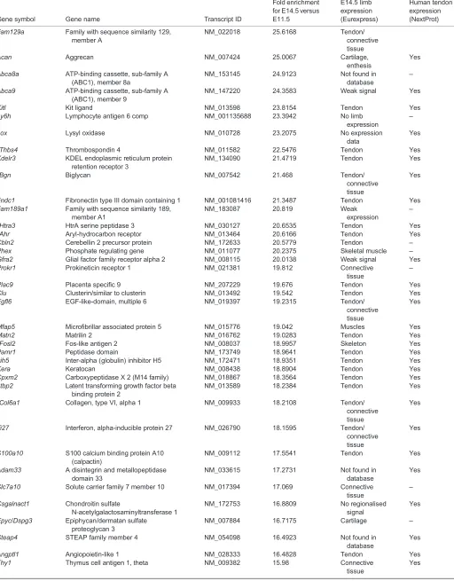
DEVEL
O
Table 3. Continued
Gene symbol Gene name Transcript ID
Fold enrichment for E14.5 versus E11.5
E14.5 limb expression (Eurexpress)
Human tendon expression (NextProt)
Fam129a Family with sequence similarity 129, member A
NM_022018 25.6168 Tendon/
connective tissue
Acan Aggrecan NM_007424 25.0067 Cartilage,
enthesis
Yes
Abca8a ATP-binding cassette, sub-family A (ABC1), member 8a
NM_153145 24.9123 Not found in
database
–
Abca9 ATP-binding cassette, sub-family A (ABC1), member 9
NM_147220 24.3583 Weak signal Yes
Kitl Kit ligand NM_013598 23.8154 Tendon Yes
Ly6h Lymphocyte antigen 6 comp NM_001135688 23.3942 No limb
expression
–
Lox Lysyl oxidase NM_010728 23.2075 No expression
data
Yes
‡Thbs4 Thrombospondin 4 NM_011582 22.5476 Tendon Yes
Kdelr3 KDEL endoplasmic reticulum protein retention receptor 3
NM_134090 21.4719 Tendon Yes
*Bgn Biglycan NM_007542 21.468 Tendon/
connective tissue
Yes
Fndc1 Fibronectin type III domain containing 1 NM_001081416 21.3487 Tendon Yes Fam189a1 Family with sequence similarity 189,
member A1
NM_183087 20.819 Weak
expression
–
‡Htra3 HtrA serine peptidase 3 NM_030127 20.6535 Tendon Yes
§
Ahr Aryl-hydrocarbon receptor NM_013464 20.6166 Tendon Yes
Cbln2 Cerebellin 2 precursor protein NM_172633 20.5779 Tendon –
Phex Phosphate regulating gene NM_011077 20.2375 Skeletal muscle –
Gfra2 Glial factor family receptor alpha 2 NM_008115 20.0138 Weak signal Yes
Prokr1 Prokineticin receptor 1 NM_021381 19.812 Connective
tissue
–
Plac9 Placenta specific 9 NM_207229 19.676 Tendon Yes
Clu Clusterin/similar to clusterin NM_013492 19.542 Tendon Yes
Egfl6 EGF-like-domain, multiple 6 NM_019397 19.2315 Tendon/
connective tissue
Yes
Mfap5 Microfibrillar associated protein 5 NM_015776 19.042 Muscles Yes
Matn2 Matrilin 2 NM_016762 19.0283 Tendon Yes
§
Fosl2 Fos-like antigen 2 NM_008037 18.9957 Skeleton Yes
Pamr1 Peptidase domain NM_173749 18.9641 Tendon Yes
Itih5 Inter-alpha (globulin) inhibitor H5 NM_172471 18.9351 Tendon Yes
Kera Keratocan NM_008438 18.8904 Tendon Yes
Cpxm2 Carboxypeptidase X 2 (M14 family) NM_018867 18.3564 Tendon Yes
Ltbp2 Latent transforming growth factor beta binding protein 2
NM_013589 18.2384 Tendon Yes
*Col6a1 Collagen, type VI, alpha 1 NM_009933 18.2108 Tendon/
connective tissue
Yes
Ifi27 Interferon, alpha-inducible protein 27 NM_026790 18.1595 Tendon/ connective tissue
Yes
S100a10 S100 calcium binding protein A10 (calpactin)
NM_009112 17.5541 Tendon Yes
Adam33 A disintegrin and metallopeptidase domain 33
NM_033615 17.2731 Not found in
database
Yes
Slc7a10 Solute carrier family 7 member 10 NM_017394 17.069 Connective tissue
–
Csgalnact1 Chondroitin sulfate
N-acetylgalactosaminyltransferase 1
NM_172753 16.8809 No regionalised
signal
Yes
Epyc/Dspg3 Epiphycan/dermatan sulfate proteoglycan 3
NM_007884 16.7175 Cartilage –
Steap4 STEAP family member 4 NM_054098 16.4923 Not found in
database
Yes
Angptl1 Angiopoietin-like 1 NM_028333 16.4828 Tendon Yes
Thy1 Thymus cell antigen 1, theta NM_009382 15.98 Connective
tissue
Yes
Continued
DEVEL
O
TGF-βpathway involvement in mouse limb tendon development
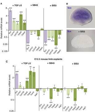
Bioinformatics analyses highlighted that, in addition to being upregulated during tendon cell differentiation between E14.5 and E12.5, components of the TGF-β pathway were also significantly upregulated between E12.5 and E11.5, i.e. during the muscle-independent phase of tendon formation (Tables 4 and 5). This suggested an involvement of the TGF-βpathway at an earlier stage of the tendon program than previously believed (Pryce et al., 2009). In order to validate our bioinformatics analysis of the tendon transcriptome, we employed an ex vivo system based on mouse forelimb explants. Forelimb bud explants were dissected either at E9.5/E10.5 to target the initial phase of tendon formation or at E12.5 to target the differentiation phase of tendon formation, and were incubated for 24 h in the presence of TGFβ2 ligand for TGF-β gain-of-function experiments or specific TGF-βinhibitors for TGF-β loss-of-function experiments. We chose TGFβ2 ligand, as opposed to TGFβ1 or TGFβ3, for the gain-of-function experiments because Tgfb2 displayed higher levels of endogenous expression than Tgfb1 and Tgfb3in mouse limbs (supplementary material Fig. S4C).
TGFβ2 was sufficient to increase Scx expression in E9.5/E10.5 mouse forelimb explants (Fig. 4A). Expression of the tendon-related
[image:6.612.50.564.73.421.2]Col1a1,Col1a2,Thbs2 andThbs4 genes was also upregulated after TGFβ2 application (Fig. 4A). In E9.5/E10.5 mouse limbs, the expression levels of Tnmd and Aqp1 were considered undetectable (above 31 PCR cycles; supplementary material Fig. S4B), and TGFβ2 was unable to increase their expression in early E9.5/E10.5 mouse limb explants cultured for 24 h (data not shown). In order to test the requirement of TGF-β signalling for the initiation of tendon gene expression in forelimb buds, we blocked the TGF-βsignalling pathway by applying specific TGF-βinhibitors (supplementary material Fig. S5). In the presence of the SB43 inhibitor, which blocks the TGF-βpathway at the level of the ALK4, ALK5 and ALK7 (ACVR1B, TGFBR1 and ACVR1C, respectively–Mouse Genome Informatics) receptors (Inman et al., 2002),Scx,Col1a1,Col1a2andThbs2 gene expression was significantly downregulated (Fig. 4A). Blockade of the SMAD2/3 intracellular pathway using the SIS3 inhibitor (Jinnin et al., 2006) also diminished the relative expression levels ofScx,Col1a1,Col1a2,Thbs2 andThbs4 compared with control limbs in E9.5/E10.5 mouse limb explants (Fig. 4A). Consistently, application of the SIS3 inhibitor abolishedScxexpression in E9.5 mouse limb explants (Fig. 4B). This showed that TGF-β was sufficient and required via the SMAD2/3 intracellular pathway for Scx, Col1a1, Col1a2, Thbs2 and Thbs4 expression in E9.5/E10.5 mouse forelimbs.
Table 3. Continued
Gene symbol Gene name Transcript ID
Fold enrichment for E14.5 versus E11.5
E14.5 limb expression (Eurexpress)
Human tendon expression (NextProt)
§
Zcchc5 Zinc-finger, CCHC domain containing 5 NM_199468 15.8492 Muscle/tendon Yes
Col8a2 Collagen, type VIII, alpha 2 NM_199473 15.3467 Tendon Yes
Itih5 Inter-alpha (globulin) inhibitor H5 NM_172471 15.336 Tendon Yes
§Atf3 Activating transcription factor 3 NM_007498 15.3012 Tendon Yes
§
Nfix Nuclear factor I/X NM_001081981 15.059 Muscle/
connective tissue
Yes
Trpm5 Transient receptor potential cation channel, subfamily M, member 5
NM_020277 15.0396 Tendon/
connective tissue
–
*Col6a3 Collagen, type VI, alpha 3 XM_00147456 15.0244 Tendon Yes
Rerg RAS-like, estrogen-regulated, growth-inhibitor
NM_001164212 14.9248 Tendon Yes
Chsy3 Chondroitin sulfate synthase 3 NM_001081328 14.8703 Not found in database
Yes
Ctsk Cathepsin K NM_007802 14.7175 Tendon/
cartilage
Yes
Cilp Cartilage intermediate layer protein, nucleotide pyrophosphohydrolase
NM_173385 14.6908 Tendon Yes
4632428N05Rik RIKEN cDNA 4632428N05 gene NM_001159572 14.6227 Not found in database
–
Fcgrt Fc receptor, IgG, alpha chain transporter NM_010189 14.4576 Cartilage Yes
*Col1a1 Collagen, type I, alpha 1 NM_007742 14.375 Tendon Yes
Olfm3 Olfactomedin 3 NM_153157 14.2809 Not found in
database
–
Ifi35 Interferon-induced protein 35 NM_027320 14.375 Cartilage Yes
Aoc3 Amine oxidase, copper containing 3 NM_009675 14.2681 Not found in database
Yes
Gpr133 G protein-coupled receptor 133 NM_007742 14.375 Weak, tendon Yes
Rnase4 Ribonuclease, RNase A family 4 NM_021472 14.1177 Limb connective tissue
–
Ggta1 Glycoprotein galactosyltransferase alpha 1, 3
NM_001145821 14.0359 Ubiquitous Yes
Genes with probe expression above 500 AU (arbitrary units) have been ordered according to fold enrichment (from high to low) of gene expression in E14.5 versus E11.5 tendon cells from the array analysis. The Eurexpress and human NextProt databases were used to define gene expression in E14.5 mouse limb tendons or in human tendons, respectively.
*Genes known to be related to tendons.
‡Genes previously not known to be tendon related and analysed byin situhybridisation or/and RT-q-PCR in the present study.
§
Transcription factors.
DEVEL
O
From E12.5, tendon differentiation is concomitant with important transcriptomic changes in mouse limb tendon cells (Table 1) and with an enriched expression of matrix and tendon genes (Tables 2 and 3). Consistently, the relative endogenous expression levels ofCol1a1, Col1a2, Thbs2, Thbs4, Tnmd and Aqp1 were significantly upregulated in E12.5 limbs compared with E9.5 or E10.5 limbs (supplementary material Fig. S4A,B). In E12.5 limb explants, TGFβ2 was sufficient to increase the relative mRNA levels ofScx,Col1a1, Thbs2,Thbs4andTnmd, but not ofAqp1(Fig. 4C). However, the blockade of TGF-βreceptors (SB43) or SMAD2/3 activity (SIS3) in E12.5 limb explants did not affectScx,Col1a2,Thbs4,TnmdorAqp1 gene expression, while decreasing that ofCol1a1andThbs2(Fig. 4C). This showed that in E12.5 mouse limbs TGF-βis sufficient for the expression of the tendon markers Scx,Col1a1,Thbs2,Thbs4 and Tnmd, while being required only forCol1a1andThbs2expression.
We conclude that TGF-βis sufficient for the expression ofScx and tendon-associated genes at different stages of limb development
from E9.5 to E12.5, whereas the intracellular SMAD2/3 pathway is required for Scxand tendon-associated gene expression in early E9.5/E10.5 limbs.
Involvement of the ERK MAPK pathway in mouse limb tendon development
[image:7.612.102.506.57.307.2]In addition to the TGF-βpathway, the MAPK signalling pathway (KEGG N°4010) also appeared to be significantly differentially regulated between E11.5 and E12.5 and between E11.5 and E14.5 (Table 5; supplementary material Fig. S3 and Table S3). MAPK pathways are activated by receptor tyrosine kinases, including FGF receptors (Mason et al., 2006). FGF signalling has been shown to positively regulateScxexpression in chick limb tendons, chick and mouse axial tendons and intermuscular tendons of chick stomach (Edom-Vovard et al., 2002; Brent and Tabin, 2004; Brent et al., 2005; Le Guen et al., 2009). No such evidence of FGF sufficiency exists during mouse limb tendon development. An observation from Fig. 2. Expression ofAqp1andHtra3genes in mouse limb tendons.(A) RT-q-PCR analyses ofTnmdandAqp1expression in tendon cells at different stages of mouse limb development. The mRNA levels of tendon cells at E11.5 or E12.5 were normalised to 1 for each comparison so that the graph shows the relative increase of mRNA levels in tendon cells between E12.5 and E11.5, E14.5 and E12.5, and E14.5 and E11.5. ***P<0.001; error bars indicate s.d. (B-E) Adjacent transverse sections of forelimbs of E14.5 mouse embryos were hybridised withScx(B,D) orAqp1(C,E) probes. Adjacent sections are shown from distal (B,C) to proximal (D,E) zeugopod limb regions. (F-H) Transverse sections of E14.5 mouse limbs hybridised withHtra3probe (blue) and immunostained for the heavy chain of myosin II (MF20 antibody; brown). u, ulna; r, radius.
Table 4. Signalling pathways (signal transduction pathways and GO tissues) modified in mouse limb tendon cells during development
Pathway (total number of genes in pathway) E11.5 versus E12.5 E12.5 versus E14.5 E11.5 versus E14.5
Signal transduction pathways
*TGF-β(640) 59 (3.42×10⁻¹⁰) 102 (2.86×10⁻⁹) 136 (6.18×10⁻⁹)
*Matrix metalloproteinase (236) 19 (2.38×10⁻³) 51 (1.88×10⁻⁹) 64 (9.60×10⁻⁹)
*Thrombospondin 1 (56) 9 (3.00×10⁻⁴) 13 (1.12×10⁻³) 17 (7.08×10⁻⁴)
‡Aurora kinase (130) – 26 (7.72×10⁻¹⁴
) 45 (2.28×10⁻²²)
‡Cell division (201) – 32 (7.72×10⁻¹⁴
) 48 (2.76×10⁻¹⁶)
Tissues
*Tendons (197) 39 (1.47×10⁻²¹) 63 (4.22×10⁻²⁷) 85 (6.30×10⁻³³)
*Ligaments (120) 35 (1.63×10⁻²⁵) 43 (4.53×10⁻²¹) 58 (5.26×10⁻²⁶)
For each comparison is shown the number of genes upregulated or downregulated with theP-value in parentheses. TGF-βis the pathway displaying the greatest number of upregulated genes in each type of comparison. Genomatix software was employed for this analysis.
*Upregulated pathways.
‡Downregulated pathways.
DEVEL
O
[image:7.612.50.564.594.698.2]the bioinformatics data is that several FGF ligands appeared significantly upregulated in mouse tendon cells during limb development (supplementary material Table S3), although none of these specific FGF ligands has been reported to be linked with
[image:8.612.48.562.72.158.2]tendon development. Another striking observation is that most of the MAP kinases displaying significant variation in the transcriptome showed a significant decrease in expression, such as Map3k4, Map2k6,Mapk8(Jnk) andMapk12(p38) (supplementary material
Table 5. Analysis of the KEGG pathway‘signal transduction’category in limb tendon cells during development
Pathway (total number of genes in pathway)
E11.5 versus E12.5 (713 regulated genes in the transcriptome)
E12.5 versus E14.5 (1767 regulated genes in the transcriptome)
E11.5 versus E14.5 (3282 regulated genes in the transcriptome)
N°04350: TGF-β(85) 11 (2.8×10⁻³) 17 (2.2×10⁻²) 27 (5.1×10⁻³)
N°04010: MAPK (259) 17 (9.21×10⁻²) 34 (NS, 0.17) 57 (5.6×10⁻²)
N°04020: Calcium (185) 13 (6.48×10⁻²) 24 (NS, 0.2179) 36 (NS, 0.356)
N°04310: Wnt (156) 12 (5.18×10⁻²) 15 (NS, 0.7958) 34 (NS, 0.175)
N°04340: Hedgehog (49) 7 (2.77×10⁻²) 5 (NS, 0.876) –
The DAVID bioinformatics resource 6.7 was used for this analysis. For each comparison is shown the number of differentially regulated genes in the pathway with theP-value in parentheses. The KEGG pathways have been ordered from high to low byP-value for the E11.5 versus E12.5 comparison. The two most significant P-values correspond to the TGF-βand MAPK pathways for the E11.5 versus E12.5 and E11.5 versus E14.5 comparisons. NS, not significant.
Fig. 3. Expression of TGF-βsignalling pathway components in mouse FACS-sorted tendon cells and limbs.(A) RT-q-PCR analyses of TGF-β-associated gene expression levels in tendon cells at different stages of development. Shown is the relative increase in mRNA levels in Scx-GFP+tendon cells at different stages of development: E12.5 versus E11.5, where mRNA levels of tendon cells at E11.5 were normalised to 1; E14.5 versus E12.5, where mRNA levels of tendon cells at E12.5 were normalised to 1; and E14.5 versus E11.5, where mRNA levels of tendon cells at E11.5 were normalised to 1. *P<0.05; **P<0.01; ***P<0.001; error bars indicate s.d. (B-M) Adjacent transverse sections of forelimbs of E14.5 mouse embryos were hybridised withScx(B-E),Thbs2(F-I) orThbs4 (J-M) probes. B,F,J, C,G,K and D,H,L are groups of adjacent sections from distal to proximal limb regions. (E,I,M) Higher magnifications of tendon shown in D,H,L. The circles delineate the same areas, highlighting differences between the Thbs2(I) andThbs4(M) expression domains in tendons. All sections are dorsal to the top and posterior to the left. u, ulna; r, radius.
DEVEL
O
[image:8.612.51.391.276.731.2]Table S3). In addition, the MAP kinase phosphataseDusp6, which is known to be a readout of active ERK1/2 (MAPK3/1) signalling during development (Lunn et al., 2007), was clearly downregulated in tendon cells during development (supplementary material Table S3). This decrease in the expression ofDusp6and of genes encoding MAP kinases in tendon cells suggested a diminution of ERK MAPK activity in tendon cells during mouse development.
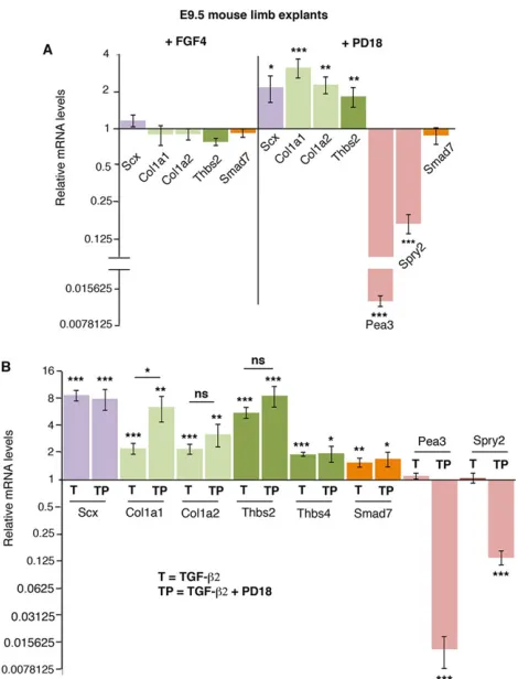
In order to validate this bioinformatics data, we blocked the ERK MAPK pathway in mouse limb explants using the PD18 inhibitor, which is known to prevent ERK phosphorylation (Bain et al., 2007). Pea3(Etv4) and sprouty 2 (Spry2) genes are transcriptional targets of ERK MAP kinases and are considered a readout of ERK activity (O’Hagan et al., 1996; Mason et al., 2006). The dramatic loss of Pea3andSpry2expression showed that ERK signalling activity was downregulated in the presence of PD18 (Fig. 5A). Consistent with the bioinformatics data, ERK inhibition led to a significant activation of the expression ofScx,Col1a1,Col1a2andThbs2in E9.5 mouse limb explants (Fig. 5A). FGF4 application to E9.5 mouse limb explants did not modify tendon gene expression (Fig. 5A). We conclude that inhibition of the ERK MAPK signalling pathway was sufficient to activate the expression of tendon genes, includingScx, in early mouse limbs.
Positive and negative cross-talk between the ERK and SMAD intracellular pathways have been highlighted in several cellular and in vivo contexts (Massague, 2012) (supplementary material Fig. S5). In epithelial cells, ERK1/2 activation inhibits SMAD3 transcriptional activity via phosphorylation in its linker region (Kretzschmar et al., 1999; Matsuura et al., 2005; Wrighton
et al., 2009). Since mutations of these phosphorylation sites increase SMAD3 activity (Wrighton et al., 2009), we hypothesise that ERK blockade could activate SMAD3 transcriptional activity in tendon cells. To assess activity of the SMAD3 pathway we used Smad7, a negative-feedback regulator that is considered a general TGF-β transcriptional target gene (Massague, 2012). We did not observe any increase inSmad7expression following PD18 application (Fig. 5A).Smad7expression was also unchanged after FGF4 application (Fig. 5A), indicating an absence of cross-talk between the FGF/ERK and SMAD3 pathways in the E9.5 mouse limb context. This indicated that tendon gene activation following ERK inhibition was not a consequence of SMAD3 activation.
[image:9.612.47.354.50.414.2]In order to determine whether the positive effect of TGF-βand of ERK inhibition on tendon gene expression could be additive, we systematically compared the TGF-βeffect with that of simultaneous TGF-β+PD18 application on the mRNA levels of tendon genes in E9.5 mouse limb explants (Fig. 5B). We did not observe any significant increase inSmad7expression levels in the presence of TGF-β+PD18 versus TGF-βalone (Fig. 5B), confirming the absence of any increase in SMAD3 transcriptional activity in the context of ERK inhibition (Fig. 5A). TGF-β receptors can activate various MAP kinases, including ERK (supplementary material Fig. S5) (Massague, 2012). However, in early mouse limb explants TGF-β did not activate Pea3orSpry2 expression (Fig. 5B) and did not prevent ERK inhibition based on similar Pea3 and Spry2 downregulation with PD18 (Fig. 5A) and TGF-β+PD18 (Fig. 5B). Regarding tendon genes, there was no difference in Scx mRNA
Fig. 4. Effects of TGF-βgain- and loss-of-function in mouse limb explants.(A) RT-q-PCR analyses of the expression levels of tendon markers in E9.5/E10.5 mouse limb explants cultured for 24 h with TGFβ2, SB43 or SIS3 inhibitors. (B)In situhybridisation forScxexpression in E9.5 mouse limb explants cultured with SIS3 inhibitor for 24 h. (C) RT-q-PCR analyses ofScx,Col1a1,Col1a2,Thbs2, Thbs4,TnmdandAqp1expression levels in E12.5 mouse limb explants cultured for 24 h with TGFβ2, SB43 or SIS3 inhibitors. For each gene, the mRNA levels of control limb explants were normalised to 1. *P<0.05; **P<0.01; ***P<0.001; error bars indicate s.e.m.
DEVEL
O
levels following TGF-β+PD18 application versus TGF-β(Fig. 5B). However, we did observe a significant difference in Col1a1 expression levels, and a non-significant tendency to increase for Col1a2 and Thbs2expression levels following TGF-β+PD18 co-treatment versus TGF-β alone in E9.5 mouse limb explants (Fig. 5B). This showed that there was no obvious additive effect onScxexpression following TGF-βapplication and ERK inhibition in E9.5 mouse limbs.
The positive effect of ERK inhibition onScxexpression observed in E9.5 limb explants was lost in E10.5 limb explants, whereas the Col1a1 and Thbs2 expression levels were still significantly elevated, following PD18 application at E10.5 (supplementary material Fig. S6A). FGF4 application did not have any significant effect on tendon gene expression in E10.5 mouse limbs, as in E9.5 limbs (supplementary material Fig. S6A). Lastly, in E12.5 mouse limb explants, ERK inhibition did not activate the expression of any tendon genes and even significantly inhibited Col1a2 expression. Consistently, FGF ligand application activatedCol1a2 expression levels in E12.5 mouse limb explants (supplementary material Fig. S6B).
We conclude that FGF does not positively regulate Scx expression in early and late mouse limbs and that ERK inhibition is sufficient to enhance the expression of tendon genes independently of the activation of TGF-βsignalling in early E9.5 mouse limbs.
TGF-β/SMAD2/3 and FGF/ERK pathway involvement in mouse mesenchymal stem cells
TGF-β and MAPK signalling pathways were modified in limb tendon cells during development (Tables 4 and 5), and TGFβ2 application or ERK MAPK inhibition was sufficient to activate tendon gene expression in mesodermal limb cells (Figs 4 and 5). In order to determine whether TGF-βligands or ERK inhibition could also drive stem cells towards the tendon lineage, we utilised mesenchymal stem cells. These cells can differentiate into various tissues of mesodermal origin, when cultured in appropriate differentiation media (Caplan, 2007).
TGF-β has been shown to have a protenogenic or
prochondrogenic effect depending on cell type or culture conditions (Lorda-Diez et al., 2009, 2013; Pryce et al., 2009). We used the multipotent murine C3H10T1/2 mesenchymal stem cells (Reznikoff et al., 1973). TGFβ2 has been shown to activate Scx expression in C3H10T1/2 stem cells (Pryce et al., 2009). Consistently, the relative levels ofScxandCol1a1gene expression were significantly elevated in the presence of TGFβ2 or TGFβ3 (Fig. 6A). In contrast to the tendon markers, the relative levels of the cartilage markerSox9were significantly decreased in the presence of TGF-βligands (Fig. 6A). We did not observe any increase in tendon or cartilage marker expression after simultaneous addition of TGFβ2 and TGFβ3 ligands compared with either ligand alone (Fig. 6A). The TGF-βeffect on tendon and cartilage marker expression was reduced in the presence of SB43 inhibitor, which blocks the TGF-βpathway at the level of the receptors, and in the presence of SIS3 inhibitor, which blocks the SMAD2/3 intracellular pathway (Fig. 6B). We conclude that TGF-β ligand has the ability to direct mouse mesenchymal stem cells towards the tendon lineage (Scx) at the expense of the cartilage lineage (Sox9) via the SMAD2/3 pathway. The outcome of FGF treatment on Scxexpression in stem cells differs between studies. FGF2 treatment in mouse stem cells activated the expression ofScx(Ker et al., 2011), whereas FGF4 treatment in mouse tendon progenitor cells inhibitsScxexpression (Brown et al., 2014). We assessed the efficiency of FGF4 and ERK inhibition (PD18) in C3H10T1/2 cells byPea3upregulation in the presence of FGF4 andPea3downregulation with PD18 (Fig. 6C). Consistent with the PD18 effect in E9.5 mouse explants (Fig. 5), ERK inhibition activated Scx and Col1a1 gene expression in C3H10T1/2 cells (Fig. 6C). PD18 treatment did not affect Smad7 expression in C3H10T1/2 cells (Fig. 6C), indicating that SMAD2/3 activity was not modified in this experimental design. ERK inhibition did not affect Sox9expression in C3H10T1/2 cells (Fig. 6C). FGF4 had the opposite effect and led to a significant inhibition of Scx and Col1a1 gene expression in C3H10T1/2 cells (Fig. 6C).
We conclude that TGF-βligand has the ability to direct mouse mesenchymal stem cells towards the tendon lineage at the expense of cartilage. ERK inhibition activatesScxexpression, whereas FGF appeared to have an anti-tenogenic effect on mouse mesenchymal stem cells.
DISCUSSION
[image:10.612.57.292.55.363.2]We have established the first transcriptome of mouse tendon cells during development. Bioinformatics analyses highlighted the TGF-β and MAPK signalling pathways as being the main signalling pathways regulated in limb tendon cells during development. Modification of these pathways in mouse limb explants or mesenchymal stem cells showed that TGF-β signalling was sufficient and required via SMAD2/3 to drive mouse mesodermal stem cells towards the tendon lineageex vivo andin vitro. FGF did not have a tenogenic effect and the inhibition Fig. 5. Effects of FGF/ERK gain- and loss-of-function in E9.5 mouse limb
explants.(A) RT-q-PCR analyses of the tendon genesScx,Col1a1,Col1a2 andThbs2in E9.5 mouse limb explants cultured for 24 h with FGF4 or PD18 inhibitor. (B) RT-q-PCR analyses ofScx,Col1a1,Col1a2,Thbs2,Thbs4, Smad7,Pea3andSpry2mRNA levels in E9.5 mouse limb explants cultured for 24 h with either TGFβ2 (condition T) or TGFβ2+PD18 inhibitor (condition TP). For each gene, the mRNA levels of control limbs were normalised to 1. *P<0.05; **P<0.01; ***P<0.001; NS, not significant; error bars indicate s.e.m.
DEVEL
O
of the ERK MAPK signalling pathway was sufficient to activate Scx in mouse limb mesodermal progenitors and mesenchymal stem cells.
Genes expressed in limb tendons cells during mouse development
We have established a list of genes that are differentially expressed in limb tendon cells during development. Differentially expressed genes displayed very high fold changes (ranging from 533 to 14 for the top 100 genes). The second most differentially expressed gene of the ordered list isTnmd, which is known to be involved in tendon formation (Docheva et al., 2005; Shukunami et al., 2006). Among the top 100 enriched genes, those encoding matrix components were found to be drastically upregulated in tendon cells during development, including Col14a1, Fmod, Dcn and Bgn. Mice mutant for these matrix genes display a tendon phenotype (Ameye et al., 2002; Ansorge et al., 2009). Among this top list,Aqp1and Htra3(which were previously not known to be related to tendon) were identified as expressed in E14.5 mouse limb tendons. The AQP1 hydrophobic transmembrane water channel protein is known to be responsible for the rapid response of cell volume to changes in plasma tonicity and is involved in cell proliferation, migration and adhesion processes of many cell types (Benga, 2012). However, AQP1 function in tendon development is not yet known. The serine protease geneHtra3, which is known to act as a tumour suppressor (Skorko-Glonek et al., 2013), has until now never been associated with tendon formation. Lastly, among the top enriched genes,
we found components of the TGF-β pathway, including the thrombospondin genes Thbs2 and Thbs4, which were expressed in mouse limb tendons. Mice that lackThbs2display connective tissue abnormalities, including in tendons (Kyriakides et al., 1998). TheDrosophilaequivalent, Thrombospondin (Tsp), is produced by tendon cells and is essential for the formation of the integrin-mediated myotendinous junction (Subramanian et al., 2007). We believe that these genes that are differentially expressed in limb tendon cells during mouse development (listed in Table 3; supplementary material Table S1) constitute an important list of tendon markers.
TGF-βsignalling pathway involvement in mouse limb tendon development
Bioinformatics data highlighted a TGF-βactivity in limb tendon cells during the muscle-independent phase (before E12.5) and the differentiation phase (after E12.5) of tendon formation. We confirmed that TGF-β was sufficient for tendon gene expression in early mouse limb explants. In addition, the SMAD2/3 intracellular pathway was required for Scx expression in early mouse limb explants. In early mouse limbs,Scxis also expressed in cartilage progenitors of the entheses (tendon attachment sites to bone) (Blitz et al., 2013; Sugimoto et al., 2013) and TGF-β signalling has been shown to be required for the specification of Scx+ enthesis progenitors (Blitz et al., 2013). The onset of Scx
[image:11.612.75.543.55.362.2]expression in mouse limbs is at E10 (Schweitzer et al., 2001; Sugimoto et al., 2013) and Scxexpression was disrupted only at Fig. 6. Effect of TGF-β/SMAD2/3 and FGF/ERK signalling in mesenchymal stem cells.(A) C3H10T1/2 cells were cultured in the presence of TGFβ2, TGFβ3 or TGFβ2+TGFβ3 for 24 h. Relative mRNA expression was examined for the tendon markersScxandCol1a1and for the cartilage markerSox9. For each gene, the mRNA levels of non-treated C3H10T1/2 cells were normalised to 1. (B) C3H10T1/2 cells were cultured in the presence of TGFβ2 (condition a), TGFβ2+SB43 inhibitor (condition b) or TGFβ2+SIS3 inhibitor (condition c) for 24 h. The mRNA levels of non-treated C3H10T1/2 cells were normalised to 1 for condition a; the mRNA levels of TGFβ2-treated C3H10T1/2 cells were normalised to 1 for conditions b and c. (C) C3H10T1/2 cells were cultured in the presence of FGF4 or PD18 inhibitor for 24 h. The mRNA levels of non-treated C3H10T1/2 cells were normalised to 1. *P<0.05; **P<0.01; ***P<0.001; error bars indicate s.d.
DEVEL
O
E12.5 in limb buds of Tgfb2−/−;Tgfb3−/−and conditionalTgfbr2 mutant mice (Pryce et al., 2009). However,ScxandCol1a1gene expression and tendon matrix organisation are altered in adult tendons ofSmad3−/−mice (Berthet et al., 2013) and it was shown that SMAD3 is recruited toScxregulatory regions in adult mouse tendons (Berthet et al., 2013), suggesting a direct role for SMAD signalling in Scx expression. Because we found a loss of Scx expression when SMAD2/3 was inhibited in mouse limb explants, we propose that in theTgfb2−/−;Tgfb3−/−and conditionalTgfbr2 mutant mice, the endogenous SMAD2/3 intracellular pathways may be activated by alternative TGF-βsuperfamily ligands or receptors to initiate Scx expression. Moreover, it has been shown that the SMAD intracellular pathways can also be activated by alternative ligands (Guo and Wang, 2009; Massague, 2012), suggesting that non-canonical TGF-β signals or receptors could drive Scx expression in early mouse limbs. Based onScxexpression, TGF-β was also able to drive mesenchymal stem cells toward the tendon lineage, via SMAD2/3. These data converge on the idea that the TGF-βsignalling pathway is sufficient and required via SMAD2/3 to initiate the commitment of undifferentiated mesodermal cells towards the tendon lineageex vivoandin vitro, but direct targeting of SMAD genes in vivowill be required to determine if, during mouse developmentScxexpression is indeed dependent exclusively on SMAD2/3 signalling.
In addition to being sufficient forScxexpression during the early stages of mouse limb development, TGFβ2 was also sufficient for tendon gene expression in late mouse limb explants. This indicated that a TGF-βsignal was involved in the positive regulation of late tendon marker expression during the tendon differentiation process. Because Tnmd mRNA expression levels were barely detectable before E12.5 in mouse limbs, we believe that a TGF-β signal participates in the initiation ofTnmd expression in E12.5 limbs. However, the intracellular SMAD2/3 pathway was only required for Col1a1 and Thbs2 expression and not for Scx, Thbs4 or Tnmd expression in E12.5 mouse limbs. This showed that other signalling pathways were required for the expression of these tendon markers during the tendon differentiation process. The calcium pathway, being the third most differentially regulated (Table 5), is a good candidate pathway. The absence ofAqp1gene regulation by TGFβ2 also indicated the involvement of other signalling pathways during tendon cell differentiation. It is likely that mechanical forces and downstream signalling pathways are important for limb tendon differentiation after E12.5.
FGF signalling in mouse limb tendon development
Bioinformatics analyses identified the MAPK pathway as being significantly modified in tendon cells during mouse limb development. The significant decrease in expression of MAP kinases and the phosphatase Dusp6 in the array suggested a reduction of MAP kinase activity in tendon cells during development. This tendency was surprising given the positive effect of FGF signal onScxexpression in chick limbs and chick and mouse somites (Edom-Vovard et al., 2002; Brent et al., 2003, 2005). In addition, the positive effect of FGF onScxexpression has been shown to occur via the Ets transcription factor PEA3 and the ERK MAP kinases in chick axial tendons (Brent and Tabin, 2004; Smith et al., 2005). In contrast to the chick model and mouse somites, we found that FGF does not activateScx expression in mouse limb explants (Fig. 5; supplementary material Fig. S6) nor in mouse mesenchymal stem cells (Fig. 6). We even observed a significant reduction ofScx andCol1a1expression in mouse C3H10T1/2 cells in the presence of FGF4 (Fig. 6). This decrease inScxexpression is consistent with that
observed in mouse tendon progenitor cells upon FGF4 treatment (Brown et al., 2014). Moreover, the inhibition of ERK MAPK signalling appeared to be sufficient for inducingScx expression in E9.5 mouse limb mesodermal progenitors and in mesenchymal stem cells. This result is consistent with the requirement of FGF loss to promote cell differentiation in many tissues (Mathis et al., 2001; ten Berge et al., 2008; Chang et al., 2013). The ability of ERK inhibition to activateScxexpression was only observed in E9.5 limb explants and not at later stages. E9.5 corresponds to the developmental time when limb mesodermal progenitor cells will commit to the tendon lineage based onScxexpression.
To date, we conclude that ERK inhibition is sufficient to prime mouse stem cells for the tendon lineage and that the FGF signalling pathway has a different role in mouse limb tendon development than that in chick limb tendon development. Experiments are underway with the aim of furthering our understanding of the differences in the involvement of the FGF/ERK pathway in limb tendon development between the chick and mouse models.
In summary, we have established a list of genes enriched in limb tendon cells during mouse development. We have shown that TGF-βsignalling is sufficient and required via SMAD2/3 to drive mouse mesodermal stem cells towards the tendon lineageex vivoand in vitro. In contrast to chick, in the mouse FGF does not have a tenogenic effect and inhibition of the ERK MAPK signalling pathway is sufficient to activateScx in mouse limb mesodermal progenitors and mesenchymal stem cells.
MATERIALS AND METHODS
Mouse lines
Scx-GFP (Pryce et al., 2007) or wild-type (Janvier, France) mouse embryos were collected after natural overnight matings. For staging, fertilisation was considered to take place at 12.00 a.m.
RNA isolation and microarray analysis
Forelimbs from E11.5, E12.5 and E14.5 Scx-GFP embryos were collected and dissociated. Cell suspensions were subjected to FACS using a MoFlo XDP flow cytometer (Beckman Coulter) with the Dako-Moflo Summit software (Dako, Agilent Technologies) or using a VantageTM SE option DiVa flow cytometer (Becton-Dickinson; laser 488 nm). The GFP+ fractions were collected in PBS containing 2 mM EDTA and 20% foetal calf serum. RNA quantity was monitored on Agilent RNA Pico LabCHips.
Fragmented biotin-labelled cRNA samples were hybridised on Affymetrix GeneChip Mouse Genome 430 2.0 arrays that contain 45,000 probe sets. Each probe set consists of 22 probes of 25 bp with 11 perfect matches and 11 mismatches. For each experimental group (E11.5, E12.5 and E14.5), three biological replicates were hybridised. Microarray analysis was performed using a high-density oligonucleotide array (Affymetrix) on the ProfileXpert core facility. Total RNA (100 ng) was amplified and biotin-labelled using GeneChip 3′IVT Express target labelling, control reagents and procedures from Affymetrix. Before amplification, spikes of synthetic mRNA at different concentrations were added to all samples; these positive controls were used to ascertain the quality of the process. Biotinylated antisense cRNA for microarray hybridisation was prepared. After final purification using magnetic beads, cRNA quantification was performed with a NanoDrop (Thermo Scientific) and quality checked with an Agilent 2100 Bioanalyzer.
Biotin-labelled cRNA samples (15 µg) were fragmented, denatured and hybridised on Affymetrix arrays for 16 h at 45°C with constant mixing by rotation at 60 rpm in a GeneChip hybridisation oven 640 (Affymetrix). After hybridisation, arrays were washed and stained with streptavidin-phycoerythrin (Invitrogen) in a Fluidic Station 450 (Affymetrix) according to the manufacturer’s instruction. The arrays were read with a confocal laser (GeneChip scanner 3000, Affymetrix). Then, CEL files were generated using Affymetrix GeneChip Command Console software 3.0. The
DEVEL
O
array has been submitted to the GEO repository with accession number GSE54207.
Statistical analysis of microarray data
The microarray data were normalised with Affymetrix Expression Console software using the MAS5 statistical algorithm. Normalised data were compared and filtered using Partek Genomic Suite software 6.5. Pairwise comparisons were performed between each developmental stage (E11.5, E12.5 and E14.5). Each sample from one group was compared with each sample from the other group and only genes showing a variation of 1.5-fold were considered significantly differentially regulated.
Bioinformatics analyses of differentially expressed genes in tendon cells
DAVID was used to identify enriched GO terms. Genomatix software was used to identify signalling pathways based on literature data mining. Consequently, a Genomatix pathway includes a larger number of components than canonical pathways. The KEGG signal transduction pathways are a collection of manually drawn pathway maps representing current knowledge on the molecular interaction and reaction networks for a wide range of biological processes. DAVID was used to identify regulated KEGG pathways.
Mouse limb explant cultures
Limb buds were dissected from E9.5, E10.5 and E12.5 mouse embryos, embedded in collagen and cultured at 37°C in 5% CO2in Optimem medium (Diez del Corral et al., 2003). Explants were treated with recombinant human TGFβ2 (R&D Systems) at 20 ng/ml or with FGF4 (R&D Systems) at 200 ng/ml, for 24 h. The TGFβ2 signalling pathway was blocked using SB431542 (SB43, Selleck Chemicals) or SIS3 (Merck) chemical inhibitors; the ERK signalling pathway was blocked using PD184352 (PD18) chemical inhibitor (Axon Medchem). All inhibitors were diluted in DMSO (Fluka) and added to the medium for 24 h at 10 µM (SB43), 20 µM (SIS3) or 3.3 µM (PD18). Media with buffers only were used as controls. After treatments, explants were fixed and processed for RT-q-PCR orin situhybridisation.
RNA isolation, reverse transcription and quantitative real-time PCR (RT-q-PCR)
Total RNAs were extracted from forelimb FACS-sorted Scx-GFP cells at different developmental stages, mouse C3H10T1/2 cells or mouse limb explants. RNA (300 ng to 1μg) was reverse transcribed using the High Capacity Retrotranscription Kit (Applied Biosystems). RT-q-PCR was performed using SYBR Green PCR Master Mix (Applied Biosystems). Primer used for RT-q-PCR are listed in supplementary material Table S4. Relative mRNA levels were calculated using the 2−ΔΔCtmethod (Livak and Schmittgen, 2001). The ΔCts were obtained from Ct normalised with Gapdh,Hprtor18Slevels in each sample. For mRNA level analyses of Scx-GFP cells at different developmental stages, three independent RNA samples originating from three FACS-sorted experiments were analysed in triplicate. For mRNA level analyses of C3H10T1/2 cell cultures, five independent RNA samples were analysed in duplicate. For mRNA level analyses in mouse limb explant cultures, 8-12 independent RNA samples were analysed in duplicate. For E9.5, E10.5 and E12.5 mouse limb explants, we pooled 14, 11 and 6 limb buds, respectively, to obtain enough material in RNA samples. Data were analysed by unpaired Student’st-test using Microsoft Excel.
In situhybridisation
Forelimbs from E14.5 wild-type mouse embryos were fixed in Farnoy and processed forin situhybridisation using 8μm wax tissue sections. Mouse limb explants were fixed in 4% formaldehyde. The digoxigenin-labelled mRNA probe for mouseScxwas used as described (Lejard et al., 2011). cDNAs forAqp1,Thbs2andThbs4were cloned by PCR in pCRII-TOPO (Invitrogen). Htra3 cDNA was cloned by PCR in pBluescript KS (Addgene). The probes were prepared by plasmid linearisation with BamHI and probe synthesis with T7 RNA polymerase for Aqp1 and Thbs4, plasmid linearisation withNotI and probe synthesis with Sp6 RNA
polymerase for Thbs2, and plasmid linearisation with SalI and probe synthesis with T7 RNA polymerase forHtra3.
Acknowledgements
We thank Sophie Gournet for illustrations and Sébastien Dussurgey for assistance with cell sorting and FACS illustrations (SFR Lyon Biosciences Gerland, UMS3444/ US8). We thank Estelle Hirsinger and Claire Fournier-Thibault for reading the manuscript.
Competing interests
The authors declare no competing financial interests.
Author contributions
D.D., E.H. and I.O-M. designed experiments; E.H., M-A.B., I.O-M. and M.R. performed experiments; E.H., N.N. and C.D. performed bioinformatic analysis; J.W., M-J.G., C.B-B., F.R and R.S. contributed reagents/analytic tools; E.H., R.S. and D.D. analysed the data and D.D. and E.H. wrote the manuscript.
Funding
This work was supported by the Fondation pour la Recherche Médicale (FRM), Centre National de la Recherche Scientifique (CNRS), Institut National de la Santé
et de la Recherche Médicale (INSERM), UniversitéPierre et Marie Curie (UPMC), Agence Nationale de la Recherche (ANR), Association Française contre les Myopathies (AFM) and the FP6 NOE Myores.
Supplementary material
Supplementary material available online at
http://dev.biologists.org/lookup/suppl/doi:10.1242/dev.108654/-/DC1
References
Ameye, L., Aria, D., Jepsen, K., Oldberg, A., Xu, T. and Young, M. F.(2002). Abnormal collagen fibrils in tendons of biglycan/fibromodulin-deficient mice lead to gait impairment, ectopic ossification, and osteoarthritis.FASEB J.16, 673-680.
Ansorge, H. L., Meng, X., Zhang, G., Veit, G., Sun, M., Klement, J. F., Beason, D. P., Soslowsky, L. J., Koch, M. and Birk, D. E.(2009). Type XIV collagen regulates fibrillogenesis: premature collagen fibril growth and tissue dysfunction in null mice.J. Biol. Chem.284, 8427-8438.
Bain, J., Plater, L., Elliott, M., Shpiro, N., Hastie, C. J., McLauchlan, H., Klevernic, I., Arthur, J. S., Alessi, D. R. and Cohen, P.(2007). The selectivity of protein kinase inhibitors: a further update.Biochem. J.408, 297-315.
Benga, G.(2012). The first discovered water channel protein, later called aquaporin 1: molecular characteristics, functions and medical implications.Mol. Aspects Med.33, 518-534.
Berthet, E., Chen, C., Butcher, K., Schneider, R. A., Alliston, T. and Amirtharajah, M.(2013). Smad3 binds Scleraxis and Mohawk and regulates tendon matrix organization.J. Orthop. Res.31, 1475-1483.
Blitz, E., Sharir, A., Akiyama, H. and Zelzer, E.(2013). Tendon-bone attachment unit is formed modularly by a distinct pool of Scx- and Sox9-positive progenitors. Development140, 2680-2690.
Bonnin, M.-A., Laclef, C., Blaise, R., Eloy-Trinquet, S., Relaix, F., Maire, P. and Duprez, D.(2005). Six1 is not involved in limb tendon development, but is expressed in limb connective tissue under Shh regulation.Mech. Dev. 122, 573-585.
Brent, A. E. and Tabin, C. J.(2004). FGF acts directly on the somitic tendon progenitors through the Ets transcription factors Pea3 and Erm to regulate scleraxis expression.Development131, 3885-3896.
Brent, A. E., Schweitzer, R. and Tabin, C. J.(2003). A somitic compartment of tendon progenitors.Cell113, 235-248.
Brent, A. E., Braun, T. and Tabin, C. J.(2005). Genetic analysis of interactions between the somitic muscle, cartilage and tendon cell lineages during mouse development.Development132, 515-528.
Brown, J. P., Finley, V. G. and Kuo, C. K.(2014). Embryonic mechanical and soluble cues regulate tendon progenitor cell gene expression as a function of developmental stage and anatomical origin.J. Biomech.47, 214-222.
Caplan, A. I.(2007). Adult mesenchymal stem cells for tissue engineering versus regenerative medicine.J. Cell. Physiol.213, 341-347.
Chang, J. Y. F., Wang, C., Liu, J., Huang, Y., Jin, C., Yang, C., Hai, B., Liu, F., D’Souza, R. N., McKeehan, W. L. et al.(2013). Fibroblast growth factor signaling is essential for self-renewal of dental epithelial stem cells.J. Biol. Chem.288, 28952-28961.
Diez del Corral, R., Olivera-Martinez, I., Goriely, A., Gale, E., Maden, M. and Storey, K.(2003). Opposing FGF and retinoid pathways control ventral neural pattern, neuronal differentiation, and segmentation during body axis extension. Neuron40, 65-79.
Docheva, D., Hunziker, E. B., Fassler, R. and Brandau, O.(2005). Tenomodulin is necessary for tenocyte proliferation and tendon maturation.Mol. Cell. Biol.25,
699-705.
DEVEL
O
Edom-Vovard, F. and Duprez, D.(2004). Signals regulating tendon formation during chick embryonic development.Dev. Dyn.229, 449-457.
Edom-Vovard, F., Schuler, B., Bonnin, M.-A., Teillet, M.-A. and Duprez, D.
(2002). Fgf4 positively regulates scleraxis and tenascin expression in chick limb tendons.Dev. Biol.247, 351-366.
Guerquin, M.-J., Charvet, B., Nourissat, G., Havis, E., Ronsin, O., Bonnin, M.-A., Ruggiu, M., Olivera-Martinez, I., Robert, N., Lu, Y. et al.(2013). Transcription factor EGR1 directs tendon differentiation and promotes tendon repair.J. Clin. Invest.123, 3564-3576.
Guo, X. and Wang, X.-F.(2009). Signaling cross-talk between TGF-beta/BMP and other pathways.Cell Res.19, 71-88.
Huang, A. H., Riordan, T. J., Wang, L., Eyal, S., Zelzer, E., Brigande, J. V. and Schweitzer, R. (2013). Repositioning forelimb superficialis muscles: tendon attachment and muscle activity enable active relocation of functional myofibers. Dev. Cell26, 544-551.
Inman, G. J., Nicolás, F. J., Callahan, J. F., Harling, J. D., Gaster, L. M., Reith, A. D., Laping, N. J. and Hill, C. S.(2002). SB-431542 is a potent and specific inhibitor of transforming growth factor-beta superfamily type I activin receptor-like kinase (ALK) receptors ALK4, ALK5, and ALK7.Mol. Pharmacol.62, 65-74.
Ito, Y., Toriuchi, N., Yoshitaka, T., Ueno-Kudoh, H., Sato, T., Yokoyama, S., Nishida, K., Akimoto, T., Takahashi, M., Miyaki, S. et al.(2010). The Mohawk homeobox gene is a critical regulator of tendon differentiation.Proc. Natl. Acad. Sci. USA107, 10538-10542.
Jinnin, M., Ihn, H. and Tamaki, K.(2006). Characterization of SIS3, a novel specific inhibitor of Smad3, and its effect on transforming growth factor-beta1-induced extracellular matrix expression.Mol. Pharmacol.69, 597-607.
Kardon, G.(1998). Muscle and tendon morphogenesis in the avian hind limb. Development125, 4019-4032.
Ker, E. D. F., Chu, B., Phillippi, J. A., Gharaibeh, B., Huard, J., Weiss, L. E. and Campbell, P. G.(2011). Engineering spatial control of multiple differentiation fates within a stem cell population.Biomaterials32, 3413-3422.
Kretzschmar, M., Doody, J., Timokhina, I. and Massague, J. (1999). A mechanism of repression of TGFbeta/Smad signaling by oncogenic Ras. Genes Dev.13, 804-816.
Kyriakides, T. R., Zhu, Y.-H., Smith, L. T., Bain, S. D., Yang, Z., Lin, M. T., Danielson, K. G., Iozzo, R. V., LaMarca, M., McKinney, C. E. et al.(1998). Mice that lack thrombospondin 2 display connective tissue abnormalities that are associated with disordered collagen fibrillogenesis, an increased vascular density, and a bleeding diathesis.J. Cell Biol.140, 419-430.
Le Guen, L., Notarnicola, C. and de Santa Barbara, P.(2009). Intermuscular tendons are essential for the development of vertebrate stomach.Development
136, 791-801.
Lejard, V., Blais, F., Guerquin, M.-J., Bonnet, A., Bonnin, M.-A., Havis, E., Malbouyres, M., Bidaud, C. B., Maro, G., Gilardi-Hebenstreit, P. et al.(2011). EGR1 and EGR2 involvement in vertebrate tendon differentiation.J. Biol. Chem.
286, 5855-5867.
Liu, W., Watson, S. S., Lan, Y., Keene, D. R., Ovitt, C. E., Liu, H., Schweitzer, R. and Jiang, R.(2010). The atypical homeodomain transcription factor Mohawk controls tendon morphogenesis.Mol. Cell. Biol.30, 4797-4807.
Livak, K. J. and Schmittgen, T. D.(2001). Analysis of relative gene expression data using real-time quantitative PCR and the 2(-Delta Delta C(T)) method.Methods
25, 402-408.
Lorda-Diez, C. I., Montero, J. A., Martinez-Cue, C., Garcia-Porrero, J. A. and Hurle, J. M.(2009). Transforming growth factors beta coordinate cartilage and tendon differentiation in the developing limb mesenchyme.J. Biol. Chem.284, 29988-29996.
Lorda-Diez, C. I., Montero, J. A., Diaz-Mendoza, M. J., Garcia-Porrero, J. A. and Hurle, J. M.(2013).βig-h3 potentiates the profibrogenic effect of TGFβsignaling on connective tissue progenitor cells through the negative regulation of master chondrogenic genes.Tissue Eng. Part A19, 448-457.
Lunn, J. S., Fishwick, K. J., Halley, P. A. and Storey, K. G.(2007). A spatial and temporal map of FGF/Erk1/2 activity and response repertoires in the early chick embryo.Dev. Biol.302, 536-552.
Mason, J. M., Morrison, D. J., Basson, M. A. and Licht, J. D.(2006). Sprouty proteins: multifaceted negative-feedback regulators of receptor tyrosine kinase signaling.Trends Cell Biol.16, 45-54.
Massague, J.(2012). TGFbeta signalling in context.Nat. Rev. Mol. Cell Biol.13, 616-630.
Mathis, L., Kulesa, P. M. and Fraser, S. E.(2001). FGF receptor signalling is required to maintain neural progenitors during Hensen’s node progression.Nat. Cell Biol.3, 559-566.
Matsuura, I., Wang, G., He, D. and Liu, F.(2005). Identification and characterization of ERK MAP kinase phosphorylation sites in Smad3. Biochemistry 44, 12546-12553.
Murchison, N. D., Price, B. A., Conner, D. A., Keene, D. R., Olson, E. N., Tabin, C. J. and Schweitzer, R.(2007). Regulation of tendon differentiation by scleraxis distinguishes force-transmitting tendons from muscle-anchoring tendons. Development134, 2697-2708.
O’Hagan, R. C., Tozer, R. G., Symons, M., McCormick, F. and Hassell, J. A.
(1996). The activity of the Ets transcription factor PEA3 is regulated by two distinct MAPK cascades.Oncogene13, 1323-1333.
Pryce, B. A., Brent, A. E., Murchison, N. D., Tabin, C. J. and Schweitzer, R.
(2007). Generation of transgenic tendon reporters, ScxGFP and ScxAP, using regulatory elements of the scleraxis gene.Dev. Dyn.236, 1677-1682.
Pryce, B. A., Watson, S. S., Murchison, N. D., Staverosky, J. A., Dunker, N. and Schweitzer, R.(2009). Recruitment and maintenance of tendon progenitors by TGFbeta signaling are essential for tendon formation. Development 136, 1351-1361.
Reznikoff, C. A., Brankow, D. W. and Heidelberger, C.(1973). Establishment and characterization of a cloned line of C3H mouse embryo cells sensitive to postconfluence inhibition of division.Cancer Res.33, 3231-3238.
Schweitzer, R., Chyung, J. H., Murtaugh, L. C., Brent, A. E., Rosen, V., Olson, E. N., Lassar, A. and Tabin, C. J.(2001). Analysis of the tendon cell fate using Scleraxis, a specific marker for tendons and ligaments.Development 128, 3855-3866.
Schweitzer, R., Zelzer, E. and Volk, T.(2010). Connecting muscles to tendons: tendons and musculoskeletal development in flies and vertebrates.Development
137, 2807-2817.
Shukunami, C., Takimoto, A., Oro, M. and Hiraki, Y.(2006). Scleraxis positively regulates the expression of tenomodulin, a differentiation marker of tenocytes. Dev. Biol.298, 234-247.
Skorko-Glonek, J., Zurawa-Janicka, D., Koper, T., Jarzab, M., Figaj, D., Glaza, P. and Lipinska, B.(2013). HtrA protease family as therapeutic targets. Curr. Pharm. Des.19, 977-1009.
Smith, T. G., Sweetman, D., Patterson, M., Keyse, S. M. and Munsterberg, A.
(2005). Feedback interactions between MKP3 and ERK MAP kinase control scleraxis expression and the specification of rib progenitors in the developing chick somite.Development132, 1305-1314.
Subramanian, A., Wayburn, B., Bunch, T. and Volk, T.(2007). Thrombospondin-mediated adhesion is essential for the formation of the myotendinous junction in Drosophila.Development134, 1269-1278.
Sugimoto, Y., Takimoto, A., Akiyama, H., Kist, R., Scherer, G., Nakamura, T., Hiraki, Y. and Shukunami, C.(2013). Scx+/Sox9+ progenitors contribute to the establishment of the junction between cartilage and tendon/ligament. Development140, 2280-2288.
ten Berge, D., Brugmann, S. A., Helms, J. A. and Nusse, R.(2008). Wnt and FGF signals interact to coordinate growth with cell fate specification during limb development.Development135, 3247-3257.
Tozer, S. and Duprez, D.(2005). Tendon and ligament: development, repair and disease.Birth Defects Res. C Embryo Today75, 226-236.
Wrighton, K. H., Lin, X. and Feng, X.-H.(2009). Phospho-control of TGF-beta superfamily signaling.Cell Res.19, 8-20.