RESEARCH ARTICLE
The cortical hem regulates the size and patterning of neocortex
Giuliana Caronia-Brown1, Michio Yoshida1,2, Forrest Gulden1, Stavroula Assimacopoulos1andElizabeth A. Grove1,*
ABSTRACT
The cortical hem, a source of Wingless-related (WNT) and bone morphogenetic protein (BMP) signaling in the dorsomedial telencephalon, is the embryonic organizer for the hippocampus. Whether the hem is a major regulator of cortical patterning outside the hippocampus has not been investigated. We examined regional organization across the entire cerebral cortex in mice genetically engineered to lack the hem. Indicating that the hem regulates dorsoventral patterning in the cortical hemisphere, the neocortex, particularly dorsomedial neocortex, was reduced in size in late-stage hem-ablated embryos, whereas cortex ventrolateral to the neocortex expanded dorsally. Unexpectedly, hem ablation also perturbed regional patterning along the rostrocaudal axis of neocortex. Rostral neocortical domains identified by characteristic gene expression were expanded, and caudal domains diminished. A similar shift occurs when fibroblast growth factor (FGF) 8 is increased at the rostral telencephalic organizer, yet the FGF8 source was unchanged in hem-ablated brains. Rather we found that hem WNT or BMP signals, or both, have opposite effects to those of FGF8 in regulating transcription factors that control the size and position of neocortical areas. When the hem is ablated a necessary balance is perturbed, and cerebral cortex is rostralized. Our findings reveal a much broader role for the hem in cortical development than previously recognized, and emphasize that two major signaling centers interact antagonistically to pattern cerebral cortex.
KEY WORDS: Neocortex, Embryonic patterning, Signaling center, Fgf8, Wnt3a, Mouse
INTRODUCTION
To understand cerebral cortical function, a key question is how the major divisions of the cortex are established during development. These divisions include broad categories of cerebral cortex, such as neocortex, paleocortex and allocortex (‘other cortex’), as well as the functionally specialized areas that form the neocortical area map (Nauta and Feirtag, 1986). Two cortical organizers have been identified in or near the embryonic cortical primordium (CP), the cortical hem in the dorsomedial telencephalon, which expresses WNT and BMP genes (Furuta et al., 1997; Grove et al., 1998), and a rostral telencephalic organizer (RTO), expressing several FGF genes, includingFgf8(Bachler and Neubuser, 2001; Borello et al., 2008; Cholfin and Rubenstein, 2008; Crossley et al., 2001; Fukuchi-Shimogori and Grove, 2001; Maruoka et al., 1998; Neubuser et al., 1997; Ohkubo et al., 2002). A third candidate signaling center is the anti-hem, a curving band of neuroepithelium at the pallial/subpallial boundary that generates a variety of signaling proteins, including
the WNT inhibitor, SFRP2, potentially antagonizing WNT signaling from the hem (Assimacopoulos et al., 2003; Kawano and Kypta, 2003; Kim et al., 2001; Rattner et al., 1997).
Patterning the cerebral cortex includes specifying regional identity, and controlling tissue growth to generate regions of the correct size. Signals from the hem and the RTO regulate both. FGF8, dispersing from the RTO in a gradient, organizes the neocortical area map along its rostrocaudal (R/C) axis, and FGF17, a member of the same FGF subfamily as FGF8, specifies areas of prefrontal cortex (Assimacopoulos et al., 2012; Cholfin and Rubenstein, 2007, 2008; Fukuchi-Shimogori and Grove, 2001; Garel et al., 2003). FGF signaling further regulates telencephalic growth (Paek et al., 2009; Storm et al., 2006, 2003). The hem induces the hippocampal primordium and orders the relative positions of the hippocampal fields, probably through a WNT signaling gradient (Galceran et al., 1999; Machon et al., 2007; Mangale et al., 2008; Zhou et al., 2004). WNT signaling from the hem additionally affects tissue growth by expanding the hippocampal progenitor cell pool (Lee et al., 2000b).
The RTO directs formation of the neocortical area map (Assimacopoulos et al., 2012; Garel et al., 2003; Toyoda et al., 2010), but no equivalently broad role has been established for the hem (Galceran et al., 2000; Yoshida et al., 2006). Yet the hem resembles in position and constituent signaling molecules a powerful patterning source in the caudal neural tube, the roofplate. WNT and BMP signals from the roofplate specify dorsal cell types in spinal cord and hindbrain and suppress ventral cell fates (Chizhikov and Millen, 2005; Dorsky et al., 2000; Lee et al., 2000a; Lewis et al., 2004; Liem et al., 2000, 1997; Muroyama et al., 2002; Ulloa and Briscoe, 2007). By analogy with the roofplate the hem would control dorsoventral (D/V) patterning across the cerebral cortex, promoting and suppressing development of dorsal and ventral regions, respectively.
To test this hypothesis, and assess other roles for the hem, cortical patterning was analyzed in mutant mice engineered to lack the hem (Yoshida et al., 2006). Because the mutant mice die at birth, cortical organization was assessed at embryonic ages. As expected, the hippocampus was absent (Galceran et al., 1999; Lee et al., 2000b; Mangale et al., 2008; Yoshida et al., 2006). Further, the dorsomedial CP showed an early decrease in cell proliferation, probably caused by loss of WNT mitogenic signals from the hem (Lee et al., 2000b; Machon et al., 2007; Megason and McMahon, 2002), and consistent with this, dorsomedial neocortex was smaller than normal in late-stage mutant embryos.
A marked shift appeared in the organization of the whole cortical hemisphere along the D/V axis. In apparent compensation for reduced dorsomedial neocortex, ventrolateral cortex expanded dorsally. The expanded region included paleocortex, namely the olfactory piriform area, as well as allocortical entorhinal cortex. These observations supported the original hypothesis, suggesting a model in which the RTO and cortical hem, respectively, organize the R/C and D/V axes of cerebral cortex.
Received 6 December 2013; Accepted 14 May 2014 1
Department of Neurobiology, University of Chicago, Chicago, IL 60637, USA. 2
RIKEN Center for Developmental Biology, Kobe, Japan.
*Author for correspondence ([email protected])
DEVEL
O
Contrary to this straightforward model, we found hem loss caused regional shifts along the R/C axis of the CP. In late-stage embryos, rostral domains of neocortex were enlarged at the expense of caudal domains. The same alteration in neocortical patterning is seen when the endogenous source of FGF8 is experimentally augmented (Fukuchi-Shimogori and Grove, 2001), but no increase in FGF8 was observed in hem-ablated brains, indicating that other mechanisms generate the R/C patterning changes. Further investigation revealed an antagonism between the hem and RTO in controlling the expression of some [Emx2,Lhx2, Dmrt3,Dmrt5(Dmrta2–Mouse Genome Informatics], but not all (Nr2f1, Sp8), transcription factor genes that regulate cortical identity and the size and position of neocortical areas. Ablating the hem downregulated expression of the first four genes, copying the gene regulatory behavior of FGF8, and implying that hem signals normally oppose FGF8.
To explore the extent of such interactions between the hem and RTO, we electroporatedFgf8andWnt3aectopically into wild-type CP and identified additional genes implicated in regional cortical development that were regulated in opposite ways by FGF8 and WNT3a. We propose that normal cortical patterning requires antagonistic interactions between the hem and RTO.
RESULTS
Genetic ablation of the hem
Wnt3aexpression is an early selective marker of the hem, appearing at embryonic day (E) 9.5 (Grove et al., 1998; Lee et al., 2000b). Mice carrying a Wnt3a-IRES-xneox-dt-a allele (Yoshida et al., 2006) were crossed withEmx1IRESCre/+mice (Gorski et al., 2002). In mice with a Wnt3aIRESxneox-dt-a/+;Emx1IRESCre/+ genotype, the hem was absent by E10 (Yoshida et al., 2006). For context, the hem is identifiable in wild-type mice from E9.5 to E13.5 (Grove et al., 1998). Mice of theWnt3aIRESxneox-dt-a/+; Emx1IRESCre/+ genotype will be referred to as hem-ablated mice. Those with a Wnt3aIRESxneox-dt-a/+; Emx1+/+ genotype had a normal hem, and were used as controls. Because recombination elsewhere in the body of double heterozygotes caused lethal defects at birth, analysis of cortical regionalization without the hem was limited to prenatal ages.
Reduced indicators of WNT and BMP signaling in hem-ablated mice
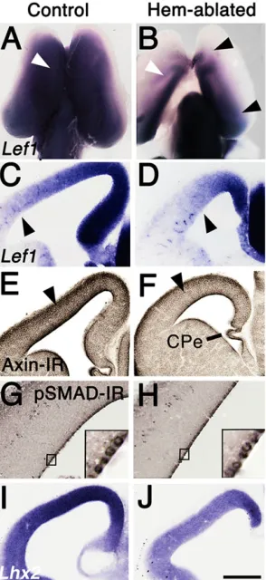
The transcription factor, lymphoid enhancer binding factor 1 (LEF1), is required for canonical WNT signaling, andLef1is also a WNT downstream target gene (Hovanes et al., 2001). In control brains at E12.5, strongLef1expression filled a large domain of the CP, showing a high dorsal to low ventral gradient (Fig. 1A,C). Notably the extent ofLef1expression was very similar to that of BAT-Gal labeling for canonical WNT signaling activity in mouse embryos of the same age (Backman et al., 2005). In the hem-ablated CP, Lef1 expression was weaker and confined more caudomedially (Fig. 1B,D). Removal of the hem thus caused a widespread drop in Lef1 expression in the rostral, lateral and ventral CP, the first indication of a broad influence of hem loss on the CP. Axin2 is another downstream gene target of canonical WNT signaling (Yan et al., 2001). Axin2 immunoreactivity (IR) was reduced in the hem-ablated CP at E12.5 (Fig. 1E,F).
Hem ablation had less obvious effects on indicators of BMP signaling, possibly because BMP ligands are secreted by choroid plexus epithelium (CPe), and are available to ventricular zone (VZ) progenitor cells from embryonic cerebrospinal fluid (Lehtinen and Walsh, 2011; Marques et al., 2011). Lack of WNT signaling from
the hem caused loss of the adjacent hippocampus, whereas lack of hem BMPs only reduced the adjacent CPe (Yoshida et al., 2006). Progenitors immunoreactive for phosphorylated SMAD1/5/8 ( pSMAD-IR) were equally dense in the VZ in mutants and controls (Fig. 1G,H) (Cheng et al., 2006). Expression of the gene encoding LIM homeobox protein 2,Lhx2, highly sensitive to BMP signaling (Monuki et al., 2001), however, was moderately downregulated in hem-ablated CP (Fig. 1I,J).
[image:2.612.366.513.295.615.2]Absence of the hippocampus and reduction of neocortex In hem-ablated mice at E18.5, the hippocampal formation was absent from the dentate gyrus to the subiculum (Fig. 2A-J), as expected from previous experimental disruption of canonical WNT signaling (Galceran et al., 2000; Lee et al., 2000b). Hippocampal fields were identified as previously by gene expression (Galceran et al., 2000; Lee et al., 2000b; Tole et al., 2000). The medial-to-lateral expansion of the neocortex was also noticeably reduced (Fig. 2K,L). Neurofilament immunoreactivity (Nfil-IR) showed a correlated reduction in the density of cortical afferent and efferent axons in the internal capsule (Fig. 2K,L).
Fig. 1. Reduced indicators of WNT and BMP signaling in hem-ablated cortex.(A-J) E12.5 mouse brains processed within situhybridization or immunohistochemistry. (A,B) Whole brains, dorsal view, rostral up, (C-J) coronal sections, medial to right. (A-D)Lef1expression in hem-ablated CP is weaker and more confined than in a control E12.5 CP. White arrowheads (A,B) indicate comparable positions in the two brains. Black arrowheads indicate low point of M/L expression gradient (B-D). (E,F) Axin-IR is lower in hem-ablated than control CP (note intensity at arrowheads). (G,H) A similar density of pSMAD-IR VZ cells in control and hem-ablated CP. Insets show higher magnification. (I,J)Lhx2expression is lower in hem-ablated than control CP. Scale bar: in J, 400 µm for A,B; 200 µm for C-F,I,J; 50 µm for G,H. CPe, choroid plexus epithelium.
DEVEL
O
Changes in regional patterning along both major axes of the CP
To evaluate changes in CP regional patterning following hem loss, we examined in coronal and sagittal brain sections gene expression patterns that demarcate CP domains and presumptive areas in perinatal mouse cortex (Assimacopoulos et al., 2012; Bishop et al., 2000; Chou et al., 2009; Fukuchi-Shimogori and Grove, 2001; Griveau et al., 2010; Miyashita-Lin et al., 1999; Rubenstein et al., 1999). At E18.5,Cdh6and Rorb were expressed in presumptive primary somatosensory cortex ( pS1) (Fig. 3A,C). In hem-ablated cortex, expression of both genes shifted dorsomedially (Fig. 3B,D),
consistent with reduction of the dorsomedial CP. The reduced region (Fig. 3C) appeared to include presumptive cingulate and retrosplenial areas, and possibly part of somatomotor cortex (Paxinos et al., 1991).
[image:3.612.95.253.199.617.2]We tested whether ventrolateral CP expands as dorsomedial CP contracts. At E18.5, the transcription factor genePou3f1(Scip) is expressed in the striatum, olfactory tubercle (OT) and neocortex, including the insular cortex (Icx) (Fig. 3E). A region of little or no Pou3f1 expression, occupied by piriform (Pir) cortex, intervenes between the OT and the ventral boundary of neocortex. This region expanded in the hem-ablated forebrain (Fig. 3E,F).Nrp2andLmo3 expression confirmed Pir dorsal expansion (Fig. 3G-J; supplementary material Fig. S2; G.C.-B., unpublished). We quantified Pir expansion in coronal sections processed to showNrp2expression. StrongNrp2
Fig. 3. Dorsomedial cortex is reduced and ventrolateral cortex expanded in hem-ablated mice.(A-J) Coronal sections, E18.5 cortex. (A-D) Strong expression ofCdh6andRorbshifts medially in hem-ablated mice (black arrowheads in A-D), demonstrating that a medial neocortical region (dotted lines, between white arrowheads in C) is reduced or lost. (E-F) A gap inPou3f1
[image:3.612.357.518.224.626.2]expression picks out the piriform area (black arrowheads) and is larger in the hem-ablated mouse. (G-J)Nrp2expression indicates dorsal expansion of presumptive piriform (white curved lines, G,H), and entorhinal areas (arrowheads, I,J). Insular cortex lies within thePou3f1expression domain. Scale bar: in J, 400 µm for A-F,I,J; 200 µm for G,H. Am, amygdala; Icx, insular cortex; ER, entorhinal; Pir, piriform.
Fig. 2. Loss of hippocampus and reduced neocortex in hem-ablated mice.
(A,B) Schematic coronal sections, control and hem-ablated E12.5 brains. Neocortical and hippocampal primordia, light and mid blue; hem is dark blue. Arrowheads point to the same anatomical landmarks in A,B as in K,L. Hem ablated-cortex lacks a hippocampus, and dorsal CP is shorter in B than in A. (C-L) Coronal sections through control (C,E,G,I,K) and hem-ablated (D,F,H,J,L) E18.5 brains processed within situhybridization (C-J) or immunohistochemistry to show Nfil-IR (K,L). (C-J) Hem-ablated brains lack gene expression indicating the dentate gyrus, CA3, CA1 and subiculum. (K,L) In hem-ablated brains, dorsal CP is shortened (arrowheads) and fewer axons cross between CP and thalamus (asterisks). Scale bar: in L, 400 µm for
C-L. DG, dentate gyrus; Sub, subiculum; Th, thalamus.
DEVEL
O
expression marks the outer layer of Pir (Fig. 3G,H; supplementary material Fig. S1). The curved outer surface of Pir was traced and measured in images of coronal sections (six coronal sections per E18.5 brain, evenly spaced along the R/C axis of one cortical hemisphere/ brain) (ImageJ, series 1.48, NIH, see Materials and Methods; supplementary material Fig. S1). The dorsoventral length of Pir was significantly greater in hem-ablated compared with control cortex (t-test, P=0.004, n=6 brains/group), extending 15-20% further dorsally than in control cortex. Entorhinal cortex (ER) is not easily discerned at E18.5, butNrp2expression was dorsally extended in caudal sections likely to contain presumptive ER (Fig. 3I,J).
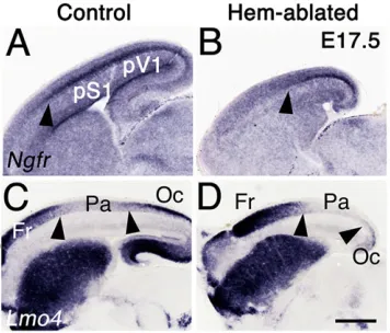
Along the R/C axis, the CP can be divided into frontal (Fr), parietal (Pa) and occipital (Oc) domains. Pa incorporates pS1 and Oc contains presumptive primary visual cortex ( pV1). In sagittal sections of E17.5 CP,Ngfrwas expressed in a distinctive band in pS1 and pV1 (Fig. 4A). This band was truncated in hem-ablated cortex (Fig. 4B, arrowhead). In controls, Lmo4 was expressed strongly in Fr and Oc, but weakly in the intervening Pa domain (Fig. 4C). In hem-ablated mice, the Pa domain shifted caudally leaving a tinyLmo4-expressing Oc domain, indicating that the most extreme reduction was in caudomedial CP containing pV1.
Estimates of the comparative size of hem-ablated and control neocortex, and the relative sizes of their Pa and Oc domains, were made using a standard method, taking measurements from images of whole brains or hemispheres in dorsal or lateral view, processed to show expression of appropriate regional markers (Armentano et al., 2007; Bishop et al., 2000; Hamasaki et al., 2004). Using images similar to those in Fig. 5, and ImageJ to measure areas, we estimated that hem-ablated neocortex was 63±6% of control size (n=18 brains/group,t-test, P<0.0001). The Pa domain was smaller than normal (60±6% of control Pa,n=6 brains/group), but consistent with the general reduction. The mean area of the Oc domain, however, was only 39±2% of control Oc (n=6 brains/domain/group) (Fig. 5A-D). Our estimation of surface area was not exact, given neocortex is three-dimensional. Nonetheless, our observations indicate a substantial reduction in the size of hem-ablated neocortex, a proportional reduction of parietal cortex, and a disproportional reduction of the Oc domain.
We measured the R/C extent of frontal neocortex (Fr) more precisely (see Materials and Methods), and found that, in contrast
to other domains, the Fr domain expanded in hem-ablated mice (Fig. 5A-D). In images of hemispheres in lateral view, the dorsal contour of the FrLmo4 expression domain was traced from the rostral pole of the telencephalon to the caudal boundary of Fr Lmo4expression, thereby accounting for differences in curvature of the cortex between mutants and controls. The R/C extent of the Fr domain in hem-ablated mice was significantly greater (t-test,P=0.005,n=6 brains per group), roughly 20% longer than in controls.
[image:4.612.355.519.201.607.2]Lateral views also highlighted the dorsal expansion of ventrolateral cortex in hem-ablated brains compared with controls (Fig. 5E,F). Both qualitative and quantitative findings therefore
Fig. 4. Caudal cortex is disproportionately reduced in hem-ablated mice.
[image:4.612.85.263.504.656.2](A-D) Sagittal sections, E17.5 cortex. (A,B) Dense band ofNgfrexpression marks pS1 and presumptive visual cortex ( pV1); this band is much shorter caudal to rostral in hem-ablated cortex (black arrowheads in A,B). (C) In control cortexLmo4expression is strong in Fr and Oc domains, virtually absent in the Pa domain containing pS1. (D) In hem-ablated cortex,Lmo4expression suggests a reduced Pa domain, and near obliteration of the Oc territory (arrowheads in C,D). Scale bar: in D, 400 µm for A-D.
Fig. 5. Hem-ablated hemispheres display shifts in cortical domain boundaries.(A-J) E18.5 forebrains, processed with whole-mountin situ
hybridization, dorsal (A,B,G,H) or lateral (C-F,I,J) view. Broken white lines (A,B) indicate the caudal boundary of neocortex, red arrowheads (C-F) mark its ventral boundary, and in E,F, neocortex is the dorsal region that does not expressNrp2. White or black arrowheads (E,F,I,J) mark the ventral edge of cortex. (A-D) FrLmo4expression expands in hem-ablated neocortex, but Oc
Lmo4expression is severely diminished. (E,F)Nrp2expression indicates expansion of ventrolateral cortex (Pir and ER). (G-H)Cdh6expression shifts caudally and towards the midline (asterisks) in hem-ablated cortex. (I,J) In lateral viewCdh6expression shifts dorsally. (K,L) Summary of regional size changes with hem ablation, neocortex is blue, ventrolateral cortex is yellow. Scale bar: in J, 500 µm for A-J.
DEVEL
O
indicated that in the absence of the hem less of the total CP was allocated to neocortex and more to ventrolateral cortex composed of the Pir and ER areas (Fig. 5C-F,I,J).
Cdh6expression provided an overview of the differences between hem-ablated and control neocortex. In control hemispheres at E18.5, Cdh6 was strongly expressed in mid-lateral neocortex, with the frontal domain and dorsomedial neocortex free of expression (Bishop et al., 2000; Takeichi et al., 1997) (Fig. 5G,I). In hem-ablated neocortex,Cdh6expression shifted caudally and medially, leaving very little expression-free caudomedial cortex but enlarging Cdh6-free frontal and lateral domains (Fig. 5I,J).
Progenitor cell proliferation altered in medial but not lateral CP
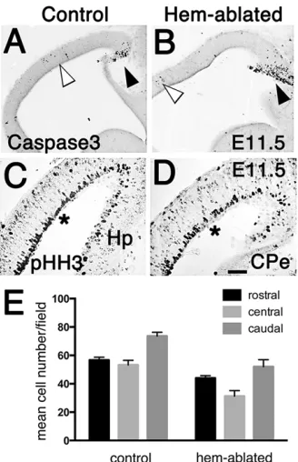
Changes in cell proliferation or cell death could alter cortical domain size. From E11.5 to E17.5, cell death was generally low in the CP, and indistinguishable between control and hem-ablated CP (Fig. 6A,B). Loss of mitogenic WNT signaling from the hem (Lee et al., 2000b) suggests cell proliferation would be reduced in the hem-ablated CP. We used phospho-histone-H3 immunoreactivity ( pHH3-IR) to identify mitotic cells in the CP from E11.5 to E17.5. Mitotic pHH3-IR cells were counted in rectangular fields of consistent size, positioned on section images, centered over the cortical VZ. At E11.5 significantly fewer pHH3-IR VZ progenitor cells appeared in the dorsomedial CP in hem-ablated mice compared with controls (t-test, P=0.002,n=6 mice/group) (Fig. 6C,D). The mean number of cells per counting field was 47 (±3) in hem-ablated mice and 71 (±1) in controls, a reduction of about 30%. No significant difference in
mitotic cell count was detected in the lateral CP between the two groups. By E13.5, pHH3-IR cells were no longer significantly reduced in hem-ablated dorsomedial CP. A similarly transient effect of a WNT gradient, emanating from the roofplate, is seen in the embryonic spinal cord, in which WNT signaling directs a D/V gradient of cell proliferation (Megason and McMahon, 2002). WNT-dependent cell proliferation decreases over time, probably because broad dispersion of the mitogen is reduced by tissue expansion (Megason and McMahon, 2002). Our findings suggest that WNT signaling no longer significantly regulates CP cell proliferation after about E12.5.
No changes in cell proliferation along the R/C axis
We next counted pHH3-IR dividing cells in consistently sized fields within sagittal sections of the E11.5 CP separated into caudal, central and rostral thirds. Two-way ANOVA tested for significant differences among mean cell counts, with genotype and R/C level as the two factors. Significant sources of variation (P=0.001) were genotype and R/C position, but no significant variation was noted in the interaction of the two (Fig. 5E). In summary, our observations of cell proliferation provided no evidence that changes in cell proliferation account for patterning shifts in hem-ablated cortex, other than the reduction in dorsomedial neocortex. Our findings pointed instead to alterations in the mechanisms that determine regional identity.
Gene manipulations that cause a cortical phenotype similar to that of hem-ablated mice
Several gene manipulations produce mice that share specific cortical patterning defects with hem-ablated animals. Rostral neocortex expands and caudal regions are reduced in mice in which the rostral FGF8 source is experimentally augmented (Fukuchi-Shimogori and Grove, 2001), and in mice deficient in Emx2, an ortholog of the Drosophila gene empty spiracles (Bishop et al., 2000; Hamasaki et al., 2004; Mallamaci et al., 2000). In mice lacking thedouble sex and mab-3-related transcription factor(Dmrt) gene,Dmrta2, lateral and rostral neocortex expand, and caudomedial cortex is diminished (Konno et al., 2012; Saulnier et al., 2012). Mice lacking the orphan nuclear hormone receptor geneNr2f1show a related but not identical phenotype in which rostral neocortex is enlarged and caudal sensory areas are almost obliterated (Armentano et al., 2007). Finally, R/C patterning shifts opposite to those seen in hem-ablated cortex arise in mice deficient inFgf8(Garel et al., 2003), or inSp8, which encodes a member of the SP1 transcription factor family, mediating or enhancing FGF8 signaling (Borello et al., 2013; Sahara et al., 2007; Zembrzycki et al., 2007).
FGF8 signaling at the RTO is not changed
[image:5.612.93.257.381.635.2]TheFgf8expression domain was similar in size in hem-ablated and control embryos at E11 to E13.5 (n=10 brains/group) (Fig. 7A,B; supplementary material Fig. S2A,B). Further, rostral expression of Spry2, a direct readout of FGF8 signaling, was not distinguishable between mutants and controls (supplementary material Fig. S2C,D, n=4 brains/group). In wild-type mice, FGF8 positions the rostral expression boundary of the patterning gene, Nr2f1, and excess FGF8 drives the boundary further caudal. The positions ofNr2f1 expression boundaries in hem-ablated and control CP were highly similar (supplementary material Fig. S3,n=7/group). Finally, FGF8 upregulates Sp8 expression (Borello et al., 2013; Cholfin and Rubenstein, 2008; Sahara et al., 2007), but in hem-ablated mice, rostral telencephalicSp8expression was lower than in control mice, rather than increased (supplementary material Fig. S2E,F, n=5/ Fig. 6. Altered cell proliferation contributes to smaller dorsomedial cortex
but not to other regional size changes.(A-D) Sagittal sections, E11.5 brains. (A,B) Sparse caspase-3-IR apoptotic cells in control and hem-ablated CP (white arrowheads), dense cells only in developing CPe (black arrowheads). (C,D) Less dense pHH3-IR apical progenitor cells in the dorsomedial CP of a hem-ablated brain compared with a control (see regions at asterisks). (E) pHH3-IR cell counts in caudal, central, rostral thirds of E11.5 CP. Two-way ANOVA indicated that mean cell counts, obtained from the six control and six hem-ablated brains, vary significantly by genotype and by R/C level, but not by
the interaction of the two factors. Scale bar: in D, 100 µm for A,B; 40 µm for C,D.
DEVEL
O
group). This reduction partially reflects loss of medial tissue that normally expresses Sp8, but the severity of reduction suggests a specific decrease following hem loss (supplementary material Fig. S2G,H; see Fig. 9). None of these findings supports increased RTO FGF8 signaling in the hem-ablated mouse.
Patterning interactions amongDmrtgenes,Emx2and WNTs In hem-ablated CP, the high caudomedial to low rostrolateral expression gradient of Emx2 remained, perhaps previously established by the telencephalic roofplate (Cheng et al., 2006). The level ofEmx2expression, however, was much weaker throughout the CP, and not detected in a substantial rostral region (Fig. 7C,D). The far-reaching effect of hem ablation onEmx2expression levels in the CP paralleled a similarly extensive reduction of Lef1 expression (Fig. 1A,B). An interaction between EMX2 and WNT signaling has been associated with deficits in the hippocampus and caudomedial neocortex ofEmx2mutants (Muzio et al., 2005; Theil et al., 2002; Tole et al., 2000). Present observations demonstrate a widespread effect of hem WNT signaling onEmx2expression throughout the neocortical primordium. Because the positioning and size of neocortical areas are sensitive to EMX2 levels (Hamasaki et al., 2004), decreasedEmx2expression in the hem-ablated mouse should reduce caudal and medial regions and allow expansion of rostral and lateral cortex (Bishop et al., 2000; Mallamaci et al., 2000) just as is observed.
Expression of the homeobox genePax6is negatively regulated by EMX2 (Muzio et al., 2002), and as expected, Pax6 expression
increased in hem-ablated CP. Cortical patterning shows little change, however, in mice with increased Pax6expression (Manuel et al., 2007), suggesting that the increase in hem-ablated mice does not contribute to their cortical phenotype.
Recent studies demonstrate that canonical WNT signaling increases expression of Dmrt3 and Dmrta2, associated with cortical patterning (Hasenpusch-Theil et al., 2012; Kikkawa et al., 2013; Konno et al., 2012; Saulnier et al., 2012). Dmrta2,Dmrt3and Emx2maintain regulatory interactions, with Dmrta2an upstream regulator ofEmx2expression (Saulnier et al., 2012).We found that CP expression of bothDmrta2andDmrt3was substantially reduced in hem-ablated mice (Fig. 7E-H). Previous and present findings therefore sketch out a cortical patterning pathway, composed of interactions among DMRTA transcription factors, EMX2, and hem WNT signaling, in which hem-ablated mice have obvious deficiencies. We propose that cortical patterning anomalies in hem-ablated mice are generated at least in part by decreased expression ofDmrt3,Dmrta2andEmx2.
Interactions between the hem and the RTO
That excess FGF8 or hem ablation cause similar cortical pattern changes suggests the hem restrains the patterning activity of FGF8, and that this restriction is required for normal cortical development. FGF8 downregulates the expression ofEmx2,Dmrta2andDmrt3, whereas canonical WNT signaling clearly upregulates expression of Emx2andDmrt3(Fig. 8A-F).
We asked if these observations indicated a more comprehensive interaction between the hem and RTO in controlling gene transcription in the CP. Using in utero microelectroporation (IUME), electroporating the CP at E10.5 and harvesting brains at E12.5, we investigated cross-regulation by FGF8 and WNT3a of several additional genes that are either known or predicted to be involved in cortical regional development.
The cortical selector gene Lhx2 (Mangale et al., 2008) has additional functions in cortical development, including regulating precise thalamic innervation of sensory neocortex (Chou and Fig. 7. Expression of genes implicated in cortical patterning.(A-D) Whole
E11.5 brains, frontal view (A,B), whole E12.5 brains, dorsal view (C,D). (E-H) Coronal sections E12.5 brains. (A,B)Fgf8expression at the RTO is not expanded in a hem-ablated brain compared with a control. (C,D)Emx2
expression gradients, caudal to rostral, and medial to lateral. In hem-ablated hemispheres,Emx2expression is lower overall. A rostral zone with little or no
Emx2expression is larger than in controls (broken lines, brains on right, C,D). (E-H)Dmrt3andDmrta2expression is reduced in hem-ablated brains compared with controls (arrowheads in E-H). Scale bar: in H, 400 µm for A,B; 1.5 mm for C,D; 300 µm for E-H. c, caudal; di, diencephalon; l, lateral; m, medial; r, rostral.
Fig. 8. Opposite control of known patterning gene expression by WNT3a and FGF8.(A-F) Coronal sections through wild-type E12.5 CD-1 mouse brains, electroporated at E10.5 with aWnt3a(A-D) orFgf8(E,F) expression construct and processed within situhybridization. Ectopic WNT3a upregulates expression ofEmx2(A,B) andDmrt3(C,D). Ectopic FGF8 downregulates
Dmrta2expression (E,F). Scale bar: in F, 300 µm for A-F.
DEVEL
O
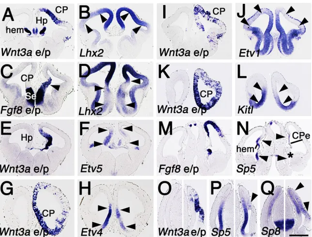
[image:6.612.354.520.473.674.2]O’Leary, 2013; Chou et al., 2009; Marcos-Mondejar et al., 2012; Roy et al., 2013; Shetty et al., 2013; Subramanian et al., 2011).Lhx2, likeEmx2,Dmrt3andDmrta2, is expressed in a caudomedial high to rostrolateral low gradient. Indicating antagonistic WNT and FGF8 regulation of this gradient, WNT3a increased Lhx2 expression (Fig. 9A,B), whereas augmenting the RTO FGF8 source extended lowLhx2expression in the rostral telencephalon (Fig. 9C,D).
Functions for the PEA3 family of ETS genes in cortical patterning have not been investigated, but seem likely given the developmental roles of PEA3 genes elsewhere in the embryo (Arber et al., 2000; Fontanet et al., 2013; Mao et al., 2009). FGF8 positively regulates all three PEA3 genes,Etv1,Etv4andEtv5, which show a nested expression along the medial telencephalon that follows the R/C FGF8 gradient (Fukuchi-Shimogori and Grove, 2003; Toyoda et al., 2010). Electroporation ofWnt3adecreased expression of all three genes (Fig. 9E-J).
The developmental function in the brain of KITL/KIT signaling, crucial for the generation of germ cells and gonads (Merkwitz et al., 2011), is unknown. KITL/KIT signaling, however, is likely to be involved in the function of FGF8 at the RTO.Kitlis expressed at the rostral telencephalic midline, prominently including the RTO (Fig. 9C), but not in the dorsal midline.Kitl expression is upregulated by ectopic FGF8 sources (Assimacopoulos et al., 2012), and, consistent with its exclusion from the hem, Kitl expression is decreased byWnt3aelectroporation (Fig. 9K,L).
WNT3a induces expression ofSp5, an SP1 family member that is expressed in the medial wall of the cortical hemisphere, and potentially contributes to medial patterning (Dunty et al., 2014; Fujimura et al., 2007; Thorpe et al., 2005) (Fig. 9N-P). Ectopic FGF8 reduces the size of the hippocampus (Shimogori et al., 2004), thereby reducing the domain of potentialSp5expression. We found, however, that after Fgf8 electroporation Sp5 expression was virtually absent in the substantial remaining medial telencephalon, indicating a true reduction of gene expression (Fig. 9N).
Expression ofSp8is upregulated by FGF8 (Borello et al., 2013; Cholfin and Rubenstein, 2008; Sahara et al., 2007) and, to our surprise, by WNT3a as well (Fig. 9O,Q), an observation supported by recent transcriptional profiling of the Wnt3a mutant mouse
(Dunty et al., 2014). Positive regulation by WNT3a is consistent with the expression of Sp8 in the medial wall of the cortical hemisphere (Fig. 9Q), and could explain reducedSp8expression in the hem-ablated mouse.
In summary, we identified several gene regulatory interactions between FGF8 and WNT3a, most of them antagonistic, in which FGF8 either up- or downregulates expression of a given gene and WNT3a does the opposite. In each case, these interactions are consistent with shaping domains of gene expression that arise during normal cortical development and contribute to patterning the cortical hemisphere. As an illustration, Etv4and Etv1expression appears at the rostromedial telencephalic midline, ending at the R/C level of the hippocampus and hem.Etv5expression continues into the most rostral hippocampus, but not into the hem. Our findings suggest that the overall pattern of PEA3 gene expression at the rostral but not caudal midline is the result of coordinated regulation by FGF8 and WNT signaling.
DISCUSSION
Findings from the present study prompt three main conclusions. First, the cortical hem regulates broad patterning of the cerebral cortex along the D/V axis, promoting dorsal cortical identity and suppressing ventral identity, reminiscent of the actions of the roofplate in the caudal neural tube. Similar to signals from the roofplate, hem signals direct both cell proliferation and the adoption of specific regional identities (Chizhikov et al., 2010; Galceran et al., 2000; Lee et al., 2000b; Liem et al., 1997; Mangale et al., 2008; Megason and McMahon, 2002). Dorsomedial neocortex is reduced in the hem-ablated mouse, because of transiently decreased cell proliferation probably caused by loss of WNT signaling from the hem. The expansion of ventrolateral cortex, however, is not explained by changes in cell proliferation, indicating that the hem directs subdivision of the cerebral cortex along the D/V axis.
[image:7.612.50.372.56.301.2]Decreased expression ofEmx2andDmrta2may account in part for expansion of ventrolateral cortex in hem-ablated mice; both Emx2 and Dmrta2mutants show enlarged lateral cortex (Bishop et al., 2000; Mallamaci et al., 2000; Saulnier et al., 2012). Diminished Lhx2 expression could also contribute to the
Fig. 9. WNT3a and FGF8 regulation of candidate patterning gene expression.(A-Q) Coronal sections through wild-type E12.5 CD-1 mouse brains, electroporated in one hemisphere at E10.5 withWnt3a
(Wnt3ae/p) orFgf8(Fgf8e/p) and processed within situ
hybridization. (A-D)Wnt3ae/p (A) increasesLhx2
expression (B, compare expression between arrowheads in each hemisphere).Fgf8e/p (C, arrowhead) expands a region of lowLhx2
expression (D, see arrowhead pairs). (E-L)Wnt3ae/p (E,G,I,K) reduces CP expression ofEtv5,4and1as well asKitl(F,H,J,L, compare expression between arrowheads in each hemisphere). (M-Q)Fgf8and
Wnt3ae/p have opposite effects on expression of Sp5 (M-P), butWnt3ae/p mimics FGF8 in upregulatingSp8
expression (Q). (O-Q) Control and e/p sides of the CP are shown in separate panels. Scale bar: in Q, 300 µm for A-F,I,J,M-Q; 200 µm for G,H,K,L.
DEVEL
O
phenotype, given that LHX2 is required for boundary formation between paleocortex and neocortex (Chou et al., 2009), but the drop in Lhx2 expression in hem-ablated cortex is modest (Fig. 1I,J); moreover, entorhinal cortex, not shown to be LHX2-dependent, is part of the enlarged ventrolateral domain.
A different possibility is that the anti-hem (Assimacopoulos et al., 2003; Kim et al., 2001) opposes the actions of the hem along the D/V axis. Supporting a patterning interaction between hem and antihem, ventrolateral cortex expands in other mice with a reduced or absent hem. In the Gli3 mutant extra-toes, patches of gene expression characteristic of piriform cortex appear throughout the D/V extent ofextra-toescortex (Vyas et al., 2003), as do patches ofSfrp2expression typical of the anti-hem (E.A.G., unpublished). Whatever the underlying cause, the shift in D/V organization of the cortical hemisphere in hem-ablated mice suggests that the hem normally controls the amount of the CP allocated to neocortex, or to a ventrolateral cortical domain that is not neocortical in character.
The second conclusion is that the hem influences R/C patterning of the neocortical primordium. Hem ablation causes rostral neocortical domains to expand at the expense of caudal domains, which are shrunken and shifted caudally. No anomalies in cell proliferation were found that could explain the R/C patterning shifts. Instead, diminished expression ofEmx2andDmrtagenes is likely to account substantially for regional shifts along the R/C axis in hem-ablated cortex. Considerable evidence supports Emx2 as crucial to neocortical patterning (Bishop et al., 2000; Hamasaki et al., 2004; Mallamaci et al., 2000), whereas theDmrtagenes are relative newcomers to this role. Studies of theEmx2mutant mouse, however, have suggested that more genes would be found with similar patterning effects toEmx2, and that their expression would respond to FGF or WNT signaling or both (Cholfin and Rubenstein, 2008; Fukuchi-Shimogori and Grove, 2003; Muzio et al., 2005). In theEmx2mutant, rostralFgf8andFgf17 expression domains expand (Cholfin and Rubenstein, 2008; Fukuchi-Shimogori and Grove, 2003), and hemWntgene expression decreases (Fukuchi-Shimogori and Grove, 2003; Muzio et al., 2005; Tole et al., 2000). Area boundary shifts in theEmx2mutant are partially reversed by reducing FGF8 or FGF17 (Cholfin and Rubenstein, 2008; Fukuchi-Shimogori and Grove, 2003), and increasing WNT signaling relieves the prominent reduction of caudomedial cortex in theEmx2 mutant (Muzio et al., 2005). These observations imply that deregulation of additional genes downstream of FGF or WNT signaling contributes to the cortical phenotype seen in the Emx2 mutant.Dmrt3andDmrta2are likely to be members of this group of genes (Konno et al., 2012; Saulnier et al., 2012), and others may be identified.
NR2F1 maintains a mutual negative regulatory relationship with FGF8, and inhibits canonical WNT signaling (Faedo et al., 2008). Cortical expression ofNr2f1was unchanged, however, by loss of WNT and BMP function in the hem-ablated mouse, or electroporation of Wnt3a into wild-type CP (supplementary material Fig. S3; S.A., unpublished).Dmrta2-deficient mice also display no change in expression of Nr2f1 (Saulnier et al., 2012). These observations indicate two partially distinct cortical patterning pathways, one implicating WNT signaling, the Dmrta genes and Emx2, but not Nr2f1, and the other centered on an antagonism between SP8 and NR2F1 (Armentano et al., 2007; Zhou et al., 2001). The third conclusion from the present study is that the hem maintains antagonistic interactions with FGF8 in regulating R/C patterning of the neocortex. Ablating the hem or increasing FGF8 levels has the same effect on early regional patterning of the neocortex. In normal development, therefore, the hem acts as a negative modulator of the rostralizing activity of FGF8; when the
hem is ablated, the brake is removed and the neocortex is rostralized. Further, opposed regulation by FGF8 and WNT3a ofSp5, which may be involved in medial cortical development, hints at a two-way antagonism. Notably, our observations do not support a simple model in which the two signaling centers independently control orthogonal axes of the cerebral cortex. Although the hem and RTO pattern the cortex along the D/V and R/C axes, respectively, the signaling sources are not independent. Interactions between the two support a more complex network of cortical regionalization signals. Finally, our findings may help to resolve a long-standing question concerning cortical regional patterning. In the spinal cord, and even in the early telencephalon, D/V patterning is controlled by two signaling centers, one producing sonic hedgehog, the other WNTs and BMPs, which both actively pattern the ventral and dorsal neural tube respectively, and make D/V patterning a more robust developmental process by antagonizing one another (Ulloa and Briscoe, 2007). To date the ‘caudal’ antagonist of the RTO has seemed to be missing. The observations reported here indicate that the antagonistic interactions that make for robust patterning of the cerebral cortex need not be between two signaling centers placed opposite one another.
MATERIALS AND METHODS Mice
Animals use followed NIH guidelines; the University of Chicago IACUC approved all mouse protocols. Hem-ablated mice were generated as described (Yoshida et al., 2006). Midday of the day of vaginal plug discovery was termed E0.5.
Immunohistochemistry
Brains were collected from embryos aged E10.5 to E18.5, fixed in 4% paraformaldehyde and sectioned with a Leica SM2000R microtome. Sections were incubated with primary antibody followed by appropriate HRP-conjugated secondary antibodies. Primary antibodies were anti-Axin2 rabbit polyclonal (Abcam), anti-phospho-Smad1/5/8 rabbit polyclonal (Cell Signaling Technology), anti-phospho Histone H3 ( pHH3) (Ser10) rabbit polyclonal (Millipore), 3A10 anti-neurofilament mouse monoclonal (Developmental Studies Hybridoma Bank), and anti-cleaved caspase 3 rabbit polyclonal antibody (Cell Signaling Technology).
In situhybridization
Whole brains or sections were processed with digoxigenin (Dig)-labeled riboprobes. cDNAs were gifts of M. Takeichi (RIKEN CDB, Kobe, Japan) (Cdh6,Cdh8), S. K. McConnell (Stanford University, CA, USA) (Rorb), and L. F. Reichardt (University of California, San Francisco, USA) (Ngfr). Other cDNAs were obtained by PCR from mouse embryo cDNA. Tissue was viewed with a Zeiss Axioscope or a Leica dissection microscope, and images captured with Axiovision cameras and software (Zeiss). For figures, digital images were adjusted for contrast, color and brightness using Adobe Photoshop CS4. Comparisons of the expression of a given gene between control and mutant mice were based on at least six brains/group/age. Genes with expression patterns used to define cortical domains at E18.5 wereCdh6 andCdh8, encoding cadherins 6 and 8,Lmo3andLmo4, encoding Lim-only domain transcription factors, Nrp2, encoding neuropilin2, a semaphorin receptor,Rorb, encoding an orphan nuclear receptor, and the nerve growth factor receptor gene,Ngfr.
Quantification
Estimates of the relative sizes of E18.5 hem-ablated and control neocortices, and their Pa and Oc domains, were obtained by taking measurements from images of whole brains, hemispheres or sections, processed to show appropriate region-specific gene expression. Consistency in the angle of view in each image was obtained by laying hemispheres from bisected brains, flat side down, on an agarose gel surface. Whole brains were placed into a depression cut into the agarose. In a given image, the total area of neocortex,
DEVEL
O
and the areas of Pa or Oc, were outlined and measured using ImageJ software (series 1.48, NIH, public domain). The expansion of frontal neocortical (Fr) in hem-ablated mice, in particular, was determined as described in the text. To confirm dorsal expansion of piriform cortex in hem-ablated mice, the D/V length of piriform cortex was determined in coronal sections from six control and six hem-ablated E18.5 forebrains (six coronal sections per cerebral cortex, evenly spaced along the R/C axis). Sections were processed within situhybridization (ISH) forNrp2expression, which demarcates piriform cortex. The outer contour of piriform cortex was drawn on section images, matched for R/C level, and measured in ImageJ. To assess differences in cell proliferation along the R/C and medial to lateral (M/L, equivalent to D/V) axes of the neocortical primordium, E11.5-E17.5 brains were sectioned sagittally or coronally and processed with immunohistochemistry to show pHH3 immunoreactivity. Mitotic pHH3-IR cells were counted in rectangular fields of consistent size, positioned on section images, centered over the cortical VZ. Statistical comparisons of measurements of cell counts in mutant and control mice were made with at-test ( paired or unpaired as appropriate), for example, when comparing cell proliferation in hem-ablated and control dorsomedial CP, or with two-way ANOVA (Prism 6, GraphPad Software), when determining whether cell proliferation varies differentially along the R/C axis between hem-ablated and control CP.
In uteromicroelectroporation
cDNAs encoding FGF8, WNT3a (Fukuchi-Shimogori and Grove, 2001), and tdTomato (Genove et al., 2005) were cloned into the pEFX expression vector (Agarwala et al., 2001). Electroporation and selection of brains with appropriate electroporation sites were as described (Assimacopoulos et al., 2012). The CP was electroporated at E10.5, and brains were collected at E12.5 and sectioned. One series of sections was processed with ISH for the electroporated gene. Others were processed to show up- or downregulation of particular genes of interest. At least six brains were processed to show expression of each gene in each experimental condition. Control electroporation constructs carried td-Tomato and eGFP. Control electroporation was repeated four to six times for each gene.
Acknowledgements
We thank members of the Grove laboratory for their advice and help with this work.
Competing interests
The authors declare no competing financial interests.
Author contributions
G.C.-B., M.Y. and E.A.G. developed the ideas and approach; G.C.-B., M.Y., F.G. and S.A. performed the experiments; G.C.-B. and E.A.G. analyzed the data and wrote the paper.
Funding
This work was supported by National Institutes of Health grants [MH059962 and MH103211] to E.A.G. Deposited in PMC for release after 12 months.
Supplementary material
Supplementary material available online at
http://dev.biologists.org/lookup/suppl/doi:10.1242/dev.106914/-/DC1
References
Agarwala, S., Sanders, T. A. and Ragsdale, C. W.(2001). Sonic hedgehog control of size and shape in midbrain pattern formation.Science291, 2147-2150.
Arber, S., Ladle, D. R., Lin, J. H., Frank, E. and Jessell, T. M.(2000). ETS gene Er81 controls the formation of functional connections between group Ia sensory afferents and motor neurons.Cell101, 485-498.
Armentano, M., Chou, S.-J., Tomassy, G. S., Leingärtner, A., O’Leary, D. D. M. and Studer, M.(2007). COUP-TFI regulates the balance of cortical patterning between frontal/motor and sensory areas.Nat. Neurosci.10, 1277-1286.
Assimacopoulos, S., Grove, E. A. and Ragsdale, C. W.(2003). Identification of a Pax6-dependent epidermal growth factor family signaling source at the lateral edge of the embryonic cerebral cortex.J. Neurosci.23, 6399-6403.
Assimacopoulos, S., Kao, T., Issa, N. P. and Grove, E. A.(2012). Fibroblast growth factor 8 organizes the neocortical area map and regulates sensory map topography.J. Neurosci.32, 7191-7201.
Bachler, M. and Neubüser, A.(2001). Expression of members of the Fgf family and their receptors during midfacial development.Mech. Dev.100, 313-316.
Backman, M., Machon, O., Mygland, L., van den Bout, C. J., Zhong, W., Taketo, M. M. and Krauss, S.(2005). Effects of canonical Wnt signaling on dorso-ventral specification of the mouse telencephalon. Dev. Biol. 279, 155-168.
Bishop, K. M., Goudreau, G. and O’Leary, D. D. M.(2000). Regulation of area identity in the mammalian neocortex by Emx2 and Pax6. Science 288, 344-349.
Borello, U., Cobos, I., Long, J. E., Murre, C. and Rubenstein, J. L. R.(2008). FGF15 promotes neurogenesis and opposes FGF8 function during neocortical development.Neural Develop.3, 17.
Borello, U., Madhavan, M., Vilinsky, I., Faedo, A., Pierani, A., Rubenstein, J. and Campbell, K.(2013). Sp8 and COUP-TF1 reciprocally regulate patterning and Fgf signaling in cortical progenitors.Cereb. Cortex24, 1409-1421.
Cheng, X., Hsu, C.-m., Currle, D. S., Hu, J. S., Barkovich, A. J. and Monuki, E. S.
(2006). Central roles of the roof plate in telencephalic development and holoprosencephaly.J. Neurosci.26, 7640-7649.
Chizhikov, V. V. and Millen, K. J.(2005). Roof plate-dependent patterning of the vertebrate dorsal central nervous system.Dev. Biol.277, 287-295.
Chizhikov, V. V., Lindgren, A. G., Mishima, Y., Roberts, R. W., Aldinger, K. A., Miesegaes, G. R., Currle, D. S., Monuki, E. S. and Millen, K. J.(2010). Lmx1a regulates fates and location of cells originating from the cerebellar rhombic lip and telencephalic cortical hem.Proc. Natl. Acad. Sci. U.S.A.107, 10725-10730.
Cholfin, J. A. and Rubenstein, J. L. R. (2007). Patterning of frontal cortex subdivisions by Fgf17.Proc. Natl. Acad. Sci. U.S.A.104, 7652-7657.
Cholfin, J. A. and Rubenstein, J. L. R.(2008). Frontal cortex subdivision patterning is coordinately regulated by Fgf8, Fgf17, and Emx2.J. Comp. Neurol. 509, 144-155.
Chou, S.-J. and O’Leary, D. D. M.(2013). Role for Lhx2 in corticogenesis through regulation of progenitor differentiation.Mol. Cell. Neurosci.56, 1-9.
Chou, S.-J., Perez-Garcia, C. G., Kroll, T. T. and O’Leary, D. D. M.(2009). Lhx2 specifies regional fate in Emx1 lineage of telencephalic progenitors generating cerebral cortex.Nat. Neurosci.12, 1381-1389.
Crossley, P. H., Martinez, S., Ohkubo, Y. and Rubenstein, J. L. R.(2001). Coordinate expression of Fgf8, Otx2, Bmp4, and Shh in the rostral prosencephalon during development of the telencephalic and optic vesicles.
Neuroscience108, 183-206.
Dorsky, R. I., Raible, D. W. and Moon, R. T.(2000). Direct regulation of nacre, a zebrafish MITF homolog required for pigment cell formation, by the Wnt pathway.
Genes Dev.14, 158-162.
Dunty, W. C., Jr, Kennedy, M. W. L., Chalamalasetty, R. B., Campbell, K. and Yamaguchi, T. P.(2014). Transcriptional profiling of Wnt3a mutants identifies Sp transcription factors as essential effectors of the Wnt/beta-catenin pathway in neuromesodermal stem cells.PLoS ONE9, e87018.
Faedo, A., Tomassy, G. S., Ruan, Y., Teichmann, H., Krauss, S., Pleasure, S. J., Tsai, S. Y., Tsai, M.-J., Studer, M. and Rubenstein, J. L. R.(2008). COUP-TFI coordinates cortical patterning, neurogenesis, and laminar fate and modulates MAPK/ERK, AKT, and beta-catenin signaling.Cereb. Cortex18, 2117-2131.
Fontanet, P., Irala, D., Alsina, F. C., Paratcha, G. and Ledda, F.(2013) Pea3 transcription factor family members Etv4 and Etv5 mediate retrograde signaling and axonal growth of DRG sensory neurons in response to NGF.J. Neurosci.33, 15940-15951.
Fujimura, N., Vacik, T., Machon, O., Vlcek, C., Scalabrin, S., Speth, M., Diep, D., Krauss, S. and Kozmik, Z.(2007). Wnt-mediated down-regulation of Sp1 target genes by a transcriptional repressor Sp5.J. Biol. Chem.282, 1225-1237.
Fukuchi-Shimogori, T. and Grove, E. A.(2001). Neocortex patterning by the secreted signaling molecule FGF8.Science294, 1071-1074.
Fukuchi-Shimogori, T. and Grove, E. A.(2003). Emx2 patterns the neocortex by regulating FGF positional signaling.Nat. Neurosci.6, 825-831.
Furuta, Y., Piston, D. W. and Hogan, B. L.(1997). Bone morphogenetic proteins (BMPs) as regulators of dorsal forebrain development. Development 124, 2203-2212.
Galceran, J., Farinas, I., Depew, M. J., Clevers, H. and Grosschedl, R.(1999). Wnt3a-/–like phenotype and limb deficiency in Lef1(-/-)Tcf1(-/-) mice.Genes Dev. 13, 709-717.
Galceran, J., Miyashita-Lin, E. M., Devaney, E., Rubenstein, J. L. and Grosschedl, R.(2000). Hippocampus development and generation of dentate gyrus granule cells is regulated by LEF1.Development127, 469-482.
Garel, S., Huffman, K. J. and Rubenstein, J. L. R. (2003). Molecular regionalization of the neocortex is disrupted in Fgf8 hypomorphic mutants.
Development130, 1903-1914.
Genove, G., Glick, B. S. and Barth, A. L.(2005). Brighter reporter genes from multimerized fluorescent proteins.Biotechniques39, 814.816, 818 passim.
Gorski, J. A., Talley, T., Qiu, M., Puelles, L., Rubenstein, J. L. and Jones, K. R.
(2002). Cortical excitatory neurons and glia, but not GABAergic neurons, are produced in the Emx1-expressing lineage.J. Neurosci.22, 6309-6314.
Griveau, A., Borello, U., Causeret, F., Tissir, F., Boggetto, N., Karaz, S. and Pierani, A. (2010). A novel role for Dbx1-derived Cajal-Retzius cells in early regionalization of the cerebral cortical neuroepithelium.PLoS Biol. 8,
e1000440.
DEVEL
O
Grove, E. A., Tole, S., Limon, J., Yip, L. and Ragsdale, C. W.(1998). The hem of the embryonic cerebral cortex is defined by the expression of multiple Wnt genes and is compromised in Gli3-deficient mice. Development 125, 2315-2325.
Hamasaki, T., Leingärtner, A., Ringstedt, T. and O’Leary, D. D. M.(2004). EMX2 regulates sizes and positioning of the primary sensory and motor areas in neocortex by direct specification of cortical progenitors. Neuron 43, 359-372.
Hasenpusch-Theil, K., Magnani, D., Amaniti, E.-M., Han, L., Armstrong, D. and Theil, T.(2012). Transcriptional analysis of Gli3 mutants identifies Wnt target genes in the developing hippocampus.Cereb. Cortex22, 2878-2893.
Hovanes, K., Li, T. W. H., Munguia, J. E., Truong, T., Milovanovic, T., Lawrence Marsh, J., Holcombe, R. F. and Waterman, M. L.(2001). Beta-catenin-sensitive isoforms of lymphoid enhancer factor-1 are selectively expressed in colon cancer.
Nat. Genet.28, 53-57.
Kawano, Y. and Kypta, R.(2003). Secreted antagonists of the Wnt signalling pathway.J. Cell Sci.116, 2627-2634.
Kikkawa, T., Obayashi, T., Takahashi, M., Fukuzaki-Dohi, U., Numayama-Tsuruta, K. and Osumi, N. (2013). Dmrta1 regulates proneural gene expression downstream of Pax6 in the mammalian telencephalon. Genes Cells18, 636-649.
Kim, A. S., Anderson, S. A., Rubenstein, J. L., Lowenstein, D. H. and Pleasure, S. J.(2001). Pax-6 regulates expression of SFRP-2 and Wnt-7b in the developing CNS.J. Neurosci.21, RC132.
Konno, D., Iwashita, M., Satoh, Y., Momiyama, A., Abe, T., Kiyonari, H. and Matsuzaki, F.(2012). The mammalian DM domain transcription factor Dmrta2 is required for early embryonic development of the cerebral cortex.PLoS ONE7, e46577.
Lee, K. J., Dietrich, P. and Jessell, T. M.(2000a). Genetic ablation reveals that the roof plate is essential for dorsal interneuron specification. Nature403, 734-740.
Lee, S. M., Tole, S., Grove, E. and McMahon, A. P.(2000b). A local Wnt-3a signal is required for development of the mammalian hippocampus.Development127, 457-467.
Lehtinen, M. K. and Walsh, C. A.(2011). Neurogenesis at the brain-cerebrospinal fluid interface.Annu. Rev. Cell Dev. Biol.27, 653-679.
Lewis, J. L., Bonner, J., Modrell, M., Ragland, J. W., Moon, R. T., Dorsky, R. I. and Raible, D. W.(2004). Reiterated Wnt signaling during zebrafish neural crest development.Development131, 1299-1308.
Liem, K. F., Jr, Tremml, G. and Jessell, T. M.(1997). A role for the roof plate and its resident TGFbeta-related proteins in neuronal patterning in the dorsal spinal cord.
Cell91, 127-138.
Liem, K. F., Jr, Jessell, T. M. and Briscoe, J.(2000). Regulation of the neural patterning activity of sonic hedgehog by secreted BMP inhibitors expressed by notochord and somites.Development127, 4855-4866.
Machon, O., Backman, M., Machonova, O., Kozmik, Z., Vacik, T., Andersen, L. and Krauss, S.(2007). A dynamic gradient of Wnt signaling controls initiation of neurogenesis in the mammalian cortex and cellular specification in the hippocampus.Dev. Biol.311, 223-237.
Mallamaci, A., Muzio, L., Chan, C.-H., Parnavelas, J. and Boncinelli, E.(2000). Area identity shifts in the early cerebral cortex of Emx2-/- mutant mice.Nat. Neurosci.3, 679-686.
Mangale, V. S., Hirokawa, K. E., Satyaki, P. R. V., Gokulchandran, N., Chikbire, S., Subramanian, L., Shetty, A. S., Martynoga, B., Paul, J., Mai, M. V. et al.(2008). Lhx2 selector activity specifies cortical identity and suppresses hippocampal organizer fate.Science319, 304-309.
Manuel, M., Georgala, P. A., Carr, C. B., Chanas, S., Kleinjan, D. A., Martynoga, B., Mason, J. O., Molinek, M., Pinson, J., Pratt, T. et al. (2007). Controlled overexpression of Pax6 in vivo negatively autoregulates the Pax6 locus, causing cell-autonomous defects of late cortical progenitor proliferation with little effect on cortical arealization.Development134, 545-555.
Mao, J., McGlinn, E., Huang, P., Tabin, C. J. and McMahon, A. P.(2009). Fgf-dependent Etv4/5 activity is required for posterior restriction of Sonic Hedgehog and promoting outgrowth of the vertebrate limb.Dev. Cell16, 600-606.
Marcos-Mondejar, P., Peregrin, S., Li, J. Y., Carlsson, L., Tole, S. and Lopez-Bendito, G.(2012). The lhx2 transcription factor controls thalamocortical axonal guidance by specific regulation of robo1 and robo2 receptors.J. Neurosci.32, 4372-4385.
Marques, F., Sousa, J. C., Coppola, G., Gao, F., Puga, R., Brentani, H., Geschwind, D. H., Sousa, N., Correia-Neves, M. and Palha, J. A.(2011). Transcriptome signature of the adult mouse choroid plexus.Fluids Barriers CNS 8, 10.
Maruoka, Y., Ohbayashi, N., Hoshikawa, M., Itoh, N., Hogan, B. L. M. and Furuta, Y.
(1998). Comparison of the expression of three highly related genes, Fgf8, Fgf17 and Fgf18, in the mouse embryo.Mech. Dev.74, 175-177.
Megason, S. G. and McMahon, A. P.(2002). A mitogen gradient of dorsal midline Wnts organizes growth in the CNS.Development129, 2087-2098.
Merkwitz, C., Lochhead, P., Tsikolia, N., Koch, D., Sygnecka, K., Sakurai, M., Spanel-Borowski, K. and Ricken, A. M.(2011). Expression of KIT in the
ovary, and the role of somatic precursor cells.Prog. Histochem. Cytochem.46, 131-184.
Miyashita-Lin, E. M., Hevner, R., Wassarman, K. M., Martinez, S. and Rubenstein, J. L. R.(1999). Early neocortical regionalization in the absence of thalamic innervation.Science285, 906-909.
Monuki, E. S., Porter, F. D. and Walsh, C. A.(2001). Patterning of the dorsal telencephalon and cerebral cortex by a roof plate-lhx2 pathway.Neuron 32, 591-604.
Muroyama, Y., Fujihara, M., Ikeya, M., Kondoh, H. and Takada, S.(2002). Wnt signaling plays an essential role in neuronal specification of the dorsal spinal cord.
Genes Dev.16, 548-553.
Muzio, L., Di Benedetto, B., Stoykova, A., Boncinelli, E., Gruss, P. and Mallamaci, A. (2002). Emx2 and Pax6 control regionalization of the pre-neuronogenic cortical primordium.Cereb. Cortex12, 129-139.
Muzio, L., Soria, J. M., Pannese, M., Piccolo, S. and Mallamaci, A.(2005). A mutually stimulating loop involving Emx2 and canonical Wnt signalling specifically promotes expansion of occipital cortex and hippocampus.Cereb.
Cortex15, 2021-2028.
Nauta, W. J. H. and Feirtag, M.(1986).Fundamental Neuroanatomy. New York: W.H.Freeman and Company.
Neubüser, A., Peters, H., Balling, R. and Martin, G. R.(1997). Antagonistic interactions between FGF and BMP signaling pathways: a mechanism for positioning the sites of tooth formation.Cell90, 247-255.
Ohkubo, Y., Chiang, C. and Rubenstein, J. L. R.(2002). Coordinate regulation and synergistic actions of BMP4, SHH and FGF8 in the rostral prosencephalon regulate morphogenesis of the telencephalic and optic vesicles.Neuroscience 111, 1-17.
Paek, H., Gutin, G. and Hebert, J. M.(2009). FGF signaling is strictly required to maintain early telencephalic precursor cell survival. Development 136, 2457-2465.
Paxinos, G., Tork, I., Tecott, L. H. and Valentino, K. L.(1991).Atlas of the
Developing Rat Brain. San Diego, CA: Academic Press.
Rattner, A., Hsieh, J.-C., Smallwood, P. M., Gilbert, D. J., Copeland, N. G., Jenkins, N. A. and Nathans, J.(1997). A family of secreted proteins contains homology to the cysteine-rich ligand-binding domain of frizzled receptors.Proc. Natl. Acad. Sci. U.S.A.94, 2859-2863.
Roy, A., Gonzalez-Gomez, M., Pierani, A., Meyer, G. and Tole, S.(2013). Lhx2 Regulates the Development of the forebrain hem system.Cereb. Cortex 24, 1361-1372.
Rubenstein, J. L., Anderson, S., Shi, L., Miyashita-Lin, E., Bulfone, A. and Hevner, R.(1999). Genetic control of cortical regionalization and connectivity.
Cereb. Cortex9, 524-532.
Sahara, S., Kawakami, Y., Izpisua Belmonte, J. C. and O’Leary, D. D. M.(2007). Sp8 exhibits reciprocal induction with Fgf8 but has an opposing effect on anterior-posterior cortical area patterning.Neural. Dev.2, 10.
Saulnier, A., Keruzore, M., De Clercq, S., Bar, I., Moers, V., Magnani, D., Walcher, T., Filippis, C., Kricha, S., Parlier, D. et al.(2012). The doublesex homolog Dmrt5 is required for the development of the caudomedial cerebral cortex in mammals.Cereb. Cortex23, 2552-2567.
Shetty, A. S., Godbole, G., Maheshwari, U., Padmanabhan, H., Chaudhary, R., Muralidharan, B., Hou, P.-S., Monuki, E. S., Kuo, H.-C., Rema, V. et al.(2013). Lhx2 regulates a cortex-specific mechanism for barrel formation.Proc. Natl. Acad.
Sci. U.S.A.110, E4913-E4921.
Shimogori, T., Banuchi, V., Ng, H. Y., Strauss, J. B. and Grove, E. A.(2004). Embryonic signaling centers expressing BMP, WNT and FGF proteins interact to pattern the cerebral cortex.Development131, 5639-5647.
Storm, E. E., Rubenstein, J. L. R. and Martin, G. R.(2003). Dosage of Fgf8 determines whether cell survival is positively or negatively regulated in the developing forebrain.Proc. Natl. Acad. Sci. U.S.A.100, 1757-1762. Epub 2003 Feb 6.
Storm, E. E., Garel, S., Borello, U., Hebert, J. M., Martinez, S., McConnell, S. K., Martin, G. R. and Rubenstein, J. L. R. (2006). Dose-dependent functions of Fgf8 in regulating telencephalic patterning centers.Development 133, 1831-1844.
Subramanian, L., Sarkar, A., Shetty, A. S., Muralidharan, B., Padmanabhan, H., Piper, M., Monuki, E. S., Bach, I., Gronostajski, R. M., Richards, L. J. et al.
(2011). Transcription factor Lhx2 is necessary and sufficient to suppress astrogliogenesis and promote neurogenesis in the developing hippocampus.
Proc. Natl. Acad. Sci. U.S.A.108, E265-E274.
Takeichi, M., Matsunami, H., Inoue, T., Kimura, Y., Suzuki, S. and Tanaka, T.
(1997). Roles of cadherins in patterning of the developing brain.Dev. Neurosci. 19, 86-87.
Theil, T., Aydin, S., Koch, S., Grotewold, L. and Ruther, U.(2002). Wnt and Bmp signalling cooperatively regulate graded Emx2 expression in the dorsal telencephalon.Development129, 3045-3054.
Thorpe, C. J., Weidinger, G. and Moon, R. T.(2005). Wnt/beta-catenin regulation of the Sp1-related transcription factor sp5l promotes tail development in zebrafish.
Development132, 1763-1772.
DEVEL
O
Tole, S., Goudreau, G., Assimacopoulos, S. and Grove, E. A.(2000). Emx2 is required for growth of the hippocampus but not for hippocampal field specification.
J. Neurosci.20, 2618-2625.
Toyoda, R., Assimacopoulos, S., Wilcoxon, J., Taylor, A., Feldman, P., Suzuki-Hirano, A., Shimogori, T. and Grove, E. A.(2010). FGF8 acts as a classic diffusible morphogen to pattern the neocortex. Development 137, 3439-3448.
Ulloa, F. and Briscoe, J.(2007). Morphogens and the control of cell proliferation and patterning in the spinal cord.Cell Cycle6, 2640-2649.
Vyas, A., Saha, B., Lai, E. and Tole, S.(2003). Paleocortex is specified in mice in which dorsal telencephalic patterning is severely disrupted.J. Comp. Neurol.466, 545-553.
Yan, D., Wiesmann, M., Rohan, M., Chan, V., Jefferson, A. B., Guo, L., Sakamoto, D., Caothien, R. H., Fuller, J. H., Reinhard, C. et al. (2001).
Elevated expression of axin2 and hnkd mRNA provides evidence that Wnt/beta -catenin signaling is activated in human colon tumors.Proc. Natl. Acad. Sci.
U.S.A.98, 14973-14978.
Yoshida, M., Assimacopoulos, S., Jones, K. R. and Grove, E. A.(2006). Massive loss of Cajal-Retzius cells does not disrupt neocortical layer order.Development 133, 537-545.
Zembrzycki, A., Griesel, G., Stoykova, A. and Mansouri, A.(2007). Genetic interplay between the transcription factors Sp8 and Emx2 in the patterning of the forebrain.Neural Dev.2, 8.
Zhou, C., Tsai, S. Y. and Tsai, M.-J.(2001). COUP-TFI: an intrinsic factor for early regionalization of the neocortex.Genes Dev.15, 2054-2059.
Zhou, C.-J., Zhao, C. and Pleasure, S. J.(2004). Wnt signaling mutants have decreased dentate granule cell production and radial glial scaffolding abnormalities.J. Neurosci.24, 121-126.