ORIGINAL RESEARCH ARTICLE
PRODUCTION AND CHARACTERIZATION OF
BIOSURFACTANT BY
BACILLUS SUBTILIS
ISOLATED FROM SOIL SAMPLES
Sanjana S. Varma, Durgesh D. Wasnik and P.M. Tumane
Post Graduate Teaching Department of Microbiology, Rashtrasant Tukadoji Maharaj Nagpur University,
L.I.T Premises, Nagpur- 440 033 (M.S)
ARTICLE INFO ABSTRACT
This study investigated the capability of a biosurfactant produced by a novel strain of Bacillus subtilis to enhance the biodegradation rates and bioavailability of organic contaminants isolated from soil sample. 10 isolates of Bacillus subtilis were isolated from three different oil contaminated soil samples. All isolates showed α – haemolysis in haemolytic test. In oil displacement test, isolates BS1, BS2, BS3, BS4, BS5, BS7 and BS8 showed zone of diameter 25mm, 13mm, 18mm, 16mm, 9mm, 11mm and 18mm respectively. The E24 % of BS1, BS2, BS3, BS4, BS5, BS7 and BS8 were 38.46%, 15.38%, 35.71%, 30.76%, 15.38%, 14.28% and 23.07% respectively. Drop collapse test were given positive by isolates BS1, BS2, BS3, BS4, BS7 and BS8. The emulsification activity of produced biosurfactant is calculated by emulsification index (E24). E24% of biosurfactant produced by BS1, BS2, BS3, BS4 and BS8 are 34.21%, 19.44%, 32.43%, 31.57% and 16.66% respectively. BS1, BS2 and BS4 showed maximum biosurfactant activity and highest dry weight found in isolate BS1 (0.05 gm/100ml). Overall, the results indicated the potential use of B. subtilis in environmental remediation processes.
*Corresponding author
Copyright ©2017,Sanjana S. Varma et al. This is an open access article distributed under the Creative Commons Attribution License, which permits unrestricted use, distribution, and reproductionin any medium, provided the original work is properly cited.
INTRODUCTION
INTRODUCTION
Surface active substances or surfactants are amphiphilic molecule that tends to lower surface tension of fluids. Also, they have the ability to orient themselves at interface of two immiscible compounds. Surfactants, owing to their surface activity find many industrial applications such as detergents, emulsifiers, de-emulsifiers, dispersants, wetting agents, foam retardant, stabilizers, gelling agent etc (Padmanabhan et al., 2014). Biologically synthesized surface active agents are known as biosurfactants (Shah et al., 2016). Biosurfactants are amphiphilic compound that have hydrophilic and hydrophobic domains. The hydrophilic domains are usually consisting of carbohydrates, amino acids and phosphate groups. Hydrophobic domains are usually made up of long chain fatty acids (Suganya, 2013). It is produced extracellularly or as part of the cell membranes by a variety of yeast, bacteria and filamentous fungi from various substances including sugars, oils and wastes (Femi-Ola et al., 2015).
These are able to reduce the surface and interfacial tensions and have the capacity to mix two immiscible solutions (Ali Diab and Shereen Gamal El Din, 2013). Biosurfactants comprise the properties of dropping surface tension, stabilizing emulsions, promoting foaming and are usually non-toxic and biodegradable. Recently interest in biosurfactant has increased because of its diversity, flexibility in operation, and more ecofriendly then chemical surfactant. Furthermore possibility of their production on large scale, selectivity, performance under intense conditions and their future applications in environmental fortification also these have been increasingly attracting the attention of the scientific and industrial community. These molecules have a potential to be used in a variety of industries like cosmetics, pharmaceuticals, humectants, food preservatives and detergents (Ali Diab and Shereen Gamal El Din, 2013). The most active biosurfactants can lower the surface tension of water from 72 to 30 mN·m 1 and the interfacial tension between water and n-hexadecane from 40 to 1 mN·m 1. Biosurfactant activities depend on the
ISSN: 2230-9926
International Journal of Development Research
Vol. 07, Issue, 08, pp.14857-14864, August,2017
Article History:
Received 28th May, 2017
Received in revised form
19th June, 2017
Accepted 20th July, 2017
Published online 30th August, 2017
Citation: Sanjana S. Varma, Durgesh D. Wasnik and P.M. Tumane., 2017. “Production and characterization of biosurfactant by Bacillus subtilis
isolated from soil samples”, International Journal of Development Research, 7, (08), 14857-14864.
ORIGINAL RESEARCH ARTICLE Open Access
Key words:
Biosurfactant,
concentration of the surface-active compounds until the critical micelle concentration (CMC) is obtained. At concentrations above the CMC, biosurfactant molecules associate to form micelles, bilayers and vesicles. Micelle formation enables biosurfactants to reduce the surface and interfacial tension and increase the solubility and bioavailability of hydrophobic organic compounds. The CMC is commonly used to measure the efficiency of surfactant. Efficient biosurfactants have a low CMC, which means that less biosurfactant is required to decrease the surface tension. Micelle formation has a significant role in microemulsion formation. Microemulsions are clear and stable liquid mixtures of water and oil domains separated by monolayer or aggregates of biosurfactants. Microemulsions are formed when one liquid phase is dispersed as droplets in another liquid phase, for example oil dispersed in water (direct microemulsion) or water dispersed in oil (reversed microemulsion). The biosurfactant effectiveness is determined by measuring its ability to change surface and interfacial tensions, stabilization of emulsions and by studying its hydrophilic-lipophilic balance (HLB). The HLB value is a measure to indicate whether a biosurfactant is related to water-in-oil or oil-in-water emulsion. This factor can be used to determine the suitable applicability of biosurfactants. Emulsifiers with low HLB are lipophilic and stabilize water-in-oil emulsification, whereas emulsifiers with high HLB have the opposite effect and confer better water solubility (Deshmukh et al., 2014).
Nowadays, biosurfactants are produced using co- and by-products of different technologies as a carbon source for microorganisms (molasses, glycerol, whey, frying oil, animal fat, soapstock and starch-rich wastes e.g. potato wastes) (Juliana et al., 2016). Several factors affect production of biosurfactants, such as the nature of carbon and nitrogen sources used, as well as the presence of phosphorus, iron, manganese and magnesium. In addition, other factors such as pH, temperature, agitation and operation mode are extremely important to quantity and quality of produced biosurfactant (Sidkey et al., 2014). Microorganisms utilize a variety of organic compounds as the source of carbon and energy for their growth. When the carbon source is an insoluble substrate like a hydrocarbon (CxHy), microorganisms facilitate their
diffusion into the cell by producing a variety of substances, the biosurfactants. Some bacteria and yeasts excrete ionic surfactants which emulsify the CxHy substrate in the growth
medium. Some examples of this group of biosurfactants are rhamnolipids which are produced by different Pseudomonas
sp., or the sophorolipids which are produced by several
Torulopsis sp.. Some other microorganisms are capable of changing the structure of their cell wall, which they achieve by synthesizing lipopolysaccharides or nonionic surfactants in their cell wall. Examples of this group are: Candida lipolytica
and C. tropicalis which produce cell wall-bound lipopolysaccharides when growing on n-alkanes and
Rhodococcus erythropolis, and many Mycobacterium sp. and
Arthrobacter sp. which synthesize nonionic trehalose corynomycolates. There are lipopolysaccharides, such as Emulsan, synthesized by Acinetobacter sp. and lipoproteins or lipopeptides, such as Surfactin and Subtilisin, produced by
Bacillus subtilis. Other effective biosurfactants are: (i) Mycolates and Corynomycolates which are produced by
Rhodococcus sp., Corynebacteria sp., Mycobacteria sp., and
Nocardia sp. and (ii) ornithinlipids, which are produced by
Pseudomonas rubescens, Gluconobacter cerinus, and
Thiobacillus ferroxidans.
In the present study, Bacillus subtilis used for the production of biosurfactants. Bacillus (genus Bacillus), any of a group of rod-shaped, gram-positive, aerobic or (under some conditions) anaerobic bacteria widely found in soil and water. Bacteria of the gram-positive genus Bacillus produce a number of cyclic lipopeptides, many of which have appreciable antibacterial or antifungal properties. There is considerable structural diversity as a consequence of differences in the nature of the fatty acid component, for example in chain-length (C6-C18) and often the
presence of hydroxyl groups and/or iso- or anteiso-methyl branches, as well as in the type, number and configuration of the amino acids in the peptide chain. For example, various strains of B. subtilis produce more than twenty different molecules with antibiotic activity including many lipopeptides. Surfactin (illustrated) in addition to its antibiotic properties is one of the most powerful biosurfactants. B. subtilis produces two further families of lipopeptide antibiotics, the iturins
(bacillomycins, iturins and mycosubtilins) and fengycins (plipastatins) (Deshmukh et al., 2014).
MATERIALS AND METHODS
Sample collection
Oil contaiminated soil samples were collected from garrages at Shatabdi square, Kukde layout and Ravi nagar in Nagpur. The samples were collected in plastic bags and taken to the laboratory.
Isolation of Bacillus subtilis from soil samples
The samples were serially diluted in distilled water and 100µl from the final three dilutions of all the samples were spreaded on HiCrome Bacillus Agar plates. Plates were incubated at 37 °C for 24 hours. After 24 hours plates were observed for colonies.
Identification of Bacillus subtilis
The isolated colonies were identified on the basis of morphology by performing Gram staining and motility, biochemical by testing sugar fermentation using Glucose, Lactose, Mannitol , Maltose, Sucrose, IMViC Test, Catalase test, Oxidase test, Triple Sugar Iron (TSI) test, Urease test and cultural characteristics by inoculating bacteria on Hichrome Bacillus Agar and Bacillus Differentiation Agar.
Screening of isolated Bacillus subtilis for Biosurfactant activity
All the isolates were inoculated in 10 ml nutrient broth and incubated at 37oC for 4 days. Followed by incubation all the tubes should be subjected to centrifugation at 5000 rpm for 30 min. Screening of surfactant producing Bacillus should be done by adopting the following tests (Juliana et al., 2016). 1. Haemolytic activity: Isolates were screened on blood
agar plates containing 5% (v/v) sheep blood and incubated at 37°C for 48 h. Hemolytic activity was detected by presence of a clear zone around bacterial colonies (Suganya, 2013).
The diameter of clear zone on the oil surface was measured and compared to 10 μL of distilled water as negative control (Rabah and Bello, 2015).
3. Emulsification assay method: The emulsifying capacity was evaluated by an emulsification index (E24). The E24 of culture samples were determined by adding 2 ml of kerosene and 2 ml of the cell-free broth in test tube, the cell-free broth obtained after incubation period and centrifuge at 4000 rpm for 4 min, after that vortex the mixture at high speed for 2 min and allowed to stand for 24h. The E24 index is given as percentage of the height of emulsified layer (mm) divided by the total height of the liquid column (mm). The percentage of emulsification index calculated by using the following equation (Sidkey
et al., 2014):
E24 = Height of emulsion formed X 100 Total height of the solution
4. Drop collapse method: A drop of the culture supernatant was placed carefully on an oil coated glass slide and observed after one minute. If the drop of supernatant collapsed and spread on the oil coated surface, it signifies the presence of biosurfactant (positive). But if the drop remains after one minute, it was documented as negative. This test was simultaneously carried out on distilled water as control (Femi-Ola et al., 2015).
Production of Biosurfactant
Each of the most active purified bacterial isolates was inoculated in 100ml autoclaved nutrient broth containing 1% (v/v) kerosene oil. The broths were then incubated at 37 °C for 48-72 hr. At the end of the incubation period the medium were centrifuged at 6000 rpm for 30 minutes for the removal of cells. The cell free broth (supernatant) was tested for the production and activitiy of biosurfactant by using ODA test and emulsification test (Ali Diab and Shereen Gamal El Din, 2013; Priya and Usharani, 2009; Sharma et al., 2014).
Extraction of Biosurfactant
After incubation the bacterial cells were removed by centrifugation at 5000 rpm, 4oC for 20 minutes. The supernatant was taken and the pH of the supernatant was adjusted to 2, using 1M H2SO4 and the acidified supernatant
was left overnight at 4 °C for the complete precipitation of the biosurfactant. After centrifugation, the precipitate was dissolved in a 0.1M NaHCO3 solution, followed by the
biosurfactant extraction step with a solvent having a (2:1) CHCl3–CH3OH ratio at room temperature (25–27°C). The
organic phase was transferred to a petriplate and left overnight for evaporation. White coloured sediments were obtained as a result i.e., the biosurfactant. Then sterile plates were taken and its weight was measured. Then the sediment was poured on the plates and their weight was measured. The dry weight was calculated using following formula: Dry weight of biosurfactant = weight of the plate after drying - weight of empty plate (Ali Diab and Shereen Gamal El Din, 2013; Sharma et al., 2014).
Characterization of Biosurfactant Thin Layer Chromatography
Preliminary characterization of the biosurfactant was done by TLC method. A portion of the crude biosurfactant was
separated on a silica gel plate using CHCl3:CH3OH:H2O
(70:10:0.5, v/v/v) as developing solvent system with different color developing reagents. Ninhydrin reagent (0.5 g ninhydrin in 100 mL anhydrous acetone) was sprayed to detect lipopeptide biosurfactant as red spots (Priya and Usharani, 2009).
Emulsification activity
E24 index is defined as percentage of the height of emulsified layer divided by the height of liquid column. In this method, 2 ml solution of produced biosurfactant and 2 ml of kerosene oil were added to a screw cap tubes and vortex at high speed for 2 min. The mixtures were incubated at room temperature for 24 hours. The emulsification index (E24) was calculated by (Sharma et al., 2014; Ghayyomi Jazeh et al., 2012):
E24 = Height of emulsion formed X 100 Total height of the solution
RESULTS AND DISCUSSION
Isolation of Bacillus subtilis from soil samples
On HiCrome Bacillus agar plates after incubation of 24 hours green and blue color colonies were obtained. The green colored colonies were streaked on HiCrome Bacillus agar slants and from these slants the isolates were further inoculated on nutrient agar slants. Total 10 isolates of Bacillus subtilis
were obtained from three soil samples. Identification of Bacillus subtilis
MORPHOLOGICAL CHARACTERISTICS
The results of morphological characteristics of all the isolates of B. subtilis are given in the following Table 1.
BIOCHEMICAL CHARACTERISTICS
The results biochemical characteristics of all the isolates of B. subtilis are given in the following Table 2 and 3.
CULTURAL CHARACTERISTICS
The results of cultural characteristics of all the isolates of B. subtilis on HiCrome Bacillus agar and Bacillus differentiation agar are given in the following Table 4 and Fig. 1-6. On the basis of morphological, biochemical and cultural characteristics it was confirmed that all the 10 isolates are of
Bacillus subtilis.
Screening of isolated Bacillus subtilis for Biosurfactant activity
All isolated Bacillus subtilis were screened for confirmation of biosurfactant producing ability in which isolates BS1, BS2, BS3, BS4, BS5 BS7 and BS8 showed good results in the following tests:
Haemolytic activity: All 10 isolates of Bacillus subtilis
showed α-haemolysis on blood agar.
Table No. 1: Morphological characteristics of Bacillussubtilis
Bacterial isolates Gram Staining Endospore staining Motility
BS1 Gram Positive, rods Positive Motile
BS2 Gram Positive, rods Positive Motile
BS3 Gram Positive, rods Positive Motile
BS4 Gram Positive, rods Positive Motile
BS5 Gram Positive, rods Positive Motile
BS6 Gram Positive, rods Positive Motile
BS7 Gram Positive, rods Positive Motile
BS8 Gram Positive, rods Positive Motile
BS9 Gram Positive, rods Positive Motile
BS10 Gram Positive, rods Positive Motile
[image:4.595.109.490.220.340.2]*BS – Bacillus subtilis
Table No. 2: Biochemical characteristics of Bacillussubtili
Bacterial isolates Indole MR VP Citrate Urease Catalase TSI
Acid Gas H2S
BS1 -ve -ve +ve +ve -ve +ve +ve +ve -ve
BS2 -ve -ve +ve +ve -ve +ve +ve +ve -ve
BS3 -ve -ve +ve +ve -ve +ve +ve +ve -ve
BS4 -ve -ve +ve +ve -ve +ve +ve +ve -ve
BS5 -ve -ve +ve +ve -ve +ve +ve +ve -ve
BS6 -ve -ve +ve +ve -ve +ve +ve +ve -ve
BS7 -ve -ve +ve +ve -ve +ve +ve +ve -ve
BS8 -ve -ve +ve +ve -ve +ve +ve +ve -ve
BS9 -ve -ve +ve +ve -ve +ve +ve +ve -ve
[image:4.595.138.460.369.491.2]BS10 -ve -ve +ve +ve -ve +ve +ve +ve -ve
Table No. 3: Sugar fermentation test of Bacillussubtilis
Bacterial isolates
Dextrose Sucrose Mannitol Lactose
Acid Gas Acid Gas Acid Gas Acid Gas
BS1 +ve +ve +ve +ve +ve +ve +ve +ve
BS2 +ve +ve +ve +ve +ve +ve +ve +ve
BS3 +ve +ve +ve +ve +ve +ve +ve +ve
BS4 +ve +ve +ve +ve +ve +ve +ve +ve
BS5 +ve +ve +ve +ve +ve +ve +ve +ve
BS6 +ve +ve +ve +ve +ve +ve +ve +ve
BS7 +ve +ve +ve +ve +ve +ve +ve +ve
BS8 +ve +ve +ve +ve +ve +ve +ve +ve
BS9 +ve +ve +ve +ve +ve +ve +ve +ve
BS10 +ve +ve +ve +ve +ve +ve +ve +ve
Table No.4: Cultural characteristics of B. subtilis on HiCrome Bacillus agar and Bacillus differentiation agar
Bacterial isolates
HiCrome Bacillus agar Bacillus differentiation agar
Shape , Margin Colour Shape, Margin Colour
BS1 Circular, smooth Green Circular, smooth Yellow
BS2 Circular, smooth Green Circular, smooth Yellow
BS3 Circular, smooth Green Circular, smooth Yellow
BS4 Circular, smooth Green Circular, smooth Yellow
BS5 Circular, smooth Green Circular, smooth Yellow
BS6 Circular, smooth Green Circular, smooth Yellow
BS7 Circular, smooth Green Circular, smooth Yellow
BS8 Circular, smooth Green Circular, smooth Yellow
BS9 Circular, smooth Green Circular, smooth Yellow
BS10 Circular, smooth Green Circular, smooth Yellow
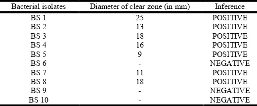
Table No. 5: Observations and results of ODA test
Bacterial isolates Diameter of clear zone (in mm) Inference
BS 1 25 POSITIVE
BS 2 13 POSITIVE
BS 3 18 POSITIVE
BS 4 16 POSITIVE
BS 5 9 POSITIVE
BS 6 - NEGATIVE
BS 7 11 POSITIVE
BS 8 18 POSITIVE
BS 9 - NEGATIVE
BS 10 - NEGATIVE
[image:4.595.94.504.524.647.2] [image:4.595.168.428.679.786.2]Figure No.1: Isolated colonies of Bacillus species
Bacillus Agar
Figure No.2: Results of emulsification assay method
Emulsification assay method: The isolates which had ability to emulsification are shown in the following Table 6.
Drop collapse method: The results of drop collapse method are given in the following Table
Production of Biosurfactant
Isolates BS1, BS2, BS3, BS4 and BS8 were used for production of biosurfactant. Production was done in 100ml
specieson HiCrome
Figure No.2: Results of emulsification assay method
The isolates which had ability to emulsification are shown in the following The results of drop collapse method are given in the following Table 7.
Isolates BS1, BS2, BS3, BS4 and BS8 were used for production of biosurfactant. Production was done in 100ml
(A)
(B)
Figure No.3 : A] Nutrient broth before incubation B] Nutrient broth after incubation
Figure No.4 : Petriplates containing dry biosurfactant
Figure No.5 : Results of TLC
nutrient broth containing 1% (v/v) kerosene oil. After incubation of 48-72 hr, broth was centrifuged to remove cells and supernatant was tested for biosurfactant production by ODA test and emulsification test. All isolates supernatant had given ODA test and emulsification test positive.
(A)
(B)
Figure No.3 : A] Nutrient broth before incubation B] Nutrient broth after incubation
Figure No.4 : Petriplates containing dry biosurfactant
Figure No.5 : Results of TLC
broth containing 1% (v/v) kerosene oil. After 72 hr, broth was centrifuged to remove cells and supernatant was tested for biosurfactant production by ODA test and emulsification test. All isolates supernatant had
Figure No.6 : Emulsification activity of produced biosurfactants
Table No. 6: Observations and results of Emulsification assay method
Bacterial isolates
Height of emulsion (in cm)
Total height of solution (in cm)
BS1 0.5 1.3
BS 2 0.2 1.3
BS 3 0.5 1.4
BS 4 0.4 1.3
BS 5 0.2 1.3
BS 6 - 1.4
BS 7 0.2 1.4
BS 8 0.3 1.3
BS 9 - 1.4
BS 10 - 1.5
Figure No.7 : Emulsification assay of isolates
Table No. 7 : Results of drop collapse
Bacterial isolates Results of drop collapse method
BS1 POSITIVE
BS 2 POSITIVE
BS 3 POSITIVE
BS 4 POSITIVE
BS 5 NEGATIVE
BS 6 NEGATIVE
BS 7 POSITIVE
BS 8 POSITIVE
BS 9 NEGATIVE
BS 10 NEGATIVE
Extraction of Biosurfactant
White coloured sediments were obtained after extraction i.e., the biosurfactant. The dry weight of biosurfactant was calculated by using following formula: Dry weight of
14862 Sanjana S. Varma et al. Production and characterization of biosurfactant by bacillus subtilis isolated from soil samples
Emulsification activity of produced
Emulsification assay method
Total height of
solution (in cm) E24%
38.46% 15.38% 35.71% 30.76% 15.38%
- 14.28% 23.07%
- -
Emulsification assay of isolates
Table No. 7 : Results of drop collapse method
Results of drop collapse method
NEGATIVE NEGATIVE
NEGATIVE NEGATIVE
White coloured sediments were obtained after extraction i.e., the biosurfactant. The dry weight of biosurfactant was calculated by using following formula: Dry weight of
biosurfactant = weight of the plate after drying empty plate (Table 8).
Table No. 8 : Dry weight of biosurfactants produced by isolates
Bacterial isolates
weight of empty plate
(in gm)
weight of the plate after drying (in
BS1 103.82
BS2 87.49
BS3 103.36
BS4 109.85
BS8 98.28
Figure No. 8 :Dry weight of biosurfactants produced by isolates
Table No. 9 : Rf values of produced biosurfactants
Bacterial isolates
Distance moved by solvent (in cm)
BS1 7.2
BS2 7.2
BS3 7.2
BS4 7.2
BS8 7.2
Table No. 10 : Emulsification index of produced biosurfactants
Bacterial isolates
Height of emulsion (in cm)
BS1 1.3
BS 2 0.7
BS 3 1.2
BS 4 1.2
BS 8 0.6
Figure No.9 : Emulsification index of produced biosurfactants
Characterization of Biosurfactant
Thin Layer Chromatography:
red spots were observed which indicate that all five biosurfactant were lipopeptide biosurfactant
Production and characterization of biosurfactant by bacillus subtilis isolated from soil samples
biosurfactant = weight of the plate after drying - weight of
Table No. 8 : Dry weight of biosurfactants produced by isolates
weight of the plate after drying (in
gm)
Dry weight of biosurfactant (in
gm/100ml)
103.87 0.05
87.51 0.02
103.40 0.04
109.89 0.04
98.31 0.03
Figure No. 8 :Dry weight of biosurfactants produced by isolates
Table No. 9 : Rf values of produced biosurfactants
Distance moved by Distance moved
by solute (in cm) Rf
5.3 0.73
5.3 0.73
5.1 0.70
5.2 0.72
5.2 0.72
Emulsification index of produced biosurfactants
Total height of
solution (in cm) E24%
3.8 34.21%
3.6 19.44%
3.7 32.43%
3.8 31.57%
3.6 16.66%
Figure No.9 : Emulsification index of produced biosurfactants
Characterization of Biosurfactant
Thin Layer Chromatography: After spraying ninhydrin, red spots were observed which indicate that all five
Emulsification activity: Emulsification index of the produced biosurfactant are given in the following Table 10.
In present study, 10 isolates of Bacillus subtilis were isolated from three different oil contaminated soil samples. All isolates were Gram positive rods, motile and endospore forming and on the basis of morphological, biochemical, and cultural characteristics it was confirmed that all the isolates are of
Bacillus subtilis. Then biosurfactant producing Bacillus subtilis were screened by haemolytic activity, oil displacement test, emulsification assay method and drop collapse method. All isolates showed α – haemolysis in haemolytic test. In oil displacement test, isolates BS1, BS2, BS3, BS4, BS5, BS7 and BS8 showed zone of diameter 25mm, 13mm, 18mm, 16mm, 9mm, 11mm and 18mm respectively [Table No.7]. The E24 % of BS1, BS2, BS3, BS4, BS5, BS7 and BS8 were 38.46%, 15.38%, 35.71%, 30.76%, 15.38%, 14.28% and 23.07% respectively [Table No.6]. Lastly, drop collapse test were given positive by isolates BS1, BS2, BS3, BS4, BS7 and BS8 [Table No.7].
The isolates BS1, BS2, BS3, BS4 and BS8 of Bacillus subtilis
were used for production of biosurfactant in nutrient broth containing 1% (v/v) kerosene oil. After incubation, the cell free broths were tested for production by ODA and emulsification test. All isolates supernatant had given ODA test and emulsification test positive. Extraction of biosurfactant was done by acid precipitation and solvent extraction method with chloroform: methanol (2:1). Amount of biosurfactant obtained after production by isolates BS1, BS2, BS3, BS4 and BS8 were 0.05 gm/100ml, 0.02 gm/100ml, 0.04gm/100ml, 0.04 gm/100ml and 0.03 gm/100ml respectively. Biosurfactants were characterized using thin layer chromatography and found to be lipopeptides, as red spots were observed after spraying ninhydrin reagent on silica gel plate. Rf value of biosurfactant produced by isolates BS1, BS2, BS3, BS4 and BS8 are 0.73, 0.73, 0.70, 0.72 and 0.72 respectively. The emulsification activity of produced biosurfactant is calculated by emulsification index (E24). E24% of biosurfactant produced by BS1, BS2, BS3, BS4 and BS8 is 34.21%, 19.44%, 32.43%, 31.57% and 16.66% respectively.
Screening of biosurfactant producing Bacillus subtilis was done by haemolytic activity, oil displacement test, emulsification assay method and drop collapse method. These methods were also followed by Anitha et al. (2015), Olteanu Violeta et al. (2011), Suresh Chander et al. (2012), and Okore Chioma et al. (2013). In this study, all isolates showed α – haemolysis in haemolytic test. Okore Chioma et al. (2013) found that out of 9 isolates of both Bacillus and Pseudomonas
7 isolates showed α – haemolysis and 2 isolates showed β – haemolysis. β – haemolysis is one of the characteristics also of biosurfactant producing microorganisms. In this study, the zone produced by BS1, BS2, BS3, BS4, BS5, BS7 and BS8 isolates of Bacillus subtilis in ODA test had diameter of 25mm, 13mm, 18mm, 16mm, 9mm, 11mm and 18mm respectively [Table No.7]. Similarly, Olteanu violeta et al.
(2011) obtained best results of zone diameter, 28 mm and 30 mm for B2 and OS17 strains of Bacillus subtilis respectively on kerosene (Olteanu Violeta et al., 2011). In studies done by, Suganya (2013), and Suresh Chander et al. (2012) Bacillus subtilis showed zone of 17mm, 3.2 mm, on kerosene oil and 2.1 cm on gingelly oil, respectively. In study of Anayata
Sharma et al. (2014), strain B1 and B2 of Bacillus showed zone of 1cm and 2cm respectively. In this study, out of 10 isolates of Bacillus subtilis seven showed emulsification activity against kerosene oil. The E24 % of BS1, BS2, BS3, BS4, BS5, BS7 and BS8 were 38.46%, 15.38%, 35.71%, 30.76%, 15.38%, 14.28% and 23.07% respectively [Table No.6]. In the study of Femi-Ola et al. (2015) Bacillus showed emulsification capacity 51.61%. Bacillus subtilis MTCC441 in Suresh Chander et al. (2012) study also showed emulsification activity against kerosene oil . In this study, drop collapse test were given positive by isolates BS1, BS2, BS3, BS4, BS7 and BS8. Femi-Ola et al. (2015) also found drop collapse test positive for Bacillus spp.
In the present study, production of biosurfactant was done in nutrient broth containing 1% (v/v) kerosene oil and after incubation cell free broths were tested for production by ODA and emulsification test. Similar was followed by Anayata Sharma et al. (2014), Priya et al. (2009) and Ali Diab et al.
(2013) . And also, ODA and emulsification test were positive indicating that biosurfactant was produced. Extraction of biosurfactant was done by acid precipitation and solvent extraction method with chloroform: methanol (2:1). Similarly, Anayata Sharma et al. (2014), J.Anitha et al. (2015), Arutchelvi et al. (2009), Dhouha Ghribi et al. (2011), Priya et al. (2009), Olteanu Violeta et al. (2011), Suganya (2013), and Ali Diab et al. (2013) also used these methods for extraction of biosurfactant. In this study, the amount of biosurfactant produced by BS1, BS2, BS3, BS4 and BS8 are 0.05 gm/100ml, 0.02 gm/100ml, 0.04gm/100ml, 0.04gm/100ml and 0.03 gm/100ml respectively [Table No.8]. Similarly, in the study of Anayata Sharma et al. (2014) the amount of biosurfactant produced by isolates B1, B2 and B3 of Bacillus spp. were 0.125g, 0.105g and 0.14g respectively. In study of Arutchelvi
et al. (2009), Bacillus subtilis was capable of producing 0.5 g L-1 of crude biosurfactant (dry weight) in the mineral salt medium. After characterization of biosurfactants, produced by isolates BS1, BS2, BS3, BS4 and BS8 by TLC, it was found that they were lipopeptide in nature. Similar result was obtained by Anitha et al. (2015), Priya et al. (2009), Olteanu Violeta et al. (2011), Samadhan Waghmode et al. (2014) and Okore Chioma et al. (2013) in their studies. Rf values of biosurfactant produced by isolates BS1, BS2, BS3, BS4 and BS8 on kerosene are 0.73, 0.73, 0.70, 0.72 and 0.72 respectively. Priya et al. (2009) obtained Rf value of 0.51 for biosurfactant produced by Bacillus subtilis on kerosene. The Rf value of biosurfactant produced by Bacillus subtilis on substrates coconut waste, soyabean waste, basal medium, and sesame waste in Samadhan Waghmode et al. (2014) study were 0.33, 0.25, 0.21 and 0.22 respectively.
The emulsification activity of produced biosurfactant is calculated by emulsification index (E24).E24% of biosurfactant produced by BS1, BS2, BS3, BS4 and BS8 are 34.21%, 19.44%, 32.43%, 31.57% and 16.66% respectively [Table No.10]. This test was also followed by Anayata Sharma
et al. (2014), Anitha et al. (2015), Juliana F.F. Secato et al. (2016), Olteanu Violeta et al. (2011), and Ali Diab et al.
(2013). Olteanu Violeta et al. (2011) strains B2 and OS17 of
Conclusion
This study concludes that Bacillus subtilis is the potent producer of lipopeptide biosrufactant and can be used as a culture for large scale production of biosurfactant. These biosurfactant are also a potent emulsifier. So, that it has wide applications in oil, food, pharmaceuticals and cosmetic industries. It can also be used in bioremediation and microbial enhanced oil recovery.
REFERENCES
Ali Diab and Shereen Gamal El Din, 2013. Production and characterization of biosurfactants produced by Bacillus spp.
and Pseudomonas spp. isolated from the rhizosphere soil of an Egyptian salt marsh plant, Nature and Science, 11(5), 103-112.
Ali Diab and Shereen Gamal El Din. Application of the biosurfactants produced by Bacillus spp. (SH 20 and SH 26) and Pseudomonas aeruginosa SH 29 isolated from the rhizosphere soil of an Egyptian salt marsh plant for the cleaning of oil - contaminataed vessels and enhancing the biodegradation of oily sludge African Journal of Environmental Science and Technology 7(7), 671-679. Anitha J., V. Jeyanthi and P. Ganesh, 2015. Production and
characterization of biosurfactant by Bacillus and its applicability in enhanced oil recovery. Int. J. Adv. Res. Biol. Sci. 2(5): 7–16
Arutchelvi J, Bhaduri S, Upparo P V, and Doble M, 2009. Production and characterization of biosurfactant from
Bacillus subtilis YB7, Journal of Applied Sciences, 9(17), 3152-3155.
Deshmukh P W, Kulkarni N S and Bodhankar M G, 2014. Production of biosurfactant of microbial origin using different raw materials, Indian Journal of Microbiology Research, 1(1), 84-90.
Femi-Ola, T. O., Oluwole, O. A., Olowomofe, T. O. and Yakubu, H., 2015. Isolation and screening of biosurfactant- producing bacteria from soil contaminated with domestic waste water, British Journal of Environmental Scienc5es, 3(1), 58-63.
Foukia E.Mouafi, Mpstfa M.Abo Elsoud Maysa E.Moharam.
2016. Optimization of biosurfactant production by Bacillus brevis using response surface methodology. Biotechnology Reports Volume 9, Pages 31-37
Ghayyomi Jazeh, M , F. Forghani and Deog-Hwan O, 2012. Biosurfactant production by Bacillus sp. isolated from petroleum contaminated soils of Sirri Island, American Journal of Applied Sciences, 9 (1), 1-6.
Juliana F. F. Secato, Diego F. Coelho, Nathália G.J. Rosa, Lucas D.L. Costa and Elias B. Tambourgi, 2016. Biosurfactant production using Bacillus subtilis and industrial waste as substrate, Chemical Engineering transactions, 49, 103-108.
Okore Chioma, 2013. Isolation and Characterization of Biosurfactants Producing Bacteria from Oil Polluted Soil. Journal of Natural Sciences Research Vol.3, No.5.
Olteanu Violeta, Sicuia Oana, Ciuca Matilda, Carstea Doina Maria, Voaides Catalina, Campeanu Gheorghe, Cornea Calina Petruta, 2011.Production of biosurfactants and antifungal compounds by new strains of Bacillus Spp. isolated from different sources Romanian Biotechnological Letters Vol. 16, No.1.
Padmanabhan G, Suresh G, Kannappan V, Jayakumar S, and Ramesh B, 2014. Estimation of bio-surfactant produced using Bacillus subtilis CS14 in emulsion using ultrasonic probing technique, International Journal of Current Microbiology and Applied Sciences, 3(7), 737-744.
Płociniczak M P, Płaza G A , Seget Z P, and Cameotra S S, 2011. environmental applications of biosurfactants: recent advances, International Journal of Molecular Sciences,12, 633-654.
Priya T and Usharani G, 2009. Comparative study for biosurfactant production by using Bacillus subtilis and
Pseudomonas aeruginosa, Botany Research International, 2 (4), 284-287.
Rabah, A B and Bello H, 2015. Production and characterization of biosurfactants from abattoirwastewater,
Journal of Biology, Agriculture and Healthcare, 5(7), 187-190.
Shah N, Nikam R, Gaikwad S, Sapre V and Kaur J, 2016. Biosurfactant: Types, detection methods, importance and applications, Indian Journal of Microbiology Research, 3(1), 5-10.
Sharma A, Soni J, Kaur G and Kaur J, 2014. A study on biosurfactant production in Lactobacillus and Bacillus sp., International Journal of Current Microbiology and Applied Sciences, 3(11), 723-733.
Sidkey, Nagwa M, Al Hadry and Eman A., 2014. Biosurfactant production by Bacillus cereus, B7 from lubricant oil waste,
International Journal of Science and Research (IJSR),
3(12), 498-509.
Suganya R S 2013. Screening, optimization and production of biosurfactants from Bacillus and Pseudomonas species,
International Journal of Current Pharmaceutical Research, 5(1), 19-23.
Suganya R.S. 2013. Screening Optimization and Production of Biosurfactants from Bacillus and Pseudomonas Species. International Journal of Current Pharmaceutical Research Vol 5, Issue 1.
Suresh Chander C. R., T. Lohitnath, D. J. Mukesh Kumar and P. T. Kalaichelvan, 2012. Production and characterization of biosurfactant from bacillus subtilis MTCC441 and its evaluation to use as bioemulsifier for food bio – preservative. Advances in Applied Science Research, 3 (3):1827-1831.
Waghmode S, Kulkarn C, Shukla S, Sursawant P, and Velhal C, 2014. Low cost production of biosurfactant from different substrates and their comparative study with commercially available chemical surfactant, International Journal of Scientific & Technology Research, 3(3), 146-149.