RESEARCH ARTICLE
Genetic basis for the evolution of organ morphogenesis: the case
of
spalt
and
cut
in the development of insect trachea
Cristina de Miguel1,2,*, Friedemann Linsler2,3,*, Jordi Casanova2,3,‡and Xavier Franch-Marro1,‡
ABSTRACT
It is not clear how simple genetic changes can account for the coordinated variations that give rise to modified functional organs. Here, we addressed this issue by analysing the expression and function of regulatory genes in the developing tracheal systems of two insect species. The larval tracheal system of Drosophila can be distinguished from the less derived tracheal system of the beetle Triboliumby two main features. First,Triboliumhas lateral spiracles connecting the trachea to the exterior in each segment, while Drosophila has only one pair of posterior spiracles. Second, Drosophila, but not Tribolium, has two prominent longitudinal branches that distribute air from the posterior spiracles. Both innovations, while considered different structures, are functionally dependent on each other and linked to habitat occupancy. We show that changes in the domains ofspaltandcutexpression in the embryo are associated with the acquisition of each structure. Moreover, we show that these two genetic modifications are connected both functionally and genetically, thus providing an evolutionary scenario by which a genetic event contributes to the joint evolution of functionally inter-related structures.
KEY WORDS: Trachea, Spiracles,Spalt,Cut, Morphogenesis, Evolution,Drosophila,Tribolium
INTRODUCTION
One of the main processes that facilitates animal habitat adaptation is organ morphology diversification. Morphogenesis is a complex genetically programmed process. Modifications in this genetic program throughout evolution allowed the formation of a wide range of morphologies. Evidence of this phenotypic diversification in evolution has been mainly collected from the study of quantitative traits. However, the genetic mechanisms that explain how simple genetic changes account for the coordinated variations that give rise to modified functional organs is still poorly understood. Here, we address this question by analysing and comparing the tracheal system development of two insects.
The tracheal system is the respiratory organ of the insects. The development of the tracheal system has been deeply studied in Drosophila, where it develops from clusters of ectodermal cells that
arises in either side of the embryo from the first thoracic (T1) to the last abdominal segment (A8) (for a review see Manning and Krasnow, 1993). Tracheal cells are specified by the activity of a set of‘tracheal inducer genes’that includes the transcription factors trachealess (trh) and ventral veinless (vvl) (for a review, see Ghabrial et al., 2003). Once specified, those tracheal cells invaginate and migrate to form a stereotypical network of tubes. This network consists of a couple of main dorsal trunks (DTs), specified by the transcription factorspalt(sal, also known assalm), which have many ramifications and are connected to the outside through specific structures called spiracles. Posterior spiracles in Drosophiladevelop from a cluster of epithelial cells in the dorsal side of the eighth abdominal segment (A8) that forms an internal multicellular tube, the spiracular chamber, which links the trachea to the exterior (Hu and Castelli-Gair, 1999). The Hox geneAbdominal B(Abd-B) specifies the formation of the posterior spiracles mainly by the activation of the transcription factorscut(ct),grain(grn) and empty spiracles(ems) (Lovegrove et al., 2006). In contrast, other insects present spiracles in every segment where trachea develops, which goes hand in hand with an overall different tracheal system morphology. Formation of such different tracheal morphologies depending on the development of few or many functional spiracles might imply an evolutionary relationship between both structures (Keilin, 1944). However, the genetic mechanisms underlying these morphologically linked changes remain elusive.
Here, we address this evolutionary question by describing the sequential branching process that gives rise to theTriboliumlarval trachea and comparing that with tracheal development in Drosophila. In contrast to Drosophila, the Tribolium tracheal system presents functional lateral spiracles and lacks dorsal trunks (DTs), suggesting that the distinct morphology of the Tribolium tracheal system is coupled with different patterns ofctandsalgene expression. Furthermore, we show that changes in the domains of sal and ct expression are associated with the acquisition of each morphological innovation and that these two genetic modifications are connected both functionally and genetically.
RESULTS AND DISCUSSION
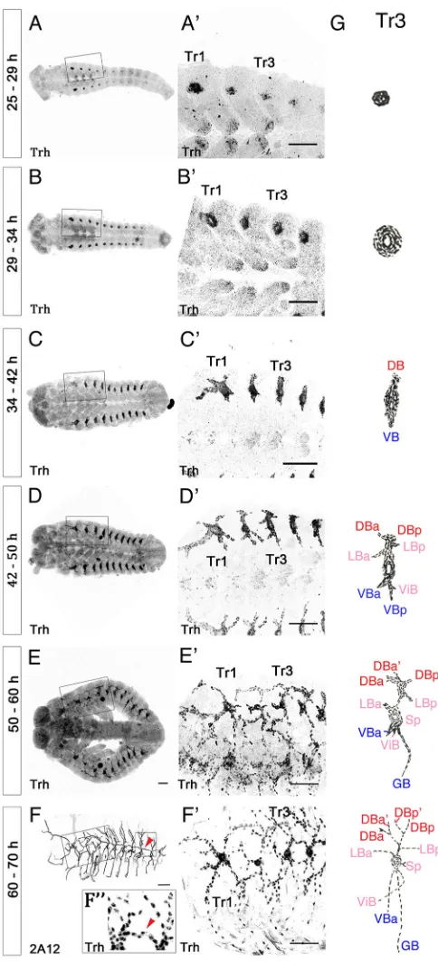
TheTriboliumtracheal system starts to develop 27 h after egg laying (AEL). It derives from 10 clusters of ectodermal cells (Tr1-Tr10) on both sides of the embryo, from the second thoracic (T2) to the eight abdominal (A8) segments (Fig. 1A,B). The specification and development of each cluster of cells starts with those in the most anterior segments and progress along the antero-posterior axis. The cells of each cluster invaginate and form an elongated sac of cells connected to the surface by a lateral spiracle (Fig. 1B,B′). A dorsal and a ventral bud then start to form and grow (Fig. 1C,C′). Four additional buds–three dorsal and one ventral–arise later, by 47 h AEL (Fig. 1D,D′). Cells from these buds migrate in various directions and to characteristic lengths and form the main tracheal primary branches that are about two cell diameters wide (Fig. 1E,E′). Received 8 January 2016; Accepted 12 August 2016
1
Institute of Evolutionary Biology (CSIC-Universitat Pompeu Fabra), Functional Genomics and Evolution, Department Passeig Marı́tim de la Barceloneta 37-49, Barcelona 08003, Spain.2Institut de Biologı́a Molecular de Barcelona (CSIC), Carrer de Baldiri Reixac 10, Barcelona, Catalonia 08028, Spain.3Institut de Recerca Biomèdica de Barcelona, Carrer de Baldiri Reixac 10, Barcelona, Catalonia 08028, Spain.
*These authors contributed equally to this work
‡
Authors for correspondence ( [email protected]; [email protected])
X.F., 0000-0002-7465-6729
DEVEL
O
However, some cells do not migrate and remain clustered around the point of the initial invagination as a sac connected to the exterior by the lateral spiracle, which in Tribolium develops in all tracheal segments except Tr2. By 65 h AEL, the dorsal branches in each segment from the two body sides fuse and form a dorsal anastomosis connecting the right and left sides of the tracheal network. Afterwards, the ventral branches from neighbouring segments fuse and connect the tracheal network along the whole embryo (Fig. 1F,F′ and Fig. S1). In addition, as the tracheal branches elongate, their cells intercalate from a side-by-side arrangement to a one cell thick row organisation, forming narrower and longer branches (Fig. 1F″and Fig. 2) that make up the final larval tracheal network. Each metamere comprises the following branches: four dorsal branches, two anterior
(DBa and DBa′) and two posterior (DBp and DBp′); two lateral branches, one anterior (LBa) and one posterior (LBp) that fuse with the lateral branches of adjacent tracheal segments to form the lateral trunk; and three ventral branches, the visceral branch (ViB) that oxygenates the gut, and the ventral (VB) and ganglionic branch (GB) that migrate towards the central nervous system (Fig. 1G).
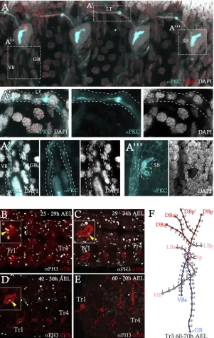
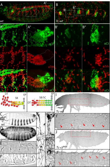
By means of the PH3 mitotic marker, we detected cell division in the tracheal cells from their determination in the ectoderm (25 h AEL) to the formation of primary branches (50 h AEL) (Fig. 2B-E). Upon these divisions, each tracheal cluster contains ∼80 cells, except for Tr1 and Tr2, which contain 140 cells. The extra number of cells in these two clusters is responsible for the formation of the additional cephalic and leg branches on the thoracic segments (Fig. 1F,F′). Tracheal cells are typically distributed among the different branches with a characteristic number of cells (Fig. 2F). Two main features distinguish the trachea of Tribolium and Drosophila larvae (Fig. 3). First, the lateral spiracles, which in Tribolium connect the tracheal branches at both sides of each segment to the exterior, are not present in Drosophila larvae (Fig. 3A,B). This observation is consistent with the long-standing notion that‘from a relative primitive, open form of tracheal system of ancient insects originated phylogenetically, the relative secondary, closed form of the present time larvae’(Keilin, 1944; Palmén, 1877). Instead,Drosophilahas two posterior spiracles in the last segment, which open to the exterior through highly specialised structures, namely the stigmatophores (Fig. 3A). During subsequent moults,Drosophilasecond and third instar larvae also develop anterior spiracles, similar openings at their anterior-most region that become functional at the pupa stage (Manning and Krasnow, 1993). In place of the lateral spiracles,Drosophilalarvae have spiracular branches, which are groups of tracheal cells connected to the inner side of the ectoderm but with no opening to the exterior (Manning and Krasnow, 1993). These branches are considered a rudiment of an ancestral tracheal system with segmental spiracles (Palmén, 1877). They consist of aggregates of progenitor adult tracheal cells (Pitsouli and Perrimon, 2010; Weaver and Krasnow, 2008) and do not participate in larval breathing.
[image:2.612.51.293.55.587.2]The other main feature that distinguishes the trachea ofDrosophila andTriboliumlarvae is the occurrence of dorsal trunks in the former. These trunks are a pair of prominent branches that run longitudinally along each side of the larva (Fig. 3C,E). In contrast to the other branches, cells in the dorsal trunks do not intercalate and therefore more than one cell contributes to the circumference of the trunks, thus accounting for their greater diameter. Dorsal trunks are not present inTribolium(Fig. 3D,F) and all tracheal cells, except those connected to the lateral spiracle, undergo intercalation and form branches of a similar diameter in this insect (Fig. 1F″).
Fig. 1. Development of the tracheal system inTribolium castaneum.
(A-F) Whole-mount embryos immunostained with anti-Trh (A-E) and 2A12 (F) antibodies at different stages of tracheal development. (A) Tracheal cell determination and placode formation 25-29 h AEL. (B) Invagination of tracheal cells 29-34 h AEL. Tracheal cell migration and branch formation 34-42 h AEL (C), 42-50 h AEL (D) and 50-60 h AEL (E). (F) Lateral trunk fusion 60-70 h AEL. A more detailed image of F is also shown in Fig. 3D. (A′-E′) Magnification of panels A-E showing tracheal metameres Tr1 to Tr4. (F′) Detail showing tracheal metameres Tr1-Tr4 of an embryo at the same stage as that in F but stained with an anti-Trh antibody. (F″) Detail of an embryo immunostained with anti-Trh antibody corresponding to the same region as the inset in F to show the nuclei organised as a row in an abdominal tracheal branch (arrowhead), an indication of cell intercalation. (G) Diagrams of tracheal development of Tr3. DBa, dorsal branch anterior; DBp, dorsal branch posterior; LBa, lateral branch anterior; LBp, lateral branch posterior; Sp, spiracular sac; ViB, visceral branch; VB, ventral branch; GB, ganglionic branch.
DEVEL
O
To address what might account for the lack of functional lateral spiracles in Drosophila, we compared the patterns of gene expression in the spiracular branches and in the posterior spiracles. InDrosophila, the gene encoding the Ct homeodomain transcription factor plays an important role in the formation of both structures–the spiracular branches and the posterior spiracles (Hu and Castelli-Gair, 1999; Pitsouli and Perrimon, 2010). At the posterior spiracles,ctis expressed in the ectodermal cells that will give rise to the spiracular chamber, the spiracular structure that connects the tracheal cells (trh -expressing cells) to the exterior (Hu and Castelli-Gair, 1999). However, a close analysis shows thatctexpression extends beyond thetrhexpression domain (Fig. 4A,A″). In contrast, the domain ofct expression coincides with that of tracheal cells in the lateral spiracular branches (Fig. 4A,A′). This finding raises the possibility that an epidermal population of ct-expressing cells next to the spiracular branches is required to build a functional spiracle.
Consistent with this hypothesis, we foundTc-ctexpression at the sites of the lateral spiracles inTribolium, in the tracheal cells ( Tc-trh-expressing cells), and also in some neighbouring ectodermal cells (Fig. 4B,B″). Remarkably, this specific Tc-ct expression is present in allTriboliumsegments but is very much reduced in the third thoracic segment (Fig. 4B,B′)–the only segment in which the lateral spiracles close as development proceeds (Bennett et al., 1999; Lewis et al., 2000). This finding thus reinforces the link between ectodermalTc-ctexpression and functional spiracles. To assess the functional role of Tc-ct in the process, we knocked down its expression by injection of dsRNA (see Materials and Methods for details) and found that the resultingTriboliumlarvae lack openings in the position of the lateral spiracles (Fig. 4I-J′).
We turned toDrosophilato further explore the role ofctin the development of spiracles. ct mutant embryos have abnormal posterior spiracles that lack an open connection (Hu and Castelli-Fig. 2. Tracheal cell organisation, division and distribution during embryogenesis.(A-A‴) Whole-mountTribolium
embryos from 60-70 h AEL immunostained with CBP (red), anti-PKC (cyan) and DAPI (white). (A′,A″) Magnification of insets in A to show the unicellular organisation of LT (A′) and the ventral branches, VB and GB (A″). (A‴) Magnification of inset in A showing the multicellular organisation of the SB. Dashed lines outline the tracheal branches. (B-E) Whole-mountTriboliumembryos 25-70 h AEL immunostained with anti-Trh (red) and anti-PH3 (white) antibodies. Insets in A-C show a high magnification of the Tr1 tracheal placode with PH3+cells (arrowheads). (D) No mitotic cells were detected at 60-70 h AEL. (E) Distribution of cells in aTriboliumtracheal hemisegment. (F) Typical positions of tracheal cell nuclei (filled circles) in Tr3 are shown. There were 79±2 cells (n=5) at 60-70 h AEL. The mean number of cells in each branch were: DBa, 8; DBa′, 4; DBp, 6; DBp′, 5; LBa, 3; LBp, 3; Sp, 11; ViB, 12; VB, 9; GB, 13.
DEVEL
O
[image:3.612.51.352.58.533.2]Gair, 1999). Forced expression ofctat the lateral ectoderm, either by broadly expressed drivers such asnullo-Gal4 or by the ectodermal 69B-Gal4(see Materials and Methods for details), promoted the development of openings at the lateral ectoderm (Fig. S2). However, these embryos were greatly distorted. We thus used grn-Gal4, a construct that drives more restricted expression in tracheal cells and in patches of the lateral ectoderm (Fig. S3). Using this driver, ectopic expression of ct also induced openings at the lateral ectoderm (Fig. 4C-F), which were connected to the internal tracheal cavities (Fig. 4G-H′), supporting the notion that these openings are similar to lateral spiracles (Fig. S4). Thus, ct expression in the ectoderm is necessary for the formation of functional lateral spiracles inTriboliumand both necessary and sufficient to induce the formation of corresponding structures inDrosophila.
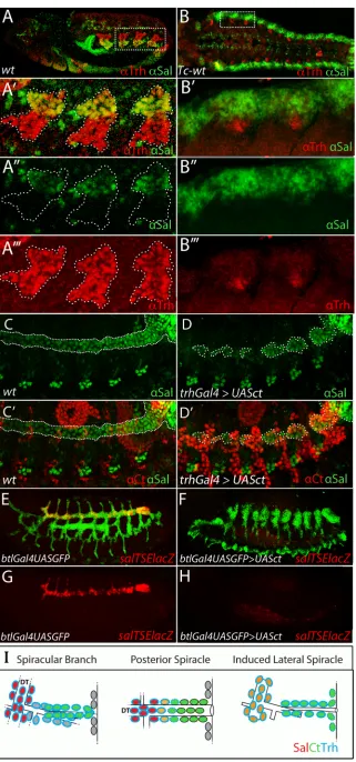
Next, we addressed what might account for the development of the prominent dorsal trunks in Drosophila and their absence in Tribolium. In this regard, the morphology of theTriboliumtrachea is reminiscent of that observed inDrosophila embryos that lack the twosalgenes (Franch-Marro and Casanova, 2002). Indeed,salacts as a repressor of cell intercalation in Drosophila (Ribeiro et al., 2004) and Drosophila sal mutants do not develop dorsal trunks (Kühnlein and Schuh, 1996), thus supporting the notion that sal expression is associated with the acquisition of this morphological innovation. To address this possibility, we comparedsalexpression in the two insects. In stage 10Drosophilaembryos,salis expressed
in broad patches of dorsal ectoderm that encompass the dorsal region of each tracheal placode (Kühnlein and Schuh, 1996). As development proceeds,sal expression declines in the ectodermal region between the placodes and becomes restricted to the dorsal region of the tracheal placodes (Kühnlein and Schuh, 1996; Fig. 5A, A‴). As the trachea develops further,salexpression persists in the dorsal branches and in the dorsal trunks and finally accumulates only in the latter (Fig. 5C,C′), where it represses cell intercalation (Kühnlein and Schuh, 1996; Ribeiro et al., 2004). Similar to its expression in Drosophila,Tc-sal is expressed in a broad strip of dorsal ectoderm inTribolium(Fig. 5B). However, and in contrast to Drosophila, the ectodermal expression ofTc-saldoes not include tracheal cells (Fig. 5B-B‴) andTc-salis not expressed during the formation ofTriboliumtracheal branches. Consistently, we did not observe any evident tracheal phenotype in Tc-sal-depleted Tribolium embryos (data not shown). Nevertheless, we detected Tc-salexpression in the Tribolium tracheal system much later, at 60 h AEL, once all the branches have already formed (Fig. S5).
[image:4.612.107.506.57.349.2]As mentioned above, both the development of dorsal trunks and the closure of lateral spiracles are adaptations to the breathing physiology ofDrosophilalarvae. Indeed, it has been reported that dorsal trunks are over-developed in certain insect larvae with few functional spiracles (Keilin, 1944). In many insect taxa, this feature was linked to the aquatic life history of larvae. As early as 1877, Palmén observed an association between the appearance of Fig. 3. Organisation of the tracheal systems inDrosophilaandTribolium.(A) Cuticle of aDrosophilaembryo at stage 16 in phase contrast; arrowhead indicates the posterior spiracle. Magnification in the inset shows a posterior spiracle in detail. (B) Cuticle of aTriboliumembryo 70 h AEL in phase contrast; arrowheads indicate the lateral spiracles. Magnification in the inset shows a lateral spiracle in detail. (C,D) Whole-mountDrosophila(C) andTribolium(D) embryos immunostained with the 2A12 antibody showing the branches of the fully developed embryonic tracheal system. Prominent dorsal trunks are present in
Drosophila, seen in a dorso-lateral view to show both dorsal trunks, one in each side of the embryo (arrowheads in C), but not inTribolium, shown in a lateral view (D). Note that panel D shows an amplification of the image of Fig. 1F for comparison withDrosophilatracheal network. (E,F) Schematic representation of the
Drosophila(E) andTribolium(F) embryonic tracheal trees. CB, cephalic branch; LBd, dorsal lateral branch; LBv, ventral lateral branch; sp., spiracle; SB, spiracular branch; GB, ganglionic branch; LB, leg branch; DBa, anterior dorsal branch; DBp, posterior dorsal branch; ViB, visceral branch; VB, ventral branch; LT, lateral trunk.
DEVEL
O
longitudinal trunks and the loss of segmental spiracles. In this regard, it is interesting to note that ectopic ct expression in Drosophila larvae not only induced the development of lateral openings but was also associated with a failure in the development of dorsal trunks when ectopic ct expression encompassed the tracheal cells (Fig. 5D,D′,F and Fig. S6). This result suggests a link between the lack of functional spiracles and the development of thick tracheal trunks. Furthermore, we observed that ectopic tracheal expression of ct was associated with the downregulation of endogenoussal, which accumulated very weakly or was absent in the tracheal cells that expressedct(Fig. 5D,D′). A similar repressive
effect of ct on sal expression has also been reported in the Drosophilaposterior spiracles (Hu and Castelli-Gair, 1999). These results suggest that ectopic tracheal expression ofctinterferes with the development of dorsal trunks by downregulatingsalexpression. However, other outcomes of ectopicctexpression, such as its role in the regulation of the pro-apoptotic genereaper(Zhai et al., 2012), are probably also responsible for the deleterious effect on dorsal trunk development, as we were unable to revert the defects upon forced expression ofctby co-expression ofsal(Fig. S7).
[image:5.612.50.411.55.601.2]Composite patterns of gene expression are often associated with distinct regions in their promoters that are specific for their Fig. 4. Lateral and posterior spiracles are associated withcutexpression.
(A) Whole-mountDrosophilaembryo at stage 15 immunostained with anti-Trh (red) and anti-Ct (green) antibodies.
(A′-A″) Magnification of insets in A and schematic representations to show the domains of Trh and Ct expression in the spiracular branches (A′) and in the posterior spiracles (A″). The different colours represent the overlapping levels of Ct and Trh expression. Note that all Ct+cells in the spiracular branches are also Trh+but some Ct+cells extend beyond the Trh domain in
the posterior spiracles. (B) Detail of a whole-mountTriboliumembryo 60 h AEL, immunostained with Trh (red) and anti-Ct (green)Drosophilaantibodies. (B′,B″) Magnification of insets in B showing lower levels of Tc-Ct protein in the Tr2 lateral spiracle compared with the Tr3 spiracle. (C) Wild-type cuticle of a
Drosophilaembryo reared at 29°C. (D) Cuticle of aDrosophilaembryo reared at 29°C in whichctis ectopically expressed with agrn-GAL4driver. (E) A magnification of D to show spiracular branches with lateral openings. (F) Detail of the cuticle of another
Drosophilaembryo of the same genotype as in D to show a transversal view of the same structures. (G-H′) Detail of transverse and surface views of a wild-type (G,G′) and agrnGal4-UASctembryo (H,H′)
immunostained with an anti-DE-cadherin antibody to show the connection of the lateral opening with the tracheal system. (I,I′) Cuticle of a wholeTriboliumembryo and detail of the inset in I to show the openings of the lateral spiracles (filled arrowheads) except Tr2 (open arrowhead). (J,J′) Cuticle of a wholeTriboliumembryo from a female injected withctdsRNA and detail of the inset in J to show the absence of lateral spiracle openings (open arrowheads); note that embryo in J is more ventrally located than that in I.
DEVEL
O
Fig. 5. Dorsal trunks are associated withsal expression, which is repressed bycut.(A) Whole-mount earlyDrosophilaembryo at germ band extension (stage 12) immunostained with Trh (red) and anti-Sal (green) antibodies. (A′-A‴) Magnification of inset in A to show the partial overlap between Trh and Sal at the domain of the tracheal placode that will develop into the dorsal trunk. Orientation of the inset is switched to show the dorsal and ventral part of the tracheal placode. (B) Whole-mountTriboliumembryo 29-34 h AEL, immunostained with anti-Trh (red) and anti-Sal (green) antibodies. (B′-B‴) Magnification of a detail from B to show the lack of overlap between Tc-Trh and Tc-Sal. (C,C′) Expression of Sal (green) and Ct (red) in a late
Drosophilaembryo (stage 14). The dorsal trunk is marked by Sal expression. Note that Ct (red) is not expressed in those cells. (D)Drosophilaembryo at stage 14 overexpressing Ct under control oftrh-GAL4reared at 29°C. Ectopic tracheal expression of Ct (red) represses Sal expression (green) in the dorsal trunk cells. In C-D′, dotted lines outline the dorsal trunk. (E-H) Expression of specificsal-TSE-lacZtracheal enhancer in wild-type (E,G) and inbtl-GAL4/UAS-ct(F,H)Drosophilaembryos at stage 15. Tracheal cells are visualised by GFP (in green) andsal-TSE-lacZby anti-β-gal staining (in red). In
btl-GAL4/UAS-ctembryos under these conditions, sal-TSE-lacZreporter expression is completely abolished. (I) Schematic representations to show the domains of Trh, Ct and Sal expression in theDrosophilaspiracular branch, posterior spiracle and induced lateral spiracle.
DEVEL
O
regulation in given domains. Such is the case for theDrosophila sal gene, which has a trachea-specific enhancer. Repression ofsalbyct in theDrosophilatrachea is mediated through its tracheal enhancer (Fig. 5E-H), thus suggesting a mechanism for the acquisition of tissue-specific gene interactions and consistent with the notion of enhancers as an opportunity for the independent evolution of gene regulation (e.g. Averof and Akam, 1995). To further assess the hypothesis thatDrosophilamight have acquired a distinct enhancer to drivesalexpression specifically in tracheal cells, we generated a Triboliumtransgenic line bearing a reporter gene under the control of theDrosophila salpromoter region (see Materials and Methods for details and Fig. S8). Unfortunately, and probably because of the phylogenetic distance between the two species, this construct gave rise to a completely unrelated expression pattern, thus impeding us from evaluating this hypothesis further.
One of the main challenges in evolutionary genetics is to unveil the molecular basis of key phenotypic innovations that ultimately permit the occupation of alternative ecological niches (e.g. Kratochwil and Meyer, 2015). Our results establish a functional correlation between changes in the domains ofctandsalexpression and the structure of the larval tracheal system. These changes are likely to be associated with the exploitation of new habitats by the fruit fly larvae. In addition, it has been a long-standing problem to consider the gradual development of such innovations because of the difficulty explaining the selective value of all hypothetical intermediate states. In this regard, it is worth noting that changes in ct expression could be sufficient to account for the differences in the number of lateral spiracles. Moreover, the issue of morphological innovation is even more complex in the case of independent innovations that are nevertheless functionally related. Our results showing crosstalk betweenctandsalprovide a link between two functionally related innovations, namely the elimination of lateral spiracles and the development of dorsal trunks. All together, these results provide an evolutionary scenario by which a single genetic event contributes to the joint evolution of functionally inter-related organs.
MATERIALS AND METHODS
Insect stocks
The San Bernardino wild-type strain ofTribolium castaneumwas used for
dsRNA injections and inmunostaining and was provided by Yoshinori Tomoyasu (Miami University). The Vermilion White strain was used for
generating the transgenic line enhancer Dsal ( pBac[3×P3-EGFP_Tc‘hsp5’
-Dsalenhancer_Gal4]) and was provided by Gregor Bucher (University of
Göttingen). All Stocks were reared under constant temperature of 29°C and 60% humidity on wholewheat flour with 5% inactivated yeast.
The followingDrosophilalines were used:btl-Gal4(Shiga et al., 2014),
nullo-Gal4(Kunwar et al., 2003),69B-Gal4(Brand and Perrimon, 1993),
sal-TSE-lacZ (Kühnlein and Schuh, 1996),grn-Gal4 (Garces and Thor,
2006),UAS-ct (Ludlow et al., 1996) andUAS-sal(Kühnlein and Schuh,
1996). Crosses with GAL4 lines were reared at either 25°C or 29°C.
Pupal injection
Tc-ct dsRNA (IB_06353) was synthesised by the Eupheria Biotech
Company. A concentration of 1 µg/µl dsRNA was injected into 40 female adults in the abdominal body cavity laterally to avoid damaging genitals according to Tomoyasu and Denell (2004). Injected females were crossed with wild-type males in order to obtain knockdown embryos.
Embryo fixation and antibody staining
Triboliumembryos were fixed according to Bucher (http://wwwuser.gwdg.
de/~gbucher1/tribolium-castaneum-beetle-book1.pdf ). For antibody staining, samples were incubated overnight in a primary cocktail in PBX (PBS plus 0.5% Triton-20X and 0.3% BSA) at 4°C, washed three times for 20 min in PBX, incubated for 1 h at room temperature with the appropriate
fluorophore-conjugated secondary antibodies, washed three times for 20 min in PBX, and mounted on Vectashield with DAPI (Vector Laboratories) for microscopic analysis. The following primary antibodies were used: rat DE-cadherin (1:20, DSHB, DCAD2), mouse anti-Cut (1:200, DSHB, 2B10), Crumbs (1:10,
DSHB, Cq4),α-Spectrin (1:5, DSHB, 3A9) and mAb2A12 (1:1, DSHB);
rabbit anti-Spalt (1:200) provided by Rosa Barrio (Barrio et al., 1999) and anti-PKC (1:100, Santa Fe Technologies, H7 sc-8393); rat anti-Trachealess (1:300, generated in the lab.) (Lebreton and Casanova, 2014). Secondary antibodies labelled with Alexa Fluor 488, 555 or 683 were obtained from Molecular Probes and with Cy5 from Jackson ImmunoResearch.
Drosophilastaining was performed according to standard protocols using
the same antibodies and concentrations as inTribolium. Micrographs were
acquired with a Leica SP5 confocal microscope and images were processed with Fiji and Adobe Photoshop CS4.
Cuticle preparation
First instar larvae cuticles were transferred to a drop of Hoyer’s-lactic acid
1:1 on a slide and incubated overnight at 60°C.
Acknowledgements
We thank R. Barrio, J. Castelli-Gair Hombrıa, Y. Tomoyasu, and G. Bucher for beetlé and fly stocks, reagents and technical support, and M. Averof, C. Desplan, C. Pitsouli and colleagues in the lab for discussions and reading the manuscript. We thank S. Chafino for technical support on rearingTriboliumcolonies.
Competing interests
The authors declare no competing or financial interests.
Author contributions
Conception and design of the project: C.d.M., F.L., J.C. and X.F.-M. Performed experiments: C.d.M. and F.L. Analysis of the data: C.d.M., F.L., J.C. and X.F.-M. The manuscript was written by J.C. with the support of X.F.-M. All authors were involved in discussions and commented on the manuscript.
Funding
This work was supported by the Ministerio de Ciencia e Innovacion (MICINN) BFU2009-07629 to C.d.M., J.C. and F.L.; Consolider-Ingenio 2010 program to J.C; BFU2009-08748 to X.F.-M; and FEDER funds]; Obra Social‘la Caixa’Foundation (F.L.); Generalitat de Catalunya to X.F-M. [CGL2014-55786-P] and to J.C.
Supplementary information
Supplementary information available online at
http://dev.biologists.org/lookup/doi/10.1242/dev.134924.supplemental
References
Averof, M. and Akam, M.(1995). Hox genes and the diversification of insect and crustacean body plans.Nature376, 420-423.
Barrio, R., de Celis, J. F., Bolshakov, S. and Kafatos, F. C.(1999). Identification of regulatory regions driving the expression of the Drosophila spalt complex at different developmental stages.Dev. Biol.215, 33-47.
Bennett, R. L., Brown, S. J. and Denell, R. E.(1999). Molecular and genetic analysis of the Tribolium Ultrabithorax ortholog, Ultrathorax.Dev. Genes Evol. 209, 608-619.
Brand, A. H. and Perrimon, N.(1993). Targeted gene expression as a means of altering cell fates and generating dominant phenotypes.Development 118, 401-415.
Franch-Marro, X. and Casanova, J.(2002). spalt-induced specification of distinct dorsal and ventral domains is required for Drosophila tracheal patterning.Dev. Biol.250, 374-382.
Ghabrial, A., Luschnig, S., Metzstein, M. M., Krasnow, M. A.(2003). Branching morphogenesis of the Drosophila tracheal system.Annu. Rev. Cell Dev. Biol.19, 623-647.
Garces, A. and Thor, S.(2006). Specification of Drosophila aCC motoneuron identity by a genetic cascade involving even-skipped, grain and zfh1. Development133, 1445-1455.
Hu, N. and Castelli-Gair, J.(1999). Study of the posterior spiracles of Drosophila as a model to understand the genetic and cellular mechanisms controlling morphogenesis.Dev. Biol.214, 197-210.
Keilin, D.(1944). Respiratory systems and respiratory adaptations in larvae and pupae of Diptera.Parasitology36, 1-66.
Kratochwil, C. F. and Meyer, A.(2015). Mapping active promoters by ChIP-seq profiling of H3K4me3 in cichlid fish - a first step to uncover cis-regulatory elements in ecological model teleosts.Mol. Ecol. Resour.15, 761-771.
DEVEL
O
Kühnlein, R. P. and Schuh, R.(1996). Dual function of the region-specific homeotic gene spalt during Drosophila tracheal system development.Development122, 2215-2223.
Kunwar, P. S., Starz-Gaiano, M., Bainton, R. J., Heberlein, U. and Lehmann, R.
(2003). Tre1, a G protein-coupled receptor, directs transepithelial migration of Drosophila germ cells.PLoS Biol.1, e80.
Lovegrove, B., Simões, S., Rivas, M. L., Sotillos, S., Johnson, K., Knust, E. et al.
(2006). Coordinated control of cell adhesion, polarity, and cytoskeleton underlies Hox-induced organogenesis in Drosophila.Curr. Biol.16, 2206-2216.
Lebreton, G. and Casanova, J.(2014). Specification of leading and trailing cell features during collective migration in the Drosophila trachea.J. Cell Sci.127, 465-474.
Lewis, D. L., DeCamillis, M. and Bennett, R. L.(2000). Distinct roles of the homeotic genes Ubx and abd-A in beetle embryonic abdominal appendage development.Proc. Natl. Acad. Sci. USA97, 4504-4509.
Ludlow, C., Choy, R. and Blochlinger, K.(1996). Functional analysis of Drosophila and mammalian cut proteins in flies.Dev. Biol.178, 149-159.
Manning, G. and Krasnow, M. A. (1993). Development of the Drosophila tracheal system. InThe Development of Drosophila melanogaster(eds M. Bate
and A.M. Arias), pp. 609-685. Cold Spring Harbor, Cold Spring Harbor Laboratory Press.
Palmén, J. A.(1877).Zur morphologie des Tracheensystems. Leipzig: Wilhelm Engelmann.
Pitsouli, C. and Perrimon, N.(2010). Embryonic multipotent progenitors remodel the Drosophila airways during metamorphosis.Development137, 3615-3624.
Ribeiro, C., Neumann, M. and Affolter, M.(2004). Genetic control of cell intercalation during tracheal morphogenesis in Drosophila.Curr. Biol.14, 2197-2207.
Shiga, Y., Tanaka-Matakatsu, M. and Hayashi, S.(1996). A nuclear GFP/β -galactosidase fusion protein as a marker for morphogenesis in living Drosophila. Dev. Growth Differen.38, e101609-8.
Tomoyasu, Y. and Denell, R. E.(2004). Larval RNAi in Tribolium (Coleoptera) for analyzing adult development.Dev. Genes Evol.214, 575-578.
Weaver, M. and Krasnow, M. A.(2008). Dual origin of tissue-specific progenitor cells in Drosophila tracheal remodeling.Science321, 1496-1499.
Zhai, Z., Ha, N., Papagiannouli, F., Hamacher-Brady, A., Brady, N., Sorge, S., Bezdan, D. and Lohmann, I.(2012). Antagonistic regulation of apoptosis and differentiation by the cut transcription factor represents a tumor-suppressing mechanism in Drosophila.PLoS Genet.8, e1002582.