insights into complement factor H–related
protein function
V. Michael Holers
J Clin Invest.
2013;
123(6)
:2357-2360.
https://doi.org/10.1172/JCI69684
.
The study in this issue of the
JCI
by Tortajada et al. demonstrates that a duplication within
the gene complement factor H–related 1 (
CFHR1
; encoding FHR1) leads to the production
of an aberrant larger form of the protein. Elegant in vitro studies of the mutant and normal
variants demonstrate an unexpected mechanism of action of FHR1, wherein
homodimeration and hetero-oligomerization with FHR2 and FHR5 generates more avid
molecules that very effectively compete with FH binding to surfaces and impair its ability to
regulate local complement activation. As variants of FHRs are linked to many human
inflammatory and autoimmune diseases, these and other recently published
structure/function studies of these proteins provide key insights into their complement
regulatory activities and likely roles in disease.
Commentary
Find the latest version:
http://jci.me/69684/pdf
23. Salama P, et al. Tumor-infiltrating FOXP3+ T regu-latory cells show strong prognostic significance in colorectal cancer. J Clin Oncol. 2009;27(2):186–192. 24. Lee AM, et al. Number of CD4+ cells and location of forkhead box protein P3-positive cells in diagnostic follicular lymphoma tissue microarrays correlates with outcome. J Clin Oncol. 2006;24(31):5052–5059. 25. Andreu P, et al. FcRgamma activation regulates
inflammation-associated squamous carcinogene-sis. Cancer Cell. 2010;17(2):121–134.
21. West NR, et al. Tumour-infiltrating FOXP3(+) lymphocytes are associated with cytotoxic immune responses and good clinical outcome in oestro-gen receptor-negative breast cancer. Br J Cancer. 2013;108(1):155–162.
22. Yoon HH, Orrock JM, Foster NR, Sargent DJ, Smyrk TC, Sinicrope FA. Prognostic impact of FoxP3+ regulatory T cells in relation to CD8+ T lymphocyte density in human colon carcinomas.
PLoS One. 2012;7(8):e42274. 18. Nomi T, et al. Clinical significance and therapeutic
potential of the programmed death-1 ligand/pro-grammed death-1 pathway in human pancreatic cancer. Clin Cancer Res. 2007;13(7):2151–2157. 19. Brahmer JR, et al. Safety and activity of anti-PD-L1
antibody in patients with advanced cancer. N Engl J Med. 2012;366(26):2455–2465.
20. Topalian SL, et al. Safety, activity, and immune correlates of anti-PD-1 antibody in cancer. N Engl J Med. 2012;366(26):2443–2454.
Human C3 glomerulopathy provides unique
insights into complement factor H–related
protein function
V. Michael Holers
Departments of Medicine and Immunology, Division of Rheumatology, University of Colorado School of Medicine, Aurora, Colorado, USA.
The study in this issue of the JCI by Tortajada et al. demonstrates that a
duplication within the gene complement factor H–related 1 (CFHR1;
encod-ing FHR1) leads to the production of an aberrant larger form of the protein.
Elegant in vitro studies of the mutant and normal variants demonstrate an
unexpected mechanism of action of FHR1, wherein homodimeration and
het-ero-oligomerization with FHR2 and FHR5 generates more avid molecules that
very effectively compete with FH binding to surfaces and impair its ability to
regulate local complement activation. As variants of FHRs are linked to many
human inflammatory and autoimmune diseases, these and other recently
published structure/function studies of these proteins provide key insights
into their complement regulatory activities and likely roles in disease.
Conflict of interest: V. Michael Holers has received consulting income and research support in the last year from Alexion Pharmaceutics.
Citation for this article: J Clin Invest. 2013; 123(6):2357–2360. doi:10.1172/JCI69684.
Introduction
Major advances have recently been made in our understanding of the biological and pathophysiological roles of the comple-ment system. Genetic association studies, deep sequencing efforts, clinical associa-tion findings, results from animal models, and markedly positive results in therapeu-tic trials in an increasing number of human diseases have refocused attention on the important pathogenic role of inappropriate complement activation in the broad scope of human diseases (1, 2). Complement inhibitors have been successfully developed for therapeutic use in two human genetic deficiency states, hereditary angioedema (HAE; ref. 3) and paroxysmal nocturnal hemoglobinuria (PNH; ref. 4), as well as the rare condition designated atypical hemolytic uremic syndrome (aHUS; ref. 5).
Positive initial clinical trial results have been reported in many additional condi-tions. Notably, major efforts are underway to understand why common polymor-phisms and rare variants of complement pathway genes whose products primarily promote activation of the alternative path-way are associated with human age-related macular degeneration (AMD; ref. 6).
The complement system is an evolution-arily ancient member of the innate immune system that is involved in many inflamma-tory and autoimmune diseases, with func-tions ranging from modulation of adaptive immunity to generation of potent injurious effector functions when endogenous con-trol mechanisms fail to restrain its activities during tissue injury (7). Complement sys-tem components are in general well under-stood with regard to their structure/func-tion characteristics. The system consists of more than 40 proteins that either function during activation through the three initia-tion pathways and the amplificainitia-tion loop or act as recognition molecules, receptors, negative regulatory proteins, or stabilizing/
activating factors (7). There are, however, a number of fundamental unanswered ques-tions with regard to certain components of the system, and high on the list are the functional roles of complement factor H– related proteins (FHRs).
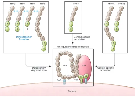
What are FHRs? As Tortajada et al. note in this issue (8), these proteins have been known for many years to be part of a struc-turally related family including the larger factor H protein (FH; encoded by CFH). FHRs are encoded by a series of genes adja-cent to CFH on human chromosome 1 and also contain short consensus repeat (SCR) domains with homology to subregions of FH (9, 10). As genetic variants character-ized by the absence of CFHR1 and CFHR3
(ΔCFHR3-CFHR1), or of CFHR1 and CFHR4
(ΔCFHR1-CFHR4), are relatively common in the
human population, the proteins encoded by these genes do not appear to be required for human development or immune com-petence under normal conditions. Nev-ertheless, an increasing number of pro-tective or risk associations of deletions or variants of these genes have been reported with human diseases (9, 10). Among these are AMD, for which ΔCFHR3-CFHR1 is a highly
penetrant protective factor (11), and aHUS, for which ΔCFHR3-CFHR1 is a disease risk factor
and is associated with autoantibodies that interfere with FH regulatory function (12).
Duplication of sequences within CFHR1 is linked to C3 glomerulopathy
either alone or in association with FHR2 and FHR5, primarily acts on surfaces to block FH binding, resulting in complement deregulation and enhanced local activation. Presumably, altered function of the FHR1 mutant promoted the development of renal disease in the two patients (8). Copy num-ber variants in FHRs consisting of inter-nal duplication and heterozygous hybrid proteins have been previously identified in patients presenting with renal disease and biopsy findings characteristic of C3G (14, 15). Thus, self-association of FHRs is undoubtedly important in their ability to interfere with FH, which is even more rel-evant because FH itself forms tetramers (16) and other types of oligomers (17) that appear to promote more avid binding to C3 fragments and other surface determinants, including glycosaminoglycans (GAG). the first four SCRs duplicated in tandem.
The authors purified wild-type nonmutated FHR1 from human plasma, and chromato-graphic analysis revealed that rather than a single expected peak, multiple peaks con-tained FHR1. Tortajada et al. showed that FHR1 homo- and hetero-oligomerized with FHR2 and FHR5, but not with FHR3 or FHR4A/B. Next, the authors demonstrated that these complexes compete for the func-tionally essential FH binding activity to surface-fixed C3 fragments that is encom-passed within SCR19–SCR20, impairing the ability of FH to block surface complement activation. Importantly, the FHR1 mutant protein in the patients with C3G assem-bled into unusually large multimeric com-plexes that even more effectively impaired FH function. The conclusion of the study was that FHR1, assembled into multimers variants and mutations. Tortajada and
col-leagues follow that approach in the unusual but informative clinical condition desig-nated C3 glomerulopathy (C3G; ref. 8). C3G is a newly classified pathological entity with glomerular immunohistochemistry that is characterized by C3 accumulation in the absence of the substantial concurrent presence of immunoglobulins (13). Nota-bly, C3G is associated with mutations in C3 and complement components that regulate C3 activation (13).
In the present article, Tortajada and col-leagues describe a mother and son who pre-sented with clinical syndromes and renal biopsies consistent with C3G (8). Extensive genetic and protein analyses demonstrated that an internal duplication within CFHR1
[image:3.585.63.536.81.420.2]resulted in an aberrant protein containing nine SCRs instead of the normal five, with
Figure 1
in controlling activation in the kidney and eye? A third question is why a deficiency state, such as ΔCFHR3-CFHR1, is protective in
one complement-related disease, like AMD, but a risk factor in another. A fourth is why the FHR family evolved, and, if pathogen interactions were involved, which were the key drivers over eons. Are there additional lessons to be learned by a study of FH and FHRs, in populations with genetic variants, similar to the insights obtained for other complement components?
Finally, other than the potential for ther-apeutic manipulation in humans, how do we learn more about the roles of individ-ual FHRs, such that the various hypothe-ses illustrated in Figure 1 could be tested in a whole animal system? Mice appar-ently express a set of FHRs with structural features similar to those of their human counterparts (25, 26). If their activities can be demonstrated to be orthologous, the roles shown in Figure 1 should be able to be effectively tested in models of ophthal-mologic, renal, arthritic, and other disease conditions in which alternative pathway activation is essential to develop tissue injury and modulation of FH activities has been shown to affect tissue injury (reviewed in ref. 1). In addition, enforcing tissue-spe-cific inhibition or overexpression will allow us to better understand unique local tissue-specific effects of these proteins on complement activation.
Acknowledgments
The author acknowledges the important contribution to this commentary of many prior and ongoing discussions with Jonathan Hannan, Joshua Thurman, Kevin March-bank, Nirmal Banda, Alexandra Antonioli, Michael Pangburn, and Viviana Ferreira. Address correspondence to: V. Michael Holers, Division of Rheumatology, Depart-ments of Medicine and Immunology, University of Colorado School of Medi-cine, Mail Stop B115, Aurora, Colorado 80045, USA. Phone: 303.724.7605; Fax: 303.724.7581; E-mail: michael.holers@ ucdenver.edu.
1. Holers VM. The spectrum of complement alter-native pathway-mediated diseases. Immunol. Rev.
2008;223:300–316.
2. Ricklin D, Lambris JD. Progress and trends in complement therapeutics. Adv Exp Med Biol.
2013;735:1–22.
3. Varga L, Farkas H. rhC1INH: a new drug for the treatment of attacks in hereditary angioedema caused by C1-inhibitor deficiency. Expert Rev Clin Immunol. 2011;7(2):143–153.
4. Rother RP, Rollins SA, Mojcik CF, Brodsky RA, Bell
nism to bypass this key innate immune rec-ognition process (reviewed in ref. 10). For example, FHR2 and FHR5 are captured by the complement regulator–acquiring pro-teins (CRASPs) of serum-resistant Borrelia burgdorferi and modulate complement activation (22). Further work to explore the roles of FHR interactions with patho-gens will benefit our understanding of the diverse roles these proteins play in the innate immune response.
Unanswered questions and potential approaches to demonstrating in vivo roles of the FHRs
Unquestionably, the work by Tortajada and colleagues (8), as well as the related report by Goicoechea de Jorge and colleagues (18), have provided important insights into the very likely primary functions of FHR1, FHR2, and FHR5 in deregulating FH on specific binding sites. These observations have built on rapidly increasing under-standing of FH structure/function in a way that has allowed investigators to use struc-tural similarities and differences compared with FHRs in order to develop innovative hypotheses. Likewise, the impressive effect on the field of the study of mutant forms of complement molecules that exhibit clin-ically important phenotypes continues to be validated by this report. Nevertheless, important questions remain with regard to the FHR family.
The first question one could ask is whether all FHRs act in the same manner. For example, FHR4 is not a part of the complexes described and has been recently demonstrated to promote complement alternative pathway activation by serving as a platform for activation (23). Indeed, the activities of FHRs may be quite con-text dependent; one way to consider the various demonstrated and postulated roles of FHRs is outlined in Figure 1. A second question concerns what surface deter-minants are important in binding of the deregulatory and regulatory FHRs. Why are the kidney, and the retina in AMD, the pri-mary targets of damage in these potentially systemically injurious processes? Why are other organs, such as the liver (where the majority of FH and FHRs are produced) or the joints (where FH plays a key role in con-trolling activation on the cartilage surface), spared (24)? Does this situation have to do with some as-yet unrecognized unique characteristics in the matrix, cell mem-brane constituents, or organ physiology that make FHRs particularly important
Other recent, highly informative structural studies of FHRs
Tortajada et al. acknowledge the impor-tance of the recent crystallography and structure/function studies by Goicoechea de Jorge and colleagues (18). Strikingly, that group recently reported that FHR1, FHR2, and FHR5 contain dimerization motifs that are highly conserved and allow head-to-tail dimer formation. They also found predominant homo- and het-erodimers of the native proteins in serum and demonstrated more avid interactions of the multimers with C3 fragments, both in vitro and in vivo. Additionally, the complexes exhibited complement deregulation by acting as competitive antagonists of FH binding. Analysis of the variant FHR5 protein, found to be associated with C3G in a Cypriot popu-lation (15), similarly demonstrated a sub-stantially increased ability to impair FH binding and deregulate surface comple-ment activation.
What are the primary roles of FHR1 and other FHRs in modulating complement activation?
In contrast to the reports by Tortajada et al. (8) and Goicoechea de Jorge et al. (18), many prior studies of FHRs have focused not on deregulating FH, but on the poten-tial role of a subset of these proteins in directly regulating complement activation and impairing or promoting activation at specific steps of the cascade. For exam-ple, FHR2 and FHR4 have been shown to enhance FH cofactor activity, and FHR5 exhibits weak cofactor and decay-accelerat-ing activities (reviewed in ref. 10). Likewise, FHR1 was previously shown to inhibit C5 convertase and terminal complex forma-tion (19), and FHR3 was demonstrated to inhibit C3 convertase and display antiin-flammatory activity by blocking C5a gen-eration (20). As the structure/function of FH has become increasingly understood (21), it has become more challenging to reconcile the reported regulatory activi-ties of FHRs with the structural determi-nants, other than through unusual steric hindrance or allosteric interaction mech-anisms. Nevertheless, these prior findings should be taken into account in the overall understanding of FHR family activities.
mecha-114(12):2439–2447.
20. Fritsche LG, et al. An imbalance of human comple-ment regulatory proteins CFHR1, CFHR3 and fac-tor H influences risk for age-related macular degen-eration. Hum Mol Genet. 2010;19(23):4694–4704. 21. Morgan HP, et al. Structural basis for engagement
by complement factor H of C3b on a self surface.
Nat Struct Mol Biol. 2011;18(4):463–470. 22. Siegel C, et al. Complement factor H-related
pro-teins CFHR2 and CFHR5 represent novel ligands for the infecction-associated CRASP proteins of Borrelia burgdorferi. PLoS One. 2010;5(10):e13519. 23. Hebecker M, Józsi M. Factor H-related protein 4 activates complement by serving as a platform for the assembly of alternative pathway C3 convertase via its interaction with C3b protein. J Biol Chem. 2012;287(23):19528–19536.
24. Banda NK, et al. Essential role of surface-bound complement factor H in controlling immune complex-induced arthritis. J Immunol. 2013; 190(7):3560–3569.
25. Vik DP, Muñoz-Cánoves P, Kozono H, Martin LG, Tack BF, Chaplin DD. Identification and sequence analysis of four complement factor H-related transcripts in mouse liver. J Biol Chem.
1990;265(6):3193–3201.
26. Hellwage J, et al. Two factor H-related proteins from the mouse: expression analysis and func-tional characterization. Immunogenetics. 2006; 58(11):883–893.
associated with lower risk of age-related macular degeneration. Nat Genet. 2006;38(10):1173–1177. 12. Skerka C, Zipfel PF. Complement factor H
related proteins in immune diseases. Vaccine.
2008;26(suppl 8):I9–I14.
13. Fakhouri F, Frémeaux-Bacchi V, Noël LH, Cook HT, Pickering MC. C3 glomerulopathy: a new clas-sification. Nat Rev Nephrol. 2010;6(8):494–499. 14. Malik TH, et al. A hybrid CFHR3-1 gene causes
familial C3 glomerulopathy. J Am Soc Nephrol.
2012;23(7):1155–1160.
15. Gale DP, et al. Identification of a mutation in com-plement factor H-related protein 5 in patients of Cypriot origin with glomerulonephritis. Lancet. 2010;376(9743):794–801.
16. Pangburn MK, Rawal N, Cortes C, Alam MN, Fer-reira VP, Atkinson MA. Polyanion-induced self-as-sociation of complement factor H. J Immunol.
2009;182(2):1061–1068.
17. Perkins SJ, Nan R, Li K, Khan S, Miller A. Com-plement Factor H–ligand interactions: Self-asso-ciation, multivalency and dissociation constants.
Immunobiology. 2012;217(2):281–297.
18. Goicoechea de Jorge E, et al. Dimerization of complement factor H-related proteins modulates complement activation in vivo. Proc Natl Acad Sci U S A. 2013;110(12):4685–4690.
19. Heinen S, et al. Factor H-related protein 1 (CFHR-1) inhibits complement C5 convertase activity and terminal complex formation. Blood. 2009; L. Discovery and development of the complement
inhibitor eculizumab for the treatment of parox-ysmal nocturnal hemoglobinuria. Nat Biotechnol.
2007;25(11):1256–1264.
5. Schmidtko J, Peine S, El-Housseini Y, Pascual M, Meier P. Treatment of atypical hemolytic uremic syndrome and thrombotic microangiopathies: a focus on eculizumab. Am J Kidney Dis. 2013; 61(2):289–299.
6. Gehrs KM, Anderson DH, Johnson LV, Hageman GS. Age-related macular degeneration — emerging pathogenetic and therapeutic concepts. Ann Med.
2006;38(7):450–471.
7. Ricklin D, Hajishengallis G, Yang K, Lambris JD. Complement: a key system for immune sur-veillance and homeostasis. Nat Immunol. 2010; 11(9):785–797.
8. Tortajada A, et al. C3 glomerulopathy–associ-ated CFHR1 mutation alters FHR oligomeriza-tion and complement regulaoligomeriza-tion. J Clin Invest.
2013;123(6):2434–2446.
9. de Cordoba SR, Tortajada A, Harris CL, Morgan BP. Complement dysregulation and disease: from genes and proteins to diagnostics and drugs.
Immuno biology. 2012;217(11):1034–1046. 10. Józsi M, Zipfel PF. Factor H family proteins and
human diseases. Trends Immunol. 2008;29(8):380–387. 11. Hughes AE, Orr N, Esfandiary H, Diaz-Torres M,
Goodship T, Chakravarthy U. A common CFH haplotype,with deletion of CFHR1 and CFHR3,is
Transplant rejection and paradigms lost
Terry B. Strom
Beth Israel Deaconess Medical Center, Harvard Medical School, Boston, Massachusetts, USA.
During transplant rejection, migrating T cells infiltrate the grafted organ,
but the signals that direct this migration are incompletely understood.
In this issue of the JCI, Walch et al. debunk two classical paradigms
concerning transplant rejection, with important consequences for the design
of antirejection therapeutics.
Transplant rejection begins with the migration and influx of recipient T cells into the transplant. Many, but not all, of these T cells bear TCRs specific for donor alloantigen. If migration to and infiltra-tion of the transplant by tissue-damag-ing anti-donor T cells can be neutralized, even temporarily, the recipient anti-donor allograft response would be significantly weakened and more amenable to low-dose or tolerizing therapies.
The current dogma explaining the initial phases of the allograft response is based on the leukocyte migration paradigm (1). Chemokines, a family of small transmem-brane proteins that attract migratory T cells bearing specific receptors for them,
are believed to draw T cells to the trans-plant. Then, the interaction of T cell sur-face integrins with molecules present on donor endothelial cells retain T cells and enable their transmigration into the trans-plant. When chemokines interact with chemokine receptors, chemokine recep-tor–associated Gαi proteins are activated,
and a pertussis toxin–sensitive signaling cascade is initiated. The downstream Gαi
signaling pathway induces conforma-tional changes in the integrins expressed on chemokine-activated T cells (2). These changes enhance the affinity of integrins such as VLA-4 for surrounding tissues, thought to be endothelial venules, of the transplant (Figure 1).
Chemokine production is markedly ampli-fied within inflamed tissues, and transplant procedures, especially those involving deceased donors, inevitably lead to ische-mia-reperfusion injury, resulting in
inflam-mation of the endothelium and chemokine expression (3, 4). Thus, it has been widely believed that chemokines, which regulate T cell traffic in lymphoid tissue, also play a pivotal role in transplant rejection.
Against this theoretical backdrop, and influenced by the surprisingly weak thera-peutic effect of antibodies directed against chemokine receptors upon transplant sur-vival (5–7), Walch et al. tested the authen-ticity of the classical leukocyte migration paradigm in mouse cardiac and renal trans-plant models (8). In short, they confirmed an important role for the integrin VLA-4, but found that the paradigm did not hold true for the role of chemokines in directing the massive influx of recipient anti-donor T cells in the allograft response.
Chemokine receptor signaling in the allograft response
To investigate the role of chemokine recep-tors in the allograft response, Walch et al. used passive T cell transfer models in which recipient strain T cells are injected into syngeneic lymphopenic hosts. They found that infiltration into allografts by pertussis toxin–treated memory or acti-Conflict of interest: The author has declared that no
conflict of interest exists.