Andrew J. Whittle, Antonio Vidal-Puig
J Clin Invest.
2012;
122(3)
:804-807.
https://doi.org/10.1172/JCI62595
.
Thermogenesis in brown adipose tissue (BAT) is well characterized as being under the
control of the sympathetic nervous system. The energy-burning capacity of BAT makes it an
attractive target for anti-obesity therapies. However, previous attempts to manipulate BAT’s
sympathetic activation have lacked specificity. In this issue of the
JCI
, Bordicchia et al.
provide new data indicating that cardiac natriuretic peptides (NPs) are also able to activate
thermogenic machinery in adipose tissue. Their findings suggest a novel strategy to
increase energy dissipation in adipose tissue, independent of adrenergic receptors.
Commentary
Find the latest version:
Medicine, The University of Tokyo, 7-3-1 Hongo, Bunkyo-ku, Tokyo 113-0033, Japan. Phone: 81.3.5841.3431; Fax: 81.3.5841.3393; E-mail: [email protected].
1. Covacci A, Telford JL, Del Giudice G, Parsonnet J, Rappuoli R. Helicobacter pylori virulence and genet-ic geography. Science. 1999;284(5418):1328–1333. 2. Blaser MJ. An endangered species in the stomach.
Sci Am. 2005;292(2):38–45.
3. Marshall BJ, Warren JR. Unidentified curved bacilli in the stomach of patients with gastritis and peptic ulceration. Lancet. 1984;1(8390):1311–1315.
4. Superbaum S, Michetti P. Helicobacter pylori infec-tion. N Engl J Med. 2002;347(15):1175–1186.
5. Chen Y, Blaser MJ. Inverse associations of Helico-bacter pylori with asthma and allergy. Arch Intern Med. 2007;167(8):821–827.
6. Strachan DP. Hay fever, hygiene, and household size. BMJ. 1989;299(6710):1259–1260.
7. Arnold IC, et al. Helicobacter pylori infection pre-vents allergic asthma in mouse models through the induction of regulatory T cells. J Clin Invest.
2011;121(8):3088–3093.
8. Oertli M, et al. DC-derived IL-18 drives Treg dif-ferentiation, murine Helicobacter pylori–specific immune tolerance, and asthma protection. J Clin Invest. 2012;122(3):1082–1096.
9. Steinman RM, Cohn ZA. Identification of a novel cell type in peripheral lymphoid organs of mice. I. Morphology, quantitation, tissue distribution.
J Exp Med. 1973;137(5):1142–1162.
10. Maldonado RA, von Andrian UH. How tolerogenic dendritic cells induce regulatory T cells. Adv Immu-nol. 2010;108:111–165.
11. Watanabe T, et al. NOD1 contributes to mouse host defense against Helicobacter pylori via induction of type I IFN and activation of the ISGF3 signaling pathway. J Clin Invest. 2010;120(5):1645–1662. 12. Gu Y, et al. Activation of Interferon-gamma
induc-ing factor mediated by Interleukin-1beta convert-ing enzyme. Science. 1997;275(5297):206–209.
13. Yoshimoto T, et al. Nonredundant roles for CD1d-restricted natural killer T cells and conventional CD4+ T cells in the induction of immunoglobulin E antibodies in response to interleukin 18 treat-ment of mice. J Exp Med. 2003;197(8):997–1005. 14. Koch MA, Tucker-Heard G, Perdue NR, Killebrew
JR, Urdahl KB, Campbell DJ. The transcription factor T-bet controls regulatory T cell homeosta-sis and function during type 1 inflammation. Nat Immunol. 2009;10(6):595–602.
15. Zheng Y, et al. Regulatory T-cell suppressor pro-gram co-opts transcription factor IRF-4 to control Th2 responses. Nature. 2009;458(7236):351–356.
16. Sakaguchi S, Yamaguchi T, Nomura T, Ono M. Reg-ulatory T cells and immune tolerance. Cell. 2008; 133(5):775–787.
17. Schreiber S, et al. The spatial orientation of Helico-bacter pylori in the gastric mucus. Proc Natl Acad Sci U S A. 2004;101(14):5024–5029.
18. Petersen AM, Krogfelt KA. Helicobacter pylori: an invading microorganism? A review. FEMS Immunol Med Microbiol. 2003;36(3):117–126.
19. Ito T, et al. Helicobacter pylori invades the gas-tric mucosa and translocates to the gastric lymph nodes. Lab Invest. 2008;88(6):664–681.
20. Nagai S, et al. Role of Peyer’s patches in the induc-tion of Helicobacter pylori-induced gastritis. Proc Natl Acad Sci U S A. 2007;104(21):8971–8976. 21. Kiriya K, et al. Essential role of Peyer’s patches in
the development of Helicobacter-induced gastritis.
Int Immunol. 2007;19(4):435–446.
NPs — heart hormones that regulate brown fat?
Andrew J. Whittle and Antonio Vidal-Puig
University of Cambridge Metabolic Research Laboratories, Institute of Metabolic Science, Cambridge, United Kingdom.
Thermogenesis in brown adipose tissue (BAT) is well characterized as being
under the control of the sympathetic nervous system. The energy-burning
capacity of BAT makes it an attractive target for anti-obesity therapies.
How-ever, previous attempts to manipulate BAT’s sympathetic activation have
lacked specificity. In this issue of the JCI, Bordicchia et al. provide new data
indicating that cardiac natriuretic peptides (NPs) are also able to activate
thermogenic machinery in adipose tissue. Their findings suggest a novel
strategy to increase energy dissipation in adipose tissue, independent of
adrenergic receptors.
Conflict of interest: The authors have declared that no conflict of interest exists.
Citation for this article: J Clin Invest. 2012; 122(3):804–807. doi:10.1172/JCI62595. Regulation of the thermogenic program via p38 MAPK
Obesity is a growing epidemic that places an impossible burden on the health care systems of both developed and develop-ing countries across the globe, due to the negative effects of excess fat accumulation on multiple organs. Fat is stored mainly in the well-described white adipose tissue (WAT), but there exists a second fat reser-voir known as brown adipose tissue (BAT). Maintenance of core body temperature is essential to all mammals, and brown fat is highly specialized to generate heat. In smaller organisms such as rodents, it is well established that brown adipocytes perform
an essential role in this process, and there is now substantial evidence that BAT depots exist and are active in adult humans (1–3). Following exposure to cold temperatures, the brain coordinates increased activation of BAT via the sympathetic nervous sys-tem (SNS). Brown adipocytes in BAT and populations of “brown-like” adipocytes in WAT respond by increasing transcription of genes important for mobilizing and oxi-dizing lipids to generate heat. This genetic response is known as the thermogenic pro-gram, the key components of which are lipases and the specialized mitochondrial transporter uncoupling protein 1 (UCP1). UCP1 enables the separation of lipid oxi-dation from ATP production, allowing a higher metabolic rate and the conversion of nutritional energy to heat (4). Under-standing the regulation of the thermogenic program is of particular importance, as its
induction is fundamental to not just ther- mogenic activity but also the differentia-tion of adipocytes toward a more “brown” cell fate (5). In this issue of the JCI, Bordic-chia, Collins, and colleagues demonstrate a novel mechanism for activating this genetic program in adipose tissue (6).
Adrenergic signaling is the classical activator of the thermogenic machin-ery. Catecholamines drive increased intracellular cAMP levels, directly acti-vating cAMP-dependant protein kinase (PKA), which subsequently phosphory-lates hormone-sensitive lipase (HSL). But as shown previously by the Collins team, PKA can also phosphorylate kinas-es in the p38 MAPK pathway (7, 8). The
UCP1 promoter contains motifs bound
by multiple transcription factors, such as activating transcription factor–2/cAMP response element–binding (ATF2/CREB) and PPARγ, the transcriptional effects of which are all enhanced by increased p38 MAPK activation. Pharmacological inhibition of p38 MAPK is sufficient to negate the effects of adrenergic stimula-tion on UCP1 transcription (7).
system have proved successful at reduc-ing body weight in overweight individuals (9, 10). However, the effective compounds (ephedrine and sibutramine) have also been found to significantly increase the risk of heart attack and stroke, particularly in obese patients (11, 12). This is largely attributed to the widespread expression of adrenergic receptors throughout the cardiovascular system and the indiscrimi-nate manner in which these compounds act upon them.
Cardiac natriuretic peptides activate thermogenesis
Natriuretic peptides (NPs) are hormones produced in the heart that traditionally act on the renal system to maintain fluid homeostasis and exist in atrial (ANP) and ventricular (BNP) forms. They signal through an active receptor (NPRA) and a clearance receptor (NPRC), with the regula- tion of both modulating NP activity. Inter-estingly, NPs increase cyclic GMP levels to activate cGMP-dependant protein kinase (PKG), which shares homology with PKA in terms of its potential substrates (13). Bordicchia et al. provide evidence that in addition to regulating hemodynamics, NP signaling is also an important part of the thermogenic response in adipocytes (6).
The authors begin by demonstrating that mice lacking NPRC — the negative regulator of NP activity — have reduced fat deposition in WAT and less lipid accumulation in BAT, perhaps suggesting increased thermogenic
activity. NPRC-null mice also possess ingui-nal WAT that displays a more “brown-like” morphology and have increased expression of thermogenic genes in both WAT and BAT. Using a human-derived adipocyte cell line, the authors proceed to show that treatment with ANP results in induction of thermogenic and mitochondrial genes and that these increases are dependent on PKG signaling. At a functional level, these transcriptional changes induced by ANP increase the capacity for uncoupled respi-ration in acutely treated adipocytes. This effect appears more potent following a lon-ger pretreatment, suggesting chronically increased ANP signaling may bring about adaptive changes in BAT (6).
Bordicchia et al. progress to show that NPs elicit this response by increasing p38 MAPK signaling and phosphorylation of ATF2, directly increasing UCP1 transcrip-tion in adipocytes. The effects of ANP on transcription of UCP1 and indeed other thermogenic genes appear to be additive to, and perhaps synergistic with, those increases seen following classical adren- ergic stimulation. Compared with physi-ological stimulation, the response to the relatively high dose of agonist used in the studies (10 nM isoproterenol) dem- onstrates a second important character-istic of adipocytes: that there is room for further amplification of the thermogenic machinery above the level resulting from a potent adrenergic stimulus, and this can
be induced using NPs. Finally, the authors add support to this hypothesis by treating mice with BNP and observing an increase in energy expenditure accompanied by increased thermogenic protein levels in both BAT and WAT (6).
An increasing pool of novel BAT activators
These new findings provide further sup- port for the hypothesis that there are addi- tional mechanisms for activating thermo-genesis in BAT that sidestep the need for manipulating the SNS. NPs appear to pro-vide an opportunity to increase the scale of the thermogenic response in BAT and WAT without altering endogenous adren-ergic stimulation.
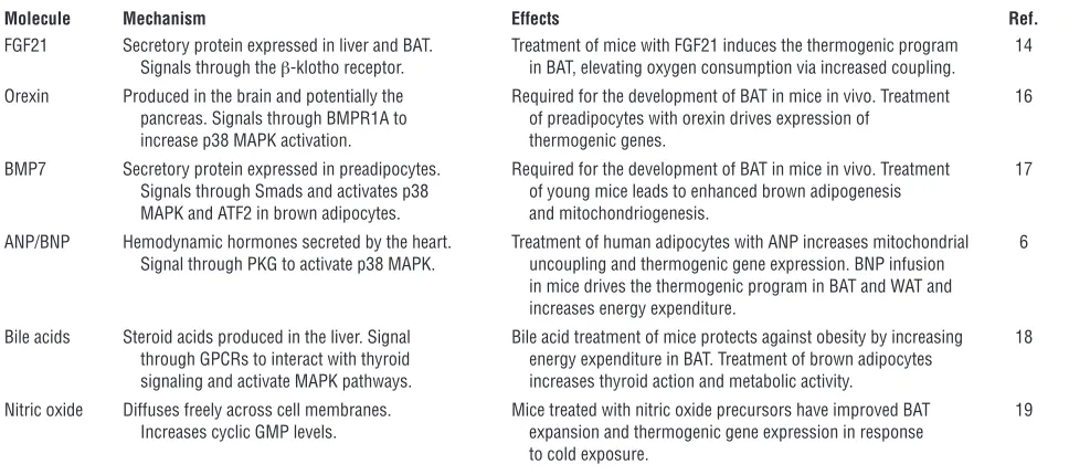
[image:3.585.52.536.114.329.2]Over the past five years, a number of molecules have also been shown to pos-sess thermogenic properties not reliant on adrenergic receptors (Table 1). In a remark-ably similar fashion to NP, fibroblast growth factor 21 (FGF21) has been found to induce thermogenic program genes and increase uncoupled respiration in brown adipocytes via the β-klotho receptor (14, 15). Orexins have been demonstrated to signal through bone morphogenetic pro-tein receptor–1a (BMPR1A) and, like NP, feed into the p38 MAPK pathway to induce thermogenic genes and drive brown adi-pogenesis (16). One bone morphogenetic protein (BMP7) itself has also been shown to stimulate a similar “browning” effect in
Table 1
Non-adrenergic activators of the thermogenic program
Molecule Mechanism Effects Ref.
FGF21 Secretory protein expressed in liver and BAT. Treatment of mice with FGF21 induces the thermogenic program 14 Signals through the β-klotho receptor. in BAT, elevating oxygen consumption via increased coupling.
Orexin Produced in the brain and potentially the Required for the development of BAT in mice in vivo. Treatment 16 pancreas. Signals through BMPR1A to of preadipocytes with orexin drives expression of
increase p38 MAPK activation. thermogenic genes.
BMP7 Secretory protein expressed in preadipocytes. Required for the development of BAT in mice in vivo. Treatment 17 Signals through Smads and activates p38 of young mice leads to enhanced brown adipogenesis
MAPK and ATF2 in brown adipocytes. and mitochondriogenesis.
ANP/BNP Hemodynamic hormones secreted by the heart. Treatment of human adipocytes with ANP increases mitochondrial 6 Signal through PKG to activate p38 MAPK. uncoupling and thermogenic gene expression. BNP infusion
in mice drives the thermogenic program in BAT and WAT and increases energy expenditure.
Bile acids Steroid acids produced in the liver. Signal Bile acid treatment of mice protects against obesity by increasing 18 through GPCRs to interact with thyroid energy expenditure in BAT. Treatment of brown adipocytes
signaling and activate MAPK pathways. increases thyroid action and metabolic activity.
Nitric oxide Diffuses freely across cell membranes. Mice treated with nitric oxide precursors have improved BAT 19
preadipocytes as they differentiate, again via increased p38 MAPK activation and induction of the thermogenic program (17). Other factors such as bile acids, which increase thyroid hormone signaling (18), and nitric oxide levels (19), which like NP increase cGMP levels, have also been shown to enhance the thermogenic program in brown adipocytes.
These developments seem to suggest that the p38 MAPK pathway exists as a physiological mechanism used by endog-enous metabolic hormones and secretory molecules to “tap into” the adrenergic axis. BAT has a particular requirement for such mechanisms because it is a highly dynamic and plastic tissue. Adaptive alter-ation of their general level of sensitivity to adrenergic stimulation would confer greater flexibility to brown adipocytes. BAT would still respond to acute CNS-informed changes in thermogenic demand
but would do so in the context of longer-term changes in environmental conditions (Figure 1). For instance, in response to a high calorie diet, thermogenesis increases due to alterations in SNS tone to BAT. The level of increase, though, is greater when animals are housed in cold environ-ments (20). One might envisage, the effect of the dietary stimulus would be reduced at lower environmental temperatures, but this is not the case, demonstrating mainte-nance of acute responsiveness alongside a sustained increase in thermogenic activity (20). In support of this mechanism, Bor- dicchia et al. show that levels of circulat-ing NP and expression of NPRA increase in BAT, while NPRC expression decreases following cold exposure (6). Of note, car-diac hypertrophy is a classical result of cold adaptation in rodents and may be a biological indicator for BAT to expand via increased NP signaling.
Potential for therapeutic activation of BAT
The authors of the present study did not investigate the long-term effects of NP on energy balance or body weight. Nor did they administer NPs to mice that were already obese to assess their effectiveness as a weight loss strategy. Another impor-tant question that remains unanswered is whether NPs alter adrenergic sensitivity in other organs. If this is the case, it may ren-der them as unfavorable as sibutramine or ephedrine for long-term use in humans.
[image:4.585.46.343.80.480.2]However, the ability of NPs to modulate the effects of endogenous SNS activation in BAT makes them an exciting candidate for further investigation. In humans, we are beginning to understand that there is a negative correlation between BMI and fat mass and the detectability of active BAT (2). Thus, any molecule that was able to “trick” BAT into sensing its well-warmed human
Figure 1
Hypothesized role of NPs as chronic regula-tors of BAT. Alterations in cardiac demand
host was actually in a long-term cold envi- ronment may well have favorable bioener-getic effects in overweight individuals.
Acknowledgments
Andrew J. Whittle and Antonio Vidal-Puig are supported by the Biotechnology and Biological Sciences Research Council (BBSRC), the Medical Research Council (MRC), Wellcome Trust, and the European Commission (FP7 BetaBAT).
Address correspondence to: Antonio Vidal-Puig, University of Cambridge Metabolic Research Laboratories, Level 4, Institute of Metabolic Science, Box 289, Addenbrooke’s Hospital, Cambridge CB2 0QQ, United Kingdom. Phone: 44.1223.762790; Fax: 44.1223.330598; E-mail: [email protected].
1. Cypess AM, et al. Identification and importance of brown adipose tissue in adult humans. N Engl J Med. 2009;360(15):1509–1517.
2. Ouellet V, et al. Outdoor temperature, age, sex, body mass index, and diabetic status determine the prevalence, mass, and glucose-uptake activity of 18F-FDG-detected BAT in humans. J Clin Endo-crinol Metab. 2011;96(1):192–199.
3. Saito M, et al. High incidence of metabolically active brown adipose tissue in healthy adult humans: effects of cold exposure and adiposity. Diabetes. 2009;58(7):1526–1531.
4. Cannon B, Nedergaard J. Brown adipose tissue: function and physiological significance. Physiol Rev. 2004;84(1):277–359.
5. Seale P, et al. PRDM16 controls a brown fat/skeletal muscle switch. Nature. 2008;454(7207):961–967. 6. Bordicchia M, et al. Cardiac natriuretic peptides
act via p38 MAPK to induce the brown fat thermo-genic program in mouse and human adipocytes.
J Clin Invest. 2012;122(3):1022–1036.
7. Cao W, et al. p38 mitogen-activated protein kinase is the central regulator of cyclic AMP-dependent tran-scription of the brown fat uncoupling protein 1 gene.
Mol Cell Biol. 2004;24(7):3057–3067.
8. Cao W, Medvedev AV, Daniel KW, Collins S. beta- Adrenergic activation of p38 MAP kinase in adipo-cytes: cAMP induction of the uncoupling protein 1 (UCP1) gene requires p38 MAP kinase. J Biol Chem. 2001;276(29):27077–27082.
9. Astrup A, Lundsgaard C, Madsen J, Christensen NJ. Enhanced thermogenic responsiveness dur-ing chronic ephedrine treatment in man. Am J Clin Nutr. 1985;42(1):83–94.
10. Finer N, Bloom SR, Frost GS, Banks LM, Griffiths J. Sibutramine is effective for weight loss and dia- betic control in obesity with type 2 diabetes: a ran-domised, double-blind, placebo-controlled study.
Diabetes Obes Metab. 2000;2(2):105–112.
11. Haller CA, Benowitz NL. Adverse cardiovascular and central nervous system events associated with dietary supplements containing ephedra alkaloids.
N Engl J Med. 2000;343(25):1833–1838.
12. Torp-Pedersen C, et al. Cardiovascular responses to weight management and sibutramine in high-risk subjects: an analysis from the SCOUT trial. Eur Heart J. 2007;28(23):2915–2923.
13. Misono KS. Natriuretic peptides and their recep-tors. FEBS J. 2011;278(11):1791.
14. Hondares E, Rosell M, Gonzalez FJ, Giralt M, Iglesias R, Villarroya F. Hepatic FGF21 expression is induced at birth via PPARalpha in response to milk intake and contributes to thermogenic acti-vation of neonatal brown fat. Cell Metab. 2010; 11(3):206–212.
15. Ogawa Y, et al. BetaKlotho is required for metabol-ic activity of fibroblast growth factor 21. Proc Natl Acad Sci U S A. 2007;104(18):7432–7437. 16. Sellayah D, Bharaj P, Sikder D. Orexin is required
for brown adipose tissue development, differentia-tion, and function. Cell Metab. 2011;14(4):478–490. 17. Tseng YH, et al. New role of bone morphogenetic
protein 7 in brown adipogenesis and energy expen-diture. Nature. 2008;454(7207):1000–1004.
18. Watanabe M, et al. Bile acids induce energy expen-diture by promoting intracellular thyroid hormone activation. Nature. 2006;439(7075):484–489. 19. Petrovic V, Buzadzic B, Korac A, Vasilijevic A,
Jankovic A, Korac B. NO modulates the molecular basis of rat interscapular brown adipose tissue ther-mogenesis. Comp Biochem Physiol C Toxicol Pharmacol. 2010;152(2):147–159.
20. Rothwell NJ, Stock MJ. Influence of environmental temperature on energy balance, diet-induced ther-mogenesis and brown fat activity in ‘cafeteria’-fed rats. Br J Nutr. 1986;56(1):123–129.
DYRK1A in Down syndrome:
an oncogene or tumor suppressor?
Yehudit Birger1 and Shai Izraeli1,2
1Functional Genomics and Department of Pediatric Hemato-oncology, Edmond and Lily Safra Children's Hospital, Sheba Medical Center,
Tel Hashomer, Ramat Gan, Israel. 2Sackler Faculty of Medicine, Tel Aviv University, Tel Aviv, Israel.
Children with Down syndrome (DS) have a markedly increased risk of
devel-oping acute megakaryoblastic leukemia (AMKL) and acute lymphoblastic
leukemia compared with that of children without DS. Despite recent
break-throughs, it is not clear which genes on chromosome 21, the chromosome
that is trisomic in individuals with DS, cause this predisposition. In this
issue of the JCI, Malinge et al. report their loss- and gain-of-function
experi-ments in mouse and human cells that show that increased expression of the
kinase encoded by the chromosome 21 gene DYRK1A suppresses the nuclear
factor of activated T cells pathway and promotes AMKL. Interestingly, the
same protein has been suggested to contribute to the reduced risk of
epi-thelial cancers in adults with DS, leading to the possibility that it could be
proleukemic in children and antitumorigenic in adults.
Down syndrome (DS) is one of the most common genetic birth defects, affect-ing about 1 in 800 newborns a year in the
United States. It is caused by constitutional trisomy 21 (cT21). The symptoms of DS are heterogeneous, ranging from mild to severe, as are the associated medical conditions. One of the associated medical conditions is leukemia — children with DS have a mark-edly higher risk of developing both acute lymphoblastic leukemia (ALL) and acute
myeloid leukemia than do children who do not have DS. This is in sharp contrast with the reduced prevalence of epithelial cancers in adult individuals with DS (1). Thus, cT21 is proleukemogenic during the pediatric period but antitumorigenic in adulthood. In this issue of the JCI , Malinge et al. pro- vide new insight into the functional contri-bution of trisomy 21 to leukemogenesisthat may have therapeutic implications (2).
DS-associated myeloid leukemias and the role of cT21
DS-associated myeloid leukemias evolve in a unique, multistep clinical pattern (Figure 1 and reviewed in ref. 3). Epide-miologic studies have indicated that five percent of newborns with DS have tran-sient myeloproliferative disorder (TMD), a condition that is characterized by the
clonal expansion of immature megakaryo-Conflict of interest: The authors have declared that no conflict of interest exists.