Utility and efficacy of navigation system use in
interventional radiology
Jatin Kaicker, Sriharsha Athreya
Department of Medical Imaging, McMaster University, Hamilton, Canada Email: [email protected]
Received 16 July 2013; revised 16 August 2013; accepted 23 August 2013
Copyright © 2013 Jatin Kaicker, Sriharsha Athreya. This is an open access article distributed under the Creative Commons Attribu-tion License, which permits unrestricted use, distribuAttribu-tion, and reproducAttribu-tion in any medium, provided the original work is properly cited.
ABSTRACT
Background: To ensure precision and accuracy dur- ing interventional radiologic (IR) procedures, naviga- tion systems are utilized. There are four main catego- ries of guidance systems that can be used to assist in IR procedures: optical system, electromagnetic (EM) tracking, Cone Bean Computer Tomography (CBCT) and Magnetic Navigation system. Objective: The pur- pose of this report is to examine some current medical literature to present an impression as to the state of navigation system use in interventional radiology. Methods: Three health databases were selected: Pub- med, Embase and OVID Medline, with the search terms “Interventional Radiology” and “Navigation System” being used. All included studies were pre- sented in English. Studies were excluded if they did not pertain to navigation systems in interventional ra- diology, were in a language other than English, pre- sented an abstract only or solely discussed interven- tional cardiology. Results: General themes emerged within the literature for the advantages of navigation system use including benefits to interventional radi- ologic procedures, increased patient accuracy and re- duced procedure time and the potential for reduction in costs. Increased radiation exposure, problems ac- counting for respiratory motion and sterility remain issues for navigation system use. Conclusion: With potential to better standardize treatment using navi- gation systems, patients can have access to up-to-date technology for treatment. To ensure the highest stan- dard of care, navigation systems should be used by in- terventional radiologists only. As indications and cli- nical efficacy are frequently being defined for naviga- tion system use in interventional radiology, continual review of the published literature and large clinical trials for each system should be pursued.
Keywords:Interventional Radiology; Navigation System; Electromagnetic tracking; Cone Bean Computer Tomography
1. INTRODUCTION
Interventional radiology (IR) has been increasingly in- volved in minimally invasive diagnostic and therapeutic care of patients. The scope of practice for IR ranges from placement of catheters and needles to the treatment of tumors in patients using image guided technology [1]. The various techniques utilized by interventional radio- logists provide symptomatic relief, treatment for certain cancers and increases patient survival [2]. Medical im- aging in interventional radiology supports image guided therapy by assisting with pre-procedural planning, intra- procedural targeting, monitoring, control and post-pro- cedure assessment [3]. Procedural execution depends on the skill set of the Interventional Radiologist. These phy- sicians need to be skilled in interventional techniques and diagnostic interpretation of images while possessing a solid foundation of anatomy and patient care [4].
Interventional radiologists use sophisticated manual skills to manipulate devices in order to target specific tis- sue. There are a few navigation systems that are being used to enhance conventional IR by helping to guiding needles and guide-wires, in some cases using real time spatial feedback [1]. There are four main categories of guidance systems that can be used to assist IR: Optical system, Electromagnetic (EM) tracking, Cone Bean Com- puter Tomography (CBCT) and Magnetic Navigation. There are some indications for the use of each navigation systems and they possess both strengths but also draw- backs in their use.
examine some of the current medical literature available on navigation systems for interventional radiologic pro- cedures. The relevance of this narrative review investiga- tion will be to present an overall impression as to the state of navigation system use in interventional radiol- ogy.
2. MATERIALS AND METHODS
For this investigation, three health databases were se- lected: Pubmed, Embase (1996 to 2012 Week 31) and OVID Medline (1946 to September 2012). All databases were searched on September 10, 2012. The search terms used were “Interventional Radiology” and “Navigation System”. This combination was used to due to its broad scope to adequately represent the parameters for this in- vestigation.
Studies were assessed if they were primary research that was related to the use of the common navigation sys- tems in interventional radiology. All included studies were presented in English. Studies were excluded if they: did not pertain to navigation systems in interventional radi- ology, or were in a language other than English. Litera- ture discussing navigation system use with surgical inter- vention as opposed to interventional radiologic proce- dure was also excluded. Additional reasons for exclusion included journal articles that were not accessible, litera- ture presented as an abstract only, or literature discussing interventional cardiology. Studies identified were initial- ly screened for inclusion or exclusion on the basis of the abstracts.
3. RESULTS
The appropriate studies were assessed for description of the advantages and drawbacks of the common navigation systems used in interventional radiology. Some consis- tent themes from these studies are presented in this sec- tion. For purposes of this report, the following navigation systems will be discussed: optical navigation devices, electromagnetic tracking, C arm based CBCT and Mag- netic Navigation systems.
Optical navigation devices are able to continuously es- timate positions of a moving target. They can be active or passive systems and have the ability to continuously estimate positions. Such devices are useful for three di- mensional trajectory planning and needle placement [5]. However, there are several drawbacks to such devices. It is a bulky apparatus and has limited application in CT guided interventions. These devices also require a free line of sight between the cameras and optical markers [5]. Finally, while the needle hub can be optically tracked, it is difficult to effectively track additional regions of in- serted needles [5].
Electromagnetic (EM) tracking functions on the basis
of sensor coils with differential magnetic fields [1]. One of the major benefits of electromagnetic tracking of ins- truments in interventional radiology is real time display of position and orientation. It also allows interventional radiologists to guide needles toward a target within pre- viously acquired three dimensional images to help avoid critical structures. Studies have found EM tracking able to improve operator confidence and increase accuracy of needle placement especially in lung biopsy and ablation procedures [6]. When other modalities present poor vi- sualization of the target such as an ultrasound, EM track- ing has been found more effective in such cases [6]. Re- peated investigations have also demonstrated enhanced accuracy for procedures using EM tracking and reduced time to procedure. These investigated correlation increas- ed accuracy with reduction in tracking error [6]. Finally, radiation doses have been found to be significantly lower in EM tracking compared with that used in CT guided procedures [6].
One drawback of electromagnetic tracking is the pa- tient’s need to be immobilized for reduction of patient error due to respiratory motion and organ shift during the procedure. Operator time, automation of system setup, ease of use, integration with angiography system and cost effectiveness also remain issues with using electro- magnetic tracking [7]. Furthermore, a trade off exists be- tween positioning fiducial markers too close to the target, jeopardizing sterility and ensuring accuracy of the pro- cedure. Finally, optimal field strength is imperative for the navigation system. EM tracking registration can be compromised by degradation of coil signal if the equip- ment is in the vicinity of metallic structures such as a CT gantry [6].
Moreover, C arm based CBCT when combined with image guided navigation is used for vertebroplasty, gas- trostomy, stenting, biopsies and ablation procedures. The system has been found to increase technical success rates for procedures involving needle placement [6]. C arm ba- sed CBCT has two main factors that hinder more wide- spread adoption of its use. The first is concern for the patients’ accumulated radiation exposure [8]. Secondly, there is an assumption of immobility and rigidity of ana- tomy that is also seen in optical tracking. Additional li- mitations for CBCT include respiratory and patient mo- tion, which can impair registration and therefore decrease accuracy.
navigation systems have also been found to be easier, more accurate and faster to use in the hands of less ex- perienced investigators while allowing for more predict- able procedure times (Table 1) [9].
4. DISCUSSION
Interventional radiology is highly dependent on the pre- cise accuracy of procedures such as needle, catheter and device placement. Navigation systems help guide inter- ventional radiologists for where these devices need to be placed, using spatial feedback in relation to imaging data. Navigation systems are useful when entry angles are challenging or visibility of specific lesions is limited. Image guided interventional treatment requires a multi- disciplinary approach to provide complete patient care. As technology changes, it is imperative to appreciate the new direction of treatment so patients have access to well trained expert interventional radiologists and imaging ex- perts working in proper environments with the right equip- ment [11]. Previous investigations have demonstrated that interventional radiology has value in adults [12]. This investigation provides evidence of the integration of navigation systems in the discipline and the benefits and potential drawbacks.
Three general themes appear to consistently emerge within the literature in regards to navigation systems: procedural changes, monetary benefits and radiation ex- posure. Both advantages and drawbacks have been noted for integrating navigation systems into interventional ra- diologic procedures. The systems are able to help guide needles accurately toward a target along a preplanned trajectory while avoiding critical structures. This is safer for the patient and can avoid compromising the integrity of vessels through potential perforations. Some systems also allow for real time display of position and orienta- tion. Furthermore, the use of navigation systems has also led to more predictable procedure times. This allows for more efficient scheduling of patients.
There are some drawbacks procedurally to the integra- tion of navigation systems in interventional radiology. Some navigation systems have made procedures less de-
pendant on operator experience and skills. A potentially troublesome trend may occur with the increasing devel- opment of IR technology and associated navigation sys- tems. With standardization of integrated treatment plans, there will be a decrease in variability of practice patterns of the administered treatment. However, this may encou- rage less experienced operators to start delivering treat- ment. To ensure the highest standard of patient care, well trained expert interventional radiologists need to ensure standardization of the procedures. Training programs en- sure a graduated system with residents in radiology lear- ning fundamental skills particularly on simulators which can be helpful.
Respiratory motion and sterility are two additional is- sues consistently associated with navigation system use. Respiratory motion has the potential to compromise the integrity of the operator’s orientation within previously acquired three dimensional images. The other issue is that fiducial pads of the electro-magnetic system can get in the way and even compromise the sterility of a clinical procedure. A trade off therefore exists between potential- ly jeopardizing sterility and ensuring accuracy of the pro- cedure
The addition of new technology to the angio suite in- evitably increases the overall cost of procedures. How- ever in the long term, it is expected that any reduction in morbidity/mortality can reduce future hospital costs. Ra- diation dose is another drawback consistently noted in the studies, especially for CBCT. Studies have found ex- posure levels of the CBCT system lay between traditio- nal CT and conventional radiograph [13]. Studies in the United States have found that most procedures can result in clinically significant radiation dose to the patient, even when performed by trained operators with use of dose- reducing technology. As a result, it is important to ensure use of the most up-to-date equipment and document pa- tient dose in the medical records.
[image:3.595.56.539.612.737.2]The results of this investigation are as expected within the interventional radiology community. Such investiga- tions can provide an overall understanding of the state of navigation system use. However, there are some limita-
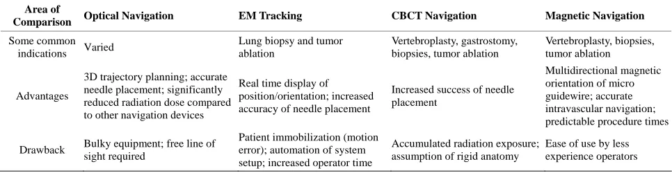
Table 1. Comparison of commonly used navigation systems. Area of
Comparison Optical Navigation EM Tracking CBCT Navigation Magnetic Navigation
Some common
indications Varied
Lung biopsy and tumor ablation
Vertebroplasty, gastrostomy, biopsies, tumor ablation
Vertebroplasty, biopsies, tumor ablation
Advantages
3D trajectory planning; accurate needle placement; significantly reduced radiation dose compared to other navigation devices
Real time display of position/orientation; increased accuracy of needle placement
Increased success of needle placement
Multidirectional magnetic orientation of micro guidewire; accurate intravascular navigation; predictable procedure times
Drawback Bulky equipment; free line of
sight required
Patient immobilization (motion error); automation of system setup; increased operator time
Accumulated radiation exposure; assumption of rigid anatomy
tions to this study. This investigation attempted to sum- marize the broad features of navigation systems used in radiology, selecting the more commonly used devices. Also, while the major medical databases were consulted for this review, studies may have been missed. As only papers presented in English were assessed, it may be ad- vantageous to investigate search engines in other lan- guages to determine if any additional literature is present.
While there are many advantages to multi-detector, thin slice, high resolution CT scanning in assessing sca- phoid fracture healing, there are some disadvantages. The first is that a higher radiation dose is needed than plain radiography. This drawback can be addressed by expos- ing only the area of interest to the radiation. Appropriate use of lead shielding, and having the patients stretch their arms above their head to limit the dose to the rest of the body can limit total body radiation exposure. The added limitation is the increased costs and reduced availability of CT scanners compared to plain radiography though is commonplace throughout hospitals in Canada and many countries worldwide. The increased cost of this investi- gation can be offset economically by definite early deter- mination of fracture healing or lack of healing in follow up patients. For patients with definitive healing, as deter- mine by the CT scan, morbidity can be reduced as they can return to work earlier by employing a short arm spica cast. For athletes in particular, CT assessment can allow for early onset exercises and decisions regarding avoid- ance of surgical intervention for the fracture.
The main strength of this pilot project lies in the novel approach of utilizing the 64 slice CT for definitive and unequivocal assessment of early scaphoid fracture heal- ing by evaluating trabecular continuity as compared to the routine practice of follow up plain radiography. The main limitation of this investigation was the sample size. The intention of the investigators was to include 12 - 15 patients and analyze the results with chi-square and one way analysis of variance methods. Due to the small sam- ple accrued, a meaningful statistical analysis could not be performed.
5. CONCLUSION
As navigation technologies evolve, their application to more complex procedures will better define their exact clinical roles and utilities. General themes appear to con- sistently emerge within the literature in regards to the ad- vantages of navigation systems including benefits to in- terventional radiologic procedures such as increased pa- tient safety, reduced procedure time and potential for re- duction in costs. Increased radiation exposure, problems accounting for respiratory motion and sterility remain issues with the use of such systems.
Larger, national clinical trials with multiple sites and
Interventional Radiologists studied over a longer period can provide greater insight into the efficacy of each sys- tem. With the potential to standardize treatment plans us- ing navigation systems, more patients can have access to up-to-date technology for treatment. However, to ensure the highest standard of care, the use of navigation sys- tems should be conducted by interventional radiologists only. With the improved accuracy, clinical utility and im- pact upon on patient outcomes, the use of navigation sys- tem in interventional radiology makes it vital to consis- tently review literature to under-stand trends and themes in the field.
REFERENCES
[1] Wood, B.J., Kruecker, J., Abi-Jaoudeh, N., et al. (2010) Navigation systems for ablation. Journal of Vascular and Interventional Radiology, 8, S257-S263.
doi:10.1016/j.jvir.2010.05.003
[2] Coldwell, D.M. and Sewell, P.E. (2005) The expanding role of interventional radiology in the supportive care of the oncology patient: From diagnosis to therapy. Semi- nars in Oncology, 32, 169-173.
doi:10.1053/j.seminoncol.2004.11.018
[3] Solomon, S.B. and Silverman, S.G. (2010) Imaging in Interventional Oncology. Radiology, 257, 624-640.
doi:10.1148/radiol.10081490
[4] Kanazawa, S. (2012) Current status of interventional on- cology. International Journal of Clinical Oncology, 17, 299-300. doi:10.1007/s10147-012-0440-6
[5] Bruners, P., Penzkofer, T., Nagel, M., Elfring, R., Gron- loh, N., Schmitz-Rode, T., Gunther, R.W. and Mahnken, A.H. (2009). Electromagnetic tracking for CT-guided spine interventions: Phantom, ex-vivo and in-vivo results. Euro- pean Radiology, 19, 990-994.
doi:10.1007/s00330-008-1227-z
[6] Abi-Jaoudeh, N., Kruecker, J., Kadoury, S., et al. (2012) Multimodality image fusion-guided procedures: Techni- que, accuracy and application. CardioVascular and Inter- ventional Radiology, 35, 986-998.
doi:10.1007/s00270-012-0446-5
[7] Wood, B.J., Zhang, H., Durrani, A., Glossop, N., Ranjam, S., Lindisch, D., Levy, E., Banovac, F., Borgert, J., Krue- ger, S., Kruecker, J., Viswanathan, A. and Cleary, K. (2005) Navigation with electromagnetic tracking for interventio- nal radiology procedures—A feasibility study. Journal of Vascular and Interventional Radiology, 16, 493-505.
doi:10.1097/01.RVI.0000148827.62296.B4
[8] Yaniv, Z. (2010) Evaluation of spherical fiducial local- ization in C-arm cone-beam CT using patient data. Me- dical Physics, 37, 5298-5305. doi:10.1118/1.3475941
[9] Schiemann, M., Killmann, R., Kleen, M., Abolmaali, N., Finney, J. and Vogl, T.J. (2004) Vascular guide wire na- vigation with a magnetic guidance system: Experimental results in a phantom. Radiology, 232, 475-481.
doi:10.1148/radiol.2322030533
plane computed-tomography-guided biopsy using a mag- netic-field-based navigation system. CVIR, 29, 108-113.
doi:10.1007/s00270-005-0041-0
[11] Wah, T., Breen, D., Patel, J., et al. (2012) Interventional oncology. Radiography, 18, 15-20.
doi:10.1016/j.radi.2011.10.001
[12] Hoffer, F.A. (2011) Interventional oncology: The future.
Pediatric Radiology, 41, S201-S206.
doi:10.1007/s00247-011-1990-x
[13] Schulze, D., Heiland, M., Thurmann, H. and Adam, G. (2004) Radiation exposure during midfacial imaging us- ing 4 and 16 slice computed tomography, cone bean com- puted tomography systems and conventional radiography.
Dentomaxillofacial Radiology, 33, 83-86.