D
E
V
E
LO
P
M
E
N
T
INTRODUCTION
Pax6 is an evolutionarily conserved transcription factor with pivotal roles in the morphogenesis of the eye, pancreas and brain. In the brain, Pax6acts as a pattern formation gene, involved in the regional specification of the telencephalon. As early as E8.5, the expression of Pax6 is confined to the neuroepithelium of the dorsal part of the telencephalic primordium (Walther and Gruss, 1991), outlining the anlage of the future cortex, while cross-repressive interactions between Pax6 and Gsh2 establish the pallial-subpallial boundary (Toresson et al., 2000). In developing cortex, Pax6 is expressed in a prominent rostrolateral-high to mediolateral-low gradient, with expression lost progressively after E15.5 (Muzio et al., 2002a; Stoykova et al., 1997; Stoykova et al., 2000). In the Pax6loss-of-function (LOF) mouse Small eye(Sey) (Hill et al., 1991), the cortical progenitors acquire ventral molecular identity starting at the ventral pallium (VP) and lateral pallium (LP), and subsequently extending dorsally and medially (Kroll and O’Leary, 2005; Muzio et al., 2002b; Stoykova et al., 1996; Stoykova et al., 2000; Torresson et al., 2000; Yun et al., 2001). Recent data indicate that the different levels of Pax6 expression in cortical progenitors along the anterior-posterior axis play a role in progenitor regionalization. Thus, in homozygous Sey/Seyembryos, the rostrolateral regions of the cortex, where Pax6 is normally expressed at the highest level, are reduced, whereas the caudomedial cortical domains, which normally show low Pax6 expression, are expanded (Bishop et al., 2000). Similarly, the high expression level of Pax6in the progenitors of the rostral VP and LP appears essential for the specification of distinct amygdalar nuclei (Tole et al., 2005).
The expression of Pax6specifies the majority of the cortical progenitors, namely the RC2-positive radial glial (RG) cells (Götz et al., 1998). These cells have been shown to act as pluripotent
progenitors, generating both neuronal and glial cells (Heins et al., 2002; Malatesta et al., 2000; Miyata et al., 2001; Noctor et al., 2004). At birth, the cortical plate (CP) of Sey/Seymice is hypocellular, with overgrown ventricular and subventricular zones (V.Z. and S.V.Z., respectively) (Schmahl et al., 1993; Stoykova et al., 1996; Stoykova et al., 2000), and the RG progenitors show defects in their mitotic cycle (Estivill-Torrus et al., 2002; Götz et al., 1998), migratory and adhesive properties (Caric et al., 1997; Hartfuss et al., 2001; Nomura and Osumi, 2004; Stoykova et al., 1997), boundary formation (Hartfuss et al., 2001; Stoykova et al., 1996) and differentiation (Götz et al., 1998; Warren et al., 1999).
In vertebrates, alternative splicing generates two different Pax6 protein isoforms, Pax6 and Pax6-5a, possibly having different sets of targets (Czerny et al., 1993; Epstein et al., 1994b; Kozmik et al., 1997). Dorsoventral patterning and boundary formation seem to be mediated exclusively by the Pax6 isoform, whereas progenitor proliferation is influenced by both isoforms (Haubst et al., 2004). Retrovirus-mediated overexpression of Pax6in RG progenitors in vitro and cell lineage experiments indicated a neurogenic activity of Pax6(Hack et al., 2004; Heins et al., 2002; Haubst et al., 2004).
Using a Cre/loxP-based recombination approach we have developed an in vivo system for conditional Pax6gain-of-function (GOF) expression in transgenic mice and studied the effects of the activation of the two Pax6isoforms in different progenitors during corticogenesis. We found that ectopic activation or overexpression of the two isoforms, Pax6and Pax6-5a, inhibits the proliferation of cortical progenitors. Furthermore, activation of transgenic Pax6 in vivo causes misregulation of the mitotic cycle, premature neurogenesis, and massive apoptosis in different progenitor pools, which seems to depend on the distinct spatiotemporal sensitivities of the cortical progenitors containing different levels of endogenous Pax6.
MATERIALS AND METHODS
Construction of plasmids and generation of transgenic mice A loxP-egfp-polyA cassette was cloned into the EcoRI site of pCAGGS (Niwa et al., 1991) followed by a loxP-(XhoI)-IRES-lacZ-polyA cassette. The coding sequence of Pax6or Pax6-5awas inserted into the XhoI site resulting in pJoP6and pJoP6-5a, respectively. The plasmids were used to generate JoP6and JoP6-5a mice by pronuclear microinjection. Transgenic
Conditional activation of
Pax6
in the developing cortex of
transgenic mice causes progenitor apoptosis
Joachim Berger1, Silke Berger1, Tran Cong Tuoc1,2, Marcello D’Amelio3, Francesco Cecconi3, Jessica A. Gorski4,
Kevin R. Jones4, Peter Gruss1and Anastassia Stoykova1,2,*
During development, Pax6is expressed in a rostrolateral-high to caudomedial-low gradient in the majority of the cortical radial
glial progenitors and endows them with neurogenic properties. Using a Cre/loxP-based approach, we studied the effect of
conditional activation of two Pax6isoforms, Pax6and Pax6-5a, on the corticogenesis of transgenic mice. We found that activation of either Pax6or Pax6-5ainhibits progenitor proliferation in the developing cortex. Upon activation of transgenic Pax6, specific
progenitor pools with distinct endogenous Pax6expression levels at different developmental stages show defects in cell cycle
progression and in the acquisition of apoptotic or neuronal cell fate. The results provide new evidence for the complex role of Pax6
in mammalian corticogenesis.
KEY WORDS: Corticogenesis, Cre/loxP, Overexpression, Pax6, Pax6-5a, Progenitor, Apoptosis, Mouse Development 134, 1311-1322 (2007) doi:10.1242/dev.02809
1Max Planck Institute for Biophysical Chemistry, D-37077 Göttingen, Germany. 2DFG, Center of Molecular Physiology of the Brain (CMPB), Göttingen, Germany. 3Dulbecco Telethon Institute at IRCCS Fondazione Santa Lucia, Rome, Italy. 4University of Colorado, Boulder, Colorado, USA.
*Author for correspondence (e-mail: [email protected])
D
E
V
E
LO
P
M
E
N
T
mice were identified by GFP fluorescence. Genomic PCR was performed with the primers JoP6F, JoP6R and JoP65aR (Table 1). pPDand pHDwere generated by cloning four PD- and five HD-consensus sequences into pGL3 (Promega) (Epstein et al., 1994a).
Immunohistochemistry
Immunohistochemistry was carried out as described (Ashery-Padan et al., 2000) and in situ hybridization analysis was according to Moorman et al. (Moorman et al., 2001). We used mouse monoclonal antibodies against Pax6 (BAbCO), 5-bromo-2-deoxyuridine (BrdU; Roche), nestin (Chemicon) and neuron-specific class III -tubulin (Tuj1; Babco), and rabbit polyclonal antisera against pHH3 (Biomol), -actin (Abcam), and activated caspase3 (Cell Signaling Technology) on 8 m cryostat sections (5 m for BrdU labeling). Secondary antibodies were from Molecular Probes (1:500). For TUNEL labeling, the ApopTag kit was used (Intergen, Purchase, NY). The BrdU labeling index was estimated as the percentage of the total cell number of BrdU+cells on DAPI-stained sections in equally sized areas in medial
pallium (MP) and dorsal pallium (DP) from cortex of both genotypes. pHH3+cells were counted and compared over the whole cortex in equally sized units of control and double-transgenic apical VZ surface. Data are shown as means±s.d. Pvalues were derived using Student’s t-test.
Luciferase reporter assay
Transient transfection of SAOS2 (ATC# HTB-85) and HeLa cells were performed using Lipofectamine2000 (Invitrogen) and 3 g of the following plasmids: pCMVand pPax6(Maulbecker and Gruss, 1993), p53Luc(Stuart et al., 1995), pmRb-pJ3Omega115ROX-p(Bernards et al., 1989), and pJoP6-5a. For in vitro overexpression of Pax6, 16 g of pPax6was used (indicated by Pax6++ in Fig. 7). Cells were subjected to the Dual-Luciferase Reporter Assay System (Promega) after 48 hours. The assay was performed in triplicate and luciferase activities were normalized to internal control activities (data shown as means±s.d.).
q-PCR analysis
Total RNA isolated from cortex was quantified by optical density and used for q-PCR with the QuantiTect Rev. Transcription Kit and the QuantiTect SYBR Green PCR Kit (Qiagen). Assays were performed in triplicate and normalized to internal 18S RNA (data as means±s.d.). Primer sequences are listed in Table 1.
RESULTS
The system for conditional activation of Pax6in vivo
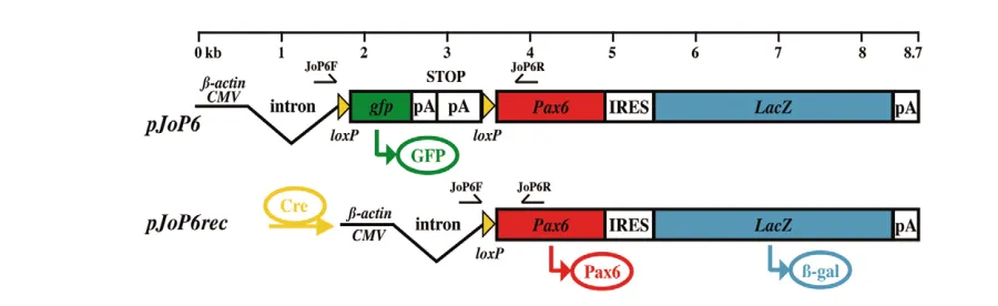
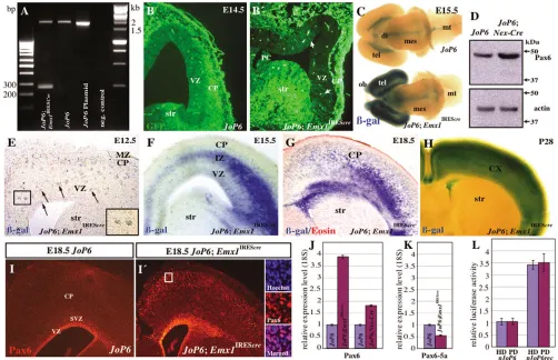
[image:2.612.48.301.73.332.2]For conditional activation of Pax6we generated a construct (pJoP6) that contains a floxed gfp-stop cassette under control of the  -actin/CMV fusion promoter (Niwa et al., 1991), driving ubiquitous expression of the gfpreporter gene (Fig. 1). Upon Cre recombination the gfp-stop cassette is excised, leading to simultaneous expression of Pax6and a second reporter, lacZ, via an IRES sequence. The transgenic mouse line generated with this construct was named JoP6. In order to test the recombination of the integrated construct, JoP6 males were crossed with female Emx1IREScremice directing recombination in most of the pallial progenitors (Gorski et al., 2002). Recombination in the JoP6;Emx1IREScre mice was monitored by PCR with the primers JoP6F and JoP6R (arrows in Fig. 1), which bind 5⬘and 3⬘of the floxed gfp-stop cassette, respectively. Genomic PCR with DNA isolated from the cortex of JoP6brains resulted in a 1831 bp DNA fragment, whereas after successful recombination an additional 261 bp DNA fragment was detected using DNA isolated from JoP6;Emx1IREScre double-transgenic cortex (Fig. 2A). Sections of JoP6 control cortex exhibited widespread GFP fluorescence in the entire brain, which
Fig. 1. The construct for transgenic activation of Pax6.The constructpJoP6contains the -actin/CMV fusion promotor and a loxP-flanked gfp -stop cassette placed in front of the Pax6and lacZcoding sequences. After Cre-mediated recombination, the gfp-stop cassette is eliminated allowing simultaneous expression of transgenic Pax6and lacZvia an IRES sequence from the resulting construct pJoP6rec. Arrows indicate the annealing sites of the primers JoP6F and JoP6R.
Table 1. Primers used in this study
Target gene Name Primer sequence (5⬘to 3⬘) or Qiagen catalog number
Pax6 F CCTGGTTGGTATCCCGGGA
R CCGCTTCAGCTGAAGTCGCA
Pax6-5a F CACAGCGGAGTGAATCAGCTT
R GCACCTGGACTTTTGCATCTG
N-CAM F ATTGTCATCTTCGTCCTGCTCC
R TCCTCCATGTCTTTGCCCTTG
Itga5 F GGATTCTGGAGTCCTCACTGTA
R GTACTCCAGAATCCGGGTGAA
R-cadherin F CTATGGTGCGGCTGCTGGTA
R GGAGTTCTCGGGAACGTTGAT
Pxn F CCGGCTGTTACTGGAGCTGAA
R GGAACTCTCCAGTTCATCCAAGA
Tnc F GCAACCAAGGACAATGTGTGTCA
R CTCCCGAATGGTGGTGGATGT
18S F
QT01036875 R
Itgb3 F
QT00128849 R
JoP6F CCTGGGCAACGTGCTGGTTATTGTGCTG
JoP6R CACAACCGTTGGATACCTGCAGAATTCGG
[image:2.612.52.502.555.693.2]D
E
V
E
LO
P
M
E
N
T
was switched off in most of the cortical cells in the VZ and CP of JoP6;Emx1IREScretransgenic mice, indicating successful excision of the gfp-stop cassette (Fig. 2B,B⬘). Remaining GFP-positive cells might correspond to GABAergic interneurons (Gorski et al., 2002). The function of the second reporter, lacZ, was tested with isolated brains after whole-mount staining for -galactosidase ( -gal). The JoP6brain showed only non-specific staining of the choroid plexus, whereas the JoP6;Emx1IREScrecortex, which is smaller than in the control, was intensively stained, indicating activation of transgenic Pax6. We further followed the expression of the lacZreporter throughout brain development. At E12.5,  -gal+aggregates as well as some individual cells were detected in
the VZ and in the thin CP (Fig. 2E). Later, at E15.5, lacZ expression was apparent in both VZ progenitors and cells migrating throughout the intermediate zone (IZ) towards the CP, invading its lower part (Fig. 2F). At E18.5, the late cortical progenitors showed strong -gal staining and the CP was massively populated by -gal+ postmitotic cells (Fig. 2G), whereas at P28 the whole cortex was extensively stained for -gal (Fig. 2H), similar to the recombination pattern in the adult Emx1IREScrebrain (Gorski et al., 2002).
[image:3.612.55.557.62.386.2]In contrast to the endogenous Pax6 expression, which was confined to VZ progenitors of the JoP6 control cortex, the JoP6;Emx1IREScrecortex exhibited – similar to the -gal staining – Fig. 2. Efficiency of recombination inJoP6mice.(A) Genomic PCR was performed with the primers JoP6F and JoP6R on DNA isolated from the
pJoP6plasmid and cortices from wild-type (negative control), JoP6 and JoP6;Emx1IREScreembryos. The PCR amplifies a 1831 bp fragment from
unrecombined DNA templates and from pJoP6plasmid and JoP6cortex, whereas an additional 261 bp DNA band is seen with the DNA sample from theJoP6;Emx1IREScrecortex. (B,B⬘) Abundant GFP fluorescence is detected on coronal sections of E14.5 JoP6cortex, whereas the majority of
the cells in the VZ and SVZ of the JoP6;Emx1IREScrecortex are GFP-negative. Arrows indicate preserved GFP+cells in the VZ. Note the preserved
fluorescence in the thin CP and the striatum (str). (C) Whole-mount staining for -gal indicates specific expression of lacZin the telencephalon (tel), olfactory bulb (ob) and scattered cells in the diencephalon (di) of E15.5 JoP6;Emx1IREScrecortex (bottom), whereas the mesencephalon (mes) and the
metencephalon (mt), as well as the JoP6control brain (top), are unstained. (D) Western blot confirms higher level of Pax6 in the E15.5 JoP6;Nex-Cre
as compared with the JoP6cortex. (E) E12.5 section of JoP6;Emx1IREScrecortex reveals weak -gal staining of individual or aggregated (arrows and
inset) progenitors. (F) At E15.5, -gal+cells are within LP/VP, IZ and the lower part of the CP. (G) Strong -gal staining is seen at E18.5 in late
progenitors as well as postmitotic cells of the CP and (H) at P28 throughout the whole cortex. (I,I⬘) Pax6 immunohistochemistry on sections of E18.5 JoP6(I) and JoP6;Emx1IREScre(I⬘) cortex, showing ectopic Pax6 expression in postmitotic cells in the IZ and the lower part of the CP. The
magnifications of the framed area in I⬘demonstrate Hoechst and Pax6 co-labeling. (J,K) q-PCRs with RNA isolated from cortex of JoP6 control,
JoP6;Emx1IREScre, and JoP6;Nex-Crenormalized to 18S RNA indicate upregulation of Pax6in the double-transgenic cortices (J) and specific elevation
of the level of Pax6transcripts in the JoP6;Emx1IREScrecortex, whereas the level of Pax6-5ais in fact diminished, as compared with the controls (K).
(L) SAOS2 cells were transiently co-transfected with a luciferase reporter construct containing either the HD (blue) or the PD (red) domain and either
D
E
V
E
LO
P
M
E
N
T
ectopic Pax6 immunoreactivity in the IZ and CP (Fig. 2I,I⬘), indicating ectopic expression. Co-labeling with Pax6 antibody and Hoechst staining confirmed Pax6 immunoreactivity exclusively in the cell nuclei (Fig. 2I⬘). The higher level of Pax6 as compared with controls was demonstrated for the JoP6;Emx1IREScrecortices by q-PCR and additionally for JoP6;Nex-Creby western blotting (Fig. 2J,D, arrow). Recent data indicate that the introduction of constructs expressing only one isoform (Pax6 or Pax6-5a) in Neuro2A and NIH3T3 cells in vitro increases the cellular levels of not only that isoform, but also of the other, indicating that positive autoregulation of the endogenous Pax6 locus occurs (Pinson et al., 2006). Interestingly, however, the results from the q-PCR assay revealed that in the cortex of the double-transgenic JoP6;Emx1IREScremice, only the level of the Pax6transcript was elevated, whereas the level of Pax6-5awas in fact diminished (Fig. 2K). The functionality of the transgenic Pax6 was indicated by co-transfection experiments demonstrating the binding activity of Pax6 to its target consensus sequences (Fig. 2L).
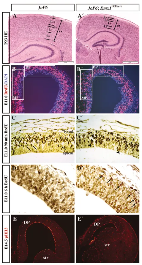
Activation of transgenic Pax6misregulates the cell cycle of early cortical progenitors
Examination of Hematoxylin and Eosin (HE) -stained histological sections from adult P23 brains revealed that the thickness of the JoP6;Emx1IREScrecortex was significantly reduced (by 43±3%) as compared with the control (P<0.001, n=12; Fig. 3A,A⬘). Despite the prominent hypocellularity, the correct positioning of the layers was not affected, as indicated by layer-specific markers [Clim1a(Ldb2), Ror and Cux2 (Cutl2); not shown]. This phenotype strongly suggests that conditional activation of transgenic Pax6might affect proliferation and/or cell survival of the cortical progenitors. Therefore, proliferation was examined at stage E11.0, when the vast majority of the cells are still proliferating. To label S-phase nuclei and determine the BrdU labeling index (percentage of BrdU+cells from the total number of DAPI+cells), a short, 30-minute pulse of BrdU was used. Significant reduction in proliferation, by 14±6% and 16±7%, was found in the medial pallium (MP) and dorsal pallium (DP) of JoP6;Emx1IREScremice, respectively (P<0.001, n=5 for MP and n=6 for DP; Fig. 3B,B⬘), two regions where recombination was strong.
[image:4.612.54.293.56.502.2]During neurogenesis, proliferating nuclei follow interkinetic nuclear migration, entering into S phase at the basal surface of VZ and progressively moving to the apical VZ surface, where they enter into M phase (Takahashi et al., 1993). After 90 minutes of BrdU incorporation, most of the progenitors in the control JoP6cortex had intensively stained (S-phase) nuclei, which were still located predominantly within the basal region of the VZ (Fig. 3C). Some nuclei with diluted BrdU content (that were at the very end of their S phase, when the BrdU pulse started) were seen at or near to the apical surface of the control VZ (arrows in Fig. 3C). In the JoP6;Emx1IREScre cortex, the S-phase labeled progenitor nuclei appeared to be distributed throughout the VZ, and only a few cells with diluted BrdU content were seen at the apical VZ (Fig. 3C⬘). In addition, faintly stained aggregates of cells were visible in the mutant VZ (arrowheads in Fig. 3C⬘). After 6 hours of BrdU labeling, the intensively stained progenitor nuclei of the control VZ reached the apical VZ, whereas in the JoP6;Emx1IREScreVZ these nuclei were retained at the basal VZ (Fig. 3D and arrows in D⬘) indicating cell cycle arrest or extended S phase. Quantitation of equally sized areas of VZ with phospho-histone H3 (pHH3) at E14.5 revealed 42±6% fewer mitotic cells at the apical VZ surface of JoP6;Emx1IREScreas compared with JoP6mice (P<0.001, n=14; Fig. 3E,E⬘). Taken together, these results indicate that in vivo activation Fig. 3. Activation of transgenic Pax6disturbs the mitotic cell
cycle.(A,A⬘) HE staining of P23 cross-sections of JoP6control and
JoP6;Emx1IREScrecortex (cx). The size of the JoP6;Emx1IREScrecortex is
significantly diminished, but cortical layering is preserved. Scale bars: 0.5 mm. (B,B⬘) Coronal 5 m sections of E11.0 JoP6control and
JoP6;Emx1IREScrecortex immunostained for BrdU and DAPI after a
30-minute BrdU pulse. Estimated BrdU labeling indices reveal a significant inhibition of the proliferation rate in the MP and DP of the
JoP6;Emx1IREScrecortex as compared with the JoP6 control. The frames
indicate the equally sized areas used for determination of the BrdU labeling index of the genotypes compared. (C-D⬘) Patterns of BrdU pulse labeling of cortical progenitors at stage E11.0 for 90 minutes (C,C⬘) and 6 hours (D,D⬘) in JoP6 control (C,D) andJoP6;Emx1IREScre
mice (C⬘,D⬘). The arrows in C point to nuclei in the control cortex that were labeled at the end of their S phase and underwent mitosis thereafter, as indicated by their diluted content of BrdU. Nuclei of such appearance are scarce in theJoP6;Emx1IREScrecortex (C⬘; arrowheads
point to cell aggregates). Note the severe distortion of the interkinetic nuclear migration in the JoP6;Emx1IREScrecortex, in which, even after a
6-hour BrdU pulse, many nuclei are still in the basal VZ (arrows in D⬘). (E,E⬘) Immunolabeling of pHH3 at stage E14.5 reveals significantly fewer mitotic cells at the apical surface of the VZ in JoP6;Emx1IREScreas
D
E
V
E
LO
P
M
E
N
T
of transgenic Pax6in the early cortical progenitors leads to a defect of interkinetic nuclear migration, a reduction in progenitor proliferation, and cortical hypocellularity.
Conditional Pax6activation enhances
neurogenesis and changes cell adhesive properties
Given the neurogenic activity of Pax6for the RG progenitors (Hack et al., 2005; Heins et al., 2002), we studied the effect of Pax6GOF on neuronal differentiation in vivo by immunostaining with monoclonal antibody (Tuj1) against neuron-specific class III -tubulin. Despite
[image:5.612.53.562.60.241.2]the 21±2% reduction in JoP6;Emx1IREScrecortical thickness compared with JoP6(P<0.001, n=12) mice, the thickness of the Tuj1+mantle zone of LP at E13.5 showed no significant difference between genotypes, suggesting enhanced neuronal differentiation in JoP6;Emx1IREScremice (Fig. 4A,A⬘). Furthermore, ectopic Tuj1+cells were detected in the apical VZ regions of the MP at E11.5 (Fig. 4C,C⬘), as well as in the LP and VP at E14.5 (Fig. 4B,B⬘), suggesting premature neurogenesis in subsets of cortical progenitors. In addition, the neuronal bHLH transcription factor gene Nex(Neurod6– Mouse Genome Informatics) showed a stronger in situ hybridization signal in the VP and LP of the E11.5JoP6;Emx1IREScrecortex as compared Fig. 4. Enhanced neurogenesis and altered adhesive properties after activation of transgenic Pax6in developing cortex.(A,A⬘) The E13.5 JoP6;Emx1IREScreVZ is significantly diminished as compared with JoP6, but the size of the Tuj1+mantle zone (M) is unchanged. Also,
premature differentiated Tuj1+cells are found ectopically in the VZ of JoP6;Emx1IREScreat E14.5 (B,B⬘) and E11.5 (C,C⬘). (D) q-PCR of total RNA from
control and JoP6;Emx1IREScrecortices normalized to 18S RNA. N-CAMand R-cadherin are upregulated, whereas Pxn, Tnc, Itgb3and Itga5are
downregulated. Error bars indicate s.d.
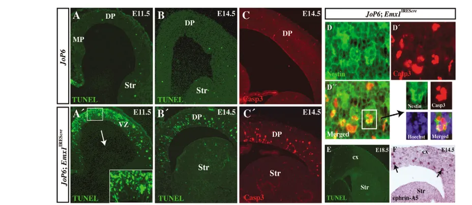
Fig. 5. Pax6GOF at early corticogenesis causes apoptosis.(A-B⬘) TUNEL assay on sections from JoP6(A,B) andJoP6;Emx1IREScre(A⬘,B⬘) embryos
at stages E11.5 (A,A⬘with higher magnification) and E14.5 (B,B⬘) illustrates apoptosis of JoP6;Emx1IREScrecortical progenitors, more severe at E11.5.
(C,C⬘) Immunolabeling against activated Casp3 confirms enhanced apoptosis in the JoP6;Emx1IREScrecortex. (D-D⬙) Double immunostaining of E13.5 JoP6;Emx1IREScrecortex against nestin (D) and activated Casp3 (D⬘) illustrates that many of the apoptotic cells are nestin+progenitors (D⬙and
magnifications counterstained with Hoechst). (E) At stage E18.5, apoptosis is no longer detected in JoP6;Emx1IREScre. (F) In situ hybridization with
[image:5.612.52.516.467.669.2]D
E
V
E
LO
P
M
E
N
T
with the control (see Fig. S1D,D⬘in the supplementary material). Together, these results suggest that upon Pax6activation in vivo, a subset of cortical progenitors, mainly those within the VP and LP, undergoes premature differentiation.
Because of the cell aggregation detected after activation of transgenic Pax6(Fig. 2E), we further analyzed the expression of genes involved in cell adhesion and cell signaling. q-PCRs performed with RNA extracted from E12.5 JoP6;Emx1IREScrecortex showed an increase in the expression of genes encoding the cell adhesion molecules N-CAM (Ncam1 – Mouse Genome Informatics) and R-cadherin (Cdh4 – Mouse Genome Informatics) as compared with the controls, whereas the expression of genes involved in cell-cell interaction and signaling such as paxillin (Pxn), tenascin C (Tnc), integrin beta 3 (Itgb3) and integrin alpha 5 (Itga5) were reduced (Fig. 4D). The change in the expression of these genes strongly suggests their involvement in the aggregation of cortical cells after activation of transgenic Pax6. However, further experiments are necessary to elucidate their individual roles.
Activation of transgenic Pax6in the developing
cortex causes apoptosis
In order to study whether the cortical hypoplasia seen in Pax6GOF in vivo might also involve an increase in cell death, we performed TUNEL reactions. At stage E11.5, massive apoptosis could be detected in the proliferating VZ of the JoP6;Emx1IREScrecortex, in contrast to the JoP6control (Fig. 5A,A⬘). Double immunolabeling against nestin, a marker for VZ progenitors associated with the cell membrane, and cytoplasmic activated caspase 3 (Casp3) showed colocalization in many cases suggesting that the cell death is confined to progenitors (Fig. 5D-D⬙). At stage E14.5, apoptosis was still detectable in progenitors of the JoP6;Emx1IREScre cortex, although at a lower level (Fig. 4B,B⬘). Labeling with antibodies against activated Casp3 implicated the caspase-dependent pathway as causing apoptosis (Fig. 4C,C⬘). At stage E18.5, the activation of transgenic Pax6expression was still maintained, as judged by the expression of the lacZreporter in the VZ and CP (see Fig. 2G); however, TUNEL reaction revealed no further apoptosis (Fig. 4E). Recent evidence has indicated that overexpression of ephrin A5 in vivo causes progenitor apoptosis in the embryonic cortex (Depaepe et al., 2005). Interestingly, both aggregates and single TUNEL+cells in the JoP6;Emx1IREScrecortex show enhanced ephrin A5 expression, suggesting that Pax6might act in an ephrin-A5-dependent manner (Fig. 4F).
When we examined whether Pax6overexpression affects the early patterning of the cortical primordium, thereby possibly initiating progenitor apoptosis, we found that neither the dorsoventral nor the mediolateral patterning in the E11.5 JoP6;Emx1IREScrecortex was affected, as indicated by the normal expression of corresponding markers (e.g. Dlx1, Wnt3a,Emx2; see Fig. S1 in the supplementary material).
Pax6GOF induces apoptosis in specific cortical progenitor pools
Several types of progenitors contribute to neurogenesis in the vertebrate cortex: neuroepithelial cells (at the beginning of neurogenesis, E11) and RG cells (after E13), which can be divided into Ngn2+/Pax6–, RC2+/Pax6+and RC2+/Pax6–(Guillemot, 2005; Hartfuss et al., 2001) (Ngn2 is also known as Neurog2, and RC2 as Ifaprc2 – Mouse Genome Informatics). By utilizing mouse lines with distinct spatiotemporal activation of the Cre recombinase, we analyzed the effect of transgenic Pax6activation in different subsets of progenitors.
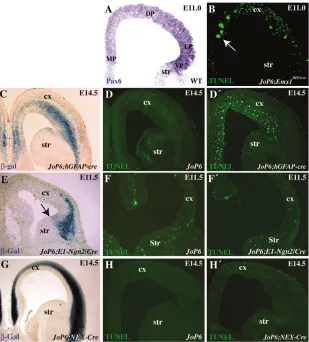
Recombination directed by Emx1IREScrestarts at E9.5 and initially proceeds at highest level in the MP progenitors (Li et al., 2003). Because the expression of Pax6in MP at this early stage is at the in situ hybridization detection limit (Muzio et al., 2002a), the massive apoptosis detected in the JoP6;Emx1IREScreMP at E11.0 involves a set of progenitors that either express endogenous Pax6at extremely low level or are Pax6-negative (Fig. 6A,B). The results suggest a high sensitivity of the early progenitors of the MP to ectopic expression or enhancement of the Pax6expression level.
To gain insight into the consequences of overexpression of Pax6 specifically in Pax6-positive RG progenitors, we used the hGFAP-creline (Zhuo et al., 2001). This line promotes activation of Cre recombinase in the majority of the RC2+/Pax6+ radial glial progenitors as early as E13.5 (Götz et al., 1998; Heins et al., 2002). After overexpression of Pax6in the JoP6;hGFAP-crecortex, as indicated by -gal staining (Fig. 6C), massive apoptosis was detected in single progenitors as well as in cell aggregates (Fig. 6D,D
⬘
).
Therefore, we conclude that overexpression of Pax6in the midgestation cortical progenitors expressing Pax6at a moderate level also leads to cell death.Interestingly, in the VP of JoP6;hGFAP-cre, where endogenous Pax6 and its target Ngn2 are expressed at a very high levels, apoptosis is seen only rarely (Fig. 6D⬘). To specifically examine the effect of Pax6overexpression in the VP and LP progenitors, we crossed JoP6 mice with the E1-Ngn2/Cre line, in which Cre recombinase is directed by the E1 enhancer element of the gene encoding transcription factor Ngn2(Berger et al., 2004). Despite significant recombination in the VP (as detected by -gal staining), neither significantly increased apoptosis nor cell aggregates were seen at stage E11.5 or E14.5 in the JoP6;E1-Ngn2/Cre double-transgenic cortex as compared with the JoP6control (Fig. 6E-F⬘and data not shown). Together, these data indicate that at the onset of neurogenesis, early cortical progenitors show differential sensitivity towards the elevation of the Pax6expression level, which correlates inversely with their endogenous Pax6expression level: the highly Pax6-positive progenitors of the rostral VP appear to be more resistant to Pax6GOF, whereas the Pax6-negative progenitors, or progenitors that are expressing Pax6at extremely low level (e.g. in the MP), undergo apoptosis.
Pax6GOF in postmitotic cells has no effect on cell survival
Although recombination and thus activation of transgenic Pax6is still detectable at E18.5 in postmitotic cells of the CP of JoP6;Emx1IREScremice, these cells do not undergo apoptosis (Fig. 5G). In order to directly assess the specific effect of the activation of transgenic Pax6expression in newly born neurons, we used the Nex-Cre mouse line, which induces Cre recombinase activity in postmitotic neurons after their exit from the mitotic cycle (Schwab et al., 2000). In the double-transgenic JoP6;Nex-Cremice cortex, where the level of Pax6expression is higher than in the controls (see Fig. 2I,J), no enhancement of apoptosis was detected at E14.5 and P21 (Fig. 6H,H⬘and data not shown). Thus, transgenic activation of Pax6in vivo specifically induces cortical progenitor apoptosis, whereas the fate of the postmitotic neurons is not affected.
Pax6-induced apoptosis does not involve
transcriptional activation of p53
D
E
V
E
LO
P
M
E
N
T
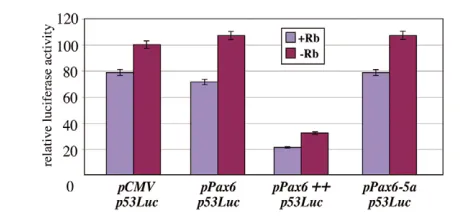
binds to the human P53(TP53) promoter with low affinity, although the effect on p53gene activity has not been assessed so far (Stuart et al., 1995). Remarkably, Pax6 also binds directly to pRb (Cvekl et al., 2004), suggesting the existence of a possible relationship between Pax6-, pRb-and p53-dependent apoptosis. In an attempt to address this issue, we studied the effect of Pax6 on the activity of the P53 -promoter in a human osteosarcoma cell line (SAOS2) lacking endogenous pRband p53. The reporter plasmid p53Luccontaining the P53promoter followed by the luciferase gene was co-transfected with pPax6or pPax6-5aexpression plasmids in the absence and presence of a mouse pRbexpression plasmid. As illustrated in Fig. 7, expression of Pax6barely influenced luciferase activation via the P53promoter, whereas upon strong overexpression of Pax6 the P53 promoter activity decreased. pRB was not able to significantly enhance the effect of Pax6. Similar results were obtained with the cell lines NIH-2H3, HeLa and HelaTAT (data not shown). Pax6-5aexpression showed no effect on the P53promoter. Together, these results suggest that the apoptosis induced by transgenic Pax6in vivo is due neither to activation of the cell death pathway through a direct transcriptional activation of p53, nor by abolishment of the pRb-dependent active repression of p53activity.
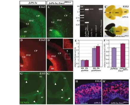
In vivo overexpression of Pax6-5amoderately
inhibits progenitor proliferation
Previous GOF experiments in primary cortical cultures indicated that retrovirus-mediated overexpression of both Pax6and Pax6-5a leads to inhibition of cell proliferation (Haubst et al., 2004). To study the in vivo activation of Pax6-5a, we generated the transgenic line JoP6-5abased on the same principles as JoP6(see Materials and
methods). Upon crossing with the Emx1IREScreline, the functionality of the JoP6-5a line was tested: PCR with primers JoP6F and JoP65aR detected recombination of genomic DNA (1955 bp and 394 bp fragments); expression of the two reporter genes gfpand lacZ was monitored by GFP fluorescence and -gal staining, respectively; and Pax6 immunostaining indicated the expression of transgenic Pax6-5a(Fig. 8A-D⬘). Co-transfection experiments of pJoP6-5arec with pHDand pPD indicated the functionality of transgenic Pax6-5a (Fig. 8E). q-PCR showed enhancement of Pax6-5aexpression in the JoP6-5a;Emx1IREScrecortex, which was much weaker compared with the Pax6 enhancement in the JoP6;Emx1IREScre cortex (Fig. 8F). Statistical analysis of the apoptotic pattern of JoP6-5a;Emx1IREScreand control cortices at stages E11.5 and E13.5 revealed no difference, and no cell aggregates were formed (n=5; Fig. 8G,G⬘). Nevertheless, estimation of the BrdU index in the E11.5 DP after 30 minutes labeling revealed a mild but significant reduction (by 10±2%) in progenitor proliferation (P<0.001, n=4; Fig. 8D,D⬘). Taken together, these results suggest a difference in the biological activity of the two Pax6 isoforms in vivo.
DISCUSSION
[image:7.612.51.360.58.400.2]To study the in vivo function of Pax6during corticogenesis we developed a conditional GOF approach that allows activation of either the Pax6or the Pax6-5aisoform. In this work, we provide evidence for a differential spatiotemporal sensitivity of cortical progenitors towards Pax6GOF in vivo. We found that different levels of Pax6 play an essential role in the regulation of cortical growth by controlling progenitor proliferation, cell cycle
Fig. 6. Activation of transgenic Pax6leads to progenitor apoptosis depending on the endogenous Pax6expression level.(A) Pax6in situ hybridization on E11.0 wild-type section demonstrates extremely faint endogenous Pax6
expression in the MP and (B) TUNEL assay on E11.0
JoP6;Emx1IREScrereveals massive apoptosis within the
same region (arrow). (C) -gal staining of section at E14.5 indicates successful recombination in the
JoP6;hGFAP-crecortex and (D,D⬘) TUNEL assay on
JoP6and JoP6;hGFAP-creshows extensive apoptosis in the Pax6+radial glial progenitors. (E) -gal staining
of an E11.5 JoP6;E1-Ngn2/Creembryo indicates recombination in the LP and VP (arrow indicates corticostriatal border). (F,F⬘) No significant difference in apoptosis is detected via TUNEL assay on sections of E11.5 JoP6(F) and JoP6;E1-Ngn2/Cre(F⬘) embryos. (G) E14.5 section of JoP6;Nex-Crecortex stained for
-gal indicates recombination in postmitotic neurons in the dorsal telencephalon. (H,H⬘) TUNEL assay did not detect additional apoptosis in the cortex of the
D
E
V
E
LO
P
M
E
N
T
progression, the acquisition of progenitor apoptotic fate and induction of neurogenesis. A similar role in controlling the proliferation of cortical progenitors was found for the Pax6-5a transcript. We show that the induced progenitor cell death evident in our assays, is unlikely to be due to a direct transcriptional regulation of the anti-tumor gene p53by Pax6.
Pax6controls cortical progenitor proliferation and cell cycle progression
We found that the adult cortex is significantly reduced in size (by 43±3%) after transgenic Pax6activation, having a preserved cortical layering and dorsoventral patterning. Subsequent analysis of progenitor proliferation at E11.0 revealed a significant reduction of the BrdU labeling index as compared with the controls: by 14±6% and 16±7% for the MP and DP, respectively. Despite the much lower level of transgenic Pax6-5aexpression, the proliferation rate of cortical progenitors inJoP6-5a;Emx1IREScremice was inhibited as well (by 10±2%). These results are in line with in vitro experiments in which Pax6transduction leads to the inhibition of progenitor proliferation (Hack et al., 2004; Heins et al., 2002; Cartier et al., 2006). Given the enhanced progenitor-proliferation rate found in the Pax6 LOF Sey/Seycortex at E10.5 (Warren et al., 1999) and E14.5-E16.5 (Götz et al., 1998), these findings confirm the pivotal role of both Pax6and Pax6-5ain controlling cortical progenitor proliferation. Remarkably, in different environmental contexts, the two Pax6 isoforms exert different control functions on progenitor proliferation. Pax6enhances proliferation in the neural retina of vertebrates (Marquardt et al., 2001) and Drosophila(Dominguez et al., 2004), but reduces proliferation of human glioblastoma cells (Zhou et al., 2005), cultivated corneal epithelial cells (Ouyang et al., 2006), and cortical progenitors in primary cell cultures (Haubst et al., 2004; Heins et al., 2002) as well as in vivo (this work). Similarly, although Pax6-5aseems to promote proliferation in the developing eye (Singh et al., 2002), it inhibits proliferation of cortical progenitors in vitro (Haubst et al., 2004) and in vivo (this study).
The majority of the S-phase and many of the M-phase progenitors express Pax6, suggesting that Pax6regulates cell cycle progression (Haydar et al., 2000). During the transition from S to M phase, the nuclei of the progenitors migrate from the basal to the apical surface of the VZ, a process termed interkinetic nuclear migration. Previous results from our laboratory (Götz et al., 1998) and from other groups
(Warren et al., 1999) indicated that in Pax6LOF, the interkinetic nuclear movement of cortical progenitors is impaired, with more cells found in the S phase as a result of a shorter cell cycle (Estivill-Torrus et al., 2002). Here we provide evidence that overexpression of Pax6in vivo leads to defects of mitotic cycle progression such that many cells seem to be stuck or prolonged in S phase and a significantly smaller proportion of progenitors undergo mitosis. In support of this conclusion, recent results from quantitative FACS analysis demonstrate that activation of Pax6in HeLa cells strongly reduces the number of cells in S and G2–M phases, indicating cell cycle arrest (Cartier et al., 2006). Similarly, overexpression of Pax6 in corneal epithelial cell lines and primary cell culture causes inhibition of cell proliferation and retardation of the cell cycle (Ouyang et al., 2006).
Experiments involvingPax6transduction in RG cell cultures and adult neurospheres (Hack et al., 2004; Heins et al., 2002) as well as in HeLa cells (Cartier et al., 2006) have revealed premature neuronal differentiation. Owing to the massive apoptosis in the JoP6;Emx1IREScremice during early corticogenesis, a quantitative estimation of possible premature neurogenesis is difficult. However, we found that although the JoP6;Emx1IREScrecortical thickness at E13.5 is significantly reduced (by 21±2%, as compared with the control), the thickness of the Tuj1+ mantle layer appeared unchanged. Furthermore, the ectopically located Tuj1+cells in the VZ, and the enhanced Nex in situ hybridization signal in the E11.5 CP of JoP6;Emx1IREScremice, suggest enhanced neurogenesis in the Pax6 GOF condition in vivo. Thus, subpopulations of early progenitors of the JoP6;Emx1IREScrecortex seem to exit prematurely from the mitotic cycle and differentiate, which would diminish the cortical progenitor pool early in development and contribute to the severe hypocellularity of the adult JoP6;Emx1IREScrecortex. It would also be of interest to test the expression of the potential Pax6 downstream target Fabp7, recently reported to be involved in maintenance of proliferation versus neuronal differentiation in cortical progenitors (Arai et al., 2005).
In addition to misregulation of the mitotic cycle, we found that transgenic Pax6activation causes aggregation of cortical progenitors. Previously, we reported on changes in the adhesive properties of isolated cortical progenitors from Sey/Sey cortex and reduced expression of R-cadherin (Stoykova et al., 1997). After Pax6GOF in vivo we found strong enhancement of the expression of N-CAM, which encodes a cell-cell adhesion molecule positively regulated by Pax6 (Holst et al., 1998; Yamaoka et al., 2000), as well as increased expression of R-cadherin. The latter result further supports the idea of R-cadherin mediating Pax6-dependent function in cell adhesion (Andrews and Mastick, 2003), possibly by direct genetic interaction of these two genes. We also found Pax6-mediated inhibition of the expression of integrin alpha 5, which contains Pax6-binding sites in its promoter (Duncan et al., 2000), as well as a decrease in the expression levels of integrin beta 3, paxillin and tenascin C, molecules involved in cell-cell interaction. Given the complex pathways in which these proteins participate, further detailed analysis is required to dissect the specific role of Pax6in these processes. However, the system for conditional activation of Pax6described here seems to be a reliable tool for such studies in vivo.
Pax6GOF in vivo induces apoptosis in specific sets of cortical progenitors
[image:8.612.53.289.58.168.2]TUNEL assays and double immunohistochemistry with antibodies against activated Casp3 and nestin revealed abundant apoptosis in the cortical progenitors of E11.5 JoP6;Emx1IREScremice. When Pax6 GOF was directed into postmitotic cells by the Nex-Cremouse line Fig. 7. Effect of Pax6 protein on activity of the P53promoter.
Pax6 protein (3 g of pPax6) does not positively regulate activity of the human P53promoter in transiently transfected SAOS2 cells, but upon overexpression (16 g pPax6, indicated by ++) does inhibit the P53
D
E
V
E
LO
P
M
E
N
T
(Schwab et al., 2000), no apoptosis could be detected. These results provide the first evidence that Pax6 is involved in progenitor apoptosis during mammalian corticogenesis.
Given that apoptosis is a very fast process, with dead cell bodies cleared out of the rat cortex in 2 hours 20 minutes (Thomaidou et al., 1997), the apoptosis at the beginning of neurogenesis in JoP6;Emx1IREScremice is massive. Although extensive apoptosis is apparent at E11.5, it weakens by E14.5, and at E18.5 no increased cell death appears to occur. In parallel, a limited number of -gal+ cells, marking recombined survived progenitors, are found at E12.5, whereas at E15.5 and E18.5 the -gal staining is progressively spreading into the IZ and CP, respectively, and at P28 the entire depth of the cortex is populated by -gal+ cells. We assume, therefore, that a significant part of the early (E9.5-E14.5) cortical
progenitors, where transgenic Pax6 is induced, undergo rapid apoptosis and are removed from the cortex before they have accumulated enough -gal to be confidently registered on thin sections, whereas the later progenitors are affected much less by Pax6GOF.
[image:9.612.56.494.60.417.2]Subpopulations of cortical progenitors survive Pax6GOF and are monitored as -gal+ cells. By using different mouse lines for regionalized recombination, we provide evidence that cortical progenitors in vivo have spatiotemporal differences in their sensitivity towards Pax6GOF. In the JoP6;Emx1IREScre cortex, activation of transgenic Pax6is initiated at E9.5 predominantly in the MP, where endogenous Pax6is only barely expressed, if at all (Muzio et al., 2002b). Thereafter, Pax6GOF progressively spreads to the majority of the glutamatergic cortical progenitors (Li et al., 2003). Therefore, Fig. 8. Conditional transgenic activation of Pax6-5avia the JoP6-5amouse line.(A,A⬘) GFP fluorescence on sections of E14.5 cortex of JoP6-5a(A) is partially lost in JoP6-5a;Emx1IREScre(A⬘). (B) Two products generated by genomic PCR with the primers JoP6F and JoP65aR (1955 bp and
394 bp) confirm successful recombination in the JoP6-5a;Emx1IREScrecortex, whereas the JoP6cortex and the pJoP6-5aplasmid show no
recombination. (C) Whole-mount -gal staining of E15.5 JoP6-5aand JoP6-5a;Emx1IREScrebrains shows strong activation of the lacZreporter in the JoP6-5a;Emx1IREScretelencephalon and olfactory bulb. (D,D⬘with inset) Immunostaining for Pax6 on E14.5 JoP6-5aand JoP6-5a;Emx1IREScrecortices
shows ectopic antibody staining indicating Cre-induced activation of Pax6-5ain the JoP6-5a;Emx1IREScreCP. (E) SAOS2 cells were transiently
co-transfected with a luciferase reporter construct containing either the HD (blue) or the PD (red) domain and either pJoP65a or pJoP65arec, which express Pax6-5a. Luciferase activities were normalized to co-transfections with the pJoP65a control plasmid. Co-transfection ofpJoP6recwith either the HD- or the PD-containing reporter construct induced an increase in luciferase activity, indicating functionality of transgenic Pax6-5a. (F) q-PCRs with RNA isolated from E12.5 cortex of JoP6-5a control and JoP6;Emx1IREScrecortex, normalized to 18S RNA, indicate only a slight enhancement of
the Pax6-5aexpression level in theJoP6-5a;Emx1IREScretelencephalon and olfactory bulb. Error bars indicate s.d.. (G,G⬘) TUNEL assay on E11.5 brain
sections of both genotypes reveals no significant difference in apoptosis (arrowheads point to endogenous apoptosis). (H,H⬘) BrdU
immunohistochemistry after a 30-minute BrdU pulse on cross-sections of E11.0 JoP6-5aand JoP6-5a;Emx1IREScrecortices reveals a significant
D
E
V
E
LO
P
M
E
N
T
the observed massive apoptosis in the MP of JoP6;Emx1IREScremice appears to be the result of either ectopic expression of Pax6in the early Pax6-negative progenitor pool (neuroepithelial cells at E9.5-E12 in MP and Pax6–RG progenitors), or overexpression of Pax6in RG cells expressing Pax6 only faintly. Also, in the JoP6;hGFAP-crecortex, where transgenic Pax6becomes activated after E13.5 exclusively in the RC2+/Pax6+radial glial cells (Malatesta et al., 2003; Zhuo et al., 2001), extensive apoptosis was detected, indicating that the overexpression of Pax6in the Pax6+midgestation cortical progenitors leads to apoptosis as well. In order to study the effect of Pax6 overexpression in the early VP progenitors, where the endogenous Pax6expression level is at its highest (Stoykova et al., 1997), we used the E1-Ngn2/Cre line (Berger et al., 2004). This line drives recombination directed by the E1-Ngn2enhancer that is activated only by a high dosage of Pax6 (Marquardt et al., 2001; Scardigli et al., 2003). No significant enhancement of apoptosis was observed in the JoP6;E1-Ngn2/Crecortex, suggesting that the VP and LP progenitors with the highest level of endogenous Pax6 are resistant to further elevation of Pax6. Collectively, these in vivo results demonstrate that the cortical progenitors have different sensitivity towards the Pax6 GOF condition, which is probably dependent upon the endogenous Pax6expression level.
The tumor suppressor gene p53is involved in the control of cell cycle arrest and apoptosis, inducing cell death upon activation (Hickman et al., 2002). In an attempt to study the molecular mechanism of the induced apoptosis in the JoP6;Emx1IREScrecortex, we tested the effect of Pax6expression on the p53promoter, which contains Pax6 target sequences (Stuart et al., 1995). Pax6 protein binds to hypophosphorylated pRb (Cvekl et al., 2004), whereas pRb allows the formation of pRb-E2F complexes, which actively repress the transcription of E2F-responsive promoters, including the pro-apoptotic gene p53(Ookawa et al., 1997; Sellers et al., 1995). Therefore, we also tested whether, upon overexpression, Pax6 could sequester pRb and abrogate pRb-E2F-dependent repression of the p53promoter, thereby inducing apoptotic fate. We found that Pax6 is unable to trigger p53activation in vitro, but, by contrast, inhibits p53transcription upon strong overexpression and independently of pRB. Therefore, although the underlying molecular mechanism of the Pax6-induced apoptosis in vivo is still unclear, our results indicate that this phenomenon is not likely to be a consequence of p53 pathway activation. It is interesting to note that after overexpression of Pax6, we detected a strong enhancement of ephrin A5 expression in apoptotic cortical progenitors. Most intriguingly, a similar apoptotic phenotype of early cortical progenitors has recently been discovered in GOF experiments for ephrin A5 in transgenic mice in vivo (Depaepe et al., 2005
),
raising the possibility of genetic interplay between the Pax6-and ephrin-A5-dependent pathways in the control of cortical progenitor cell death.Accumulating evidence supports the view that Pax6is involved in tissue growth, not only by modulating progenitor proliferation and cell cycle progression, but possibly also by the involvement of apoptosis. Evidence has been presented that a high copy-number of transgenic Pax6leads to microphtalmia in mice (Schedl et al., 1996). Pax6 overexpression in cultivated corneal epithelial cells slows down cell cycle progression and causes apoptosis (Ouyang et al., 2006). Ectopic activation of Pax6in undifferentiated and mature pancreatic -cells of transgenic mice inhibits progenitor proliferation and leads to apoptosis (Yamaoka et al., 2000), and activation of Pax6suppresses tumorigenicity of glioblastoma cells inducing apoptosis as well (Zhou et al., 2005). In addition, the Casp3 target gene Parpacts as a regulator of Pax6expression in neuroretina (Plaza et al., 1999) and, in developing Xenopus, expression of Pax6
at the neural-fold stage overlaps with TUNEL-positive cells (Hensey and Gautier, 1998). In the Pax6LOF mutant Sey/Sey, the failure in the transition of the nasal ectoderm into nasal placode has been attributed to abnormal apoptosis (Fukuda et al., 2000), but no enhanced apoptosis is detected in the developing cortex (Grindley et al., 1995) (data not shown). Therefore, further research is required to reveal the biological significance of the apoptosis induced byPax6 GOF in vivo in different cellular and experimental contexts.
We were unable to detect apoptosis in the cortical progenitors of JoP6-5a;Emx1IRESCremice. It should be noted, however, that in contrast to the substantial elevation of the level of Pax6transcripts in JoP6;Emx1IRESCre mice (3.8-fold higher, compared with the controls), the increase in the level of Pax6-5a in JoP6-5a;Emx1IRESCremice was much less evident (1.2-fold, as compared with the controls). The possibility remains that the Pax6-5a level necessary to induce apoptosis was not achieved in this assay. Therefore, presently, it cannot be stated whether the detected progenitor apoptosis is a specific feature of the in vivo elevation of the Pax6 isoform only. However, in agreement with results from Pax6and Pax6-5aGOF experiments in vitro (Haubst et al., 2004), we find that the two Pax6 isoforms successfully repress progenitor proliferation in the developing cortex.
Taken together with all the evidence available so far, the results presented in this study support the view that during corticogenesis, the modulation of Pax6expression levels is crucial for progenitor cell fate acquisition as this influences cell proliferation, differentiation and apoptosis. Using the Pax6GOF approach described here, we provide new in vivo evidence for a complex role of the Pax6gene during multiple phases of mammalian corticogenesis.
We thank K.-A. Nave, S. Goebbels and M. Schwab for the Nex-Creline; J. Frisen for the ephrin A5 probe; M. Crescenzi for pRb; W. Luc for HelaTaT; V. DeLaurenzi for SAOS2 cells; A. Jacoby for helpful comments; and M. Daniel and F. Fausti for outstanding technical assistance. F.C. is an Associate Telethon Scientist supported by Telethon and Compagnia di San Paolo. K.R.J. was supported by EY014998.
Supplementary material
Supplementary material for this article is available at http://dev.biologists.org/cgi/content/full/134/7/1311/DC1
References
Andrews, G. L. and Mastick, G. S.(2003). R-cadherin is a Pax6-regulated, growth-promoting cue for pioneer axons. J. Neurosci. 23, 9873-9880.
Arai, Y. A., Funatsu, N., Numayama-Tsuruta, K., Nomura, T., Nakamura, S. and Osumi, N.(2005). Role of Fabp7, a downstream gene of Pax6, in the maintenance of neuroepithelial cells during early embryonic development of the rat cortex. J. Neurosci. 25, 9752-9761.
Ashery-Padan, R., Marquardt, T., Zhou, X. and Gruss, P.(2000). Pax6 activity in the lens primordium is required for lens formation and for correct placement of a single retina in the eye. Genes Dev.14, 2701-2711.
Berger, J., Eckert, S., Scardigli, R., Guillemot, F., Gruss, P. and Stoykova, A.
(2004). E1-Ngn2/Cre is a new line for regional activation of Cre recombinase in the developing CNS. Genesis40, 195-199.
Bernards, R., Shackleford, G. M., Gerber, M. R., Horowitz, J. M., Friend, S. H., Schartl, M., Bogenmann, E., Rapaport, J. M., McGee, T., Dryja, T. P. et al.(1989). Structure and expression of the murine retinoblastoma gene and characterization of its encoded protein. Proc. Natl. Acad. Sci. USA86, 6474-6478.
Bishop, K. M., Goudreau, G. and O’Leary, D. D.(2000). Regulation of area identity in the mammalian neocortex by Emx2 and Pax6. Science288, 344-349.
Caric, D., Gooday, D., Hill, R. E., McConnell, S. K. and Price, D. J.(1997). Determination of the migratory capacity of embryonic cortical cells lacking the transcription factor Pax-6. Development124, 5087-5096.
Cartier, L., Laforge, T., Feki, A., Arnaudeau, S., Dubois-Dauphin, M. and Krause, K. H.(2006). Pax6-induced alteration of cell fate: Shape changes, expression of neuronal (tubulin, postmitotic phenotype, and cell migration. J. Neurobiol. 66, 421-436.
D
E
V
E
LO
P
M
E
N
T
Czerny, T., Schaffner, G. and Busslinger, M.(1993). DNA sequence recognition by Pax proteins: bipartite structure of the paired domain and its binding site.
Genes Dev.7, 2048-2061.
Depaepe, V., Suarez-Gonzalez, N., Dufour, A., Passante, L., Gorski, J. A., Jones, K. R., Ledent, C. and Vanderhaeghen, P.(2005). Ephrin signalling controls brain size by regulating apoptosis of neural progenitors. Nature435, 1244-1250.
Dominguez, M., Ferres-Marco, D., Gutierrez-Avino, F. J., Speicher, S. A. and Beneyto, M.(2004). Growth and specification of the eye are controlled independently by Eyegone and Eyeless in Drosophila melanogaster. Nat. Genet. 36, 31-39.
Duncan, M. K., Kozmik, Z., Cveklova, K., Piatigorsky, J. and Cvekl, A.(2000). Overexpression of PAX6(5a) in lens fiber cells results in cataract and upregulation of (alpha)5(beta)1 integrin expression. J. Cell Sci. 113, 3173-3185.
Epstein, J., Cai, J., Glaser, T., Jepeal, L. and Maas, R.(1994a). Identification of a Pax paired domain recognition sequence and evidence for DNA-dependent conformational changes. J. Biol. Chem.269, 8355-8361.
Epstein, J. A., Glaser, T., Cai, J., Jepeal, L., Walton, D. S. and Maas, R. L.
(1994b). Two independent and interactive DNA-binding subdomains of the Pax6 paired domain are regulated by alternative splicing. Genes Dev.8, 2022-2034.
Estivill-Torrus, G., Pearson, H., van Heyningen, V., Price, D. J. and Rashbass, P.(2002). Pax6 is required to regulate the cell cycle and the rate of progression from symmetrical to asymmetrical division in mammalian cortical progenitors.
Development129, 455-466.
Fukuda, T., Kawano, H., Osumi, N., Eto, K. and Kawamura, K.(2000). Histogenesis of the cerebral cortex in rat fetuses with a mutation in the Pax-6 gene. Brain Res. Dev. Brain Res. 120, 65-75.
Gorski, J. A., Talley, T., Qiu, M., Puelles, L., Rubenstein, J. L. and Jones, K. R.
(2002). Cortical excitatory neurons and glia, but not GABAergic neurons, are produced in the Emx1-expressing lineage. J. Neurosci. 22, 6309-6314.
Götz, M., Stoykova, A. and Gruss, P.(1998). Pax6 controls radial glia differentiation in the cerebral cortex. Neuron21, 1031-1044.
Grindley, J. C., Davidson, D. R. and Hill, R. E.(1995). The role of Pax-6 in eye and nasal development. Development121, 1433-1442.
Guillemot, F.(2005). Cellular and molecular control of neurogenesis in the mammalian telencephalon.Curr. Opin. Cell Biol. 17, 639-647.
Hack, M. A., Sugimori, M., Lundberg, C., Nakafuku, M. and Gotz, M.(2004). Regionalization and fate specification in neurospheres: the role of Olig2 and Pax6. Mol. Cell. Neurosci.25, 664-678.
Hack, M. A., Saghatelyan, A., de Chevigny, A., Pfeifer, A., Ashery-Padan, R., Lledo, P. M. and Gotz, M.(2005). Neuronal fate determinants of adult olfactory bulb neurogenesis.Nat. Neurosci. 8, 865-872.
Hartfuss, E., Galli, R., Heins, N. and Gotz, M.(2001). Characterization of CNS precursor subtypes and radial glia. Dev. Biol. 229, 15-30.
Haubst, N., Berger, J., Radjendirane, V., Graw, J., Favor, J., Saunders, G. F., Stoykova, A. and Gotz, M.(2004). Molecular dissection of Pax6 function: the specific roles of the paired domain and homeodomain in brain development.
Development131, 6131-6140.
Haydar, T. F., Wang, F., Schwartz, M. L. and Rakic, P.(2000). Differential modulation of proliferation in the neocortical ventricular and subventricular zones. J. Neurosci. 20, 5764-5774.
Heins, N., Malatesta, P., Cecconi, F., Nakafuku, M., Tucker, K. L., Hack, M. A., Chapouton, P., Barde, Y. A. and Gotz, M.(2002). Glial cells generate neurons: the role of the transcription factor Pax6.Nat. Neurosci.5, 308-315.
Hensey, C. and Gautier, J.(1998). Programmed cell death during Xenopus development: a spatio-temporal analysis. Dev. Biol. 203, 36-48.
Hickman, E. S., Moroni, M. C. and Helin, K.(2002). The role of p53 and pRB in apoptosis and cancer. Curr. Opin. Genet. Dev. 12, 60-66.
Hill, R. E., Favor, J., Hogan, B. L., Ton, C. C., Saunders, G. F., Hanson, I. M., Prosser, J., Jordan, T., Hastie, N. D. and van Heyningen, V.(1991). Mouse small eye results from mutations in a paired-like homeobox-containing gene.
Nature354, 522-525.
Holst, B. D., Vanderklish, P. W., Krushel, L. A., Zhou, W., Langdon, R. B., McWhirter, J. R., Edelman, G. M. and Crossin, K. L.(1998). Allosteric modulation of AMPA-type glutamate receptors increases activity of the promoter for the neural cell adhesion molecule, N-CAM. Proc. Natl. Acad. Sci. USA95, 2597-2602.
Kozmik, Z., Czerny, T. and Busslinger, M.(1997). Alternatively spliced insertions in the paired domain restrict the DNA sequence specificity of Pax6 and Pax8.
EMBO J. 16, 6793-6803.
Kroll, T. T. and O’Leary, D. D.(2005). Ventralized dorsal telencephalic progenitors in Pax6 mutant mice generate GABA interneurons of a lateral ganglionic eminence fate. Proc. Natl. Acad. Sci. USA102, 7374-7379.
Li, H. S., Wang, D., Shen, Q., Schonemann, M. D., Gorski, J. A., Jones, K. R., Temple, S., Jan, L. Y. and Jan, Y. N.(2003). Inactivation of Numb and Numblike in embryonic dorsal forebrain impairs neurogenesis and disrupts cortical morphogenesis. Neuron40, 1105-1118.
Malatesta, P., Hartfuss, E. and Götz, M.(2000). Isolation of radial glia cells by fluorescent-activated cell sorting reveals a neuronal lineage. Development127, 5253-5263.
Malatesta, P., Hack, M. A., Hartfuss, E., Kettenmann, H., Klinkert, W.,
Kirchhoff, F. and Gotz, M.(2003). Neuronal or glial progeny: regional differences in radial glia fate. Neuron37, 751-764.
Marquardt, T., Ashery-Padan, R., Andrejewski, N., Scardigli, R., Guillemot, F. and Gruss, P.(2001). Pax6 is required for the multipotent state of retinal progenitor cells. Cell105, 43-55.
Maulbecker, C. C. and Gruss, P.(1993). The oncogenic potential of Pax genes.
EMBO J.12, 2361-2367.
Miyata, T., Kawaguchi, A., Okano, H. and Ogawa, M.(2001). Asymmetric inheritance of radial glial fibers by cortical neurons. Neuron31, 727-741.
Moorman, A. F., Houweling, A. C., de Boer, P. A. and Christoffels, V. M.
(2001). Sensitive nonradioactive detection of mRNA in tissue sections: novel application of the whole-mount in situ hybridization protocol. J. Histochem. Cytochem.49, 1-8.
Muzio, L., DiBenedetto, B., Stoykova, A., Boncinelli, E., Gruss, P. and Mallamaci, A.(2002a). Conversion of cerebral cortex into basal ganglia in Emx2(–/–) Pax6(Sey/Sey) double-mutant mice.Nat. Neurosci.5, 737-745.
Muzio, L., DiBenedetto, B., Stoykova, A., Boncinelli, E., Gruss, P. and Mallamaci, A.(2002b). Emx2 and Pax6 control regionalization of the pre-neuronogenic cortical primordium. Cereb. Cortex12, 129-139.
Niwa, H., Yamamura, K. and Miyazaki, J.(1991). Efficient selection for high-expression transfectants with a novel eukaryotic vector. Gene108, 193-199.
Noctor, S. C., Martinez-Cerdeno, V., Ivic, L. and Kriegstein, A. R.(2004). Cortical neurons arise in symmetric and asymmetric division zones and migrate through specific phases.Nat. Neurosci.7, 136-144.
Nomura, T. and Osumi, N.(2004). Misrouting of mitral cell progenitors in the Pax6/small eye rat telencephalon. Development131, 787-796.
Ookawa, K., Tsuchida, S., Adachi, J. and Yokota, J.(1997). Differentiation induced by RB expression and apoptosis induced by p53 expression in an osteosarcoma cell line. Oncogene14, 1389-1396.
Ouyang, J., Shen, Y. C., Yeh, L. K., Li, W., Coyle, B. M., Liu, C. Y. and Fini, M. E.(2006). Pax6 overexpression suppresses cell proliferation and retards the cell cycle in corneal epithelial cells. Invest. Ophthalmol. Vis. Sci.47, 2397-2407.
Pinson, J., Simpson, T. J., Mason, J. O. and Price, D.(2006). Positive autoregulation of the transcription factor Pax6 in response to increase levels of either of its major isoforms, Pax6 or Pax6(5a), in cultured cells. BMC Dev. Biol.6, 25.
Plaza, S., Aumercier, M., Bailly, M., Dozier, C. and Saule, S.(1999). Involvement of poly (ADP-ribose)-polymerase in the Pax-6 gene regulation in neuroretina. Oncogene18, 1041-1051.
Scardigli, R., Baumer, N., Gruss, P., Guillemot, F. and Le Roux, I.(2003). Direct and concentration-dependent regulation of the proneural gene Neurogenin2 by Pax6. Development130, 3269-3281.
Schedl, A., Ross, A., Lee, M., Engelkamp, D., Rashbass, P., van Heyningen, V. and Hastie, N. D.(1996). Influence of PAX6 gene dosage on development: overexpression causes severe eye abnormalities. Cell86, 71-82.
Schmahl, W., Knoedlseder, M., Favor, J. and Davidson, D.(1993). Defects of neuronal migration and the pathogenesis of cortical malformations are associated with Small eye (Sey) in the mouse, a point mutation at the Pax-6-locus. Acta Neuropathol.86, 126-135.
Schwab, M. H., Bartholomae, A., Heimrich, B., Feldmeyer, D., Druffel-Augustin, S., Goebbels, S., Naya, F. J., Zhao, S., Frotscher, M., Tsai, M. J. et al.(2000). Neuronal basic helix-loop-helix proteins (NEX and BETA2/Neuro D) regulate terminal granule cell differentiation in the hippocampus. J. Neurosci. 20, 3714-3724.
Sellers, W. R., Rodgers, J. W. and Kaelin, W. G., Jr(1995). A potent transrepression domain in the retinoblastoma protein induces a cell cycle arrest when bound to E2F sites. Proc. Natl. Acad. Sci. USA92, 11544-11548.
Singh, S., Mishra, R., Arango, N. A., Deng, J. M., Behringer, R. R. and Saunders, G. F.(2002). Iris hypoplasia in mice that lack the alternatively spliced Pax6(5a) isoform. Proc. Natl. Acad. Sci. USA99, 6812-6815.
Stoykova, A., Fritsch, R., Walther, C. and Gruss, P.(1996). Forebrain patterning defects in Small eye mutant mice. Development122, 3453-3465.
Stoykova, A., Gotz, M., Gruss, P. and Price, J.(1997). Pax6-dependent regulation of adhesive patterning, R-cadherin expression and boundary formation in developing forebrain. Development124, 3765-3777.
Stoykova, A., Treichel, D., Hallonet, M. and Gruss, P.(2000). Pax6 modulates the dorsoventral patterning of the mammalian telencephalon. J. Neurosci. 20, 8042-8050.
Stuart, E. T., Haffner, R., Oren, M. and Gruss, P.(1995). Loss of p53 function through PAX-mediated transcriptional repression. EMBO J. 14, 5638-5645.
Takahashi, T., Nowakowski, R. S. and Caviness, V. S., Jr(1993). Cell cycle parameters and patterns of nuclear movement in the neocortical proliferative zone of the fetal mouse. J. Neurosci. 13, 820-833.
Thomaidou, D., Mione, M., Cavanagh, J. F. R. and Parnavelas, J. G.(1997). Apoptosis and its relation to the cell cycle in the developing cerebral cortex. J. Neurosci.17, 1075-1085.
Tole, S., Remedios, R., Saha, B. and Stoykova, A.(2005). Selective requirement of Pax6, but not Emx2, in the specification and development of several nuclei of the amygdaloid complex. J. Neurosci. 25, 2753-2760.
Toresson, H., Potter, S. S. and Campbell, K.(2000). Genetic control of dorsal-ventral identity in the telencephalon: opposing roles for Pax6 and Gsh2.
D
E
V
E
LO
P
M
E
N
T
Walther, C. and Gruss, P.(1991). Pax-6, a murine paired box gene, is expressed in the developing CNS. Development113, 1435-1449.
Warren, N., Caric, D., Pratt, T., Clausen, J. A., Asavaritikrai, P., Mason, J. O., Hill, R. E. and Price, D. J.(1999). The transcription factor, Pax6, is required for cell proliferation and differentiation in the developing cerebral cortex. Cereb. Cortex9, 627-635.
Yamaoka, T., Yano, M., Yamada, T., Matsushita, T., Moritani, M., Ii, S., Yoshimoto, K., Hata, J. and Itakura, M.(2000). Diabetes and pancreatic tumours in transgenic mice expressing Pa x 6. Diabetologia43, 332-339.
Yun, K., Potter, S. and Rubenstein, J. L.(2001). Gsh2 and Pax6 play
complimentar roles in dorsoventral patterning of the mammalian telencephalon.
Development128, 193-205.
Zhou, Y. H., Wu, X., Tan, F., Shi, Y. X., Glass, T., Liu, T. J., Wathen, K., Hess, K. R., Gumin, J., Lang, F. et al.(2005). PAX6 suppresses growth of human glioblastoma cells. J. Neurooncol.71, 223-229.