N A N O E X P R E S S
Two-Functional Direct Current Sputtered Silver-Containing
Titanium Dioxide Thin Films
J. MusilÆ M. LoudaÆR. Cerstvy ÆP. Baroch Æ I. B. DittaÆA. Steele ÆH. A. Foster
Received: 23 October 2008 / Accepted: 30 December 2008 / Published online: 27 January 2009
Óto the authors 2009
Abstract The article reports on structure, mechanical, optical, photocatalytic and biocidal properties of Ti–Ag–O films. The Ti–Ag–O films were reactively sputter-depos-ited from a composed Ti/Ag target at different partial pressures of oxygenpO2 on unheated glass substrate held on floating potential Ufl. It was found that addition of
*2 at.% of Ag into TiO2film has no negative influence
on UV-induced hydrophilicity of TiO2 film. Thick
(*1,500 nm) TiO2/Ag films containing (200) anatase
phase exhibit the best hydrophilicity with water droplet contact angle (WDCA) lower than 10°after UV irradiation for 20 min. Thick (*1,500 nm) TiO2/Ag films exhibited
a better UV-induced hydrophilicity compared to that of thinner (*700 nm) TiO2/Ag films. Further it was found that hydrophilic TiO2/Ag films exhibit a strong biocidal
effect under both the visible light and the UV irradiation with 100% killing efficiency of Escherichia coli ATCC 10536 after UV irradiation for 20 min. Reported results show that single layer of TiO2 with Ag distributed in its
whole volume exhibits, after UV irradiation, simulta-neously two functions: (1) excellent hydrophilicity with WDCA\10° and (2) strong power to kill E. coli even under visible light due to direct toxicity of Ag.
Keywords TiO2Ag additionMechanical properties
HydrophilicityBiocidal activitySputtering
Introduction
In recent years, a considerable attention was devoted to the development of transparent, anatase TiO2thin films with
strong hydrophilicity induced by UV light irradiation with the aim to use them in self-cleaning, antifogging and bio-cidal (self-disinfection) applications [1, 2]. In view of a potential industrial utilization of the photocatalytic anatase TiO2thin films, the investigation was concentrated mainly
on solution of three problems: (1) high-rate deposition with deposition rateaDC 50 nm/min (economically acceptable
production), (2) low-temperature deposition at tempera-tures B150°C down to*100 °C (to allow deposition on heat sensitive substrates such as polymer foils, polycar-bonate, etc.) [3, 4 and references therein] and (3) photocatalytic TiO2-based thin films operating under
visi-ble (vis) light irradiation (to increase the efficiency of photocatalyst in the visible region with the aim to avoid the need for irradiation with special UV lamps). In spite of a great effort, the last problem has not yet been overcome. The solution to this problem requires an increase in the absorption of visible light by the TiO2and thus decrease
the optical band gapEg. There have been many attempts to
shift the photocatalytic function of TiO2films from UV to
visible light by addition of different elements into TiO2
films [5–8].
The addition of elements into TiO2, often called
‘‘dop-ing’’ of TiO2 with carefully selected elements, has also
been successfully used for improvement of UV-induced photocatalytic activity of TiO2-based thin films [9–21].
Such films after UV irradiation exhibit the following UV-induced functions: (1) self-cleaning, (2) photodecomposi-tion of organic compounds and (3) self-disinfecphotodecomposi-tion. The following elements Ag [10, 11,19–21], Cu [13], Sb [12] were incorporated into TiO2film with the aim to improve
J. Musil (&)M. LoudaR. CerstvyP. Baroch
Department of Physics, Faculty of Applied Sciences, University of West Bohemia, Univerzitnı´ 22, 306-14 Plzenˇ, Czech Republic e-mail: [email protected]
I. B. DittaA. SteeleH. A. Foster
Biomedical Sciences Research Institute, University of Salford, Salford M5 4WT, UK
UV-induced biocidal function. Ag was not actually inte-grated into the bulk of TiO2film but only as a sublayer or a
thin top layer [20]. Preliminary experiments indicated that a more compact and maybe a more efficient biocidal film could be Ag-containing TiO2film with Ag homogeneously
distributed through the whole bulk of TiO2film. Therefore,
the subject of this article is the formation of Ag-containing TiO2 films with the aim to investigate the effect of Ag
addition on its physical and photocatalytic properties, and biocidal activity. The effect of Ag on mechanical proper-ties of TiO2/Ag film is also reported.
Experimental Details
Ti–Ag–O films were reactively sputter-deposited in Ar?O2 sputtering gas mixture using an unbalanced
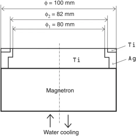
magnetron equipped with (i) composed Ti/Ag target of diameter 100 mm and (ii) NdFeB magnets. The composed target consists of Ti plate with Ag and Ti fixing ring, see Fig.1. The amount of Ag incorporated in Ti–Ag–O film was set by the inner diameter of the Ti fixing ring. The amount of Ag incorporated into TiO2film almost does not
depend on partial pressure of oxygenpO2 used in reactive sputter-deposition of TiOx films. In all Ti–Ag–O films described in this article, the amount of Ag was*2 at.%.
Films were sputter-deposited under the following condi-tions: magnetron discharge currentId=2 A, substrate bias
Us =Ufl, substrate-to-target distance ds–t=120 mm,
par-tial pressure of oxygen ranging from 0 to 1.5 Pa, and total pressure of sputtering gas mixturepT¼pArþpO2 =1.5 Pa;
Uflis the floating potential. Films were deposited on
unhe-ated glass substrates (2091091 mm3). The thicknessh of Ti–Ag–O films ranged from*500 to 2,800 nm.
The thickness of Ti–Ag–O films was measured by a stylus profilometer DEKTAK 8 with a resolution of 1 nm. The structure of film was determined by PANalytical X’Pert PRO diffractometer working in Bragg–Bretano geometry using a Cu Ka (40 kV, 40 mA) radiation. The water droplet contact angle (WDCA) on the surface of the TiO2film after its irradiation by UV light (Philips TL-DK
30W/05, Wir=0.9 mW/cm2 at wavelength k=365 nm)
was measured by a Surface Energy Evaluation System made at the Masaryk University in Brno, Czech Republic. The film surface morphology was characterized by an atomic force microscopy (AFM) using AFM-Metris-2000 (Burleigh Instruments, USA) equipped with an Si3N4
probe. The surface and cross-section film morphology was characterized by SEM Quanta 200 (FEI, USA) with a resolution of 3.5 nm at 30 kV.
The bioactivity of Ti–Ag–O film was determined using a modified standard test described by BS:EN 13697:2001 [22]. Coated samples were shaken in 100% methanol for 40 min. Samples were removed aseptically and placed in a UVA transparent disposable plastic Petri dish, film side uppermost. The coated samples were then pre-irradiated by placing those under 3915 W UVA bulbs with a 2.24 mW/cm2output for 24 h.
Escherichia coli ATCC 10536 was subcultured into nutrient broth (Oxoid, Basingstoke, UK) and inoculated onto cryobank beads (Mast Diagnostics, Liverpool, UK) and stored at -70°C. Beads were subcultured onto nutri-ent agar (Oxoid) and incubated at 37°C for 24 h and stored at 5 °C. A 50 ll loopful was inoculated into 20 ml nutrient broth and incubated for 24 h at 37°C. Cultures were centrifuged at 5,0009g for 10 min in a bench centrifuge, and the cells were washed in de-ionised water three times by centrifugation and re-suspension. Cultures were re-suspended in water and adjusted to OD 0.5 at 600 nm in a spectrometer (Camspec, M330, Cambridge, UK) to give *2 9108 colony forming units (cfu) ml-1. Fifty micro-litre of this suspension was inoculated on to each test sample and spread out using the edge of a flame sterilized microscope cover slip.
The prepared samples were then UV activated. Four samples were exposed to three 15 W UVA lamps at 2.29 mW/cm2. At time zero, a sample was removed immediately and the remaining samples removed at regular intervals. Four samples exposed to UVA but covered with a polylaminar UVA protection film (Anglia Window Film, UK) to block UVA but not infra-red, acted as controls. The samples were then immersed in 20 ml of sterile de-ionised water and vortexed for 60 s to re-suspend the bacteria. A viability count was performed by serial dilution and plating
Magnetron
Water cooling
[image:2.595.53.287.462.697.2]onto nutrient agar in triplicate and incubation at 37°C for 48 h. Each experiment was performed in triplicate.
Results and Discussion
Deposition Rate
The deposition rateaDof Ti–Ag–O film reactively
sputter-deposited in a mixture of Ar?O2 decreases with
increasing partial pressure of oxygenpO2. It is the lowest in the oxide mode of sputtering. Under conditions used in our experiment, the deposition rateaDof TiO2/Ag films formed
in the oxide mode is*4.5 nm/min (see Fig.2). Structure
Effect of Partial Pressure of Oxygen
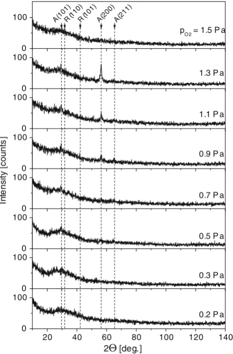
The structure of Ti–Ag–O film strongly depends on the partial pressure of oxygenpO2. An evolution of XRD patterns from sputter-deposited thin Ti–Ag–O films with increasing pO2is displayed in Fig.3. The change in the structure of film is connected with increasing energy delivered to it during growth mainly by bombarding ions with increasingpO2due to decrease ofaD(see Fig.2). It follows from the formula of
energyEbidelivered to the unit volume of growing film by
bombarding ions:Ebi=Ei/e(is/aD) = (Up-Ufl)is/aD[4,23];
hereEiis the energy of ion incident on a floating substrate,e
is the electron charge,UpandUflare the plasma and floating
potential of substrate, respectively. In our experiment, under the assumption of zero collisions the energyEi&30 eV
becauseUp&?20 V andUfl&-10 V.
Therefore, at the end of transition mode of sputtering dominated by relatively high values ofaDC6.6 nm/min at
pO2\0:3 Pa, relatively low energiesEbiare delivered to the growing film. It results in the formation of amorphous Ti–Ag–O films atpO2\0:3 Pa. As the film deposition rate aDdecreases more energy is delivered to the growing film
and the Ti–Ag–O films crystallize.
A nanocrystallization of Ti–Ag–O film, characterized by low-intensity X-ray reflections from the anatase phase, is observed at aDB 5.5 nm/min. The nanocrystallization
occurs as a consequence of longer deposition time td
nee-ded to form Ti–Ag–O film with the same thicknesshat low values of aD. It indicates that the film nanocrystallization
was very probably due to a higher total energy ET=
Ebi?Eca?Echdelivered to the growing film in the oxide
mode compared to that delivered to the film sputter-deposited at higher values of aD in the transition and
metallic (pO2 ! 0) modes of sputtering; Eca(pT) and Ech(pO2) are the energy delivered to the film by fast con-densing atoms and by the heat evolved in the formation of Fig. 2 Deposition rateaDof reactively sputter-deposited Ti–Ag–O
films as a function ofpO2. Deposition conditions:ID=2 A,US=Ufl,
ds–t=120 mm,pT=1.5 Pa
20 40 60 80 100 120 140
0 100
R(110)R(101)A(200)A(211)
In
te
nsi
ty
[
c
ount
s
]
2Θ [deg.]
A(101)
0 100 0 100 0 100 0 100 0 100 0 100 0 100
1.3 P a pO2 = 1.5 P a
0.7 P a
0.2 P a 0.5 P a
0.3 P a 0.9 P a 1.1 P a
[image:3.595.312.540.58.403.2] [image:3.595.53.288.494.677.2]oxide (exothermic reaction), respectively. From Fig.3, it is seen that the crystallinity of Ti–Ag–O film improves with increasing pO2; compare films of the same thickness h=600 nm sputter-deposited at pO2 =0.9, 1.1 and 1.3 Pa. BecauseaDof the film is almost constant for pO2 ranging from 0.9 to 1.3 Pa, this experiment indicates that a main component of energy ET delivered to the growing
film is probablyEch, i.e. the heat evolved in formation of
the oxide. The nanocrystalline Ti–Ag–O films exhibit the anatase structure with A(200) preferred crystallographic orientation. The development of WDCA and optical band gapEgof Ti–Ag–O films with increasing partial pressure of
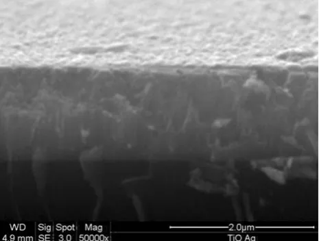
oxygenpO2 is shown in Table1. Surface morphology and film cross-section of thick Ti–Ag–O film prepared at pO2 =0.5 Pa are shown in Fig. 4. It can be seen that dense featureless structure with relatively smooth surface is developed.
The nanocrystallization of anatase phase strongly improves the hydrophilicity of the surface of Ti–Ag–O film after its UV irradiation. Almost all films sputter-deposited atpO20:5 to Pa exhibit superhydrophilicity (see Table2). The Ti–Ag–O film sputter-deposited in a pure oxygen, i.e. atpO2 =1.5 Pa, exhibits an X-ray amorphous structure. In spite of this fact also this film is still quite well hydrophilic. Effect of Film Thickness
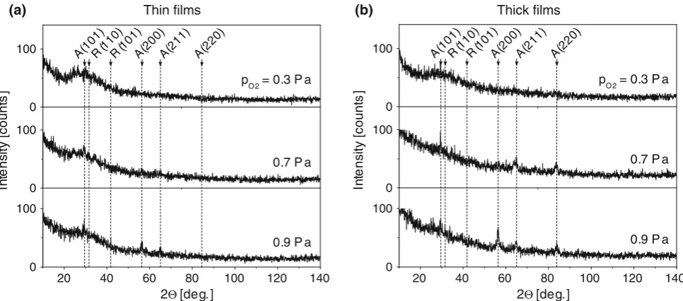
The crystallinity of TiO2 films improved not only with
increasing pO2 but also with increasing film thickness h (see Fig.5). From this figure, it can be seen that thick (*1,500 nm) films exhibited better crystallinity compared to thin (*700 nm) films sputter-deposited at the same value ofpO2. It is due to a longer deposition timetd, which enables to deliver a higher total energyETto the growing
film at the same deposition rate aD. More details on the
evolution of intensities of XRD pattern from sputter-deposited TiO2films are given in the reference [3]. Thicker
TiO2/Ag films also exhibited (i) a better UV-induced
hydrophilicity, (ii) lower values of the optical band gapEg
and (iii) higher roughness of the films (see Table2 and Fig.6, respectively). The decrease ofEgof TiO2film with
increasing crystallinity is in agreement with our previous results [3,4]. The anatase TiO2films with A(200) preferred
crystallographic orientation exhibit the best hydrophilicity (see Table2). The hydrophilicity of TiO2/Ag,
character-ized with WDCA after UV irradiation, is fully comparable with that of pure TiO2film which exhibits WDCA of*10°
or less, see for instance [3,4,25].
Hydrophilicity of Transparent TiO2/Ag Films
The hydrophilicity is characterized by a WDCA on the surface of TiO2/Ag film. The development of WDCA in
thin (*700 nm) and thick (*1,500 nm) TiO2/Ag films,
[image:4.595.309.542.60.236.2]sputter-deposited in the oxide mode of sputtering, before and after UV irradiation with increasingpO2is displayed in Fig.7. From this figure, it is clearly seen that a short (20 min) time of UV irradiation was sufficient to induce
Table 1 Deposition rateaD, thicknessh, WDCA after UV irradiation for 20, 60 and 300 min and optical band gap
Egof*500–700 nm thick TiO2 films reactively sputter-deposited atId=2 A,
pT=1.5 Pa,Us=Uflon unheated glass substrate as a function of partial pressure of oxygenpO2
Egwas determined using the formula given in [24]
pO2 (Pa) h(nm) aD(nm/min) WDCA (°) after UV irradiation for Eg(eV)
20 min 60 min 300 min
0.2 600 12.5 89 84 71 –
0.3 700 6.6 38 18 11 3.21
0.5 600 5.4 15 13 13 3.21
0.7 700 5.2 19 12 8 3.19
0.9 600 4.4 20 16 11 3.11
1.1 600 4.4 16 13 9 3.12
1.3 600 4.6 22 16 11 3.06
1.5 500 4.4 23 14 8 3.11
Fig. 4 Cross-section SEM image of thick (*1,500 nm) Ti–Ag–O film sputter-deposited on unheated substrate atID=2 A,US=Ufl,
[image:4.595.178.539.570.711.2]high hydrophilicity. The WDCA decreased below 10° in thick (*1,500 nm) films.
UV–Vis Transmission Spectra and Optical Band Gap of TiO2/Ag Films
Ultraviolet–visible (UV–vis) light transmission spectra were measured on the TiO2/Ag films sputter-deposited in
the oxide mode on unheated glass substrates. The trans-mission spectra were measured for thin (*700 nm) and thick (*1,500 nm) TiO2/Ag films (see Fig. 8). Thicker
films exhibit a decrease in the transmission of incident light and clear shift of the absorption to higher wavelengths k. As expected, this fact results in the decrease of (i) the optical band gap Egand (ii) WDCA of thicker films (see
Table2 and Fig.7). In spite of a stronger absorption of light atk=550 nm in thicker films, the reactively sputter-deposited TiO2/Ag films with thicknessh&1,500 nm still
[image:5.595.51.543.85.177.2] [image:5.595.57.540.204.417.2]remain semitransparent.
Table 2 Deposition rateaD, thicknessh, WDCA after UV irradiation and optical band gapEgof thin (*500 nm) and thick (*1,500 nm) TiO2 films reactively sputter-deposited atId=2 A,pT=1.5 Pa,Us=Uflon unheated glass substrate
pO2 (Pa) Thin films Thick films
h(nm) aD(nm/min) WDCA after UV irradiation Eg(eV) h(nm) aD(nm/min) WDCA after UV irradiation Eg(eV)
20 min 60 min 300 min 20 min 60 min 300 min
0.3 700 6.6 38 18 11 3.21 1,500 6.3 18 11 8 3.06
0.7 700 5.2 19 12 8 3.19 1,200 5.8 10 9 9 3.04
0.9 600 4.4 20 16 11 3.11 1,500 5.2 8 9 8 2.86
Egwas determined using the formula given in [24]
0 100
20 40 60 80 100 120 140
0 100 0 100
A(211)
0.9 P a 0.7 P a
pO 2 = 0.3 P a
In
te
ns
it
y
[
c
ount
s
]
2Θ [deg.] A(101)R(110)R(101)A(200) A(220)
Thick films Thin films
(a) (b)
0 100
20 40 60 80 100 120 140
0 100 0
100 A(211)
0.9 P a 0.7 P a pO 2 = 0.3 P a
In
te
ns
it
y
[
c
ount
s
]
2Θ [deg.]
A(101)R(110)R(101)A(200) A(220)
Fig. 5 Comparison of X-ray structure ofa thin (*700 nm) and bthick (*1,500 nm) Ti–Ag–O films sputter-deposited on unheated glass substrate atID=2 A,US=Ufl,ds–t=120 mm,pT=1.5 Pa and three values ofpO2 =0.3, 0.7 and 0.9 Pa
Fig. 6 Comparison of AFM surface topography ofathin (*700 nm) and b thick (*1,500 nm) Ti–Ag–O films sputter-deposited on unheated glass substrate at ID=2 A, US=Ufl, ds–t=120 mm,
[image:5.595.55.286.467.667.2]Also, it is worthwhile to note that in spite of the decrease of Eg and the shift of the absorption of electromagnetic
waves into visible region, the hydrophilicity of surface of Ti–Ag–O film must be induced by UV light (see Fig.7). A very short (B20 min) UV irradiation time was sufficient to induce hydrophilicity. The need for surface activation by UV, however, indicates that the decreasing of Egand the
shifting of absorption into vis region are not sufficient conditions to prepare hydrophilic TiO2-based films under
visible light. The key parameters, which affect the photo-induced hydrophilicity of TiO2-based films under visible
light are not known so far. Recent experiments performed in our laboratory indicate that the film nanostructure could be of a key importance for the creation of hydrophilic
TiO2-based films operating under visible light only, i.e.
without UV irradiation. Mechanical Properties
The microhardness H, effective Young’s modulus E* and resistance to plastic deformation, which is proportional to the ratio H3/E*2 [26] were measured for *950 nm thick Ti–Ag–O films as a function of partial pressure of oxygen pO2 (see Fig.9). All quantities vary only slightly withpO2 increasing above 0.5 Pa. The values ofHare low of about 4–5 GPa. The resistance to plastic deformation character-ized by the ratioH3/E*2is also very low of about 0.01. The hardnessHneeds to be increased and it could be achieved
300 min 60 min
No UV
UV irradiation 20 min
0.0 0.4 0.8 1.2 1.6 2.0
0 20 40 60 80 100
(a) (b)
WD
CA
[deg.]
0 20 40 60 80 100
WD
CA
[deg.]
pO2 [Pa]
0.0 0.4 0.8 1.2 1.6
pO2 [Pa]
No UV
20 min
60 min 300 min UV irradiation
Fig. 7 Characterization of water droplet contact angle WDCA on the surface of a thin (*700 nm) and bthick (*1,500 nm) Ti–Ag–O films under UV irradiation for 20, 60 and 300 min as a function of
partial pressure of oxygen pO2. Deposition conditions: ID=2 A,
US=Ufl,ds–t=120 mm, unheated glass substrate
300 400 500 600 700 800 900 1000
0 20 40 60 80 100
pO 2 [Pa] h [µm]
1.3 2.8 0.9 1.6 0.7 1.2 0.5 1.5 0.3 1.5 Glass
Transmittance [%]
λ [nm]
300 400 500 600 700 800 900 1000
0 20 40 60 80 100
(a) (b)
pO 2 [Pa] h [µm]
1.5 0.5 1.3 0.6 1.1 0.6 0.9 0.6 0.7 0.7 0.5 0.6 0.3 0.7 Glass
Transmittance [%]
λ [nm]
[image:6.595.309.542.68.248.2] [image:6.595.57.545.318.479.2]by substrate biasing. However, such experiment has not been performed so far and is the subject of our next investigations.
Antibacterial Properties
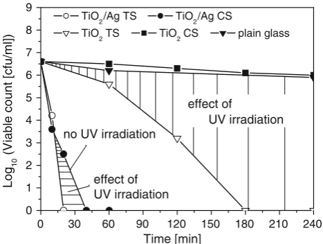
The bioactivity of Ti–Ag–O films was tested by killing the bacteriumE. coliATCC 10536 on the surface of 500 nm thick TiO2/Ag single layer sputter-deposited in the oxide
mode on unheated glass substrate during UV irradiation for a given time tir. The results are shown in Fig. 10. For
comparison, the killing of E. coli bacteria on uncoated plain glass and plain glass-coated with TiO2layer is also
given. The glass coated with TiO2/Ag single layer exhibits
the fastest killing; 20 min of UV irradiation was sufficient for 100% kill (six orders of magnitude reduction).
Figure10 further shows a comparison of the biocidal activity of TiO2 and TiO2/Ag films. There was a big
difference in biocidal activity of TiO2 test sample (TS)
(irradiation under UV lamp by both UV?IR) and control TiO2 sample (CS) (irradiated by IR only; the sample is
covered with a polylaminar UVA protection film, which blocks UV from UV lamp); here IR is the infra-red radi-ation. A strong effect of UV irradiation on killing activity is clearly seen. The 100% kill ofE. colion TiO2surface is
seen after 180 min of UV irradiation while no killing is observed on TiO2 surface without UV irradiation after
240 min.
In contrast, 100% kill of E. colion TiO2/Ag surface is
seen not only after UV irradiation (20 min) but also without UV irradiation (40 min). This result indicates that the killing ofE. colion TiO2/Ag surface is probably due to
a combination of direct toxicity of Ag- and UV-induced photocatalytic activity. Results shown in Fig. 10 indicate that the direct toxicity of Ag was probably dominant. The dashed areas in Fig.10denote the effect of UV irradiation on killing of the bacterium E. coli on TiO2 and TiO2/Ag
surface.
Conclusions
The main results of investigation of physical and functional properties of sputter-deposited Ti–Ag–O thin films with low (B2 at.%) content of Ag can be summarized as fol-lows. TiO2/Ag films with anatase phase and small amount
(*2 at.%) of Ag exhibited an excellent UV-induced hydrophilicity. The added Ag due to strong toxicity also very rapidly killed E. coli on TiO2/Ag surface. This
shows that the surface of TiO2/Ag film can be
simulta-neously hydrophilic and antibacterial. Therefore, crystalline
0.000 0.005 0.010 0.015 0.020 0.025 0.030
H
3 /E 2 [GPa
]
0.2 0.4 0.6 0.8 1.0 1.2 1.4
0 2 4 6 8 10
(a) (b)
0 20 40 60 80 100 120 140 160
E [GPa]
H [GPa]
pO2 [Pa]
0.2 0.4 0.6 0.8 1.0 1.2 1.4
pO2 [Pa]
H
[image:7.595.55.542.58.228.2]E
Fig. 9 aMicrohardnessHand effective Young’s modulusE*andbratioH3/E2of*950 nm thick Ti–Ag–O films as a function of partial pressure of oxygenpO2. Deposition conditions:ID=2 A,US=Ufl,ds–t=120 mm
0 30 60 90 120 150 180 210 240
0 1 2 3 4 5 6 7 8 9
effect of UV irradiation
TiO2/Ag TS TiO2/Ag CS
TiO2 TS TiO2 CS plain glass
Log
10
(V
iable count [cfu/m
l])
Time [min] effect of
UV irradiation no UV irradiation
[image:7.595.53.287.479.655.2]TiO2/Ag film can be used as two-functional material. One
hundred per cent kill ofE. coli on the surface of TiO2/Ag
film was observed under visible light in 40 min. No UV-induced irradiation was needed. Formation of crystal-line Ti–Ag–O film required a minimum total energyETto
be delivered to the growing film. Therefore, the crystal-linity of TiO2/Ag film improves with its increasing
thicknessh. A longer deposition timetdneeded to form a
thicker film at the same deposition rateaDresults in greater
total energy ET delivered to the growing film.
Nanocrys-talline TiO2/Ag films exhibit excellent hydrophilicity
(B10°) already after a short (20 min) time of UV irradia-tion. Nanocrystallization of TiO2/Ag film sputter-deposited
in the oxide mode on floating unheated glass substrate (Us =Ufl) is very probably induced by the heat evolved
during formation of oxide (exothermic reaction).
Based on the results given above, the next investigation in this field should be concentrated on the physical and functional properties of nanocrystalline TiO2-based films.
Acknowledgements This work was supported in part by the Min-istry of Education of the Czech Republic under Project MSM# 4977751302, in part by Project PHOTOCOAT No. GRD1-2001-40701 funded by the European Community and in part by the Grant Agency of the Czech Republic under Project No. 106/06/0327. Authors would like to thank also to Mgr. Zdenek Stryhal, Ph.D. and Ing. Rostislav Medlin for performing AFM and SEM analysis, respectively.
References
1. N. Sakai, A. Fujishima, T. Watanabe, K. Hashimoto, J. Phys. Chem. B107, 1028 (2003). doi:10.1021/jp022105p
2. K. Hashimoto, H. Irie, A. Fujishima, Jpn J. Appl. Phys.44(Part 1), 8269 (2005). doi:10.1143/JJAP.44.8269
3. J. Musil, D. Herman, J. Sicha, J. Vac. Sci. Technol. A24, 521 (2006). doi:10.1116/1.2187993
4. J. Musil, J. Sicha, D. Herman, R. Cerstvy, J. Vac. Sci. Technol. A 25(4), 666 (2007). doi:10.1116/1.2736680
5. R. Asahi, T. Morikawa, T. Ohwaki, K. Aoki, Y. Taga, Science 293, 269 (2001). doi:10.1126/science.1061051
6. M. Anpo, M. Takeuchi, J. Catal.216, 505 (2003). doi:10.1016/ S0021-9517(02)00104-5
7. S. Yang, L. Gao, J. Am. Ceram. Soc. 87, 1803 (2004). doi: 10.1111/j.1551-2916.2004.00733.x
8. Y.H. Tseng, C.S. Kuo, C.H. Huang, Y.Y. Li, P.W. Chou, C.L. Cheng, M.S. Wong, Nanotechnology 17, 2490 (2006). doi: 10.1088/0957-4484/17/10/009
9. C. He, Y. Xiong, X. Zhu, Thin Solid Films422, 235 (2002). doi: 10.1016/S0040-6090(02)00892-1
10. J. Wang, J. Li, L. Ren, A. Zhao, P. Li, Y. Leng, H. Sun, N. Huang, Surf. Coat. Technol. 201, 6893 (2007). doi:10.1016/ j.surfcoat.2006.09.109
11. H.Q. Tang, H.J. Feng, J.H. Zheng, J. Zhao, Surf. Coat. Technol. 201, 5633 (2007). doi:10.1016/j.surfcoat.2006.07.171
12. H.J. Zhang, D.Z. Wen, Surf. Coat. Technol.201, 5720 (2007). doi:10.1016/j.surfcoat.2006.07.109
13. X.B. Tian, Z.M. Wang, S.Q. Yang, Z.J. Luo, R.K.Y. Fu, P.K. Chu, Surf. Coat. Technol. 201, 8606 (2007). doi:10.1016/ j.surfcoat.2006.09.322
14. I.M. Arabatzis, T. Stergiopoulos, M.C. Bernard, D. Labou, S.G. Neophytides, P. Falaras, Appl. Catal. Environ. 42, 187 (2003). doi:10.1016/S0926-3373(02)00233-3
15. F. Falaras, I.M. Arabatzis, T. Stergiopoulos, M.C. Bernard, Int. J. Photoenergy5, 123 (2003). doi:10.1155/S1110662X03000230 16. S.X. Liu, Z.P. Qu, X.W. Han, C.L. Sun, Catal. Today93–95, 877
(2004). doi:10.1016/j.cattod.2004.06.097
17. Y. Liu, C. Liu, Q. Rong, Z. Zhang, Appl. Surf. Sci.230, 7 (2003). doi:10.1016/S0169-4332(03)00836-5
18. Y.L. Kuo, H.W. Chen, Y. Ku, Thin Solid Films515, 3461 (2007). doi:10.1016/j.tsf.2006.10.085
19. M. Stir, R. Nicula, E. Burkel, J. Eur. Ceram. Soc. 26, 1542 (2006). doi:10.1016/j.jeurceramsoc.2005.03.260
20. L.A. Brook, P. Evans, H.A. Foster, A. Steele, D.W. Sheel, H.M. Yates, J. Photochem. Photobiol. A187, 53 (2007). doi:10.1016/ j.jphotochem.2006.09.014
21. L.A. Brook, P. Evans, H.A. Foster, M.E. Pemble, D.W. Sheel, A. Steele, H.M. Yates, Surf. Coat. Technol.201, 9373 (2007). doi: 10.1016/j.surfcoat.2007.04.020
22. Anon, BS EN 13697:2001, Chemical disinfectants and antisep-tics. Quantitative non-porous surface test for the evaluation of bacterial and/or fungicidal activity of chemical disinfectants used in food, industrial, domestic and institutional areas. Test method and requirements without mechanical action. British Standards Institute, London, 2001
23. J. Musil, J. Suna, Mater. Sci. Forum502, 291 (2005)
24. P.M. Kumar, S. Badrinarayanan, M. Sastry, Thin Solid Films 358, 122 (2000). doi:10.1016/S0040-6090(99)00722-1
25. T.Y. Tsui, G.M. Pharr, W.C. Oliver, C.S. Bhatia, R.L. White, S. Anders, A. Anders, I.G. Brown, Mater. Res. Soc. Symp. Proc. 383, 447 (1995)