Thesis by Maraia Emily Ener
In Partial Fulfillment of the Requirements for the degree of
Doctor of Philosophy
CALIFORNIA INSTITUTE OF TECHNOLOGY Pasadena, California
2014
First I would like to thank my doctoral advisor, Harry Gray. Harry’s charisma, irreverence, enthusiasm for life, and passion for chemistry are unparalleled, and I am honored to be a part of the extensive Gray Nation. I have greatly benefitted from the supportive, collaborative environment that Harry fosters in the group, and I am grateful for all of the opportunities he has given me to present my research to the bioinorganic community, including a number of international conferences. This Thesis, Harry, is for you. I love it!!!!!!
I would also like to thank Jay Winkler, who (whether he intended or not) has been every bit as much a mentor. I am constantly amazed by Jay’s brilliance, wealth of knowledge, and ability to build almost anything. The work in this Thesis would not have been possible without his guidance and expertise, and I continue to strive to be a more thorough, more rigorous, (and sometimes more skeptical!) scientist because of it. Don’t panic. Just think.
Many thanks also go out to my other committee members: Professors Mitchio Okumura, Theodor Agapie, and Doug Rees. I am lucky to have such a supportive team, and have benefitted from our conversations at my candidacy exam, 4th year meeting, props exam, and thesis defense. Extra thanks to Mitchio for being my committee chair, and for the five pounds of delicious green grapes that Kana and I picked at his home.
with-my-life” morning discussions over coffee, Jeff has made me laugh, infuriated me, inspired me, and reminded me countless times that “it’s going to be okay.” No one else has so consistently and so vocally expressed confidence in my scientific abilities; for that, I am ever grateful. Oliver has been my little brother counterpart (this is my table quadrant, keep your feet out!). He is always around to lend a hand, share a drink (or five), enjoy a laugh, and has obligingly agreed to take over all of my BILRC duties - I am confident that they are in good hands (p.s. Oliver, you still owe me a beer). Kana has been a fantastic office mate, confidante, and friend. She has patiently fielded my questions in physical chemistry, laser operation, and diplomacy, has read numerous iterations of my research proposals, and has watered my plants when I’m not in town. Thanks, Kana!
And now, from the end… back to be beginning. Before I even joined the group, Nicole Bouley Ford, Gretchen Keller, and Bert Lai convinced me that Caltech was the place to be. Bert Lai became my office mate, and instantly made me feel I was part of the subbasement/bio crew. His advice that “there will always be one more experiment” has never felt more relevant than now, as I write this Thesis.
I am deeply grateful to Lionel Cheruzel, who, in his last few months at Caltech, was a patient and effective teacher, trained me on everything P450, and handed me a working project just as things were starting to get really interesting. I continue to benefit from communication and collaboration with The Frenchman.
think more deeply about fundamental aspects of inorganic chemistry. It was incredibly rewarding to teach Ch153 side-by-side. I still find myself trying to fill Jillian and Alec’s enormous shoes.
In the realm of Ch153, Alexis Komor has been a wonderful Co-TA for Ch153 for the past two years, and Bryan Hunter has done a fantastic job taking up the reins. You got this, guys!
When I joined the Gray group, the BioInorganic subbasement was a particularly rockin’ place to be. Special thanks go to Kyle Lancaster, Charlotte Whited, Nicole Bouley Ford, and Gretchen Keller for bringing, the class, the sass, the fast-talking and the shit-talking. Charlotte founded our two-person Team Subgroup for heme enzymes, and taught me that grad school is a balance between collaboration and independence: “It’s your Thesis, you figure it out!” Through the chaos and darkness, Gretchen Keller reminds me that there is a world outside of grad school. Her creativity, artistry, and insights are an inspiration. It was fantastic to have another partner in crime in the world of heme enzymes (go Team Haem Team!), and I’m honored to have become her friend. In the twilight of the subbasement days, Peter Agbo’s quiet presence, dry humor, and sharp intellect have been a comfort.
forgiven him for being a St. Olaf student, and he has shown himself to be a calm and patient teacher, and reliable resource to turn to. The farmers market trips and Saturday brunches with James, Judy and Co. made Caltech feel more like home my first year in California. Speaking of brunches, I also want to thank the other members of Team House: Alex Goldberg (and his delicious cardamom coffee) and Ethan Van Arnam.
I have had the opportunity to work with a number of talented undergraduate students, including Katja Luxem, Megan Jackson, Rocio Mercado. They continue to inspire me, and remind me that sometimes otter pops are the answer to everything. Special thanks to Katja Luxem for being my and Jeff’s first SURF (a modicum of the data she collected is in this Thesis). Based on her record of international escapades thus far, I’m pretty sure Katja will take over the world - way to go, lady! Extra special thanks to Megan for the chocolate birthday cheesecake.
I’ve been fortunate to work closely with some fantastic postdocs in the Gray group. First, I want to thank Wes Sattler, who is rarely found without a smile on his face, and who has come through a number of lab dramas with optimism and resolve. I may never end up being an organometallic chemist, but it’s not due to any lack of effort on his part! By extension, I want to thank his biological clone, Aaron Sattler. In particular, Aaron’s collection of antique Macbook Pro dongles has saved me more than once. James Blakemore has been another wonderful addition to the Group, and I have benefitted immensely from our conversations on electrochemistry.
Stubbert, Astrid Mueller, Matt Hartings, Josh Palmer, Paul Oblad, Heather Williamson, Yan Choi, Melanie Yen, Carl Blumenfeld, Mike Rose, Mike Lichterman, Kate Pletneva. Seiji Yamada’s time in our group is now the stuff of legend.
My graduate experience wouldn’t have been complete without other members of the Caltech staff, and Chemistry Department. Catherine May, Rick Jackson, and Pat Anderson keep the world spinning. I will never forget Catherine’s welcome on my first day “who are you?”, Rick’s magical cabinet stocked with tea and pick-me-up candies, and Pat’s immaculate fashion sense. Joe Drew and Steve Gould keep us supplied, and a fantastic team keeps the BI sparkling. It goes without saying that the Caltech faculty are amazing. I’m giving John Bercaw a special shout-out here for knowing where Carleton is, and for saying hi to me in the hallway. A number of non-Gray group researchers have also made important contributions to my graduate experience, including (but again, not limited to): Leslie O’Leary, Ian Tonks, Samantha MacMillan, Rachel Klet, and Maddy Radlauer.
I also want to thank a number of collaborators in Frances Arnold’s group. Pete Heinzelman was very helpful in my first few months at Caltech, Eric Brustad is a P450 BM3-crystallizing genius, and one of my proudest achievements in grad school (zero to reproducible air-sensitive redox titrations in < 30 days) came about because of collaborations with Eric, Pedro Coelho, and Jane Wang. Frances Arnold herself is an inspiration, and has been supportive of my efforts.
the character…). Because of them, I have made it to the end of my PhD; because of them, I never considered that I couldn’t. I have two phenomenal siblings, Cjuneyt and Amir Ener, who make me proud every day. Their beautiful and brilliant lady friends, Eloise Galligan and Lydia Larson, have been a wonderful addition to our family, and Eloise is now officially my sister-in-law! To my BFFs Jess Wenstrom and Erica Hoaglund – can’t wait to see you again!
Acknowledgments ...iv
Abstract... x
Table of Contents...xiii
List of Figures...xix
List of Tables ...xxv
Chapter 1: Cytochrome P450: From pursuit of reactive intermediates to engineering novel reactivity... 1
1.1 Cytochrome P450: a remarkable metabolic enzyme... 2
1.2 P450: structural features that direct function... 4
1.3 P450 activity: reactions and mechanisms ... 9
Oxygen activation...10
P450 active hydroxylating agents: ferryl compounds I and II ...14
1.4 Photo-triggered electron transfer in proteins ...15
Photosensitizers...18
Small molecule quenchers ...21
1.5 Photo-triggered ET in P450: two pathways toward reactive heme species...23
Precedence for photochemical heme oxidation...24
P450 considerations: the buried heme ...26
1.6 New Frontiers: Novel P450 Active Species for Non-Native Catalysis ..26
1.7 Conclusions ...30
1.8 References ...31
Chapter 2: Photo-triggered oxidation of Ru-modified cytochrome P450...35
2.1 Background: Toward high-valent P450 intermediates...36
2.2 Motivation and selection of the photochemical system ...40
Photosensitizer...40
Exogenous oxidative quencher...41
P450 mutants...41
2.3 Results...43
2.3.1 Characterization...43
UV-visible absorbance...43
Steady-state luminescence...44
X-ray crystal structure analysis...45
2.3.2 Laser flash-quench experiments...49
Time-resolved luminescence...49
Transient absorption...52
Kinetics analysis of TA data ...57
2.4 Discussion ...59
2.7 Materials and Methods ...66
Chemicals...66
Procedures...66
2.7.1 Ru photosensitizer ...66
Synthesis...66
Characterization...67
2.7.2 Mutagenesis and expression of P450 BM3 mutants ...67
Plasmid...67
Mutagenesis...67
Expression ...68
Extraction and purification ...68
2.7.3 Ru-P450 conjugation ...69
2.7.4 Crystallization and structure determination...69
2.7.5 Preparation of laser samples ...70
2.8 References ...71
Chapter 3: Photo-triggered electron transfer through tryptophan in Ru-P450 systems...74
3.1 Background: Light to ET ...75
3.1.1 P450 systems: Motivation and selection ...80
3.2 Results and Analysis ...84
3.2.1 Characterization of mutants ...84
UV-visible absorbance...84
X-ray crystal structure analysis of C97-BM3(WH)...85
3.2.2 Ru-P450 luminescence...88
3.2.3 Ru-P450 transient absorption...92
P450-BM3 mutants: effect of tryptophan 96...92
ET reactivity of CYP119 mutants ...94
Temperature dependence...97
Search for the tryptophan radical cation...98
3.3 Discussion ...99
Final thoughts and avenues for future work...101
3.4 Conclusions ...102
3.5 Acknowledgments ...102
3.6 Materials and Methods ...102
Materials...102
Procedures...103
3.6.1 Photosensitizer synthesis...103
Synthesis...103
Characterization...104
3.6.2 Protein mutagenesis, expression and purification ...104
3.6.3 Conjugation of the Ru-photosensitizer...105
3.6.4 Crystallization and structure determination...105
3.6.5 Preparation of laser samples ...106
3.7 References ...107
Chapter 4: Multistep electron transfer: “hopping maps” tutorial and application...109
4.1 Electron transfer through proteins...110
Method for examining photochemical ET in proteins: photochemical triggering...112
4.2 Single step electron tunneling: semiclassical theory ...114
Experimental measurements...118
4.3 Multistep electron transfer ...119
4.4 Construction of hopping maps: an example for Re-azurin ...124
4.5 ET parameters: selection process, effects, and limitations...129
Temperature...130
Distance...131
Tunneling decay constant, β...133
Reorganization energy, λ...134
Driving forces...136
4.6 Hopping map limitations ...137
4.7 Application to the Ru-W-P450 system ...138
Estimates and challenges...139
Single-step tunneling...141
Hopping analysis...142
Varying distance...143
Varying β and λ ...145
4.8 Conclusions ...147
4.9 Acknowledgments ...147
4.10 References ...148
Chapter 5: Photochemical heme reduction and gas binding in cytochrome P450...152
5.1 Background: reductive activation of dioxygen ...153
5.2 Results and Analysis ...159
5.2.1 Reductive flash-quench...159
Luminescence...159
Single wavelength transient absorption ...161
Assignment of intermediates...163
Fitting...165
5.2.2 Reductive ET in the presence of CO...168
5.2.3 CO photolysis and rebinding...170
Picosecond transient absorption measurements...178
Non-negative least squares fitting...179
Eyring analysis...185
5.3 Discussion, conclusions, and future work ...186
5.4 Acknowledgments ...187
5.5 Materials and Methods ...187
Chemicals...187
Ru-P450 conjugates...188
Synthesis/purification of reductive quencher...188
Preparation of samples for flash-quench heme reduction...188
Preparation of samples for CO photolysis and rebinding...189
5.6 References ...191
Chapter 6: Axial-ligand influence on P450 reduction potentials: implications for catalysis...194
6.1 Background: Controlled electron flow through P450...195
Enzymes...200
6.2 Methods development for redox titrations...202
Spectrophotometric titration...202
Electrochemical titration...205
Potentiometric titration...209
Test: Wild-type P450 BM3...210
6.3 Results...213
6.3.1 Wild-type Enzymes: P450 BM3 and CYP119...213
Wild Type P450 BM3...213
Wild Type CYP119...214
6.3.2 Potentiometric titration of mutants for in vivo cyclopropanation...216
C400S axial mutation...217
Engineered cyclopropanation mutants...218
6.3.3 Additional axial mutants ...219
6.4 Discussion ...223
6.5 Conclusions and future work...224
6.6 Acknowledgments ...225
6.7 Materials and Methods ...225
Chemicals...225
Instrumentation...226
Procedures...226
Preparation of samples for redox titration...226
Potentiometric redox titration...227
6.8 References ...228
Appendix A: Photochemical oxidation of nitric oxide synthase...230
Selection of NOS mutant and Ru photosensitizer...235
Characterization of RuNOS...237
A.2.2 Ru-NOS Luminescence ...239
Luminescence quenching...242
A.2.3 Transient Absorption...244
A.3 Discussion ...247
A.4 Conclusion ...249
A.5 Acknowledgments ...250
A.6 Materials and Methods ...250
Expression protocols...250
Extraction and purification...250
Ru-NOS conjugation and purification...251
A.7 References...253
Appendix B: Common Protocols...254
B.1 Instrumentation...255
B.2 Site-directed mutagenesis ...255
B.3 Transformation protocol ...256
B.4. Amplification and purification of plasmid DNA...256
B.5 P450 overexpression in E. coli, extraction and purification ...257
Expression...257
Extraction and purification...257
B.6 Ru-P450 conjugation ...258
B.7 Preparation of laser samples...259
B.8 Laser details ...260
Nanosecond-to-second transient spectroscopies...260
Picosecond-to-nanosecond transient spectroscopy...263
B.9 Data workup...265
Appendix C: Chapter-specific notes...266
C.1 Notes for Chapter 2 ...267
Ru-P450 luminescence: dependence on concentration...267
Low temperature experiments ...267
C.2 Notes for Chapter 3 ...269
Search for the tryptophan radical cation intermediate...269
C.3 Notes for Chapter 5 ...271
Selection of probe wavelength for CO rebinding kinetics...271
Overlay of PSI and NSI kinetics traces...272
Nonlinear least squares fitting of CO rebinding kinetics at various temperatures...273
C.4 Notes for Chapter 6 ...275
Determination of ferrous/ferric ratios...275
D.2 Time resolved single-wavelength data workup ...279
D.2.1 Time-zero adjustment ...279
xadjuster...279
D.2.2 Log-compression ...281
compress...281
logtimej ...283
D.3 Data splicing ...285
overlayer...285
combine...288
D.4 Singular Value Decomposition ...291
svder1...291
svderMulti...292
D.5 Multiexponential fitting...293
nonlinear_fitter4...293
autoresider...295
MExpGFitter...296
MExpG...297
MExpGvalues...298
D.6 Nonnegative least squares analysis ...299
nnls_prep...299
nnls_grad_reg_r2_KT...301
Panalyzer...310
Pmoments...312
D.7 Hopping Maps...313
MapMaker...313
MapPlotter...315
MapValues...316
tauM...318
tauETM...319
D.8 Ferric/ferrous deconvolution...320
Number Page Figure 1.1 Structures of P450 BM3 heme domain from Bacillus
megaterium and CYP119 from Sulfolobus acidocaldarius ... 5
Figure 1.2 P450 BM3 active site ... 6
Figure 1.3 CYP119 active site ... 7
Figure 1.4 Overlay of substrate-free and substrate-bound forms of P450 BM3 ... 8
Figure 1.5 Various reactions catalyzed by cytochrome P450...10
Figure 1.6 Canonical P450 catalytic cycle ...11
Figure 1.7 Structure of P450 BM3 oxygenase and reductase domains...13
Figure 1.8 [Ru(bpy)3]2+ absorption and emission spectra in water, at room temperature ...16
Figure 1.9 [Ru(bpy)3]2+ photochemistry...17
Figure 1.10 Redox active model proteins ...18
Figure 1.11 An example of a perfluorinated ruthenium wire...19
Figure 1.12 Photosensitizers for site-specific surface labeling at amino acids ...20
Figure 1.13 Flash-quench ET cycles...22
Figure 1.14 Paths for formation of high-valent CI and CII ...24
Figure 1.15 Flash-quench oxidation of a heme protein active site...25
Figure 1.16 Histidine-ligated hemes ...25
Figure 1.17 P450-catalyzed cyclopropanation of styrene ...28
Figure 1.18 Native and engineered P450 catalytic schemes ...28
Figure 2.1 Catalytic cycle for P450-catalyzed hydroxylation reactions...36
Figure 2.2 [RuII(bpy)3]2+ flash-quench and oxidation of the a heme protein active site...39
Figure 2.3 [Ru(bpy)2(IAphen)]2+ tethering to cysteine, to form the conjugate Cys-Ru(bpy)2(Aphen)...40
Figure 2.4 K97 labeling site...42
Figure 2.5 Absorption spectra of [Ru(bpy)2(IAphen)]+2, P450-BM3 C62A/C156S/K97C, RuK97C-P450BM3 at approximately equal concentrations ...44
Figure 2.6 Steady-state luminescence spectra of Ru photosensitizers in deoxygenated water ...45
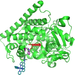
Figure 2.7 The RuK97C-P450BM3 structure. ...47
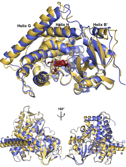
Figure 2.8 Overlay of RuK97C-P450BM3 with wild type substrate-free and substrate-bound forms ...47
Figure 2.11 Luminescence decays of RuK97C-P450BM3 (pH 8) at various
concentrations of [Ru(NH3)6]3+ quencher...51
Figure 2.12 Stern-Volmer quenching of RuK97C-P450BM3 with [Ru(NH3)6]3+ at three pH values...52
Figure 2.13 Single-wavelength transient absorption of RuII K97C-FeIIIP450 in the absence of quencher ...53
Figure 2.14 Transient Absorption data for flash-quench of [Ru(bpy)2(IAphen)]2+ with 17 mM [Ru(NH3)6]3+...54
Figure 2.15 Single-wavelength transient absorption following flash-quench (λex= 480 nm) of RuIIK97C-FeIIIP450 at pH 8 ...55
Figure 2.16 pH dependence of RuK97C-P450BM3 transient absorption features...56
Figure 2.17 tgSVD of TA data for RuIIK97C-FeIIIP450 with [Ru(NH3)6]3+ at six wavelengths ...57
Figure 2.18 Global fitting of RuII K97C-FeIIIP450 TA data at pH 8...59
Figure 2.19 Scheme for photochemical oxidation of cytochrome P450 ....60
Figure 2.20 Extracted difference spectra of intermediate species ...61
Figure 2.21 Photo-triggered cycle for flash-quench oxidation of RuIII K97C-FeIII P450...64
Figure 3.1 Photo-excitation of P680 chromophores in Photosystem II triggers oxidation of the oxygen evolving complex (OEC)...75
Figure 3.2 Photo-excitation of the tethered ruthenium complex triggers oxidation of the P450 heme active site ...76
Figure 3.3 Formation of high-valent CI and CII...77
Figure 3.4 Multistep ET in rhenium-labeled azurin...79
Figure 3.5 Multistep ET in ruthenium-labeled cytochrome P450 ...80
Figure 3.6 Putative multistep ET pathway in RuC97-BM3(W) ...81
Figure 3.7 Structures of P450 BM3 and CYP119 ...83
Figure 3.8 Photosensitizer conjugation sites in P450-BM3 and CYP119 ..84
Figure 3.9 UV-visible absorbance spectra of P450 mutants...85
Figure 3.10 Overlay of C97-BM3(WH) (purple) with open and closed WT BM3 structures ...86
Figure 3.11 C97-BM3(WH) active site...87
Figure 3.12 Time-resolved luminescence decays of Ru-BM3 conjugates in the presence and absence of 17 mM [Ru(NH3)6]3+ quencher ...89
Figure 3.13 Time-resolved luminescence decays of Ru-CYP119 conjugates in the presence and absence of 17 mM [Ru(NH3)6]3+ quencher...90
Figure 3.14 Overlay of luminescence decays for four Ru-P450 conjugates... ...91
conjugates in the presence of [Ru(NH3)6]3+, following excitation at 480 nm
...96
Figure 3.18 TA data of RuC77-CYP119(HW) at variable temperature ...97
Figure 3.19 UV-visible absorbance spectra P450, Ru photosensitizer ...99
Figure 4.1 Multistep ET in biological energy conversion systems. ...110
Figure 4.2 Sequential ET steps in the photo-triggered oxidation of Ru-P450 conjugates ...112
Figure 4.3 Photosensitizers and metallo-proteins...113
Figure 4.4 Ribonucleotide reductase from E. coli...114
Figure 4.5 Energy diagram illustrating thermodynamic parameters for an ET reaction ...115
Figure 4.6 Energy diagram illustrating diabatic and adiabatic states, and the coupling parameter HAB ...116
Figure 4.7 Tunneling timetables for driving force optimized ET reactions. ...118
Figure 4.8 The ReH124-W122-Azurin hopping system...124
Figure 4.9 Prompt #1: ET parameters...125
Figure 4.10 Prompt #2: Hopping Map parameters ...126
Figure 4.11 Sample hopping map for ReH124-W122-Azurin ...127
Figure 4.12 Temperature dependence of the ReH124-W122-Azurin hopping map ...130
Figure 4.13 Hopping maps for ReH124-W122-Azurin with differing distance formulations...132
Figure 4.14 Hopping maps for ReH124-W122-Azurin with differing values of β ...133
Figure 4.15 Hopping maps for ReH124-W122-Azurin with differing values of λ...135
Figure 4.16 Square scheme for tryptophan and tyrosine, including relevant reduction potentials and acid dissociation constants ...137
Figure 4.17 Hopping system in RuK97C-W96-P450BM3 ...139
Figure 4.18 Model of the Ru-P450 surface...140
Figure 4.19 Hopping maps for RuC97-W96-P450BM3 heme oxidation: Distance formulations ...144
Figure 4.20 Hopping maps for RuK97C-P450BM3 photochemical heme oxidation: Altering β and λ...145
Figure 4.21 Hopping maps for RuK97C-P450BM3 photochemical heme oxidation: Worst case hopping scenario ...146
Figure 5.1 Canonical P450 catalytic scheme, highlighting the reductive ET events that activate dioxygen...153
Figure 5.2 Structure of the Ru-P450BM3 conjugate ...155
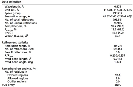
Number Page Table 1.1 Relevant reduction potentials of wild type P450s and redox cofactors ...12 Table 1.2 Parameters for a selection of oxidative quenchers ...23 Table 2.1 X-ray crystallographic data collection, refinement statistics, and validation ...46 Table 2.2 Observed rate constants (γ1-5, s-1) extracted from global fitting of single-wavelength TA at six wavelengths (390-440 nm) ...59 Table 2.3 Extracted kinetics parameters ...62 Table 3.1 Excited state lifetimes of four Ru-P450 conjugates in the absence and presence of 17 mM [Ru(NH3)6]3+ quencher...91 Table 4.1 Experimentally-determined reorganization energies for various proteins ...135 Table 4.2 Calculated single-step tunneling times for porphyrin oxidation, using a variety of ET parameters ...141 Table 4.3 Minimum driving forces necessary to obtain an ET time of 1.3 μs for each hopping map...142 Table 5.1 Luminescence lifetimes, taken from biexponential (unquenched) and monoexponential (quenched) fits ...160 Table 5.2 Rates of heme reduction extracted from global fitting of transient absorption data ...166 Table 5.3 Thermodynamic activation parameters for CO rebinding in CYP119 and various heme enzymes...186 Table 6.1 Reduction potentials of ferric P450 and redox active cofactors
Chapter 1
CYTOCHROME P450:
FROM PURSUIT OF REACTIVE INTERMEDIATES
TO
1.1. Cytochrome P450: a remarkable metabolic enzyme
Every living organism is composed of cells that are alive with chemical reactions,
the processes that allow them to grow, reproduce, and respond to the
environment. Beneficial nutrients are broken down to provide energy and
synthetic building blocks, new molecules are built from scratch or modified for
specific purposes, and unwanted or harmful chemicals are detoxified and excreted.
Each of these metabolic processes must be accomplished in a very precise,
controlled fashion; this often requires surmounting a significant reaction barrier.
Thus, cells employ enzymatic machinery to catalyze these reactions.
Metabolic enzymes face a number of significant challenges. Splitting up large
molecules into smaller or more useful components requires activating, breaking,
and functionalizing some of the strongest and most inert chemical bonds found in
organic molecules. Additionally, a series of reduction and oxidation reactions
(redox) is often required, in which electrons are specifically transferred out of one
chemical bond and into a new one. All of these transformations must occur in a
tightly controlled fashion in order to synthesize complex molecules with specific
information encoded in the molecular structure. Furthermore, they must be
accomplished on a wide variety of substrates; exposure to toxins and xenobiotics in
the environment provides a constant variety of sizes, geometries, and
functionalities.
A remarkable class of enzymes called cytochrome P450 (often abbreviated as just
“P450”), plays a critical role in these metabolic processes. These enzymes possess
three characteristics that make them indispensible. First, the P450 center houses a
thiolate-ligated heme active site capable of generating extremely reactive iron-oxo
common and inert bonds found in organic molecules. Secondly, the amino acids
that directly surround the active site orient substrates in proximity to the reactive
center, allowing the enzyme to form new bonds in a controlled fashion with high
levels of regio- and stereoselectivity. Finally, flexible protein architecture around
the active site allows P450s to envelop substrates with a wide variety of sizes,
geometries, and functionalities, and thus, metabolize myriad small molecules.
Because of these three features, P450s are found in every living organism, from
bacteria and archaea, to plants, fungi, and animals; there are even a number of
viruses that carry the P450 genetic code. Since their initial discovery in rat liver
microsomes in the 1940s, advances in genomics have led to the discovery of over
20,000 versions of P450,1 and this number keeps rising. The remarkable reactivity
displayed by P450s has made them a highly active field of research, involving many
disciplines from medicine and toxicology to molecular biology, biochemistry, and
biophysics. In addition, they are inspiration for the development of inorganic
catalysts and synthetic methodologies. A fundamental question continues to
fascinate scientists: how do these enzymes catalyze such difficult transformations?
Specifically, what is the nature of the catalytically active species? How does the
enzyme control the fundamental electron transfer (ET) events that are required to
form the active species, and for it to then react with substrates? Furthermore, can
we harness and modify P450s to drive catalysis in an entirely new way?
This Chapter begins by discussing the key features of P450s that contribute to their
remarkable reactivity, including the nature of reactive intermediates along the
catalytic pathway. Two soluble, bacterial enzymes are highlighted in particular:
P450 BM3 from the soil bacterium Bacillus megaterium, and CYP119 from the
thermophilic hot-springs archaeum Sulfolobus acidocaldarius. These two variants
Chapters 2-6. This first Chapter next addresses a rapid, photochemical method for
triggering ET within enzyme systems in order to generate and investigate reactive
species. Implementation of this method is discussed in Chapters 2-5. Finally, this
Chapter discusses a recent report of engineered P450-catalyzed cyclopropanations.
By manipulating electron flow, these artificial P450s can be reprogrammed to
catalyze cyclopropanation of styrene within the context of an E. coli cell.
Electrochemical measurements that quantify this electronic manipulation are
presented in Chapter 6.
1.2. P450: structural features that direct function
The first X-ray crystallographic characterization of a P450 was achieved in 1987
for the soluble, bacterial P450CAM from Pseudomonas putida.2 Bacterial variants
such as P450CAM often are stable, soluble, and easier to study. In contrast,
mammalian variants are delicate and frequently are membrane bound. Due in
large part to improvements to crystallization techniques, there are almost 600 P450
structures in the protein data base (PDB) to date, including 107 structures of
human variants. This increasing wealth of data allows identification of common
features and individual peculiarities. Entire volumes have been written on the
relationship between P450 structure and function,3 so this Chapter only briefly
highlights the features that are pertinent to Chapters 2-6 of this Thesis.
Cytochrome P450s are largely α-helical proteins, with a tertiary structure that is
highly conserved and unique to the superfamily; no non-P450 proteins share this
fold.4 The structures of P450 BM3 and CYP119 are shown in Figure 1.1. It is
perhaps unsurprising that the most highly conserved regions are those adjacent to
the active site, while those associated with substrate specificity may differ
At the heart of cytochrome P450 lies the heme, iron protoporphyrin IX, that drives
P450 chemistry. The iron center is ligated on the proximal face by an absolutely
conserved cysteine (Figure 1.2, green); this ligation is absolutely critical for P450
function. This cysteine is part of a signature β-bulge containing the conserved
sequence FxxGx(H/R)xCxG; this lies at the end of helix L, which runs beneath the
heme (Figure 1.2, blue). This region of the protein fold is critical for holding the
cysteine in place. Three hydrogen bonds from nearby backbone amide N-H
donors also stabilize the thiolate ligand. This ligation and hydrogen bonding
arrangement is shared with two other heme monooxygenases: chloroperoxidase
[image:31.612.116.536.332.548.2]and nitric oxide synthase.
Figure 1.2. P450 BM3 active site. The heme is colored pale pink. The L helix (blue), and contains cysteine400 (green) which ligates the heme. In bright pink: arginine398 and tryptophan96 form hydrogen bonds with one of the heme propionates. The long I helix (purple) above the heme contains threonine268 (orange). The axial water molecule, and an additional, structured water molecule above the heme are shown as a black dot.
The P450 heme is not covalently bound to the protein structure, but rather is held
between the heme propionates and amino acid side chains. The precise nature of
the hydrogen bonding partners differs among P450 variants. For P450 BM3, this
includes a tryptophan96 and arginine398; site directed mutagenesis studies that
replaced tryptophan96 with non-hydrogen bonding residues (e.g., alanine) saw
greatly diminished heme incorporation in the expressed mutants. CYP119
contains a more extensive hydrogen bond network between the protein and the
heme propionates (Figure 1.3). Histidine76, histidine315, and arginine80 form
hydrogen bonds with one of the heme propionates, and arginine259 forms a
[image:32.612.116.538.311.526.2]hydrogen bond with the other.
The distal face of the P450 heme is in contact with the substrate binding pocket. In
the absence of substrate, a water molecule acts as the distal axial ligand, making the
iron center six coordinate and low spin. The long I helix (Figure 1.2 and Figure
1.3, purple) runs along this distal face, and contains a conserved threonine side
chain that is involved in oxygen activation, as discussed in Section 1.3. The
substrate binding pocket of cytochrome P450 is largely hydrophobic, with key
residues positioned to align substrate. While the heme active site is deeply buried,
a long substrate-access channel provides a pathway for molecules to enter. Upon
substrate binding, large conformation changes can be seen for many P450 variants.
For example, in the presence of long chain fatty acids such as palmitic acid, the
P450 BM3 G, H, and B´ helices tighten down on the pocket (Figure 1.4).
1.3. P450 activity: reactions and mechanisms
Broadly, P450s catalyze the metabolism of xenobiotics and the biosynthesis of
signaling molecules. The most common and well-studied transformations are
monooxygenase reactions, such as hydroxylation of aliphatic and aromatic
carbons or heteroatoms, and epoxidations.5 Dehydrations, ring formations and
expansions, and reductive halogenations among many others have also been
observed. As will be discussed in Section 1.6, the heme active site can also be
Figure 1.5. Various reactions catalyzed by cytochrome P450s.
Oxygen Activation
P450 monooxygenase activity requires binding and activation of molecular oxygen
(O2) with the input of high-energy electrons. Once the O–O bond is broken, a
single oxygen atom is incorporated into an organic substrate molecule; the other is
released as water. A canonical P450 catalytic cycle is shown in Figure 1.6; black
rhombs represent the active site heme. Each step in this catalytic process relies on
critical features in the P450 architecture.
The stepwise, reductive activation of dioxygen involves transient generation of
reactive intermediates that include superoxide, peroxide, and high valent
iron-O H H O OH H HO O n HO O n OH n = 10, 12, 14
O
X R X
oxos. In order to prevent deleterious generation and release of these species, many
P450s have an important gating mechanism prevents the flow of electrons, and
thus, the activation of dioxygen, in the absence of substrate; both P450 BM3 and
CYP119 fall into this class. In these enzymes, the resting state contains a
six-coordinate, ferric (FeIII) heme, which is ligated by the axial thiolate on the
proximal face, and a loosely bound water molecule on the distal face (species 1 of
Figure 1.6). Strong interactions of the iron 3d orbitals with these ligands cause the
resting state to be low-spin.
Figure 1.6. Canonical P450 catalytic cycle.
Due in large part to the strongly donating character of the axial thiolate, the resting
state FeIII/II reduction potential is too negative to be reduced by native redox
cofactors such as reduced nicotinamide adenine dinucleotide phosphate
(NADPH), and reduced flavin adenine dinucleotide (FADH2) (Table 1.1).
FeIII S Cys O H H FeIII S Cys
OO!
FeIII S Cys FeII S Cys FeIII S Cys
OOH
FeIV S Cys O H FeIV S Cys O O2
1
2
3
4
5
6
7
e-, H+ e-Compound I Protonated Compound II Peroxide Shunt H+ H 2OR-H
R-H
R!
!+
H O2 R-OH
Substrate binding causes displacement of the axial water molecule,6 forming a
five-coordinate, ferric heme in which the iron center is displaced below the ring of the
porphyrin (species 2).7 The weaker ligand field in this geometry favors the high
spin iron state, and shifts the FeIII/II reduction potential 50-150 mV positive,8
facilitating one-electron reduction of the heme (species 3).
Table 1.1. Relevant reduction potentials of wild type P450s and redox cofactors8–10
a: ref 9. b: ref 10. c: ref 8. d: Chapter 6 of this Thesis
The requisite electrons from NAD(P)H, and are transferred to the heme by various
electron relays. For the subset of P450s known as “Class I” enzymes, a flavoprotein
reductase acquires the two electrons from NADPH and dispatches them
individually to an iron-sulfur protein, which in turn reduces the P450 oxygenase.
Mammalian P450s associated with steroid synthesis and the majority of bacterial
enzymes fall into this category. In Class II enzymes, a single flavoprotein reductase
transfers the electrons from NADPH to the P450 oxygenase via the associated
flavin mononucleotide cofactor (FMN).11 The mammalian P450s involved in drug
metabolism are an example of this class.11 P450 BM3 belongs to a unique,
“self-sufficient” class, in which the reductase and oxygenase domains are fused. This
rates: 930 min-1 for lauric acid, and 1470 min-1 for arachidonic acid.12 For
comparison, CYP119 oxidizes lauric acid at a rate of 11 min-1.13
Figure 1.7. Structure of P450 BM3 oxygenase and reductase domains (PDB: 1BVY). The oxygenase domain is colored green (heme: red), and the reductase domain is colored blue (flavin: purple).
The ferrous heme readily binds oxygen, forming a ferric superoxide (species 4).
Further reduction by one electron and protonated forms a hydroperoxy species
(species 5). Two main factors affect heterolysis of the O–O bond at the heme active
site. First, the “thiolate-push” from the strongly-donating thiolate ligand assists in
heterolytic bond cleavage.14 Second, a well-organized proton relay facilitates
threonine side chain on helix I, as well as organized water molecules within the
active site (Figure 1.2). The doubly-protonated distal oxygen departs as a water
molecule, generating a ferryl, ligand-radical-cation species known as Compound I
(CI, Figure 1.6, species 6).
P450 active hydroxylation agents: ferryl compounds I and II
CI is an extremely reactive oxidant, which allows P450 to functionalize strong and
unactivated C–H bonds. CI abstracts a hydrogen atom from substrate to form
Compound II (CII, Figure 1.6 7), which then undergoes radical recombination
with substrate to produce hydroxylated product. Release of product from the
active site and re-ligation by a water molecule returns the enzyme to its resting
state.
The native steps of substrate binding, ET, and O–O bond cleavage are slower than
the reaction of CI with substrate. However, CI can be generated from the resting
state by reaction with chemical oxygen-atom donors, including meta
-chloroperoxybenzoic acid (mCPBA). This chemical oxidation was accomplished
cleanly for highly purified samples of CYP119, and CI was spectroscopically
characterized by UV-visible absorption, Mössbauer, and electron paramagnetic
resonance (EPR) spectroscopies.15,16 The apparent second order rate constant for
reaction of CI with lauric acid (dodecanoic acid) is 1.1 x 10⁷ M-1s-1.15
It is remarkable that a wide range of specific chemical transformations can be
achieved by very similar reactive heme-oxygen intermediates. It is equally
remarkable that an enzyme can house a reactive species capable of activating
reactive species without reacting with the much weaker bonds that make up the
protein itself?
One factor is the gating mechanism described earlier; many P450s cannot be
reduced (and therefore cannot activate dioxygen to form CI) in the absence of
substrate. Another factor is the thiolate ligation; this causes CII to be more basic
than other heme enzymes which are ligated by histidine. Characterization of
chloroperoxidase and P450 CII by Mössbauer spectroscopy indicates a protonated,
FeIV-OH species.17,18 CII in CYP158 from Streptomyces coelicolor has a reported
pKa of almost 12,18 much higher than the pKa of ~3.5 for histidine-ligated proteins
such as horseradish peroxidase and myoglobin.19 Abstraction of the hydrogen
atom from substrate requires transfer of both the electron and the proton. By
facilitating proton transfer through basicity of CII, a less-oxidizing CI is required.
Much progress has been made to understand the native P450 catalytic cycle.
Chemical generation, trapping, and spectroscopic characterization of the CI and
CII active species have been huge steps in the understanding of cytochrome P450
reactivity. However, the fundamental nuclear reorganization that drives O–O
bond scission to form CI, and the events of hydrogen atom abstraction and radical
rebound to form hydroxylated substrate, have yet to be observed directly.
Formation of these intermediates requires rapid and precisely controlled ET.
1.4. Photo-triggered electron transfer in proteins
Photo-triggered processes provide a high degree of temporal precision for the
gating of reactions. Absorption of a photon occurs nearly instantaneously, and
light energy can be delivered in discrete packets using pulsed lasers. In these cases,
the precision of temporal gating is limited by the length of a laser pulse; this can be
or as little as a few femtoseconds. In order to translate the energy from photons
into a desired reaction, such as ET, an additional component is needed: a
photosensitizer.
When photosensitizers absorb a photon of the appropriate wavelength, a reactive
electronic excited state is generated. Ruthenium tris(2,2´-bipyridine)
([Ru(bpy)3]2+) is a classic example.20,21 The metal-to-ligand charge transfer
(MLCT) absorption band (λmax = 453 nm) is responsible for the orange-red color of
this complex (Figure 1.8). Excitation into the MLCT band generates a reactive
excited state that is both oxidizing at the metal center and reducing at the ligand
(Figure 1.9, left). The excited state can relax by emission of a photon (λmax = 626
nm), or can be “quenched” by ET to or from a redox partner.
Figure 1.8. [Ru(bpy)3]2+ absorption (blue, solid) and emission (red, dashed)
Figure 1.9. [Ru(bpy)3]2+ photochemistry. Left: MLCT excited state of
[Ru(bpy)3]2+. Right: Modified Latimer diagram for [Ru(bpy)3]2+; potentials are vs.
the normal hydrogen electrode (NHE), in water.20,21
In some cases, the photosensitizer’s excited state reduction potential is sufficient to
accomplish a desired ET reaction within the lifetime of the excited state (e.g., 600
ns for [Ru(bpy)3]2+ in deoxygenated water). However, in cases where additional
driving force is required, or where the ET reaction is slower than luminescent
relaxation of the photosensitizer, a method called “flash-quench” can be employed.
In our example of [Ru(bpy)3]2+, a “flash” of light, such as a laser pulse, generates
the photosensitizer excited state; this in turn is “quenched” by ET with exogenous
small molecules. By selection of an appropriate quencher, either the more
oxidizing [Ru]3+ or more reducing [Ru]1+ can be generated; this new species can be
used to achieve ET with a system of interest. Flash-quench generates a
photosensitizer state with additional driving force, and the separation of charges
extends the lifetime of the reactive species.
By partnering photosensitizers with redox-active proteins, flash quench can be
used to trigger biologically-relevant ET. In particular, the Gray lab has focused on
systems in which the electron acceptor (or donor) is a metal center, such as copper
in azurin, or heme iron in cytochromes, including cytochrome P450 (Figure 1.10).
visible region. Furthermore, the absorption bands are sensitive to oxidation state
of the metal center; ET reactions can be monitored using transient absorption
spectroscopies. By varying the nature of the photosensitizer and the protein
structure, the Gray lab and others have probed the mechanistic aspects of
biological ET. Curious readers are directed toward a number of excellent reviews
for further details.22–24 An extension of this analysis, multistep ET (“hopping”) is
discussed in Chapter 4.
Figure 1.10. Redox active model proteins. Left to right: P. aeruginosa azurin (PDB: 1JZG, Cu in blue), horse heart cytochrome c (PDB: 1HRC, heme Fe in red), cytochrome b562 (PDB: 256B, heme Fe in red).
The bulk of this Thesis (Chapters 2-5) focus on the extension of these
photo-triggered methods to generate redox intermediates and investigate ET mechanisms
in cytochrome P450. To set the stage for the design of photosensitizer-P450
systems, we outline strategies for the selection of appropriate photochemical
elements.
Photosensitizers
A wide variety of photosensitizers have be used for initiating ET within proteins.
The Gray lab and others have employed ruthenium, rhenium, and osmium
photosensitizers with a variety of ligand frameworks to study ET processes within
photosensitizer for each specific application requires assessment of several
photochemical and photophysical aspects.
Electronic communication between the protein and photosensitizer is critically
important. For systems in which the protein redox cofactor is solvent exposed,
simple photosensitizers such as [Ru(bpy)3]2+ can be implemented. However, when
the redox cofactor is buried inside the protein framework, more elaborate methods
must be employed. Photosensitizer “wires” that bind noncovalently to the protein
surface, or within the substrate channel, can be used (Figure 1.11).
Figure 1.11. An example of a perfluorinated ruthenium wire.
A potential disadvantage of these wires is that noncovalent binding results in a
distribution of free and bound photosensitizers. For enhanced control over
binding location, the photosensitizer can be tethered to the protein surface (Figure
1.12).
N
N N
N N N RuII
N N
F F
F F F
F
Figure 1.12. Photosensitizers for site-specific surface labeling at amino acids (blue). Left: [Re(dimethyl-bpy)(CO)3(imidazole)]1+ is ligated by hisitidine. Center:
[Ru(bpy)2(imidazole)]2+ is ligated by histidine. Right:
[Ru(bpy)2(iodoacetamidophenanthroline)]2+ is tethered to cysteine.
This can be achieved by direct coordination of the photosensitizer metal center by
histidine amino acids (Figure 1.12 left and center). Alternatively, nucleophilic
residues such as cysteine can be modified with an iodoacetamido linker attached to
the photosensitizer ligand, in order to generate a covalent thioether bond between
protein and photosensitizer (Figure 1.12, right). Selective photosensitizer
conjugation to a specific site on the protein surface is accomplished by removal of
native histidine/cysteine residues, and installation of a single labeling site, using
site directed mutagenesis.
Selection of the photosensitizer/protein conjugation method is also important. The
cysteine tethering reaction is more rapid, and can be accomplished at low
temperature (4 °C) within the span of a few hours.26 By comparison, histidine
labeling often takes days or weeks, and may require room- or elevated temperature
(37 °C).27,28 However, the more flexible iodoacetamido linker (cysteine-labeling)
results in a photosensitizer-metalloprotein distance that is not fixed, as it is in
direct metal ligation by histidine. For experiments in which knowledge of discrete
photosensitizer-protein distance is important, histidine labeling may be
advantageous. Additionally, it is important to consider the native amino acid
composition of the protein. For example, for large proteins that contain a high
number of exposed histidine residues, cysteine labeling is more practical.
The photosensitizer must also provide sufficient driving force to accomplish the
desired ET. Varying the metal center and the ligand framework provides a range of
driving forces that span over 1 V.29
Absorption (of both photosensitizer and protein) is another key factor. It is
desirable to excite the photosensitizer at a wavelength that is not also absorbed by
the protein or its cofactors. Protein absorption reduces the number of photons
available to excite the photosensitizer; it could cause undesired sample heating as
excited protein molecules relax by thermal processes; or (particularly in the case of
heme enzymes) it could cause protein photochemistry or luminescence that
interfere with observation of the desired ET processes.
Small molecule quenchers
Flash-quench methods require an exogenous small molecule ET quencher (Q). For
protein experiments, Q must be water soluble up to millimolar concentrations at
mild pH (6-8), and must have an appropriate reduction potential to rapidly
(ideally, diffusion limited) oxidize or reduce the photosensitizer.
In addition, the quencher must not have significant absorbance at wavelengths of
interest for the photosensitizer-protein system; this is analogous to what was
described for protein absorbance. Laser-triggered flash-quench ET process can be
quencher not absorb at the probe wavelengths (e.g., the Soret region for heme
proteins: 390 – 440 nm).
A final consideration for quencher selection is redox reversibility. In a reversible
cycle (such as depicted in Figure 1.13), the stable, reduced quencher serves as an
electron source to complete the ET cycle and return the system back to its resting
state. In an ideal reversible system, the flash-quench cycle can be repeated ad
infinitum, with no degradation or persistent accumulation of intermediates.
Reversibility is most desirable for rapid transient absorption laser studies, in which
the averaging of many shots is required to achieve a sufficient signal-to-noise ratio.
Alternatively, irreversible quenchers serve as a sacrificial oxidant; this supplies
more time for slow reaction events, and allows for accumulation (and detection) of
oxidized species. Irreversible quenchers are usually desirable for steady-state
flash-quench processes, such as light-driven catalysis. Parameters for three water-soluble
ET quenchers are given in Table 1.2.
Table 1.2. Parameters for a selection of oxidative quenchers.30–33 Potentials are
reported vs. NHE.
1.5. Photo-triggered ET in P450: two pathways toward reactive heme species
Photosensitizers and flash-quench can be used in place of native ET partners to
generate and investigate the active species CI and CII. Reductive flash-quench
supplies the electrons necessary to activate dioxygen (Figure 1.14, blue arrows),
much more rapidly than the ET with native redox partners. Alternatively,
assuming microscopic reversibility of reaction in the catalytic cycle, one could
envision a novel pathway in which direct oxidation of the ferric aquo heme (loss of
one electron and one proton) generates CII, followed by a second one-electron
one-proton oxidation to generate CI (Figure 1.14, red arrows). An attractive
aspect of this “reverse,” oxidative route is that the oxygen atom in CI, which would
be incorporated into substrate, is derived not from dioxygen or reactive oxygen
species, but from water. This method was previously implemented to generate CII
and CI in histidine-ligated heme enzymes and model systems (vide infra). Inspired
by these successes, we began the pursuit of P450 CI using the oxidative route; these
experiments are discussed in Chapters 2-4. Reductive ET and associated reactions
Figure 1.14. Pathways for formation of high valent CI and CII. Blue arrows: reductive activation of dioxygen (native catalytic cycle). Red arrows: oxidative activation of water. Purple arrows: hydroxylation of substrate (RH).
Precedence for photochemical heme oxidation
Photochemical heme oxidation was first achieved for the model system
microperoxidase-8 (MP8), the heme-containing 8-amino acid fragment of
horseheart cytochrome c (Figure 1.16, right). Laser flash-photolysis of a sample
composed of [Ru(bpy)3]2+, an excess of [Ru(NH3)6]3+ as an oxidative quencher, and
MP8 resulted in one-electron oxidation of the exposed heme. The position, shape,
and intensity of the heme Soret and Q-bands are sensitive to oxidation state and
heme environment. CII formation was observed by a shift to higher energy of the
Soret band at pH values above 7. In more acidic solutions, CII was not produced,
but rather an FeIII porphyrin radical cation species was characterized by loss of the
Soret absorbance (a “bleach”) (Figure 1.15). Analogous flash-quench oxidation
was also achieved for the histidine-ligated enzyme horseradish peroxidase (HRP)
Figure 1.15. Flash-quench oxidation of a heme protein active site. Blue arrow indicates excitation with blue light (e.g., 480 nm), the red arrow indicates emission of red light (e.g., 630 nm).
Figure 1.16. Histidine-ligated hemes. Left: Horseradish Peroxidase (HRP, PDB: 1HCH). Right: Microperoxidase-8 from horseheart cytochrome c.
N N N N Fe O O O O S O N N O H N Thr O N H Val O H N Glu O O NH S O H3N
P450 considerations: the buried heme
The analogous use of [Ru(bpy)3]2+ was unsuccessful for oxidation of the P450
heme, likely due to deep burial of the active site within the protein scaffold.
Ruthenium “wires” were synthesized as substrate mimics; rapid flash-quench
heme reduction was accomplished using an imidazole-terminated perfluorinated
ruthenium wire (Figure 1.11).34 However, this method both blocks the substrate
channel and displaces the axial water molecule that is necessary for oxidative
formation of CII. Covalent tethering of the photosensitizer to the P450 surface has
finally enabled the desired oxidative ET (Chapter 2).
1.6. New Frontiers: Novel P450 Active Species for Non-Native Catalysis
Cytochrome P450s offer a seemingly boundless platform in which to design and
examine redox catalysis. As described earlier, the catalytic scope of native P450
catalysis is already vast (Figure 1.5). With small adjustments from directed
evolution and rational design, P450 catalysis can be extended further to include the
biodegradation of halocarbons,35 and oxidation of gaseous hydrocarbons.36–38 By
modifying the surface with photosensitizers, P450 catalysis can even be driven with
light.39,40 As described in the previous section, the development of photochemical
methods may be used to accomplish P450 catalysis by activating water, instead of
oxygen or other reactive oxygen species.
However, the extent of catalysis is restricted by the nature of the active species;
atom-transfer reactions catalyzed by CI are limited to oxygenation reactions.
Development of alternative atom transfer methods would greatly expand utility of
One particularly attractive new reaction target is enzymatic cyclopropanation. The
controlled formation of new carbon-carbon bonds is one of the most fundamental
challenges in synthetic chemistry. Specifically, generation of the strained
cyclopropane motif often requires the formation of two new C–C bonds in a
regioselective fashion, as well as control of the stereochemistry at all three carbon
centers. Cyclopropanes are found in myriad organic compounds including natural
products, pyrethroid insecticides, fragrances, and therapeutics,41 and the
development of cheap, efficient, selective, and sustainable cyclopropanation
methods is an active area of research.42 The development of enzymatic
cyclopropanation routes can take advantage of selectivity imparted by the protein
scaffold under mild temperatures and pressures. However, this reactivity requires
formation of a novel active species.
Inspiration for P450-catalyzed carbene transfer came from small molecule late
transition metal porphyrins (primarily Rh, Fe, Ru, Os), which catalyze the
cyclopropanation of alkenes in the presence of diazo carbene precursors.43–47 The
reaction is thought to proceed via initial formation of a metallo-carbenoid,
followed by carbene transfer to the alkene.
Early in 2013, Coelho et al. reported carbene transfer from ethyldiazoacetate
(EDA) to styrene to form cyclopropanated products catalyzed by cytochrome P450
BM3.48 Both wild-type and engineered P450 mutants were capable of catalyzing
the reaction, but specific mutants achieved high turnover numbers. In particular,
the single T268A mutation was sufficient to raise the turnover number from 5
(wild-type) to 323, with high selectivity for trans 1:99 cis:trans ratio and 96%
enantioselectivity for the transS,S diastereomer. Additional mutations to the active
selectivity was reported as 92:8 cis:trans, with 97% S,S enantioselectivity for the cis
form.
Figure 1.17. P450-catalyzed cyclopropanation of styrene.
In analogy to small molecule metalloporphyrin studies,47 the authors proposed a
mechanistic scheme that employs a high-valent carbenoid active species (Figure
1.18, right).
Figure 1.18. Native and engineered P450 catalytic schemes. Left: Consensus P450 catalytic cycle for oxygenations, showing hydroxylation of a fatty acid. Right: Proposed catalytic scheme for the cyclopropanation of styrene.
Many details of the proposed mechanism have yet to be experimentally verified,
including the identity of the active cyclopropanating agent. However, there is O OEt O OEt O OEt O OEt
cis R,S cis S,R trans R,R trans S,S
OEt O H N N P450 N2 HO O HO O OH H O OEt H N OEt O N FeII S FeIII S FeIV S O FeIII S O O FeIII S O O O2 2H+ e -e -FeII S Fe S e -H O OEt OH2 FeIII S OH2 N2 H2O
n
significant evidence that ferrous (not ferric) P450 is required to activate
ethyldiazoacetate (EDA). Cyclopropanation activity required the presence of
strong reductant, such as dithionite, and the net reaction is inhibited by carbon
monoxide, which readily binds to ferrous hemes.49
While this in method offers an exquisite proof-of-concept for the development of
enzymatic cyclopropanations, the extension of this reactivity to in vivo systems
would avoid time consuming isolation and purification steps. P450 BM3 is an ideal
candidate for such development, as this “self-sufficient” enzyme contains both
heme oxygenase and flavin reductase domains fused in a single polypeptide chain.
No additional redox partner proteins are required. However, the biological
electron source for heme reduction (a critical step for activation of EDA) is
NADH. The aforementioned mutants show little to no cyclopropanation activity
in the presence of this cofactor.
This observed inactivity is due to a substrate gating mechanism described
previously (Section 1.3); styrene binds with low affinity (KM ~ 5 mM)48 and is
inefficient at effecting the low-to-high spin state change. Enzyme engineering to
increase the binding affinity might facilitate gating; however, this would need to be
achieved for every individual substrate. Incorporation of inert perfluorinated
substrate mimics has been implemented to artificially prop the gate open and
facilitate oxidation of tiny, ill-binding substrates such as ethane and methane.38
However, use of these decoy molecules in vitro is expensive, and implementation
for in vivo systems has not been established.
Alternatively, the enzyme could be reprogrammed to bypass this substrate gating
describe a series of electrochemical experiments that probe electron flow in a new
generation of P450 mutants engineered for cyclopropanation.
1.7. Conclusions
Rapid and efficient ET is critical for the formation of the reactive P450
intermediates responsible for catalysis, as well as for their selective
functionalization of organic substrates and biological molecules. The remaining
Chapters in this Thesis explore the details of photochemical generation of
high-valent heme intermediates in ruthenium-P450 conjugates, photochemical heme
reduction and gas binding as ways to monitor the native P450 cycle, and
electrochemical characterization of P450 axial mutants in relation to
1.8. References
(1) Nelson, D. R. Cytochrome P450 Stats
http://drnelson.uthsc.edu/P450.statsfile.html (accessed Nov 23, 2013).
(2) Poulos, T. L.; Finzel, B. C.; Howard, A.J. High Resolution Crystal Structure of Cytochrome P450cam. J. Mol. Biol. 1987, 195, 687–700.
(3) Ortiz de Montellano, P. Cytochrome P450: Structure, Mechanism and Biochemistry; 3rd ed.; Kluwer Academic/Plenum Publishers: New York, 2005.
(4) Poulos, T. L.; Johnson, E. F. Structures of Cytochrome P450 Enzymes. In
Cytochrome P450: Structure, Mechanism, and Biochemistry; Kluwer
Academic/Plenum Publishers: New York, 2005.
(5) Guengerich, F. P. Common and Uncommon Cytochrome P450 Reactions Related to Metabolism and Chemical Toxicity. Chem. Res. Toxicol. 2001, 14, 611–650.
(6) Denisov, I. G.; Makris, T. M.; Sligar, S. G.; Schlichting, I. Structure and Chemistry of Cytochrome P450. Chem. Rev. 2005, 105, 2253–2278.
(7) Shaik, S.; De Visser, S. P. Computational Approaches to Cytochrome P450 Function. In Cytochrome P450: Structure, Mechanism, and Biochemistry; Kluwer Academic/Plenum Publishers: New York, 2005; pp. 45–80.
(8) Ost, T. W. B.; Miles, C. S.; Munro, A. W.; Murdoch, J.; Reid, G. A.; Chapman, S. K. Phenylalanine 393 Exerts Thermodynamic Control over the Heme of Flavocytochrome P450 BM3. Biochemistry 2001, 40, 13421–13429. (9) Harris, D. C. Quantitative Chemical Analysis; 6th Edition.; W. H. Freeman
and Company: New York, 2003.
(10) Dunford, A. J.; Girvan, H. M.; Scrutton, N. S.; Munro, A. W. Probing the Molecular Determinants of Coenzyme Selectivity in the P450 BM3 FAD/NADPH Domain. Biochim. Biophys. Acta 2009, 1794, 1181–1189. (11) Meunier, B.; De Visser, S. P.; Shaik, S. Mechanism of Oxidation Reactions
Catalyzed by Cytochrome P450 Enzymes. Chem. Rev. 2004, 104, 3947–3980. (12) Munro, A. W.; Daff, S.; Coggins, J. R.; Lindsay, J. G.; Chapman, S. K. Probing
Electron Transfer in Flavocytochrome P-450 BM3 and Its Component Domains. Eur. J. Biochem. 1996, 239, 403–409.
(13) Lim, R.; Eun, C.-Y.; Park, H.-G.; Han, S.; Han, J.-S.; Cho, K. S.; Chun, Y.-J.; Kim, D. Regioselective Oxidation of Lauric Acid by CYP119, an Orphan Cytochrome P450 from Sulfolobus Acidocaldarius. J. Microbiol. Biotechnol. 2010, 20, 574–578.


![Figure 2.5. Absorption spectra of [Ru(bpy)2(IAphen)]+2 (yellow), P450-BM3 C62A/C156S/K97C (blue), RuK97C-P450BM3 (green), at approximately equal concentrations](https://thumb-us.123doks.com/thumbv2/123dok_us/8590172.863444/69.612.113.537.67.387/figure-absorption-spectra-iaphen-yellow-approximately-equal-concentrations.webp)