R E S E A R C H A R T I C L E
Open Access
The effect of different screw-rod design on
the anti-rotational torque: a biomechanical
comparison of three conventional
screw-rod constructs
Zifang Huang, Chongwen Wang, Hengwei Fan, Wenyuan Sui, Xueshi Li, Qifei Wang and Junlin Yang
*Abstract
Background:Screw-rod constructs have been widely used to correct spinal deformities, but the effects of different screw-rod systems on anti-rotational torque have not been determined. This study aimed to analyze the biomechanical effect of different rod-screw constructs on anti-rotational torque.
Methods:Three conventional spinal screw-rod systems (Legacy, RF-F-10 and USSII) were used to test the anti-rotational torque in the material test machine. ANOVA was performed to evaluate the anti-rotational capacity of different pedicle screws-rod constructs.
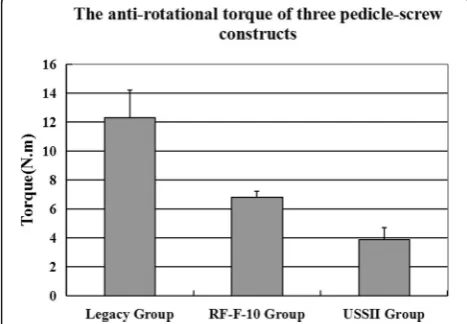
Results:The anti-rotational torque of Legacy group, RF-F-10 group and USSII group were 12.3 ± 1.9 Nm, 6.8 ± 0.4 Nm, and 3.9 ± 0.8 Nm, with aPvalue lower than 0.05. This results indicated that the Legacy screws-rod construct could provide a highest anti-rotation capacity, which is 68% and 210% greater than RF-F-10 screw-rod construct and USSII screw-rod respectively.
Conclusions:The anti-rotational torque may be mainly affected by screw cap and groove design. Our result showed the anti-rotational torque are: Legacy system > RF-F-10 system > USSII system, suggesting that appropriate rod-screw constructs selection in surgery may be vital for anti-rotational torque improvement and preventing derotation correction loss.
Keywords:Anti-rotational torque, Rod-screw construct, Biomechanics
Background
Adolescent idiopathic scoliosis (AIS) is a complex three-dimensional (3D) anomaly of the spine in the coronal, sagittal, and axial planes. Rotational deformity is an im-portant part of AIS and can affect mental health and cause cosmetic defects. Cotrel-Dubousset (CD) instrumentation and rod derotation are excellent techniques for the coronal and sagittal realignment of deformities, but it pro-vides poor rotational improvement for a weak posterome-dialization effect [1, 2]. Modern instrumentation systems with pedicle screws are able to provide both real vertebral rotational correction and rib hump correction with the use of direct vertebral rotation (DVR) [3, 4], direct
vertebral body derotation (DVBD) [5–8], vertebral copla-nar alignment (VCA) [9, 10], or vertebral column manipu-lator (VCM) techniques [11–13]. Two recent clinical studies [14, 15] reported correction loss in the axial plane despite the use of powerful spinal screw-rod instrumenta-tion, this phenomenon was also found in our clinical prac-tice (Fig. 1). Biological or mechanical factors could both play roles, but to our knowledge, research on this topic is limited.
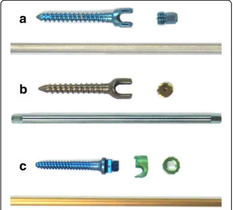
The purpose of this study was to evaluate anti-derotational torque, that is, how much tolerance a screw-rod construct has when twisting the rod in the pedicle screw groove. Here, we assessed the anti-rotational capacities of three different screw-rod con-structs (Legacy, RF-F-10, and USSII, as shown in Fig. 2), which were commonly used in China.
* Correspondence:[email protected]
Department of Orthopaedics, The 1st Affiliated Hospital of Sun Yat-sen University, NO.58, Zhongshan Er Road, Guangzhou, Guangdong, China
Methods
Three testing groups included different conventional spinal screw-rod fixation systems (Legacy, Medtronic Inc., Minneapolis, MN, USA; RF-F-10 screws, Kanghui Inc., China; and USSII, Synthes Inc., West Chester, PA, USA, respectively). Each group comprised seven mono-axial pedicle screws and one round rod (Table 1). One pedicle screw was used from in group in a preliminary study (for testing the machine and adjusting the custom jig before the experiment), and the remaining six pedicle screws were tested in the experiment. Each time, one screw was tightened on the rod with constant torque.
The tightening torque is created by twisting off the end cap (11–12.5 Nm) in Legacy group and tightening by 12 Nm in RF-F-10 group and USSII group. Then, part of screw thread was fixed inferiorly with methyl methacryl-ate to a custom jig in the mmethacryl-aterial test machine (MTS 858 System Inc., Minneapolis, MN, USA), with the free end of rod (3 cm) vertically aligned with the screw axis. Finally, the rod was twisted in the pedicle screw groove at a speed of 10°/min until the torque stopped increas-ing, and this value was recorded (Fig. 3).
Statistical analysis
Statistical comparisons were was carried out using SPSS 17 software (SPSS Inc., Chicago, IL, USA). The anti-rotational torque of three groups are presented as mean ± S.D. Analyses of variance (ANOVAs) were per-formed to evaluate the posthoc anti-rotational capacities of different pedicle screw-rod constructs.P-values <0.05 were considered significant.
Results
The mean anti-rotational torques of Legacy group, RF-F-10 group and USSII group were 12.3 ± 1.9 Nm, 6.8 ± 0.4 Nm, and 3.9 ± 0.8 Nm respectively, which were significantly different (P< 0.01). Posthoc testing comparing pairs of groups revealed that all were statistically different (all P < 0.01). The anti-rotational torque of the Legacy Fig. 1Corrective loss of the relative apical vertebral rotation (AVR) angle in a 16-year-old female AIS (Lenke V) with Risser 4. The preoperative relative
AVR anglewas 14.5°, which was calculated from the difference of rotational angles between the pelvis and apical vertebra. The relativeAVR anglewas corrected to 3.2° after surgery, and a 2.8° loss was measured at the 1.5-year follow-up visit
Fig. 2Illustrations of the screw cap and groove characteristics.a
The Legacy screw includes a“U”groove and a cap with a tip at the
[image:2.595.56.541.88.254.2]bottom;bThe RF-F-10 screw has a“U”groove and a cap with a hole in thecenter;cThe US II screw consists of an“L”groove and“U”cap
Table 1Instrument parameters
Group Screw-rod construct
Number of screws
Screw length and diameter (mm)
Rod diameter (mm)
A Legacy 7 6.5 × 50 5.5
B USSII 7 6.0 × 45 5.5
[image:2.595.56.292.472.685.2] [image:2.595.305.538.666.732.2]screw-rod construct was larger than that of RF-F-10, and the USSII screw-rod construct was the lowest (Fig. 4).
Discussion
Adolescent idiopathic scoliosis (AIS) is a complex 3D anomaly of the spine. Deformity in a single plane does not develop in isolation; rather, it is dependent on the co-development of curvature, translation, and rotation in other planes [16–18]. Curve progression, secondary thoracic cage deformity, and rib hump are always associ-ated with spinal rotational deformity [19–22]; therefore, vertebral derotation is an important consideration when correcting AIS.
A good 3D correction for scoliosis requires both spinal instrumentation systems and corrective techniques. In the early 1980s, Cotrel-Dubousset (CD) instrumentation with rod derotation was introduced to enable 3D scoli-osis correction surgery. However, recent reports suggest that rotational correction is variable (<25%) [23]. Com-pared to hooks, pedicle screw fixation provides better three-column fixation and 3D correction, which signifi-cantly improves the correction rates in the coronal and sagittal planes, particularly for rotational correction of the axial plane Lee et al. (2004), Gabriel et al. (2008),
and Huang et al. [3, 9, 11] reported 42.5%, 56%, and 55.2% apical derotations by DVR, VCA, and VCM com-bined with segmental pedicle instrumentation in AIS, re-spectively. All three of these techniques also reduce the rib hump, in some cases eliminating the need for a thor-acoplasty. This result had been substantiated in other studies [3, 10, 13]. However, Fu et al. [14] reported a re-cent study of vertebral rotation correction in AIS treated with four different techniques and anchors in which the patients were evaluated by the RAml method on com-puted tomography scan after 2 years. The authors found that rotation losses in the hook, wire, screw, and anterior groups were 20, 19.4, 17.1, and 12%, respectively. A similar result was described by Cui et al. [15], who re-ported a mean 2.1° correction loss of apical vertebral ro-tation angle 2 years after 27 AIS patients were treated with segmental pedicle screw rod constructs. However, to our knowledge, the difference in the anti-rotational capacities of different screw-rod constructs has not been previously described.
In this study, we biomechanically tested the anti-rotational torque of three different pedicle screws-rod constructs. The Legacy screw-rod construct provided the best anti-rotation capacity compared with the RF-F-10 and USSII screw-rod constructs by 68% and 2RF-F-10%, re-spectively. This suggests that the Legacy screw-rod con-struct can increase derotational power and better sustain potential derotational correction than the other two spinal instrumentation systems. As the same type of round rods are used in all the three, it is suspected that the anti-rotational torque differences may be caused by differences in pedicle screw design, especially in the screw head and cap part. Indeed, the cap of the Legacy screw is considered to be the main reason for its excel-lent anti-rotational torque.
The biomechanical testing results demonstrated that the Legacy screw-rod construct provides greater capacity for anti-rotational torque compared to the other two systems. This desirable property is likely affected by different screw cap and groove designs; thus, selecting an appropriate screw-rod system is very important for increasing the Fig. 3Anti-rotational torque testing. Each rod was twisted (10°/min) in the pedicle screw groove until we were no longer able to measure anti-rotational torque
[image:3.595.58.539.87.198.2] [image:3.595.56.290.542.704.2]anti-rotational torque of a screw-rod construct, and dero-tational correction loss after AIS surgery is thereby re-duced. However, the actual anti-rotational capacity of these three constructs need to be confirmed in clinical comparisons.
It is important to discuss the limitations of the current study. Only one pedicle screw with straight rod was tested in each simulation, which is both biologically and mechanically different from the situation in which mul-tiple pedicle screws connected with curved rod during surgery. The second limitation was that polyaxial screws test was not considered in this study, although we thought it will more useful for easying the rod place-ment in clinical practice but not derotation. Further-more, our findings do not clarify whether the resulting stress concentration by the cap tip on the rod in the Legacy screw-rod construct would cause fatigue failure in the rods (Fig. 5). Thus, further biomechanical evalua-tions of different systems’ fatigue failure and polyaxial screw’s anti-rotational effect may be needed.
Conclusions
In summary, we successfully assessed three conventional spinal screw-rod fixations for anti-rotational torque with the MTS 858 Testing System. The results showed that Leg-acy screws-rod construct can provide better anti-rotational capacity, possibly due to the screw cap and groove designs. The preliminary experimental results demonstrated that appropriate rod-screw construct selection in the clinic is very important for improving anti-rotational torque and preventing derotation correction loss.
Abbreviations
AIS:Adolescent idiopathic scoliosis; DVBD: Direct vertebral body derotation; DVR: Direct vertebral rotation; VCA: Vertebral coplanar alignment; VCM: Vertebral column manipulator
Acknowledgments
No funds were received in support of this work. No benefits in any form have been or will be received from a commercial party related directly or indirectly to the subject of this manuscript.
Availability of data and materials
We had full access to all of the data in the study and take responsibility for the integrity of the data and the accuracy of the data analysis. The data is available on request from the corresponding author.
Competing interests
The corresponding author declares the absence of any conflict of interest regarding the submitted manuscript. The funders had no role in study design, data collection and analysis, decision to publish or preparation of the manuscript.
Publisher’s Note
Springer Nature remains neutral with regard to jurisdictional claims in published maps and institutional affiliations.
Received: 1 November 2016 Accepted: 17 July 2017
References
1. Cotrel Y, Dubousset J, Guillaumat M. New universal instrumentation in spinal surgery. Clin Orthop Relat Res. 1988;227:10–23.
2. Krismer M, Bauer R, Sterzinger W. Scoliosis correction by Cotrel-Dubousset instrumentation. The effect of derotation and three-dimensional correction. Spine. 1992;17:S263–9.
3. Lee SM, Suk SI, Chung ER. Direct vertebral rotation: a new technique of three-dimensional deformity correction with segmental pedicle screw fixation in adolescent idiopathic scoliosis. Spine. 2004;29:343–9.
4. Mladenov KV, Vaeterlein C, Stuecker R. Selective posterior thoracic fusion by means of direct vertebral derotation in adolescent idiopathic scoliosis: effects on the sagittal alignment. Eur Spine J. 2011;20:1114–7. 5. Shah SA. Derotation of the spine. Neurosurg Clin N Am. 2007;18:339–45. 6. Hwang SW, Samdani AF, Gressot LV. Effect of direct vertebral body derotation
on the sagittal profile in adolescent idiopathic scoliosis. Eur Spine J. 2012;12:31–9.
7. Hwang SW, Dubaz OM, Ames R, et al. The impact of direct vertebal body derotation on the lumbar prominence in Lenke Type 5C curves. J Neurosurg Spine. 2012;17(4):308–13.
8. Hwang SW, Samdani AF, Lonner B, et al. Impact of direct vertebral body derotation on rib prominence: are preoperative factors predictive of changes in rib prominence? Spine (Phila Pa 1976). 2012;37(2):E86–9.
9. Vallespir GP, Flores JB, Trigueros IS. Vertebral coplanar alignment: a standardized technique for three dimensional correction in scoliosis surgery: technical description and preliminary results in Lenke type 1 curves. Spine. 2008;33: 1588–97.
10. Qiu Y, Zhu F, Wang B. Comparison of surgical outcomes of Lenke type 1 idiopathic scoliosis: vertebral coplanar alignment versus derotation technique. J Spinal Disord Tech. 2011;12:492–9.
11. Huang Z, Wang Q, Yang J, et al. Vertebral derotation by vertebral column manipulator improves postoperative radiographs outcomes of Lenke 5C patients for follow up minimum 2 year. J Spinal Disord Tech. 2014;4. [Epub ahead of print].
12. Yang J, Huang Z, Grevitt MP, et al. Double-curve synchronous derotation with convex correction: a new corrective technique for adolescent idiopathic scoliosis with double curves. J Spinal Disord Tech. 2014;27(1):E32–6. 13. Sun L, Song Y, Liu L, et al. Bilateral apical vertebal derotation technique by
vertebral column manipulation compare with vertebral coplanar alignment technique in the correction of Lenke I type idiopathic scoliosis. BMC Musculoskelet Disord. 2013;14:175.
14. Fu G, Kawakami N, Goto M. Comparison of vertebral rotation corrected by different techniques and anchors in surgical treatment of adolescent thoracic idiopathic scoliosis. J Spinal Disord Tech. 2009;22:182–9.
15. Cui G, Watanabe K, Nishiwaki Y, et al. Loss of apical vertebral derotation in adolescent idiopathic scoliosis: 2-year follow-up using multi-planar reconstruction computed tomography. Eur Spine J. 2012;21(6):1111–20.
[image:4.595.55.292.88.162.2]16. Cholewicki J, Crisco JJ 3rd, Oxland TR. Effects of posture and structure on three-dimensional coupled rotations in the lumbar spine. Spine. 1996;21: 2421–8.
17. Perdriolle R, Vidal J. Morphology of scoliosis: three-dimensional evolution. Orthopedics. 1987;10:909–15.
18. Stokes IA. Axial rotation component of thoracic scoliosis. J Orthop Res. 1989; 7:702–8.
19. Weinstein SL, Zavala DC, Ponseti IV. Idiopathic scoliosis: long-term follow-up and prognosis in untreated patients. J Bone Joint Surg Am. 1981;63:702–12. 20. Aronsson DD, Stokes IA, Ronchetti PJ. Surgical correction of vertebral axial
rotation in adolescent idiopathic scoliosis: prediction by lateral bending films. J Spinal Disord. 1996;9:214–9.
21. Behensky H, Cole AA, Freeman BJ. Fixed lumbar apical vertebral rotation predicts spinal decompensation in Lenke type 3C adolescent idiopathic scoliosis after selective posterior thoracic correction and fusion. Eur Spine J. 2007;16:1570–8.
22. Perdriolle R, Vidal J. Thoracic idiopathic scoliosis curve evaluation and prognosis. Spine. 1985;10:785–91.
23. Steib JP, Dumas R, Mitton D. Surgical correction of scoliosis by in situ contouring: a detorsion analysis. Spine. 2004;29:193–9.
• We accept pre-submission inquiries
• Our selector tool helps you to find the most relevant journal • We provide round the clock customer support
• Convenient online submission • Thorough peer review
• Inclusion in PubMed and all major indexing services • Maximum visibility for your research
Submit your manuscript at www.biomedcentral.com/submit