Staphylococcus aureus
Strain-Typing Systems by Use of Kaplan-Meier
Survival Analysis
Matthew V. N. O’Sullivan, Vitali Sintchenko, Gwendolyn L. Gilbert
Centre for Infectious Diseases and Microbiology, Sydney Medical School and Sydney Emerging Infections and Biosecurity Institute, University of Sydney, Westmead Hospital, New South Wales, Australia
Knowledge concerning stability is important in the development and assessment of microbial molecular typing systems and is
critical for the interpretation of their results. Typing system stability is usually measured as the fraction of isolates that change
type after several
in vivo
passages, but this does not necessarily reflect
in vivo
stability. The aim of this study was to utilize
sur-vival analysis to provide an informative quantitative measure of
in vivo
stability and to compare the stabilities of various
tech-niques employed in typing methicillin-resistant
Staphylococcus aureus
(MRSA). We identified 100 MRSA pairs (isolated from
the same patient
>
1 month apart) and typed them using multilocus sequence typing (MLST), phage-derived open reading frame
(PDORF) typing, toxin gene profiling (TGP), staphylococcal cassette chromosome
mec
(SCC
mec
) subtyping, pulsed-field gel
electrophoresis (PFGE), and
spa
sequence typing. Discordant isolate pairs, belonging to different MLST clonal complexes, were
excluded, leaving 81 pairs for analysis. The stabilities of these methods were examined using Kaplan-Meier survival analysis, and
discriminatory power was measured by Simpson’s index of diversity. The probability percentages that the type remained
un-changed at 6 months for
spa
sequence typing, TGP, multilocus variable number of tandem repeats analysis (MLVA), SCC
mec
subtyping, PDORF typing, and PFGE were 95, 95, 88, 82, 71, and 58, respectively, while the Simpson’s indices of diversity were
0.48, 0.47, 0.70, 0.72, 0.89, and 0.88, respectively. Survival analysis using sequential clinical isolates adds an important
quantita-tive dimension to the measurement of stability of a microbial typing system. Of the methods compared here, PDORF typing
pro-vides high discriminatory power, comparable with that of PFGE, and a level of stability suitable for MRSA surveillance and
out-break investigations.
T
he stability of a new typing method is one of the key
parame-ters defining its utility, along with discriminatory power,
re-producibility, ease of use and interpretation, cost, throughput,
and concordance with both epidemiologic data and existing
typ-ing systems (
1
). Discriminatory power is usually estimated by
Simpson’s index of diversity (
2
), concordance is usually measured
by Wallace or Rand coefficients (
3
), and reproducibility is usually
measured by Cohen’s kappa.
Stability relates to the likelihood that the measured
character-istic of an organism will change with time or in subsequent
gen-erations of the organism. If a typing system has low stability, it may
incorrectly identify related isolates as being unrelated. It is
impor-tant to define the stability of a typing system so that guidelines for
the interpretation of results can be established. If isolates typed by
a highly stable system differ at one locus, it may indicate that they
are unrelated strains, whereas if they are typed by an unstable
system, they may need to differ at multiple loci before
nonrelat-edness can be inferred.
Stability has been most commonly measured by observing the
fraction of isolates in which the genotype remains unchanged after
a fixed number of
in vitro
passages (
1
). However, this approach is
less applicable to epidemiologic studies and does not provide
much relevant information about the natural rate of change over
time. Here, we describe a method of measuring stability using
Kaplan-Meier survival analysis and its application to the
estima-tion of
in vivo
stability of typing methods used to study the
short-term epidemiology of methicillin-resistant
Staphylococcus aureus
(MRSA) in hospital and community settings.
MATERIALS AND METHODS
Isolate collection.A collection of MRSA isolates from clinical and screen-ing samples obtained between July 2005 and March 2009, routinely stored at the Centre for Infectious Diseases and Microbiology, Westmead Hos-pital, Sydney, Australia, was used in the study. This collection was sur-veyed to find multiple isolates from the same patient; patients were en-rolled if they had two isolates collectedⱖ1 month apart, irrespective of the anatomical site or type of specimen from which they were isolated. The multilocus sequence type (MLST) was predicted using kinetic PCR for informative single-nucleotide polymorphisms (SNPs) (4), and patients were excluded if their isolates belonged to different MLST clonal com-plexes (as it was assumed that this represented acquisition of a new strain rather than evolution of the initial strain).
DNA preparation.For PCR-based methods, one or two colonies from a pure subculture were suspended in 400l of molecular-grade water, which was boiled for 10 min and frozen. After thawing, the suspension was centrifuged at 10,000 rpm for 5 min, and the supernatant was used for a DNA template. The same lysate was used for all methods. A sweep of the pure subculture was used to make suspensions for pulsed-field gel elec-trophoresis (PFGE).
Received26 May 2012Returned for modification30 August 2012
Accepted15 October 2012
Published ahead of print24 October 2012
Address correspondence to Matthew V. N. O’Sullivan, matthew.osullivan @swahs.health.nsw.gov.au.
Copyright © 2013, American Society for Microbiology. All Rights Reserved. doi:10.1128/JCM.01406-12
on May 16, 2020 by guest
http://jcm.asm.org/
Molecular typing.PFGE of SmaI-digested genomic DNA was per-formed according to the Harmony protocol (5) and analyzed using BioNumerics (Applied Maths NV, Belgium). A comparison of PFGE pat-terns in BioNumerics was performed using the Dice coefficient with the position tolerance set at 1.5% (change toward end of the fingerprint, 0.75%), and a dendrogram was constructed using the unweighted-pair group method using arithmetic averages (UPGMA). The isolates were grouped using two similarity cutoffs: 100% (indistinguishable patterns) and 80% (PFGE-100 and PFGE-80, respectively), as 80% is commonly used to define PFGE patterns that are likely to be epidemiologically related (6). Multilocus variable number of tandem repeats analysis (MLVA) was performed as reported previously (7). Each MLVA locus was amplified using single-primer-pair PCRs, and the amplification products were de-tected using gel electrophoresis with isolate pairs in adjacent lanes. The band sizes and matching bands between isolates were determined using BioNumerics and confirmed by visual inspection. MLVA patterns were
considered to be different if the molecular weights of one or more loci varied by at least the size of one repeat for that locus.spasequence typing was performed as described previously (8), andspatypes were assigned using the SpaServer (Ridom Bioinformatics) (9). Three multiplex PCR/ reverse line blot (mPCR/RLB) binary typing systems were employed (10, 11): (i) toxin gene profiling to targetsea,seb,sec,sed,see,seg,seh,sei,eta, etb,etd,tst, andlukS-PV(Panton-Valentine leukocidin) genes (8), (ii) phage-derived open reading frame typing (PDORF) to examine 16 loci derived from integrated prophages (12), and (iii) staphylococcal cassette chromosomemec(SCCmec) subtyping to determine themecclass andccr type and to interrogate 14 loci in the three junkyard regions (13). Refer-ence strains that, in combination, were positive for each probe and a DNA-free control were used as the positive and negative controls, respec-tively.
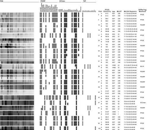
Data analysis.Stability was assessed by Kaplan-Meier survival analysis using SAS for Windows 9.3. For the purposes of survival analysis, an FIG 1Comparison of genotyping methods for 23 pairs, which differed by only one method (as indicated in the right-hand column). TGP, toxin gene profiling; PDORF, phage-derived open reading frame typing; MLVA, multilocus variable number of tandem repeats analysis. The MLVA repeats are for (from left to right) sspA,spa,sdrC,sdrD,sdrE,clfA, andclfB.
on May 16, 2020 by guest
http://jcm.asm.org/
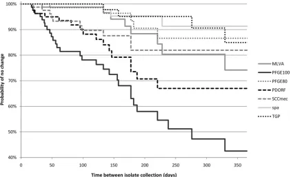
[image:2.585.48.543.67.504.2]“event” was considered to have occurred when the members of the isolate pair had different results depending on the typing method under analysis. The time at which the “event” occurred was arbitrarily considered to be the midpoint between the collection times of the two isolates. An isolate pair was considered “censored” (at the time of collection of the second isolate) if the results for the typing method for the two isolates were in-distinguishable, i.e., the time required for a change in molecular type was unknown for that patient, but it was longer than the period of observation. Survival analysis was used to estimate the probability that a typing method would remain unchanged for an isolate after 6 months. The log rank2
test was used to assess the differences in survival curves between methods, andPvalues were adjusted for multiple comparisons using the Šidák method (14). The initial isolate of each pair was used to calculate Simp-son’s index of diversity (2) of the typing method.
RESULTS
One hundred pairs of MRSA isolates were identified from the
culture collection, and the time between isolate collections for
each of the pairs varied between 1 month and 2.7 years. The
mem-bers of 19 isolate pairs belonged to different MLST clonal
com-plexes and therefore were excluded from further analysis.
Of the remaining 81 isolate pairs, 37 had concordant results
with all the methods (PFGE with 100% similarity [PFGE-100],
PFGE with 80% similarity [PFGE-80], phage-derived open
read-ing frame typread-ing [PDORF], SCC
mec
subtyping, toxin gene
profil-ing [TGP],
spa
sequence typing, and MLVA). Twenty-three isolate
pairs differed by only 1 method (excluding PFGE-80): 13 for
PFGE-100, 8 for PDORF, and 1 each for MLVA and SCC
mec
sub-typing (
Fig. 1
). Fourteen isolate pairs differed by 2 methods, and
the remaining 7 differed by 3 or more methods. Five isolate pairs
differed by PFGE-80: one of these pairs was concordant for all
non-PFGE methods, and the remaining four differed by 1, 2, 3,
and 5 non-PFGE methods.
Kaplan-Meier survival curves show a clear separation of
PFGE-100, PDORF, and SCC
mec
subtyping from the other more stable
methods after as little as 3 months (
Fig. 2
). PFGE-100 was
signif-icantly less stable than all other methods tested (probability of no
change at 6 months, 58%; 95% confidence interval [CI], 43 to
70%).
spa
typing was the most stable (probability of no change at
6 months, 95%; 95% CI, 82 to 99%), but this higher stability was
significant only when compared with PFGE-100 and PDORF (
P
⬍
0.0001 and
P
⫽
0.03, respectively). PFGE-80 was significantly
more stable but less discriminatory than PFGE-100. In general,
there was an expected inverse relationship between stability and
Simpson’s index of diversity (
Table 1
). However, despite having a
fractionally higher discriminatory power than PFGE-100
(Simp-son’s index of diversity, 0.89 versus 0.88), PDORF also had
signif-icantly higher stability, with a probability of no change at 6
months of 71% (95% CI, 55 to 82%) (
Table 1
).
DISCUSSION
Stability has an inverse relationship with discriminatory power
and is a function of the molecular “clock speed” of the genetic loci
being interrogated by a typing system. Housekeeping genes, for
example, which are utilized for multilocus sequence typing
(MLST), have a low molecular clock speed, resulting in high
sta-bility but low discriminatory power. Genetic loci on mobile
ge-netic elements demonstrate a high molecular clock speed; utilizing
these for typing may result in systems with low stability (possibly
leading to misleading inferences about the relationships between
isolates) but potentially high discriminatory power. The stability
of a typing method has traditionally been measured by sampling
strains at two time points and determining the fraction of strains
that have the same results at each time point. Most commonly,
sampling is performed before and after a given number of serial
FIG 2Kaplan-Meier survival curves for different genotyping methods. PFGE-100, PFGE-80: PFGE with similarity cutoffs of 100% and 80%, respectively. For other typing method abbreviations, see theFig. 1legend.on May 16, 2020 by guest
http://jcm.asm.org/
[image:3.585.82.504.66.323.2]passages of the organism in the laboratory (
in vitro
stability); less
commonly, it is performed by culturing an organism in its natural
environment at different time points, e.g., conducting repeat
cul-tures on a patient known to be colonized with the organism (
in
vivo
stability). Measuring stability
in vitro
may lead to
overestima-tion of the stability of a pathogen in its natural host environment,
since the opportunities for genetic changes through
transforma-tion, the acquisition of mobile genetic elements, or in response to
environmental conditions, such as antibiotic therapy or immune
pressure, will be absent.
Our findings suggest that the consideration of stability of an
MRSA typing system is especially important when one aims to
measure the relatedness of MRSA isolates obtained in time
inter-vals of more than 3 months. With studies examining shorter time
periods (e.g., during a hospital outbreak), highly discriminatory
but relatively unstable methods, such as PDORF or PFGE, will be
suitable for examining the microepidemiology of strains which
may otherwise be closely related. Studies examining longer time
periods (macroepidemiology) benefit from more stable methods,
such as
spa
sequence typing or PFGE, which have similarity cutoffs
of 80%. These findings are consistent with those of a recent study
that utilized next-generation sequencing to examine the
micro-evolution of one geographically widespread MRSA clone (ST239)
and which found an estimated molecular clock speed of one SNP
mutation in the core genome every 6 weeks (
15
).
Simultaneous carriage of multiple MRSA strains, or loss of one
strain and recolonization with another, are potentially
confound-ing factors in studies of
in vivo
stability. Major changes in
coloniz-ing strains have been infrequent in other
in vivo
stability studies
with MRSA (
16
–
18
), but the reported frequency of simultaneous
carriage of multiple strains has varied in the literature (
19
). One
report suggested that while simultaneous carriage of multiple
me-thicillin-sensitive
Staphylococcus
aureus (MSSA) strains may be
common, this is not the case for MRSA (
20
). Of 100 isolate pairs
initially identified in our study, 19 differed by their MLST clonal
complex and so were assumed to represent
recolonization/infec-tion with different strains.
The potential limitations of this study include the lack of serial
sampling of isolates, which may have impaired the accuracy of the
stability measures; more frequent sampling would require a
pro-spective clinical study. While attempts were made to exclude
re-infection/colonization by a different strain by excluding the
iso-late pairs belonging to different MLST clonal complexes, it is
possible that some of the changes between isolate pairs
repre-sented reinfection with a different strain within the same clonal
complex rather than the evolution of the initial isolate or perhaps
the carriage of multiple strains at different body sites, since a
com-bination of clinical and screening isolates was utilized for this
study. This limitation may have led to an underestimate of
stabil-ity, particularly that of methods with higher discriminatory
power. However, the majority of discordant pairs differed by only
one method, suggesting that reinfection with a different isolate
was probably infrequent in this cohort. Despite the limitations,
the estimates produced in this study should reliably reflect the
relative stability of different genotyping methods.
In conclusion, PDORF offered greater stability than PFGE-100
over a 12-month period but had a similar ability to discriminate
between apparently unrelated isolates from different individuals;
this indicates that PDORF may be a suitable substitute for PFGE in
the investigation of MRSA outbreaks and infection control
sur-veillance. It also has additional advantages over PFGE, such as
lower cost, faster turnaround time, higher portability of results,
and easier interpretation. All other methods were relatively more
stable but had correspondingly limited discriminatory power.
Survival analysis techniques provide a useful quantitative measure
of stability for the assessment of novel genotyping targets.
ACKNOWLEDGMENT
M.V.N.O. was supported by Australian National Health and Medical Re-search Council Postgraduate Scholarship no. 512029.
REFERENCES
1.Struelens MJ.1996. Consensus guidelines for appropriate use and evalu-ation of microbial epidemiologic typing systems. Clin. Microbiol. Infect. 2:2–11.
2.Hunter PR, Gaston MA.1988. Numerical index of the discriminatory ability of typing systems: an application of Simpson’s index of diversity. J. Clin. Microbiol.26:2465–2466.
3.Carrico JA, Silva-Costa C, Melo-Cristino J, Pinto FR, de Lencastre H, Almeida JS, Ramirez M.2006. Illustration of a common framework for relating multiple typing methods by application to macrolide-resistant Streptococcus pyogenes. J. Clin. Microbiol.44:2524 –2532.
4.Huygens F, Inman-Bamber J, Nimmo GR, Munckhof W, Schooneveldt J, Harrison B, McMahon JA, Giffard PM.2006.Staphylococcus aureus genotyping using novel real-time PCR formats. J. Clin. Microbiol.44: 3712–3719.
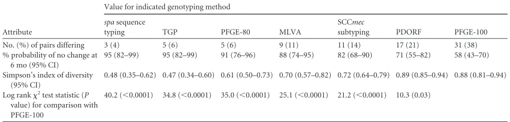
[image:4.585.38.547.78.200.2]5.Murchan S, Kaufmann ME, Deplano A, de Ryck R, Struelens M, Zinn CE, Fussing V, Salmenlinna S, Vuopio-Varkila J, El Solh N, Cuny C, Witte W, Tassios PT, Legakis N, van Leeuwen W, van Belkum A, Vindel A, Laconcha I, Garaizar J, Haeggman S, Olsson-Liljequist B, Ransjo U, Coombes G, Cookson B.2003. Harmonization of pulsed-field gel elec-trophoresis protocols for epidemiological typing of strains of methicillin-resistantStaphylococcus aureus: a single approach developed by consensus TABLE 1Stability and diversity measures of different genotyping methods
Attribute
Value for indicated genotyping method
spasequence
typing TGP PFGE-80 MLVA
SCCmec
subtyping PDORF PFGE-100
No. (%) of pairs differing 3 (4) 5 (6) 5 (6) 9 (11) 11 (14) 17 (21) 31 (38)
% probability of no change at 6 mo (95% CI)
95 (82–99) 95 (82–99) 91 (76–96) 88 (74–95) 82 (68–90) 71 (55–82) 58 (43–70)
Simpson’s index of diversity (95% CI)
0.48 (0.35–0.62) 0.47 (0.34–0.60) 0.61 (0.50–0.73) 0.70 (0.57–0.82) 0.72 (0.64–0.79) 0.89 (0.85–0.94) 0.88 (0.81–0.94)
Log rank2test statistic (P
value) for comparison with PFGE-100
40.2 (⬍0.0001) 34.8 (⬍0.0001) 35.0 (⬍0.0001) 25.1 (⬍0.0001) 21.2 (⬍0.0001) 10.3 (0.03)
on May 16, 2020 by guest
http://jcm.asm.org/
in 10 European laboratories and its application for tracing the spread of related strains. J. Clin. Microbiol.41:1574 –1585.
6.McDougal LK, Steward CD, Killgore GE, Chaitram JM, McAllister SK, Tenover FC.2003. Pulsed-field gel electrophoresis typing of oxacillin-resistantStaphylococcus aureusisolates from the United States: establish-ing a national database. J. Clin. Microbiol.41:5113–5120.
7.Sabat A, Krzyszton-Russjan J, Strzalka W, Filipek R, Kosowska K, Hryniewicz W, Travis J, Potempa J. 2003. New method for typing Staphylococcus aureusstrains: multiple-locus variable-number tandem re-peat analysis of polymorphism and genetic relationships of clinical iso-lates. J. Clin. Microbiol.41:1801–1804.
8.Cai Y, Kong F, Wang Q, Tong Z, Sintchenko V, Zeng X, Gilbert GL. 2007. Comparison of single and multilocus sequence typing and toxin gene profiling for characterization of methicillin-resistantStaphylococcus aureus(MRSA). J. Clin. Microbiol.45:3302–3308.
9.Harmsen D, Claus H, Witte W, Rothgänger J, Claus H, Turnwald D, Vogel U.2003. Typing of methicillin-resistantStaphylococcus aureusin a university hospital setting by using novel software forsparepeat determi-nation and database management. J. Clin. Microbiol.41:5442–5448. 10. Kong F, Gilbert GL.2006. Multiplex PCR-based reverse line blot
hybrid-ization assay (mPCR/RLB)—a practical epidemiological and diagnostic tool. Nat. Protoc.1:2668 –2680.
11. O’Sullivan MV, Zhou F, Sintchenko V, Kong F, Gilbert GL. 2011. Multiplex PCR and reverse line blot hybridization assay (mPCR/RLB). J. Vis. Exp. (54):2781. doi:10.3791/2781.
12. O’Sullivan MV, Kong F, Sintchenko V, Gilbert GL.2010. Rapid identi-fication of methicillin-resistantStaphylococcus aureustransmission in hos-pitals by use of phage-derived open reading frame typing enhanced by multiplex PCR and reverse line blot assay. J. Clin. Microbiol.48:2741– 2748.
13. Cai L, Kong F, Wang Q, Wang H, Xiao M, Sintchenko V, Gilbert GL. 2009. A new multiplex PCR-based reverse line-blot hybridization (mPCR/ RLB) assay for rapid staphylococcal cassette chromosome mec (SCCmec) typing. J. Med. Microbiol.58:1045–1057.
14. Šidák Z.1967. Rectangular confidence regions for the means of multivar-iate normal distributions. J. Am. Stat. Assoc.62:626 – 633.
15. Harris SR, Feil EJ, Holden MTG, Quail MA, Nickerson EK, Chantratita N, Gardete S, Tavares A, Day N, Lindsay JA, Edgeworth JD, de Len-castre H, Parkhill J, Peacock SJ, Bentley SD.2010. Evolution of MRSA during hospital transmission and intercontinental spread. Science327: 469 – 474.
16. Hartstein AI, Phelps CL, Kwok RY, Mulligan ME.1995. In vivo stability and discriminatory power of methicillin-resistantStaphylococcus aureus typing by restriction endonuclease analysis of plasmid DNA compared with those of other molecular methods. J. Clin. Microbiol.33:2022–2026. 17. Kuhn G, Francioli P, Blanc DS.2007. Double-locus sequence typing usingclfBandspa, a fast and simple method for epidemiological typing of methicillin-resistantStaphylococcus aureus. J. Clin. Microbiol.45:54 – 62. 18. Sakwinska O, Blanc DS, Lazor-Blanchet C, Moreillon M, Giddey M, Moreillon P.2010. Ecological temporal stability ofStaphylococcus aureus nasal carriage. J. Clin. Microbiol.48:2724 –2728.
19. Bloemendaal AL, Fluit AC, Jansen WT, Vriens MR, Ferry T, Amorim JM, Pascual A, Stefani S, Papaparaskevas J, Borel Rinkes IH, Verhoef J. 2009. Colonization with multipleStaphylococcus aureusstrains among pa-tients in European intensive care units. Infect. Control Hosp. Epidemiol. 30:918 –920.
20. O’Brien FG, Coombs GW, Pearman JW, Gracey M, Moss F, Christian-sen KJ, Grubb WB.2009. Population dynamics of methicillin-susceptible and -resistantStaphylococcus aureusin remote communities. J. Antimi-crob. Chemother.64:684 – 693.