JOURNAL OFCLINICALMICROBIOLOGY, Mar. 2008, p. 1026–1035 Vol. 46, No. 3 0095-1137/08/$08.00⫹0 doi:10.1128/JCM.02027-07
Copyright © 2008, American Society for Microbiology. All Rights Reserved.
Selection and Validation of a Multilocus Variable-Number
Tandem-Repeat Analysis Panel for Typing
Shigella
spp.
䌤
†
Olivier Gorge
´,
1* Ste
´phanie Lopez,
1Vale
´rie Hilaire,
1Olivier Lisanti,
1Vincent Ramisse,
1and Gilles Vergnaud
1,2Department of Analytical Microbiology, Centre d’Etudes du Bouchet, BP3, F-91710 Vert-le-Petit, France,1and
Universite´ Paris-Sud, Institut de Ge´ne´tique et Microbiologie, and CNRS, Orsay, F-91405, France2
Received 17 October 2007/Returned for modification 17 December 2007/Accepted 14 January 2008
The Shigella genus has historically been separated into four species, based on biochemical assays. The
classification within each species relies on serotyping. Recently, genome sequencing and DNA assays, in particular the multilocus sequence typing (MLST) approach, greatly improved the current knowledge of the
origin and phylogenetic evolution ofShigellaspp. TheShigellaandEscherichiagenera are now considered to
belong to a unique genomospecies. Multilocus variable-number tandem-repeat (VNTR) analysis (MLVA) provides valuable polymorphic markers for genotyping and performing phylogenetic analyses of highly
homo-geneous bacterial pathogens. Here, we assess the capability of MLVA forShigellatyping. Thirty-two potentially
polymorphic VNTRs were selected by analyzing in silico fiveShigellagenomic sequences and subsequently
evaluated. Eventually, a panel of 15 VNTRs was selected (i.e., MLVA15 analysis). MLVA15 analysis of 78
strains or genome sequences ofShigellaspp. and 11 strains or genome sequences ofEscherichia coli
distin-guished 83 genotypes.Shigellapopulation cluster analysis gave consistent results compared to MLST. MLVA15
analysis showed capabilities forE. colityping, providing classification among pathogenic and nonpathogenic
E. colistrains included in the study. The resulting data can be queried on our genotyping webpage (http://mlva
.u-psud.fr). The MLVA15 assay is rapid, highly discriminatory, and reproducible forShigellaandEscherichia
strains, suggesting that it could significantly contribute to epidemiological trace-back analysis of Shigella
infections and pathogenic Escherichia outbreaks. Typing was performed on strains obtained mostly from
collections. Further studies should include strains of much more diverse origins, including all pathogenicE.
colitypes.
Shigellosis is a widespread disease occurring mainly in de-veloping countries, often in association with poor sanitation, and is responsible for about 600,000 deaths per year in the world, two-thirds of them concerning ⬍10-year-old children (2). The implementation of treatment and, to some extent, control strategies is a significant challenge, especially in Asia, because antibiotic-resistant strains of different species and se-rotypes have emerged and because the distribution ofShigella
species and serotypes is heterogeneous (46). The low infectious dose (10 to 100 cells) allows the organism to spread effectively by contaminated food or water but also by person-to-person contact, and a specific virulence plasmid that encodes a type III secretion system plus the invasion plasmid antigens is respon-sible for epithelial cell wall invasiveness. Finally, it is worth noting that Shigella, like other bacteria responsible for food and waterborne diseases, has been classified as a potential biological threat agent due to its infection route and environ-mental stability (38).
Shigellasp. strains are currently classified into four species:
S. dysenteriae,S. flexneri,S. boydii, andS. sonnei. Three of them
are apparently antigenically heterogeneous, comprising several serotypes, whereasS. sonneiis antigenically homogeneous.
Shigella typing relies on phenotypic characteristics, but discrimination between the four species can be difficult. Serotyping is not always able to provide a correct species identification, due to cross-reactions or the absence of ag-glutination. New serotypes are regularly discovered (28, 49) and are sometimes found to cross-react withEscherichia coli
strains.
In addition, the discriminatory power of phenotypic tools and serotyping is limited and requires the manipulation of the live agent. The introduction of DNA-based molecular typing methods, such as ribotyping (5), plasmid profile analysis, re-striction fragment length polymorphism (25, 26), and pulsed-field gel electrophoresis (PFGE) (37), has greatly improved the ability of researchers to discriminate between epidemiologi-cally related and unrelated isolates in outbreaks. In the United States, PulseNet, a network of laboratories implicated in food-borne disease surveillance (42), uses PFGE typing coupled with strict quality control procedures in order to ensure inter-laboratory reproducibility, but this approach remains labor-intensive for routine clinical strain typing, so cheaper alterna-tives are actively being pursued. Multilocus sequence typing (MLST) is a very powerful approach, and it provides a clear view of the population structure (52). However, it is not yet appropriate for the routine, first-line genotyping of a large number of isolates. Recently, PulseNet members acknowledged that multilocus variable-number tandem-repeat (VNTR) analysis * Corresponding author. Mailing address: Department of Analytical
Microbiology, Centre d’Etudes du Bouchet, BP3, F-91710 Vert-le-Petit, France. Phone: 33-1-69-90-82-23. Fax: 33-1-69-90-83-35. E-mail: [email protected].
† Supplemental material for this article may be found at http://jcm .asm.org/.
䌤Published ahead of print on 23 January 2008.
1026
on May 16, 2020 by guest
http://jcm.asm.org/
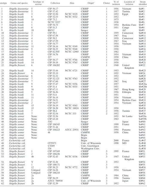
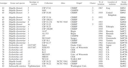
TABLE 1. List of strains used in this study
Genotype Genus and species Serotype or
serovar Collection Alias Origin
a Cluster Yr of
isolation
Location of isolation
Strain identifier
1 Shigella dysenteriae 5 CIP 56.17 CRBIP 1 1956 Vietnam Sd#1 2 Shigella dysenteriae 5 CIP 57.42 CRBIP 1 1957 Tunisia Sd#2 3 Shigella boydii 3 CIP 52.50 NCTC 9329 CRBIP 1 1952 Egypt Sb#3 4 Shigella boydii 6 CIP 52.53 NCTC 9332 CRBIP 1 1952 Sb#4
5 Shigella boydii 10 CIP 73.2 CRBIP 1 1973 Sb#5
6 Shigella boydii 1 NCTC 9327 CRBIP 1 1938 Sb#6
7 Shigella boydii 1 CIP 54.73 CRBIP 1 1954 Burkina Faso Sb#7 8 Shigella boydii 8 CIP 51.3 CRBIP 1 1951 Congo Sb#8
9 Shigella boydii 18 BS512 Sanger 1 AZ Sb#9
10 Shigella dysenteriae 3 CIP 59.1 CRBIP 1 1959 Cameroon Sd#10 11 Shigella dysenteriae 3 CIP 67.58 CRBIP 1 1967 Iraq Sd#11 12 Shigella dysenteriae 4 CIP 59.2 CRBIP 1 1959 Cameroon Sd#12 13 Shigella dysenteriae 4 CIP 67.59 CRBIP 1 1967 Iraq Sd#13 14 Shigella dysenteriae 3 CIP 54.77 CRBIP 1 1954 Vietnam Sd#14 15 Shigella dysenteriae 11 CIP 58.16 NCTC 9349 CRBIP 1 1958 Sd#15 16 Shigella dysenteriae 9 CIP 58.25 NCTC 9348 CRBIP 1 1958 Sd#16 17 Shigella boydii 2 CIP 82.50T NCTC 12985 CRBIP 1 1982 Sb#17 18 Shigella boydii 4 CIP 54.76 CRBIP 1 1954 Vietnam Sb#18 19 Shigella boydii 4 227 ChMPH 1 1950 China Sb#19 20 Shigella boydii 14 CIP 58.17 NCTC 9766 CRBIP 1 1958 Sb#20 21 Shigella boydii 14 CIP 58.18 NCTC 9765 CRBIP 1 1958 Sb#21 22 Shigella flexneri 6 CIP 106202 CRBIP 1 1940 United
Kingdom
Sf#22
23 Shigella boydii 14 CIP 53.44 NCTC 6721 CRBIP 1 1953 Sb#23 24 Shigella flexneri 6 CIP 55.19 CRBIP 1 1955 Vietnam Sf#24 25 Shigella dysenteriae 6 CIP 52.32 CRBIP 1 1952 Sd#25 26 Shigella dysenteriae 7 CIP 52.123 NCTC 9763 CRBIP 1 1952 Sd#26 27 Shigella dysenteriae 7 CIP 67.60 CRBIP 1 1967 Iraq Sd#27 28 Shigella boydii 9 CIP 57.47 NCTC 9356 CRBIP 2 1957 Sb#28a 28 Shigella boydii 9 CIP 51.4 NCTC 9355 CRBIP 2 1951 Sb#28b 29 Shigella boydii 16 CIP 67.11 CRBIP 2 1967 Hong Kong Sb#29 30 Shigella boydii 5 CIP 56.36 CRBIP 2 1956 Ethiopia Sb#30 31 Shigella boydii 11 CIP 51.6 CRBIP 2 1951 Sb#31 32 Shigella boydii 11 CIP 56.18 CRBIP 2 1956 Vietnam Sb#32 33 Shigella dysenteriae 2 CIP 54.51 CRBIP 2 1954 Burkina Faso Sb#33 34 Shigella dysenteriae 2 CIP 54.57 CRBIP 2 1954 Vietnam Sd#34 35 Shigella boydii 15 CIP 58.19 NCTC 9365 CRBIP 2 Sb#35 36 Shigella boydii 15 CIP 58.20 NCTC 10024 CRBIP 2 1958 Sb#36 37 Shigella boydii 7 CIP 58.4 CRBIP 2 1958 Tunisia Sb#37 38 Shigella boydii 7 CIP 52.54 NCTC 333 CRBIP 2 1952 Sb#38 39 Shigella sonnei None CIP 55.56 CRBIP 1955 Sri Lanka Ss#39a 39 Shigella sonnei None CIP 63.10 CRBIP 1963 Ss#39b
40 Shigella sonnei None 53G Sanger Japan Ss#40
41 Shigella sonnei None CIP 106345 CRBIP 1999 France Ss#41 42 Shigella sonnei None CIP 51.44 CRBIP 1951 Ss#42 43 Shigella sonnei None CIP 104223 ATCC 25931 CRBIP 1994 Panama Ss#43 44 Shigella sonnei None 46 ChMPH 1950 China Ss#44
45 Shigella sonnei None Be´gin Ss#45
46 Shigella sonnei None Be´gin Ss#46
47 Escherichia coli Be´gin 2000 France Ec#47
48 Escherichia coli CFT073 Univ. of Wisconsin 1990 MD Ec#48
49 Escherichia coli UTI89 Univ. Goettingen Ec#49
50 Escherichia coli 536 Washington Univ. Ec#50
51 Shigella flexneri 1 CIP 107169 CRBIP 3 1995 France Sf#51
52 Shigella flexneri 2 CIP 106236 CRBIP 3 Sf#52
53 Shigella flexneri 4b CIP 52.43 NCTC 8336 CRBIP 3 1947 United Kingdom
Sf#53
54 Shigella flexneri Y CIP 52.36 CRBIP 3 1952 Sf#54
55 Shigella flexneri 1 CIP 106171 CRBIP 3 Sf#55
56 Shigella boydii 12 CIP 58.23 NCTC 9772 CRBIP 3 1958 Sb#56 57 Shigella flexneri Untyped CIP 54.58 CRBIP 1954 Vietnam Sf#57 58 Shigella flexneri Untyped CIP 106210 CRBIP Sf#58 59 Shigella flexneri 2a 301 ChMPH 3 1984 China Sf#59 60 Shigella flexneri 2a CIP 56.19 CRBIP 3 1956 Tunisia Sf#60 61 Shigella flexneri 2a ATCC 700930 Univ. of Wisconsin 3 Japan Sf#61 62 Shigella flexneri 2b CIP 52.39 CRBIP 3 1952 Sf#62
Continued on following page
on May 16, 2020 by guest
http://jcm.asm.org/
(MLVA) is a highly promising typing tool, likely to replace PFGE in the coming years (9). MLVA typing is being actively developed by a number of laboratories, together with the as-sociated Internet-based query tools and databases (3, 6, 17, 18), to genotype several bacterial pathogens, in particular, po-tential biothreat agents, such asBacillus anthracis(18),Yersinia pestis (18, 31), Legionella pneumophila (32, 33), Salmonella enterica (36), Brucella spp. (19), Francisella tularensis (11),
Burkholderia mallei, andB. pseudomallei(43). MLVA is also increasingly recognized as a future reference technique for bacterial genotyping allowing systematic typing of all isolates for a number of other pathogens of high public health interest (including Mycobacterium tuberculosis [17], Pseudomonas aeruginosa[30, 47],Streptococcus pneumoniae[13], and Staph-ylococcus aureus[40]) (the technique is reviewed in references 21, 44, and 45).
The present work aimed to set up MLVA for theShigella
genus to facilitate epidemiological follow-up, by providing easy-to-use typing tools, in countries suffering from a recru-descence of Shigella outbreaks. Recently, Liang et al. (20) proposed an MLVA for molecular typing ofS. sonnei and explored the capability of a 26-VNTR set for detecting clus-ters of infection. Because the proposed VNTRs have very short repeat units, this assay requires a high precision of DNA length measurement, as provided, for instance, by microcapillary electrophoresis and fluorescent markers. This is a strong limitation for routine typing because of the cost of such equipment and the associated consumables. In contrast, the present investigation favors markers with larger repeat units in order to provide an assay widely
ac-cessible to research and public health laboratories, particu-larly in developing countries.
MATERIALS AND METHODS
Strains and DNA preparation.Sixty-eightS. boydii,S. dysenteriae,S. flexneri, andS. sonneistrains and fourE. colistrains were obtained from the Centre de Ressources Biologiques de l’Institut Pasteur (CRBIP), the American Type Cul-ture Collection, the Hoˆpital d’Instruction des Arme´es (Be´gin, France), and the Centre d’Etudes du Bouchet (CEB) strain collections (Table 1). Strains were cultured overnight at 30°C on Trypticase soy agar (reference number 43011; bioMe´rieux, Marcy L’Etoile, France) before DNA extraction.S. flexneri,S. son-nei, andS. boydiiDNA extractions were processed in a biosafety level 2 lab by use of lysozyme, sodium dodecyl sulfate, and proteinase K followed by phenol-chloroform extraction and ethanol precipitation, as described elsewhere (41).S. dysenteriaeserotype 1 DNA extraction was done in a biosafety level 3 lab by use of the genomic DNA 500/G extraction kit (Qiagen GmbH, Hilden, Germany), according to the manufacturer’s instructions. In several cases, the presence of high quantities of polysaccharides was suspected after lysis and cetyltrimethyl-ammonium bromide extraction was used to remove this excess (41). The average size of the extracted DNA was checked on a 0.8% agarose gel. Nucleic acid concentration and purity were quantified with an ND-1000 UV-Vis spectropho-tometer (NanoDrop Technologies, Wilmington, DE).
MLVA setup.A multiple comparison ofShigellagenome sequences available at the onset of the study was done through the Microorganism Tandem Repeats database (3, 6), accessible as a Web service (http://minisatellites.u-psud.fr/). The methods previously described (3, 18) and the genome sequence data for strains
S. flexneri2457T (GenBank accession no. AE005174) (48),S. flexneri 301 (GenBank accession no. NC_004337) (10),S. sonnei46 (GenBank accession no. NC_007384),S. dysenteriae197 (GenBank accession no. CP000034), andS. boydii227 (GenBank accession no. NC_007613) (50) were used to identify presumably polymorphic tandem repeats (TRs).
[image:3.585.46.543.80.344.2]Loci with repeat units longer than 9 bp were favored in order to facilitate allele repeat number calling by a variety of DNA amplicon length estimation devices (24) and as a complement to a previous investigation (20). Selected primers pairs were validated in silico with available genome sequence data forShigellaandE.
TABLE 1—Continued
Genotype Genus and species Serotype or
serovar Collection Alias Origin
a Cluster Yr of
isolation
Location of isolation
Strain identifier
63 Shigella flexneri 5 CIP 67.61 CRBIP 3 1967 Iraq Sf#63
64 Shigella flexneri 5 8401 ChMPH 3 Sf#64
65 Shigella flexneri 3 CIP 54.40 CRBIP 3 1919 United Kingdom
Sf#65
66 Shigella flexneri X CIP 52.34 CRBIP 3 1952 Sf#66
67 Shigella flexneri 3a CIP 106211 CRBIP 3 Sf#67
68 Shigella dysenteriae 8 CIP 53.134 NCTC 9345 CRBIP 1953 Sd#68 69 Shigella flexneri 4c CIP 52.25 CRBIP 3 1952 Sf#69 70 Shigella dysenteriae 1 CIP 56.33 CRBIP 1956 Ethiopia Sd#70 71 Shigella dysenteriae 1 CIP 106200 CRBIP 1919 Sd#71 72 Shigella dysenteriae 1 A147 Be´gin 1994 Rwanda Sd#72 73 Shigella dysenteriae 1 A1-313 Be´gin 1994 Rwanda Sd#73 74 Shigella dysenteriae 1 197 ChMPH 1950 China Sd#74a 74 Shigella dysenteriae 1 M131649 Sanger 1970 Guatemala Sd#74b 75 Shigella dysenteriae 1 CIP 58.1 CRBIP 1958 Tunisia Sd#75a 75 Shigella dysenteriae 1 CIP 62.17 CRBIP 1962 Cameroon Sd#75b 76 Escherichia coli O157:H7 Sakaı¨ Osaka Univ. 1996 Japan Ec#76 77 Escherichia coli O157:H7 EDL933 Univ. of Wisconsin 1982 MI Ec#77
78 Escherichia coli K12 CEB 1922 CA Ec#78a
78 Escherichia coli K12 CEB 1922 CA Ec#78b
78 Escherichia coli MG1655 Univ. of Wisconsin 1922 CA Ec#78c
79 Escherichia coli HB101 CEB 1922 CA Ec#79
80 Escherichia coli W3110 NARA IST 1922 CA Ec#80
81 Shigella boydii 13 CIP 58.21 NCTC 9361 CRBIP 1958 Sb#81 82 Shigella boydii 13 CIP 58.22 NCTC 9362 CRBIP 1958 Sb#82 83 Salmonella enterica Typhimurium LT2 Washington Univ. 1940s Se#83
aBe´gin, Hoˆpital d’Instruction des Arme´es, Be´gin, France; ChMPH, Microbial Genome Center of the Chinese Ministry of Health; CRBIP, Centre de Ressources Biologiques de l’Institut Pasteur; NARA IST, Nara Institute of Science and Technology; Univ., University.
1028 GORGE´ ET AL. J. CLIN. MICROBIOL.
on May 16, 2020 by guest
http://jcm.asm.org/
coli strains by using the multiple-PCR-primer BLAST Web service (http: //minisatellites.u-psud.fr/) which provides the expected size and sequence of the PCR products. The resulting in silico typing data were integrated into the MLVA.
Some VNTR loci ofE. coliO157:H7 described by Keys et al. (12) are present in theShigellagenus genome sequences. Five of them (11, 13, O157-25, O157-33, and O157-34), selected for their relative ease of use (repeat unit size,⬎6 bp) or high rate of polymorphism, were evaluated in this study.
VNTR amplification and genotyping. PCRs were performed as previously described (18) and using an annealing temperature of 60°C. DNA from theS. flexneri2457T strain was used as an internal control to ensure high-quality size assignments as described previously (36). The PCR products were run on agarose gels, stained with ethidium bromide, visualized under UV, and photographed as previously described (19).
Sequence-based typing.To allow comparison between MLVA and MLST assays, a region of thethrCgene described by Pupo et al. that carries enough information to discriminate the collection in the clusters previously described (35) was selected. Primers were designed with Primer3 software (39) and led to a 351-bp-long amplicon. This region was amplified in all strains of the present collection, and PCR products were sent to MWG-BIOTECH AG (Ebersberg, Germany) for sequencing.
Data analysis.Gel electrophoresis images were analyzed by using the Bionu-merics software package, version 5.0 (Applied-Maths, Sint-Martens-Latem, Bel-gium), as previously described (19). The number of repeats in each allele was deduced from the amplicon size. The resulting data were analyzed as a character data set with Bionumerics software. Clustering analysis was done by using the categorical parameter and the unweighted-pair group method with arithmetic averages (UPGMA) coefficient. The same weight was given to large and small numbers of differences in the repeats at each locus. The categorical parameter was also used to calculate the minimum spanning tree (MST). MST is a conve-nient complementary tool to cluster multiple isolates and visualize the relative diversity within different lineages. Polymorphism was quantified by the Hunter-Gaston diversity index (HGDI) (7). MLST sequences published by Pupo et al. (35) were imported into the Bionumerics program.
RESULTS
The in silico multiple-genome comparison of five genomes identified 32 TRs (repeat unit size,⬎9 bp; repeat sequence conservation, ⬎80%) predicted to show at least two alleles among the five available genomes. One of the 32 loci was previously investigated (22). Five VNTR loci described by Keys et al. (12) for MLVA typing ofE. coli O157:H7 were added to this panel owing to their presence and their polymor-phism in bothShigellaspp. andE. coliO157:H7. All VNTR loci considered herein are located on the chromosome. Specific primers for these VNTRs were checked using an initial selec-tion of 12 bacterial strains (1S. dysenteriaestrain, 4S. flexneri
strains, 2S. sonnei strains, 4 S. boydii strains, and 1 E. coli
strain) and led to the elimination of 18 markers (8 failed to amplify DNA satisfactorily and 10 did not show polymor-phism) (see Table S1 in the supplemental material). Analysis of the collection of 72Shigellastrains (Table 1) eliminated four additional markers that amplified less than 90% of the strains (see Table S1 in the supplemental material), leading to a final set of 15 markers (Table 2), including 3 from the panel de-scribed by Keys et al.
The sizes of the amplification products for the sequenced strainS. flexneri2457T were as expected for each marker, and DNA from this strain was used as the reference in all analyses (Fig. 1). Four VNTR loci yielded an allele assignation for all
Shigellaand Escherichia strains investigated, while 11 others yielded no PCR product in some strains in spite of repeated attempts, suggesting locus absence and/or sequence polymor-phism at priming sites.
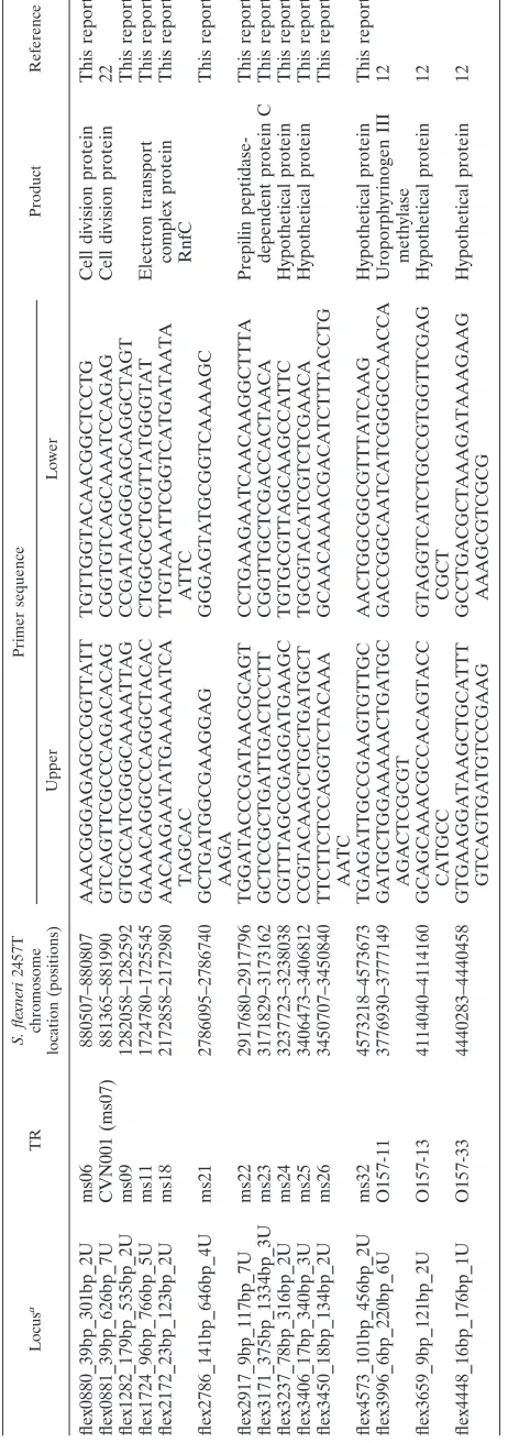
Table 3 highlights characteristics and diversity of the 15
selected VNTR loci. Ten loci are located within coding or putative coding regions, and five are intergenic. Repeat unit lengths ranged from 6 to 375 base pairs. These VNTRs were also tested in silico by using the 17 available sets of genomic sequence data ofShigella,E. coli, andSalmonella enterica se-rovar Typhimurium (Table 1).
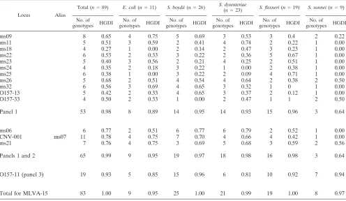
The HGDI (7) of each VNTR was calculated for the com-plete collection and for each species. Three different panels of VNTR loci (panel 1 included 11 loci, panel 2 included 3 loci, and panel 3 included 1 locus) were defined according to their HGDI value and were subsequently combined in a composite data set using three different levels of weight (individual marker weight of 20, 10, or 1) along the line previously pro-posed forBrucellaMLVA (1). The rationale for this approach is that the diversity index indirectly reflects mutation rate and homoplasy level at each locus. Markers with a higher ho-moplasy level have a lower phylogenetic value. Panel 1 in-cluded 11 VNTRs (individual weights of 20) with HGDIs be-low 0.75 (ms09, ms11, ms18, ms22, ms23, ms24, ms25, ms26, ms32, O157-13, and O157-33), panel 2 included 3 VNTRs (individual weights of 10) with HGDIs ranging from 0.75 to 0.9 (ms06, CNV-001, and ms21), and panel 3 is limited to the hypervariable VNTR, O157-11 (individual weight of 1), with an HGDI higher than 0.9.
Diversity indexes differ within the genus Shigella and the speciesE. coli(Table 3). As expected, the least variable group of organisms is theS. sonneigenus, with an HGDI of 0.64 when the highly variable locus O157-11 is not included (MLVA14, comprising panels 1 and 2). Locus O157-11 alone has an HGDI of 0.94 forS. sonneiand leads to a general HGDI of 0.97 for this species. S. flexneri, S. boydii, and S. dysenteriae
show much greater MLVA14 diversity, correlated with the higher number of serotypes (12, 15, and 10, respectively) in these species. RegardingE. coli, the limited number of strains included in the study does not allow comparison with the others.
By use of the MLVA14 assay (panels 1 and 2), the 89 strains (72 DNA samples tested and 17 sets of sequence data) were differentiated into 65 genotypes. When the O157-11 VNTR is used, the discriminatory power of the assay is significantly increased, with 83 genotypes numbered 1 to 83 in the dendro-gram produced (Fig. 2). With the low relative weight given in the clustering analysis to this very highly variable marker, the two clusters are highly similar. All strains are identified by species acronym and genotype number (e.g., Sd#01 or Sf#70). Several strains sharing the same genotype will be additionally differentiated by a letter (e.g., a, b, etc.).
Cluster analysis shows that most of theShigellastrains fall into four main clusters. Sb#81 and Sb#82 stand as outliers, Se#83 being an outgroup. Sb#81 and Sb#82 belong to theS. boydiiserotype 13 (B13 in reference 35) and have an MLVA15 profile very different from those of all the otherShigellastrains, with seven cases lacking amplification and two alleles observed only in these strains. Two strains ofS. sonnei, two strains ofS. boydii, three strains ofE. coli, and two pairs ofS. dysenteriae
strains share the same genotype. Additional markers might allow further differentiation of Shigella strains, supposed to have been isolated in different places at different times (20). The HGDIs, however, are high and, by adding in the cluster analysis markers discarded previously, the strains remain
on May 16, 2020 by guest
http://jcm.asm.org/
TABLE 2. List of TRs selected Locus a TR S. flexneri 2457T chromosome location (positions) Primer sequence Product Reference Upper Lower flex0880_39bp_301bp_2U ms06 880507–880807 AAACGGGAGAGCCGGTTATT TGTTGGTACAACGGCTCCTG Cell division protein This report flex0881_39bp_626bp_7U CVN001 (ms07) 881365–881990 GTCAGTTCGCCCAGACACAG CGGTGTCAGCAAATCCAGAG Cell division protein 22 flex1282_179bp_535bp_2U ms09 1282058–1282592 GTGCCATCGGGCAAAATTAG CCGATAAGGGAGCAGGCTAGT This report flex1724_96bp_766bp_5U ms11 1724780–1725545 GAAACAGGCCCAGGCTACAC CTGGCGCTGGTTATGGGTAT Electron transport This report flex2172_23bp_123bp_2U ms18 2172858–2172980 AACAAGAATATGAAAAATCA TAGCAC TTGTAAATTCGGTCATGATAATA ATTC complex protein RnfC This report flex2786_141bp_646bp_4U ms21 2786095–2786740 GCTGATGGCGAAGGAG AAGA GGGAGTATGCGGTCAAAAGC This report flex2917_9bp_117bp_7U ms22 2917680–2917796 TGGATACCCGATAACGCAGT CCTGAAGAATCAACAAGGCTTTA Prepilin peptidase-This report flex3171_375bp_1334bp_3U ms23 3171829–3173162 GCTCCGCTGATTGACTCCTT CGGTTGCTCGACCACTAACA dependent protein C This report flex3237_78bp_316bp_2U ms24 3237723–3238038 CGTTTAGCCGAGGATGAAGC TGTGCGTTAGCAAGCCATTC Hypothetical protein This report flex3406_17bp_340bp_3U ms25 3406473–3406812 CCGTACAAGCTGCTGATGCT TGCGTACATCGTCTCGAACA Hypothetical protein This report flex3450_18bp_134bp_2U ms26 3450707–3450840 TTCTTCTCCAGGTCTACAAA AATC GCAACAAAACGACATCTTTACCTG This report flex4573_101bp_456bp_2U ms32 4573218–4573673 TGAGATTGCCGAAGTGTTGC AACTGGCGGCGTTTATCAAG Hypothetical protein This report flex3996_6bp_220bp_6U O157-11 3776930–3777149 GATGCTGGAAAAACTGATGC AGACTCGCGT GACCGGCAATCATCGGGCCAACCA Uroporphyrinogen III methylase 12 flex3659_9bp_121bp_2U O157-13 4114040–4114160 GCAGCAAACGCCACAGTACC CATGCC GTAGGTCATCTGCCGTGGTTCGAG CGCT Hypothetical protein 12 flex4448_16bp_176bp_1U O157-33 4440283–4440458 GTGAAGGATAAGCTGCATTT GTCAGTGATGTCCGAAG GCCTGACGCTAAAGATAAAGAAG AAAGCGTCGCG Hypothetical protein 12 aLocus names were created as follows: four letters for the species strain used as a reference, four digits for genome location in the sequenced referenc e strain, TR size, locus size in the reference strain, and number of TR in the reference strain, separated by underscore sign.
1030 GORGE´ ET AL. J. CLIN. MICROBIOL.
on May 16, 2020 by guest
http://jcm.asm.org/
[image:5.585.175.406.72.728.2]distinguishable (data not shown), supporting the idea that they are in all likelihood epidemiologically related.
To estimate the validity of the clustering observed by VNTR typing, selected strains were analyzed by sequence typing using published MLST data (35). Strains provided amplification products of the expected size, regarding the segment of the
thrCgene. Sequences were imported and aligned in the
Bionu-merics program, and a cluster analysis was produced (see Ta-ble S2 in the supplemental material). A single discrepancy between clusters inferred from known genera and serotypes on one hand and sequence analysis on the other hand was ob-served due to the incongruence of phylogeny at the different loci.S. dysenteriaeCIP 53.134 (D8 in reference 35) fell within cluster 3 when only thethrCsequence data were analyzed. It is thetrpC-trpBsequence used in the MLST assay, in particular, which excludes that strain from cluster 3. Subsequently, all strains from the collection for which thethrClocus was inves-tigated were characterized by their relevance to the clusters classified by Pupo et al., as indicated in Fig. 2.
This sequence analysis and comparison with published se-quence data show that three of these clusters do correspond to the clusters classified by Pupo et al., and they are numbered accordingly in Fig. 2. Cluster 1 groupedS. boydiiserotypes 1 to 4, 6, 8, 10, 14, and 18,S. dysenteriaeserotypes 3 to 7, 9, and 11, and S. flexneri serotype 6. Cluster 1 was the most diverse, grouping representatives of allShigellaspecies exceptS.sonnei. This cluster could be divided into three subclusters, i.e., SC-1A, SC-1B, and SC-1C. Strains sharing the same serotype are usually discriminated by MLVA15 typing.
Cluster 2 comprisesS. boydiiserotypes 5, 7, 9, 11, 15, and 16,
[image:6.585.44.286.68.193.2]S. dysenteriaeserotype 2, and allS. sonneiserotypes. The twoS. boydiiserotype 9 displayed the same MLVA15 pattern. TheS. sonneistrains are located into two branches, one with the two representatives ofS. boydiiserotype 7. Three uropathogenicE. colistrains are included in cluster 2, together with anE. coli
FIG. 1. Illustration of the MLVA assay setup. The PCR products of an amplification using ms06 primers were loaded on an agarose gel, electrophoresed, and stained with ethidium bromide. Lanes M show a 100-bp-ladder molecular weight marker. Lanes 1 and 7 correspond to theS. flexneristrain 2457T; lanes 2 to 6 and lanes 8 to 12 correspond to strains from our assay. The image illustrates how the number of units can be directly deduced by manual reading. The marker name below the gel provides the repeat unit size of the TR, the expected PCR product size in theS. flexneri2457T genome, and the correspond-ing number of units in theS. flexneri2457T genome.
TABLE 3. Diversity indexes calculated for MLVA panels and individual markers in the fourShigellaspecies andE. colia
Locus Alias
Total (n⫽89) E. coli(n⫽11) S. boydii(n⫽26) S. dysenteriae
(n⫽23) S. flexneri(n⫽19) S. sonnei(n⫽9)
No. of
genotypes HGDI No. of
genotypes HGDI No. of
genotypes HGDI No. of
genotypes HGDI No. of
genotypes HGDI No. of
genotypes HGDI
ms09 8 0.65 4 0.75 5 0.69 3 0.53 3 0.4 2 0.22
ms11 5 0.51 3 0.59 2 0.41 4 0.74 2 0.22 1 0.00
ms18 4 0.27 1 0.00 2 0.14 2 0.47 3 0.23 1 0.00
ms22 6 0.53 2 0.33 3 0.22 2 0.36 5 0.67 1 0.00
ms23 5 0.40 3 0.56 2 0.21 4 0.25 2 0.51 1 0.00
ms24 4 0.35 2 0.18 3 0.22 1 0.00 2 0.38 1 0.00
ms25 6 0.38 1 0.00 3 0.22 2 0.09 4 0.71 1 0.00
ms26 5 0.68 2 0.51 4 0.54 4 0.64 2 0.38 2 0.50
ms32 6 0.56 3 0.69 4 0.65 3 0.32 1 0 1 0.00
O157-13 5 0.42 2 0.33 4 0.65 3 0.37 2 0.12 1 0.00
O157-33 4 0.50 2 0.33 1 0.00 2 0.47 1 1 2 0.50
Panel 1 53 0.98 8 0.89 14 0.95 14 0.93 15 0.96 3 0.64
ms06 6 0.77 2 0.51 6 0.77 6 0.79 2 0.52 1 0.00
CNV-001 ms07 11 0.78 4 0.75 7 0.70 4 0.66 4 0.42 1 0.00
ms21 7 0.76 4 0.75 3 0.69 5 0.68 3 0.59 2 0.56
Panels 1 and 2 65 0.99 9 0.95 19 0.97 18 0.98 16 0.98 3 0.64
O157-11 (panel 3) 19 0.93 5 0.85 15 0.96 6 0.81 10 0.92 7 0.94
Total for MLVA-15 83 1.00 9 0.95 25 1.00 21 0.99 19 1.00 8 0.97
a
n, number of strains.
on May 16, 2020 by guest
http://jcm.asm.org/
[image:6.585.44.540.430.718.2]1032
on May 16, 2020 by guest
http://jcm.asm.org/
strain isolated from a patient at Hoˆpital d’Instruction des Arme´es, Be´gin, France (Ec#47).
Cluster 3 groups allS. flexneristrains (with the exception of
S. flexneriserotype 6, located in cluster 1),S. boydiiserotype 12, andS. dysenteriaeserotype 8 (Sd#56). Sd#56 is weakly asso-ciated with subcluster 3A together withS. flexneriserotypes 3, 3a, 5, and X. A second subcluster, tentatively called 3B (Fig. 2), comprisesS. boydiiserotype 12 andS. flexneriserotypes 1, 2, 2a, 2b, 4b, and Y.S. flexneriserotype 4c is more distantly related. Each strain has a unique genotype, although several serotypes were represented by more than one strain.
Consequently, and with the exception of Sd#56 (Sd8), the composition of the clusters is in remarkable agreement with the report of Pupo et al., although the underlying approaches are quite different (35).
We propose here to define a fourth cluster. Cluster 4 would include all theS. dysenteriaeserotype 1 strains, five represen-tatives of the E. coliK-12 strain (including two genomic se-quences), and the twoE. coliO157:H7 strains included in the study. TheE. coli O157:H7 representatives are located in a quite distinct branch, slightly closer to theS. dysenteriae sero-type 1 branch than to theE. coli K-12 branch. The eight S. dysenteriaeserotype 1 strains are separated into six genotypes, showing the discriminative power of the VNTR panel. Among those strains, two were isolated from patients during an out-break in a refugee camp in Rwanda in 1994. They show exactly the same MLVA15 pattern. Three of theE. coliK-12 strains were wild-type K-12 and two were derivatives (14). The three K-12 wild types share exactly the same genotype, while deriv-atives present different patterns at locus ms22, ms32, or O157-11. The O157:H7 strains exhibited two differences among them, at loci ms11 and O157-11.
In very rare instances, a given allele is strongly associated with a specific cluster, at least in the limited collection of strains investigated here. The three-repeat-unit allele at locus ms06 is observed only in cluster 2, and locus O157-33 has more than one repeat unit only in clusters 2 and 4 (associated with theE. colistrains investigated here).
ms25 and ms26 each gave rise to a very large amplicon in two strains and four strains, respectively, and these amplicons cor-respond to alleles with more than 60 repeat units (compared to the usual allele size ranges of one to three and two to four repeat units, respectively) (see Table S2 in the supplemental material). The six alleles were sequenced. The ms25 sequenc-ing revealed that two different insertion sequences (ISs) were present, IS629 and IS2 for Sf#22 and Sd#26, respectively. IS629is a member of the IS3family of transposable elements. The ms26 allele sequencing led to the finding that only one IS, known as IS630, was present in this TR (27), and it was in-serted at the same position and orientation in the four strains Sd#33, Sd#34, Sb#35, and Sb#36, confirming the close rela-tionship between the four strains already suggested by MLVA15 clustering, in spite of a different species assignment.
As a complementary analysis, an MST analysis was per-formed. MST analysis is a convenient tool to cluster multiple isolates and illustrate the relative diversity within different lin-eages (Fig. 3). This kind of analysis is applicable to categorical data sets. The creation of hypothetical types further minimizes the summed distance of all branches of the tree. The MST was drawn without locus O157-11, which is too variable to address correct relatedness within different lineages, especially since this analysis does not currently allow the assignment of differ-ent relative weights to markers. In cluster 1, the three main subclusters are conserved, with only minor changes. The twoS. dysenteriaeserotype 7 strains, Sd#26 and Sd#27, are located in the vicinity of subcluster A but interspaced by Sb#04 and Sb#05. The composition of cluster 2, withS. sonnei andS. boydiiserotype 7 in the same branch, is also found by this approach. Sd#56 is not included in cluster 3, which is in accordance with results obtained from other molecular methods and reflects the absence of amplification at several loci, which is taken into account in this analysis. The only change in cluster 4 is the location ofE. coli O157:H7 rep-resentatives that are far from other members of the cluster and weakly linked to cluster 2.
DISCUSSION
TheShigellagenus has been classified into species and se-rotypes for a long time and for practical reasons, but MLST (15, 16, 34, 52) and genome comparisons (51) have emphasized the strong genetic homogeneity between the different species. Most importantly, MLST has further strongly suggested that in some instances, the current species designations are not com-patible with the actual phylogeny of the Shigellagenus. It is now considered more likely thatShigellasp. strains, as well as enteroinvasiveE. colistrains, are derived from multiple origins within the speciesE. coli(50).
The purpose of the present study was to develop an MLVA assay forShigellaand evaluate if the resulting data would be in agreement with the complex concept of the Shigella-E. coli
genomospecies, as revealed by MLST data. We investigated TR loci predicted to be polymorphic by comparing fiveShigella
genome sequences. The selection of 15 VNTRs allowed us to discriminate 89 strains, covering the large majority of known diversity inShigellaspecies and severalE. colistrains (77 Shi-gellaspp., 11 E. colistrains, and 1 S. entericastrain), into 83 genotypes. Strains sharing the same serotype are rarely from the same genotype, with difference ranging from one marker (Sd#1 and Sd#2, ofS. dysenteriaeserotype 5) to five markers (Sd#26 and Sd#27, ofS. dysenteriaeserotype 7). On the other hand, the two strains of S. dysenteriaeserotype 1 isolated in Rwanda in 1994 during a dysentery outbreak in refugee camps are identical.
Through MLVA15 analysis, four main clusters were defined, together with some outliers. Among them, the strains of S.
FIG. 2. Dendrogram with all the typed strains, including the ones corresponding to sequenced strains that were included in the data set by their allele numbers as calculated theoretically from the expected amplicon sizes of all 15 loci based on their genome sequences. Clustering analysis was done using the categorical and UPGMA options. The columns indicate the genotype number, the genus, the species, the serotype, the strain identification number (collection), the cluster number as defined by Pupo et al., and the year and place of isolation.
on May 16, 2020 by guest
http://jcm.asm.org/
boydiiserotype 13 were clearly distinguished from the other
Shigellastrains. Some cases lacking amplification, as well as unique sizes for several loci, illustrate the wide distance be-tweenS.boydiiserotype 13 and otherShigellarepresentatives. According to Hyma et al. (8),S. boydiiserotype 13 is closely related to a differentEscherichiaspecies,E. albertii. It supports the hypothesis of an ancient separation of lineages,S.boydii
serotype 13, as well asS. entericaLT2, being a clear outgroup. This finding also corroborated the results of MLST investiga-tions by Pupo et al. (35) and Lan et al. (15).
In order to be able to compare our MLVA15 clustering data with published MLST data, part of the thrC gene was se-quenced. In this way, the strains used in this study could be assigned to the different clusters defined by Pupo et al. (35), and it very clearly appears that MLVA15 clustering provides the same view of theShigellapopulation structure as MLST. Recently, Yang et al. (52) conducted another MLST analysis, based on 23 housekeeping genes. The greater number of genes
analyzed allowed them to define three subclusters inside clus-ter 1. MLVA15 clusclus-tering reveals the same fine substructure. Such striking similarity demonstrates the potential usefulness of MLVA forShigellatyping. While the general patterns are highly similar, there are some differences between MLST and MLVA approaches. In the present study, the nineS. sonnei
strains are assigned to cluster 2, close toS. boydiiserotype 7. Yang et al. (52) placed the singleS. sonneiinvestigated close to cluster 1 by MLST. TheS. sonneistrain investigated by Esco-bar-Paramo et al. (4) was found to be closer to cluster 2 and cluster 3 by analysis of four other housekeeping genes. Pupo et al. (35) placed the singleS. sonneistrain they analyzed far from the three clusters. Thus, the definitive location of S. sonnei
strains remains undefined. Further MLST analysis ofS. sonnei
might eventually permit us to revisit the relative evolutionary position of this species.
[image:9.585.45.542.66.441.2]Considering that Shigella clusters are now believed to have independently arisen several times fromE. colispecies FIG. 3.Shigellapopulation modeling. The number in each circle indicates the corresponding genotype identified in Fig. 2. The empty circle indicates a hypothetical genotype (not present in the population analyzed) created to minimize overall distances between neighboring genotypes. The distance between neighboring genotypes is expressed as the number of allelic changes and is outlined by different shapes of lines: a hatched bold line indicates one change, a full gray line indicates two changes, a black dotted line indicates three changes, and a gray dotted line indicates more than four changes. Gray hatched lines are used to separate subgroups inside clusters. The tree calculation was made without locus O157-11, owing the location of different genotypes in the same circle.
1034 GORGE´ ET AL. J. CLIN. MICROBIOL.
on May 16, 2020 by guest
http://jcm.asm.org/
while acquiring pathogenicity factors (16, 35, 52), the un-derrepresentation of E. coli investigated here led to the representation of small groups ofE. colistrains arising from
Shigella clusters, while, in fact, the situation would be the opposite. Such analysis should be done by including a Shi-gella collection in a larger E. coli collection to obtain a general overview ofE. coli/Shigellasp. genomospecies (such work is in progress).
Combining UPGMA cluster analysis and MST provides an overview ofShigellaintraspecies relationships. In agreement with previous investigations, we can infer from this combina-tion thatShigellataxonomy is of little phylogenetic value. This is illustrated for instance byS. flexneriserotype 6 being asso-ciated with cluster 1 by MLVA or MLST, whereas the otherS. flexneriserotypes are assigned to cluster 3 (together with the two serologically untypedS. flexneristrains). Four strains clus-tered together by MLVA share the same rare IS insertion event at the same position, while they are assigned to different species, reinforcing the fact that current classification does not reflect the genetic relatedness within theShigellagenus. It is now believed thatShigellaorganisms arose several times within
E. colispecies (35, 52), leading to three clusters not related to the currently recognizedShigellaspecies taxonomy based upon biochemical and serological characteristics. The three main clusters are also identified here with a fourth corresponding essentially toS. dysenteriaeserotype 1.
This study augments the results relative to discrimination power, specificity, and sensitivity of the MLVA approach to the
Shigellaspecies. Moreover, the MLVA15 panel shows some capabilities to be applied toE. colifor typing purposes, with a wider range of use than considered before in several published studies, mainly focused on Shiga toxin-producingE. coli(12, 23, 29). Another MLVA assay was validated for O157:H7 outbreak detection (12). It showed high discrimination be-tween the E. coliO157 strains and appeared to have equal sensitivity to that of PFGE and specificity superior to that of PFGE. Lindstedt et al. (22) described a seven-TR panel de-signed to type the ECOR collection and pathogenic E. coli
species. SeveralShigellastrains were included in the study and typed. However, all serotypes were not available, and in one case, the TR was absent, and in another, the TR was mono-morphic. Our panel primarily intended to cover all Shigella
species, and its satisfactory performance indicates also good capabilities to discriminate between nonpathogenic E. coli, uropathogenicE. coli, and enterohemorrhagicE. colistrains.
As suggested previously (45), the proposed combination of well-selected independent polymorphic TR loci is highly dis-criminatory and provides a relevant clustering compared with the currently accepted classification. The panel of 15 markers has been divided into three panels according to the polymor-phism of each locus. It is important to keep in mind that the very high discriminatory power of some markers usually results in a very high homoplasy level. For this reason, such markers must be given a lower weight when similarity matrices are calculated. Here, the different weights proposed for each panel were empirically determined, but it is very likely that in future MLVA developments, special attention will be given to the optimization of these coefficients. For this to be done, much larger collections of strains, with detailed epidemiological data, will need to be typed first. It can only be hoped that in the
future, funding bodies will support international consortiums of laboratories aimed at producing high-quality data of this kind. VNTR typing could then be widely accessible for re-search and public health laboratories, particularly in develop-ing countries, where the majority of cases ofShigella occur, since this method is highly suitable for sharing results and for the generation of databases as previously demonstrated (http: //mlva.u-psud.fr).
ACKNOWLEDGMENTS
We thank Sabrina Gausson and Claudette Simoes-Miranda for tech-nical help. We thank Jean-Didier Cavallo (Hoˆpital d’Instruction des Arme´es, Be´gin, France) for providing some of the strains used in this study. O. Gorge´ also acknowledges Rozenn Bernard and Philippe Le Fle`che for many useful discussions and ideas.
Work on the typing of collections of dangerous pathogens and the making of reference genotype databases was supported by De´le´gation Ge´ne´rale pour l’Armement (PEA02-36-01).
REFERENCES
1.Al Dahouk, S., P. Le Fle`che, K. Nockler, I. Jacques, M. Grayon, H. C. Scholz, H. Tomaso, G. Vergnaud, and H. Neubauer.2007. Evaluation ofBrucella
MLVA typing for human brucellosis. J. Microbiol. Methods69:137–145. 2.Chaignat, C.2004. Shigellosis, p. 527–531.InD. L. Heyman (ed.), Control of
communicable diseases manual, 18th ed. American Public Health Associa-tion, Washington, DC.
3.Denoeud, F., and G. Vergnaud.2004. Identification of polymorphic tandem repeats by direct comparison of genome sequence from different bacterial strains: a web-based resource. BMC Bioinformatics5:4.
4.Escobar-Paramo, P., C. Giudicelli, C. Parsot, and E. Denamur.2003. The evolutionary history ofShigellaand enteroinvasiveEscherichia colirevised. J. Mol. Evol.57:140–148.
5.Faruque, S. M., K. Haider, M. M. Rahman, A. R. Abdul Alim, Q. S. Ahmad, M. J. Albert, and R. B. Sack.1992. Differentiation ofShigella flexneristrains by rRNA gene restriction patterns. J. Clin. Microbiol.30:2996–2999. 6.Grissa, I., P. Bouchon, C. Pourcel, and G. Vergnaud.On-line resources for
bacterial micro-evolution studies using MLVA or CRISPR typing. Bio-chimie, in press.
7.Hunter, P. R., and M. A. Gaston.1988. Numerical index of the discrimina-tory ability of typing systems: an application of Simpson’s index of diversity. J. Clin. Microbiol.26:2465–2466.
8.Hyma, K. E., D. W. Lacher, A. M. Nelson, A. C. Bumbaugh, J. M. Janda, N. A. Strockbine, V. B. Young, and T. S. Whittam. 2005. Evolutionary genetics of a new pathogenicEscherichiaspecies: Escherichia albertiiand relatedShigella boydiistrains. J. Bacteriol.187:619–628.
9.Hyytia-Trees, E., S. C. Smole, P. A. Fields, B. Swaminathan, and E. M. Ribot.2006. Second generation subtyping: a proposed PulseNet protocol for multiple-locus variable-number tandem repeat analysis of Shiga toxin-pro-ducing Escherichia coli O157 (STEC O157). Foodborne Pathog. Dis.3:118– 131.
10.Jin, Q., Z. Yuan, J. Xu, Y. Wang, Y. Shen, W. Lu, J. Wang, H. Liu, J. Yang, F. Yang, X. Zhang, J. Zhang, G. Yang, H. Wu, D. Qu, J. Dong, L. Sun, Y. Xue, A. Zhao, Y. Gao, J. Zhu, B. Kan, K. Ding, S. Chen, H. Cheng, Z. Yao, B. He, R. Chen, D. Ma, B. Qiang, Y. Wen, Y. Hou, and J. Yu.2002. Genome sequence ofShigella flexneri2a: insights into pathogenicity through compar-ison with genomes ofEscherichia coliK12 and O157. Nucleic Acids Res. 30:4432–4441.
11.Johansson, A., J. Farlow, P. Larsson, M. Dukerich, E. Chambers, M. Bystrom, J. Fox, M. Chu, M. Forsman, A. Sjostedt, and P. Keim.2004. Worldwide genetic relationships amongFrancisella tularensisisolates deter-mined by multiple-locus variable-number tandem repeat analysis. J. Bacte-riol.186:5808–5818.
12.Keys, C., S. Kemper, and P. Keim.2005. Highly diverse variable number tandem repeat loci in theE. coliO157:H7 and O55:H7 genomes for high-resolution molecular typing. J. Appl. Microbiol.98:928–940.
13.Koeck, J. L., B. M. Njanpop-Lafourcade, S. Cade, E. Varon, L. Sangare, S. Valjevac, G. Vergnaud, and C. Pourcel.2005. Evaluation and selection of tandem repeat loci forStreptococcus pneumoniaeMLVA strain typing. BMC Microbiol.5:66.
14.Kuhnert, P., J. Nicolet, and J. Frey.1995. Rapid and accurate identification ofEscherichia coliK-12 strains. Appl. Environ. Microbiol.61:4135–4139. 15.Lan, R., M. C. Alles, K. Donohoe, M. B. Martinez, and P. R. Reeves.2004.
Molecular evolutionary relationships of enteroinvasiveEscherichia coliand
Shigellaspp. Infect. Immun.72:5080–5088.
16.Lan, R., and P. R. Reeves.2002.Escherichia coli in disguise: molecular origins ofShigella. Microbes Infect.4:1125–1132.
on May 16, 2020 by guest
http://jcm.asm.org/
17.Le Fle`che, P., M. Fabre, F. Denoeud, J. L. Koeck, and G. Vergnaud.2002. High resolution, on-line identification of strains from theMycobacterium tuberculosiscomplex based on tandem repeat typing. BMC Microbiol.2:37. 18.Le Fle`che, P., Y. Hauck, L. Onteniente, A. Prieur, F. Denoeud, V. Ramisse, P. Sylvestre, G. Benson, F. Ramisse, and G. Vergnaud.2001. A tandem repeats database for bacterial genomes: application to the genotyping of
Yersinia pestisandBacillus anthracis. BMC Microbiol.1:2.
19.Le Fle`che, P., I. Jacques, M. Grayon, S. Al Dahouk, P. Bouchon, F. Denoeud, K. Nockler, H. Neubauer, L. A. Guilloteau, and G. Vergnaud.2006. Evalu-ation and selection of tandem repeat loci for aBrucellaMLVA typing assay. BMC Microbiol.6:9.
20.Liang, S. Y., H. Watanabe, J. Terajima, C. C. Li, J. C. Liao, S. K. Tung, and C. S. Chiou.2007. Multilocus variable-number tandem repeat analysis for molecular typing ofShigella sonnei. J. Clin. Microbiol.45:3574–3580. 21.Lindstedt, B. A.2005. Multiple-locus variable number tandem repeats
anal-ysis for genetic fingerprinting of pathogenic bacteria. Electrophoresis26: 2567–2582.
22.Lindstedt, B. A., L. T. Brandal, L. Aas, T. Vardund, and G. Kapperud.2007. Study of polymorphic variable-number of tandem repeats loci in the ECOR collection and in a set of pathogenic Escherichia coli and Shigella isolates for use in a genotyping assay. J. Microbiol. Methods69:197–205.
23.Lindstedt, B. A., E. Heir, E. Gjernes, T. Vardund, and G. Kapperud.2003. DNA fingerprinting of Shiga-toxin producingEscherichia coliO157 based on multiple-locus variable-number tandem-repeats analysis (MLVA). Ann. Clin. Microbiol. Antimicrob.2:12.
24.Lista, F., G. Faggioni, S. Valjevac, A. Ciammaruconi, J. Vaissaire, C. le Doujet, O. Gorge´, R. De Santis, A. Carattoli, A. Ciervo, A. Fasanella, F. Orsini, R. D’Amelio, C. Pourcel, A. Cassone, and G. Vergnaud.2006. Geno-typing of Bacillus anthracisstrains based on automated capillary 25-loci multiple locus variable-number tandem repeats analysis. BMC Microbiol. 6:33.
25.Litwin, C. M., A. L. Storm, S. Chipowsky, and K. J. Ryan.1991. Molecular epidemiology ofShigellainfections: plasmid profiles, serotype correlation, and restriction endonuclease analysis. J. Clin. Microbiol.29:104–108. 26.Liu, P. Y., Y. J. Lau, B. S. Hu, J. M. Shyr, Z. Y. Shi, W. S. Tsai, Y. H. Lin,
and C. Y. Tseng.1995. Analysis of clonal relationships among isolates of
Shigella sonneiby different molecular typing methods. J. Clin. Microbiol. 33:1779–1783.
27.Matsutani, S., H. Ohtsubo, Y. Maeda, and E. Ohtsubo.1987. Isolation and characterization of IS elements repeated in the bacterial chromosome. J. Mol. Biol.196:445–455.
28.Melito, P. L., D. L. Woodward, J. Munro, J. Walsh, R. Foster, P. Tilley, A. Paccagnella, J. Isaac-Renton, J. Ismail, and L. K. Ng.2005. A novelShigella dysenteriaeserovar isolated in Canada. J. Clin. Microbiol.43:740–744. 29.Noller, A. C., M. C. McEllistrem, A. G. Pacheco, D. J. Boxrud, and L. H.
Harrison.2003. Multilocus variable-number tandem repeat analysis distin-guishes outbreak and sporadicEscherichia coliO157:H7 isolates. J. Clin. Microbiol.41:5389–5397.
30.Onteniente, L., S. Brisse, P. T. Tassios, and G. Vergnaud.2003. Evaluation of the polymorphisms associated with tandem repeats forPseudomonas aeruginosastrain typing. J. Clin. Microbiol.41:4991–4997.
31.Pourcel, C., F. Andre-Mazeaud, H. Neubauer, F. Ramisse, and G. Vergnaud. 2004. Tandem repeats analysis for the high resolution phylogenetic analysis ofYersinia pestis. BMC Microbiol.4:22.
32.Pourcel, C., Y. Vidgop, F. Ramisse, G. Vergnaud, and C. Tram.2003. Char-acterization of a tandem repeat polymorphism inLegionella pneumophila
and its use for genotyping. J. Clin. Microbiol.41:1819–1826.
33.Pourcel, C., P. Visca, B. Afshar, S. D’Arezzo, G. Vergnaud, and N. K. Fry. 2007. Identification of variable-number tandem repeat sequences in Legio-nella pneumophilaand development of an optimized MLVA typing scheme. J. Clin. Microbiol.45:1190–1199.
34.Pupo, G. M., D. K. Karaolis, R. Lan, and P. R. Reeves.1997. Evolutionary relationships among pathogenic and nonpathogenicEscherichia colistrains inferred from multilocus enzyme electrophoresis andmdhsequence studies. Infect. Immun.65:2685–2692.
35.Pupo, G. M., R. Lan, and P. R. Reeves.2000. Multiple independent origins ofShigellaclones ofEscherichia coliand convergent evolution of many of their characteristics. Proc. Natl. Acad. Sci. USA97:10567–10572.
36.Ramisse, V., P. Houssu, E. Hernandez, F. Denoeud, V. Hilaire, O. Lisanti, F. Ramisse, J. D. Cavallo, and G. Vergnaud.2004. Variable number of tandem repeats inSalmonella entericasubsp.entericafor typing purposes. J. Clin. Microbiol.42:5722–5730.
37.Ribot, E. M., M. A. Fair, R. Gautom, D. N. Cameron, S. B. Hunter, B. Swaminathan, and T. J. Barrett.2006. Standardization of pulsed-field gel electrophoresis protocols for the subtyping of Escherichia coliO157:H7,
Salmonella, andShigellafor PulseNet. Foodborne Pathog. Dis.3:59–67. 38.Rotz, L. D., A. S. Khan, S. R. Lillibridge, S. M. Ostroff, and J. M. Hughes.
2002. Public health assessment of potential biological terrorism agents. Emerg. Infect. Dis.8:225–230.
39.Rozen, S., and H. Skaletsky.2000. Primer3 on the WWW for general users and for biologist programmers. Methods Mol. Biol.132:365–386. 40.Sabat, A., J. Krzyszton-Russjan, W. Strzalka, R. Filipek, K. Kosowska, W.
Hryniewicz, J. Travis, and J. Potempa.2003. New method for typing Staph-ylococcus aureusstrains: multiple-locus variable-number tandem repeat anal-ysis of polymorphism and genetic relationships of clinical isolates. J. Clin. Microbiol.41:1801–1804.
41.Sambrook, J., and D. Russell.2001. Molecular cloning, a laboratory manual, 3rd ed. Cold Spring Harbor Laboratory Press, Cold Spring Harbor, NY. 42.Swaminathan, B., T. J. Barrett, S. B. Hunter, and R. V. Tauxe. 2001.
PulseNet: the molecular subtyping network for foodborne bacterial disease surveillance, United States. Emerg. Infect. Dis.7:382–389.
43.U’Ren, J. M., J. M. Schupp, T. Pearson, H. Hornstra, C. L. Friedman, K. L. Smith, R. R. Daugherty, S. D. Rhoton, B. Leadem, S. Georgia, M. Cardon, L. Y. Huynh, D. DeShazer, S. P. Harvey, R. Robison, D. Gal, M. J. Mayo, D. Wagner, B. J. Currie, and P. Keim.2007. Tandem repeat regions within the
Burkholderia pseudomalleigenome and their application for high resolution genotyping. BMC Microbiol.7:23.
44.van Belkum, A.2007. Tracing isolates of bacterial species by multilocus variable number of tandem repeat analysis (MLVA). FEMS Immunol. Med. Microbiol.49:22–27.
45.Vergnaud, G., and C. Pourcel.2006. Multiple locus VNTR (variable number of tandem repeat) analysis (MLVA), p. 83–104.InE. Stackebrandt (ed.), Molecular identification, systematics and population structure of pro-karyotes, 1st ed. Springer-Verlag, New York, NY.
46.von Seidlein, L., D. R. Kim, M. Ali, H. Lee, X. Wang, V. D. Thiem, G. Canh do, W. Chaicumpa, M. D. Agtini, A. Hossain, Z. A. Bhutta, C. Mason, O. Sethabutr, K. Talukder, G. B. Nair, J. L. Deen, K. Kotloff, and J. Clemens. 2006. A multicentre study ofShigelladiarrhoea in six Asian countries: disease burden, clinical manifestations, and microbiology. PLoS Med.3:e353. 47.Vu-Thien, H., G. Corbineau, K. Hormigos, B. Fauroux, H. Corvol, A.
Clem-ent, G. Vergnaud, and C. Pourcel.2007. Multiple-locus variable-number tandem-repeat analysis for longitudinal survey of sources ofPseudomonas aeruginosainfection in cystic fibrosis patients. J. Clin. Microbiol.45:3175– 3183.
48.Wei, J., M. B. Goldberg, V. Burland, M. M. Venkatesan, W. Deng, G. Fournier, G. F. Mayhew, G. Plunkett III, D. J. Rose, A. Darling, B. Mau, N. T. Perna, S. M. Payne, L. J. Runyen-Janecky, S. Zhou, D. C. Schwartz, and F. R. Blattner.2003. Complete genome sequence and comparative genomics ofShigella flexneriserotype 2a strain 2457T. Infect. Immun.71: 2775–2786.
49.Woodward, D. L., C. G. Clark, R. A. Caldeira, R. Ahmed, G. Soule, L. Bryden, H. Tabor, P. Melito, R. Foster, J. Walsh, L. K. Ng, G. B. Malcolm, N. Strockbine, and F. G. Rodgers.2005. Identification and characterization ofShigella boydii20 serovar nov., a new and emerging Shigella serotype. J. Med. Microbiol.54:741–748.
50.Yang, F., J. Yang, X. Zhang, L. Chen, Y. Jiang, Y. Yan, X. Tang, J. Wang, Z. Xiong, J. Dong, Y. Xue, Y. Zhu, X. Xu, L. Sun, S. Chen, H. Nie, J. Peng, J. Xu, Y. Wang, Z. Yuan, Y. Wen, Z. Yao, Y. Shen, B. Qiang, Y. Hou, J. Yu, and Q. Jin.2005. Genome dynamics and diversity ofShigellaspecies, the etiologic agents of bacillary dysentery. Nucleic Acids Res.33:6445–6458.
51.Yang, J., L. Chen, J. Yu, L. Sun, and Q. Jin.2006. ShiBASE: an integrated database for comparative genomics ofShigella. Nucleic Acids Res.34:D398– D401.
52.Yang, J., H. Nie, L. Chen, X. Zhang, F. Yang, X. Xu, Y. Zhu, J. Yu, and Q. Jin.2007. Revisiting the molecular evolutionary history ofShigellaspp. J. Mol. Evol64:71–79.
1036 GORGE´ ET AL. J. CLIN. MICROBIOL.