OUTCOME ASSESSMENT OF TEETH WITH NECROTIC PULPS AND APICAL PERIODONTITIS TREATED WITH LONG-TERM CALCIUM HYDROXIDE
Christopher L. Ammons
A thesis submitted to the faculty at the University of North Carolina at Chapel Hill in partial fulfillment of the requirements for the degree of Master of Science in the Program in the Adams
School of Dentistry (Endodontics).
Chapel Hill 2020
Approved By: Peter Z. Tawil
© 2020
ABSTRACT
Christopher L. Ammons: Outcome Assessment of Teeth with Necrotic Pulp and Apical Periodontitis Treated with Long-Term Calcium Hydroxide
(Under the direction of Peter Z. Tawil, Glen A. Karunanayake, and Sigurdur R. Saemundsson)
The aim of this retrospective study is to assess clinical outcomes of teeth via the
periapical index (PAI) system to grade healing of periradicular tissues after long-term (30+ days) use of calcium hydroxide. 242 cases diagnosed with pulpal necrosis and apical periodontitis were treated with long-term calcium hydroxide under a standardized protocol. All cases were re-evaluated between one and three months until radiographic healing was observed. Clinical and radiographic evaluations were performed annually. Pre- and post-operative periapical
ACKNOWLEDGMENTS
I would like to thank my research advisor and mentor, Dr. Peter Z. Tawil, for his
continuous support, professional guidance and generosity through the completion of this research project and my residency training. I wish to extend a special thanks to Dr. Sheldon Best for his generous contribution to this project. I would also like to thank my committee members,
TABLE OF CONTENTS
LIST OF TABLES ... vii
LIST OF FIGURES ... viii
Chapter THESIS INTRODUCTION ...1
REVIEW OF LITERATURE ...5
i. History ...5
ii. Antimicrobial Effects ...5
iii. Anti-inflammatory Effects ...6
iv. Denaturation of Endotoxins ...6
v. Bacterial Resistance to Calcium Hydroxide ...8
vi. In-vitro Claims on Decreased Fracture Resistance ...9
vii. Post-Treatment Pain/Flare-ups ...10
viii. Periapical Index (PAI) Score System ...11
ix. Healing Outcomes of Apical Periodontitis ...12
x. Gaps in Knowledge ...14
Methods...15
ii. Inclusion Criteria...15
iii. Exclusion Criteria ...16
iv. Treatment Protocol ...16
v. Radiographic and Clinical Evaluation...17
vi. Calibration of Examiners ...18
vii. Outcome Measures and Data Analysis ...18
Results ...20
Discussion ...22
Conclusion ...30
LIST OF TABLES
Table 1 – Healing Rates According to PAI Score ...31
Table 2 – Distribution of Teeth and PAI Score at Baseline ...31
Table 3 – Summary of Previous Studies ...32
Table 4 – Case Series Data ...32
LIST OF FIGURES
Figure 1 – Periapical Index (PAI) Score Reference ...47
Figure 2 – Frequency distribution of healing with Long-Term Calcium Hydroxide ...47
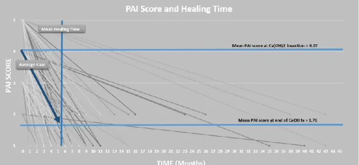
Figure 3 – PAI Score and Healing Time ...48
I. THESIS INTRODUCTION
The focus of endodontics is the prevention and elimination of apical disease. In 1965, Kakehashi et al. clearly established the role of bacterial infection in pulpits and apical
periodontitis (1). Outcomes of root canal therapy are reduced when a radiographic radiolucency is present pre-operatively (2-5). Although there’s still an ongoing debate in regards to the clinical effectiveness of intracranal medicaments in regards to outcome (6), calcium hydroxide have clearly proven itself efficient in reducing the bacterial count inside a root canal system (5).
Calcium hydroxide (Ca(OH)2) is a white, odorless powder with a pH of 12.5 - 12.8 (7). Inherently, the pure, powder form has a high pH to exert bacteriostatic effects (8). This material was introduced to endodontics as pulp capping agent in 1920 by B.W. Herman (9). Over time, it became a multi-purpose agent for inter-appointment intracanal medicaments, endodontic sealers, apexification, and pulpotomies (8).
deep into the canal space.(7). It is available in various forms, such as liquid, paste, or premixed with radiopacifiers delivered by syringe. The percentage concentrations of the calcium hydroxide varies between 10-40% depending on the medium mixture (i.e.: glycerin, distilled water,
methylcellulose, oil-based vehicles, etc.) and the commercial product (11, 12).
Calcium Hydroxide has wide spectrum, antibacterial activity against most endodontic bacteria (7). In addition to its antimicrobial effects, this material exerts anti-inflammatory effects, detoxifies bacterial endotoxins, and induces healing of the apical periodontium by osseous repair (13-15). However, there are reports of its limitations, such as enterococcus faecalis resistance (16) and the inability to minimize pain (17). The benefits of using this material seem fitting for periradicular lesions that are driven by diseased pulpal tissues with a high microbial
environment. To achieve resolution of apical periodontitis, the advantages that are offered by this intra-canal medicament outweigh the few drawbacks.
There are claim originating from bench top studies that insinuates that this material could have deleterious effects on the organic components of the root dentin structure, which alters the mechanical properties such as fracture resistance, microhardness, modulus of elasticity, and flexural strength and predispose teeth to fracture (22-24). However, there are no clinical studies demonstrating the association between long term calcium hydroxide exposure and fractures. Let alone, the influence of altered mechanical properties on clinical outcomes have not been
established.
Success rates of using calcium hydroxide for teeth diagnosed with pulpal necrosis and apical periodontitis range between 67% and 88.97%. Commonly, the success of calcium
hydroxide treated teeth was evaluated in comparison with teeth that were endodontically treated in one visit (25-29). The consensus from these reports is that there were no statistical significant difference in outcomes between single-visit and multi-visit endodontic treatment with the use of an interappointment dressing. These conclusions imply calcium hydroxide therapy to be
purposeless in treating apical periodontitis.
II. REVIEW OF LITERATURE
i. History:
Calcium Hydroxide (Ca(OH)2) is an inorganic compound derived from limestone, a natural rock found in mountains and sea water composed of calcium carbonate (CaCO3) (30). When mixed with water via a process called "slaking", "limewater" is formed (31). In pure form, it comes as a white, odorless powder with an inherently strong alkaline pH of 12.5 -12.8 (12). It was introduced to endodontics as a pulp capping agent in 1920 by a German dentist named B.W. Herman (11). Over time, it became a multi-purpose material for inter-visit intracanal
medicaments, endodontic sealers, apexification, and pulpotomies (12).
ii. Antimicrobial effects:
Naturally, the pure, powder form has a high pH to exert bacteriostatic effects (8).
The antimicrobial effects of calcium hydroxide are based on its ability to release hydroxyl ions in an aqueous environment. It is an alkaline material with a pH of ~12.5; a level where most
In addition to its antimicrobial effect, calcium hydroxide has been associated with favorable healing and repair of periradicular tissues (13-15) and shown to chemically cauterize (35) and dissolve pulp tissue (36). Both authors, Turkun et al (35) and Hasselgren et al (36) agreed that when necrotic pulp tissues is pre-treated with Ca(OH)2, the tissue dissolution ability of 0.5% Sodium Hypochlorite is enhanced. To achieve this effect, the medicament has to be in contact with tissue long-term (24hrs or 7 days) (36).
iii. Anti-inflammatory effects:
Pulp tissues are substrates for bacteria to perpetuate endodontic disease (37). This disease process entails cellular and humoral responses that can lead to pulp death. As a consequence, apical periodontitis, which is an inflammatory response to an infected root canal, begins to manifest. (32-34). An in-vitro study by Khan et al (13) tested the effects of calcium hydroxide on pro-inflammatory cytokines and neuropeptides. Within the limitations of that study, she
discovered that endogenous inflammatory mediators, such as Interleukin-1 (IL-1), tumor necrosis factor- alpha (TNF-a), and calcitonin gene related peptide (CGRP) were effectively denatured when incubated with calcium hydroxide and significantly reduced to 50-100% in 7 days. Essentially, calcium hydroxide has shown to have anti-inflammatory properties.
iv. Denaturation of Endotoxins:
the association of endotoxins and endodontic pathosis. In their study, forty patients (n=40) were included and divided into 4 groups: (1) vital pulp (asymptomatic); (2) vital pulp (symptomatic); (3) pulpless (asymptomatic); (4) pulpless (symptomatic). Then divided into 2 radiographic groups: (1) no radiolucent areas and (2) radiolucent areas. Samples were collected under aseptic conditions by negative syringe aspiration and processed via limulus lysate test. They discovered that teeth with symptomatic, necrotic pulps had higher quantities of LPS, which correlated with periapical radiolucencies on radiographs.
In an animal study, LPS was found to be a key component in the progression of pulpal and periapical inflammation. It's a key component to induce an inflammatory response by stimulating cytokine production by macrophages in cats by radiographic evaluation and histologic analysis (45). Even in the absence of viable bacteria, LPS alone can initiate apical periodontitis (46) by eliciting bone destruction of the periradicular tissues due to interleukin-1 (IL-1) and prostaglandin- E2 (PGE2) release (40, 47).
cause radiographic, periapical lesions, but when treated with calcium hydroxide, LPS is detoxified. Both authors support the use of calcium hydroxide for treating teeth with necrotic pulps and apical periodontitis.
v. Bacterial Resistance to Calcium Hydroxide:
Shimon Friedman coined the term "post-treatment disease”, which includes persistent, recurrent, and emerged apical periodontitis associated with root canal treated teeth (51). There are a few causes of post-treatment disease such as intraradicular infection, extraradicular infection, vertical root fracture, true cysts, fibrous scar tissue or foreign body reaction (52-54). Out of all of reasonable factors mentioned, there is a generalized notion that one of the common causes of post-treatment disease is persistent bacteria. Non-healing, apical lesions can often times be very challenging to treat with conventional endodontic treatment. Despite the most stringent infection control protocol, adequate chemo-mechanical instrumentation and root filling placement in these cases, disease will continue to prevail (52).
damage is lipoteichoic acid (LTA) (62). Furthermore, in the presence of a high pH of <11.5, this particular microbe has a proton pump that acidifies the cytoplasm to resist killing and prolong survival (63).
Calcium Hydroxide has wide spectrum, antibacterial activity against most endodontic bacteria (7). Aside from its advantages, studies have shown that e. faecalis is resistant to the antimicrobial effects (64-67). Chlorhexidine Gluconate, a cationic bisguanide that acts on gram negative and gram positive bacteria, has shown superior antibacterial effects against e. faecalis compared to calcium hydroxide (68). It's mechanism of action is by adsorption of the cell wall and causing intracellular components to leak. The advantage of using this material as an endodontic medicament is antimicrobial substantivity (68). Unlike calcium hydroxide,
chlorhexidine has the potential to demonstrate antimicrobial substantivity for as long as 21 days when used as a medicament for 7 days. Published studies have investigated the efficacy of combining chlorhexidine and calcium hydroxide. Delgado et al (66) determined an increase in antimicrobial activity when both medicaments are combined compared to calcium hydroxide alone. On the other hand, a few authors found chlorhexidine alone to be an effective root canal disinfectant. Essentially, there was no advantage in mixing calcium hydroxide with
chlorhexidine (93, 94).
vi. In-vitro Claims on Decreased Fracture Resistance:
mechanical properties of dentin (22). The concept lies that by denaturing the dentin structure, the tooth will be more prone to fracture which can lead to a decreased outcome. One has to note that this fear is based on a bench-stop study that used immature mandibular incisors from sheep, which does not replicate a valid biomechanical clinical scenario of an adult mature human root. Andresen et al (22) was the first to test the hypothesis, ex-vivo, that calcium hydroxide reduces fracture strength over time. He concluded that the fracture strength is reduced by 50% in 1-year. Since this discovery, further investigation on the effects of calcium hydroxide on endodontically treated teeth has been an area of interest.
Additional in-vitro studies have been conducted to substantiate these claims by looking at the effect of long-term use of calcium hydroxide on fracture strength. Rosenberg et al (69) reported that microtensile fracture strength of teeth treated with calcium hydroxide reduces 23-49% between 7 and 84 days. An in-vitro study by Sahebi et al (70) found that after 30 days, endodontically treated teeth exposed to calcium hydroxide required less compressive force to fracture compared to untreated teeth. Doyon et al (71) sectioned human extracted teeth that were exposed to calcium hydroxide into dentin discs and found that the fracture load decreases after 180 days.
vii. Post-Treatment Pain/Flare-ups:
of sufficient severity that there is disruption of the patient's lifestyle to the point where the patient contacts the dentist. After which, an unscheduled visit and active treatment is performed" (72). The incidence of a flare-up has been reported to be 1.9 -3.17% (72, 73). Post-treatment pain can be an unpleasant, emotional experience after initiation or completion of root canal treament. Reports on the incidence of post-operative pain varies between 3.9 – 47.6% (74-77). Often times, this can be managed with analgesics and reassurance from the practitioner.
In regards to pain, calcium hydroxide has been reported to have no significant effect on post-treatment pain (78) and ups (73). Trope et al evaluated the relationship between flare-ups and intracanal medicaments (Calcium Hydroxide, Ledermix, Formocresol) and found a 2.5% incidence of flare-ups with no significant difference between the materials included in their study (73). Another prospective study by Walton et al evaluated the effects of calcium hydroxide on post-treatment pain in patients with or without the inter-appointment dressing (78). They found no differences in pain incidence or pain levels between groups (p<0.05). Interestingly, Ledermix, which is a corticosteroid paste, has been reported the only intracanal medicament to minimize pain after the initial visit compared to calcium hydroxide (79). Given the current evidence, it is appropriate to conclude that the intent of using calcium hydroxide should not be to eliminate or minimize post-operative pain.
viii. Periapical Index (PAI) Score System:
histological healing occurred in 7% of the cases. Essentially, signs of radiographic healing can over-estimate the healing status of apical periodontitis.
The periapical index (PAI) was introduced by Dag Orstavik (81) in 1986. It is a scoring system consisting of 5 categories used to register apical periodontitis radiographically. His study was based on Ingrid Brynolf's work to create a simple, classification that was accurate and reliable in assessing apical disease. In a subsequent study, the author assessed the reliability of the PAI and concluded that it exhibits a high degree of reliability (82). Based on the findings of these studies, the author suggests the use of this tool for clinical trials and epidemiological studies.
Since its introduction to endodontics, this tool has become increasingly popular in assessing endodontic outcomes. For over 2 decades, more than 50 studies, if not more, have utilized the PAI system to objectively determine if the apical lesion has reduced, remained, or increased in size (83). Aside from scientific data to support the use of the PAI system, its
popularity further confirms its validity and reliability in determining treatment outcomes for root filled teeth.
ix. Healing Outcomes of Apical Periodontitis:
Furthermore, 66.7% of the teeth showed progression of healing or were fully healed within 1 year of using calcium hydroxide as an intermediate root filling material. At 18 months, 79.5% were fully healed or exhibited healing progression.
Single- versus multi-visit treatment has been a controversial topic in endodontics. The multi-visit approach usually involves an inter-appointment dressing until the root canal filling is completed. Previous studies have evaluated the influence of calcium hydroxide therapy on outcomes by comparing one- and two-visit treatment. Trope et al (26) evaluated outcomes of apical periodontitis comparing 1- versus 2-visit treatment by using the PAI system. By
controlling for the pre-operative apical status, they found no significant difference between the two treatment groups. In 52 weeks, cases that were treated in 2-visits with calcium hydroxide had an 81% healing rate.
Friedman et al (27) prospectively assessed 4- to 6-years outcome of endodontic treatment of teeth with and without apical periodontitis. For vital and necrotic cases without apical
A randomized clinical trial by Penesis et al (28) assessed necrotic teeth with apical periodontitis by comparing 1- and 2-visit treatment at 1-year. PAI scores were not registered prior to treatment, but instead, registered post-treatment to assess radiographically healing. Like the Friedman et al study, they did not record PAI scores pre-operatively. However, their baseline PAI scores were recorded immediately post-treatment. For the 2-visit group treated with calcium hydroxide, they reported a low healing rate of 70%. Paredes Vierya et al (29), evaluated the success rate of 1- and 2-visit endodontic treatment of teeth with apical periodontitis at 2-years and reported a success rate of 88.97%. None of the authors showed a statistically significant difference between 1- and 2-visit treatments with the use of calcium hydroxide.
The conclusions from these studies create the assumption that pulpless teeth with periradicular lesions have a similar prognosis as pulpless teeth without evidence of apical periodontitis, let alone, vital pulps. The culture in performing root canal treatment for teeth with obvious signs of apical periodontitis in one-visit is perpetuated based on limited evidence. Consequently, the use of calcium hydroxide to initiate resolution of apical pathosis is reduced.
x. Gaps in knowledge:
III. METHODS & MATERIALS:
Ethical Considerations and Enrollment:
Informed written signed consent was acquired from all participants according to the Declaration of Helsinki, and exempt status was approved by the Institutional Review Board Office of Human Research Ethics at the University of North Carolina at Chapel Hill (17-0518). All eligible participants were informed in detail about the procedures, post-operative care, follow-up examinations, and alternative treatment options available to them. Consecutively, 242 patients who met inclusion criteria were enrolled in the study from the 15th of December 2004 to the 15th of December 2011.
Inclusion Criteria:
Exclusion Criteria:
Exclusion criteria included the following:
Under 18 years of age
Medically compromised health status (American Society of Anesthesiologists classification III-V)
Teeth with a vital pulp status confirmed by diagnostic tests (i.e.: electric pulp test, cold) and no evidence of apical periodontitis
Clinical or radiographic evidence of advanced periodontal disease
No evidence of apical periodontitis
Suggestive signs of coronal fractures either prior or during removal of caries/previous restorations that propagate across the pulpal floor or beyond the canal orifice level.
Previous endodontic treatment on the involved tooth.
Treatment Protocol:
All patients were treated in a private practice setting by one, board certified endodontist. After rubber dam isolation, removal of caries and previous restorations were performed with a dental operating microscope (Global G6 Microscope, Global Surgical Corporation, St Louis, MO). Endodontic access cavities were made and inspected for cracks. Following endodontic access preparation, 5% sodium hypochlorite irrigation was used throughout the treatment and canals were instrumented to a minimum size of 30/04 taper (K3, Kerr Corporation, Orange, CA). After drying the canals with paper points, injectable flowable Ca(OH)2 (Ultracal™ XS,
was closed with a 3mm layer of CAVIT™ (3M, St. Paul, MN) on the floor, followed by sterile pink Teflon tape (Blue Hawk, Gilbert, AZ) and a permanent coronal restoration. Anterior cases were restored with composites (Filtek™, 3M, St. Paul, MN) and posterior cases had amalgams (Patterson, Warrenville, IL). All cases were re-evaluated between one and three months to assess radiographic healing, monitor symptoms, and clinical signs of apical periodontitis, such as sinus tracts, probing defects of endodontic origin and acute apical abscesses. All participants were re-evaluated 30-90 days from the initial visit. If healing was not seen at the subsequent follow-up visit, paaticipants were re-evaluated monthly until signs of healing was observed. When
radiographic healing was observed, roots were filled with gutta-percha (Patterson, Warrenville, IL) and zinc-oxide cement (Pulp Canal Sealer™, Kerr Corporation, Orange, CA) via warm vertical condensation. If no radiographic signs of healing was observed at 1 year after the start of treatment, the lesion was considered non-healed in this study and alternative treatment options (i.e: periapical surgery, extraction) were discussed with the patient. After case completion, participants were re-examined annually to assess radiographic healing of periradicular tissues (Carestream, Atlanta, GA).
Radiographic and Clinical Evaluation
Calibration of Examiners:
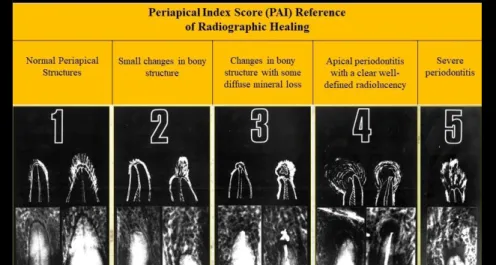
Two board-certified endodontists were calibrated using the periapical index (PAI) kit introduced by Orstavik et al (81). Instructions on grading radiographs are presented in Fig. 2. A series of 100 reference radiographs that was included in the kit were evaluated and graded on an ordinal scale from 1-5. There are 5 reference radiographs correlated with the inflammatory status of periapical tissues confirmed from histology (80). Prior to evaluating the radiographs in the study, examiners had participated in a calibration course for the PAI system. All cases on the 100 reference radiographs were scored twice by assigning one of the five PAI scores corresponding to the visual reference in Fig. 1.
The initial scoring session of the calibration data-set was completed from both examiners and were compared to a "silver standard” that was established by Orstavik (81). Three weeks later, a second reading was accomplished by the examiners that was also compared to the “silver standard” to establish the intra-rater comparison between the first and second readings.
Furthermore, the second readings were used to establish the inter-rater comparison between both examiners. The level of agreement within and between examiners was determined by Cohen's Kappa statistics.
Outcome Measures and Data Analysis:
assess the periapical healing status. Radiographs acquired pre-operatively and immediately post-treatment were evaluated. By the latest follow-up, the PAI was dichotomized as follows:
A. No Lucency B. Lucency
A "no lucency" status corresponds to PAI scores of 1 or 2, which indicates healthy apical tissues. A "lucency" status corresponds to PAI scores of 3, 4, or 5, which indicates diseased apical
tissues. In case of disagreement in scores, examiners re-evaluated the image(s) until a unanimous agreement on the PAI score was reached. Consensus scores on all images were considered "true scores" and used for statistical analysis.
To achieve calibration and assess rater agreement prior to examining experimental radiographs, Cohen's kappa coefficient (k) was used to measure their inter- and intra-rater reliability. Reproducibility was measured based on criteria created by Landis & Koch (84). Strength of agreement are as follows: poor (<0.00), slight (0.00-0.20), fair (0.21-0.40), moderate (0.41-0.60), substantial (0.61-0.80), and almost perfect (0.81-1.00). According to Orstavik et al (81), examiners must obtain a kappa value of 0.61 or higher in order to participate in evaluation of experimental materials.
IV. RESULTS
A total of 242 consecutive cases with necrotic pulp and apical periodontitis in need for root canal therapy were included using long-term calcium hydroxide through a standardized protocol. Of these cases, 23 patients were lost to follow-up, which gives a follow-up rate of 90.5%. 219 patients completed their treatment with follow-up. The distribution of teeth according to type, location, and arch position, are listed in table 2.
The average calcium hydroxide time was 5.4 months with a range of 1 - 43 months. Most cases demonstrated healing around the 3-month mark (Fig.3). The overall mean pre-op and post-op PAI scores were 4.07 (± 0.80) and 1.76 (± 0.70), respectively. At baseline for healed and diseased cases, student's t-test revealed a mean PAI score of 4.04 (± 0.80) and 4.36 (± 0.73), respectively (p = 0.068). The correlation between healing status and baseline PAI score is presented in Table 1.
same. Of these “diseased” cases, eight had to have apical microsurgery, one was extracted and 14 patients elected to not have treatment.
V. DISCUSSION
The objectives in this study were two-fold to: to assess outcomes of pulpless teeth with apical periodontitis using calcium hydroxide as an intracanal medicament for at least 30 days and detect fractures associated with hypothetical mechanical weakening of dentin following calcium hydroxide use. Exclusively, we assessed outcomes of teeth diagnosed with clear signs of apical periodontitis that were medicated with long-term, calcium hydroxide in order to have a better understanding of healing time and outcomes compared to previous studies. One of the drawbacks of this study were the lack of a control, comparing the outcome and tooth fracture predilection on teeth treated without the use of calcium hydroxide. In addition, we did not compare vital or necrotic teeth without apical lesions as they did not meet inclusion criteria to fit the aims of this study. However, the data supports the hypothesis that using calcium hydroxide as a long term, temporary root filling material had no unfavorable effects on endodontic outcomes, such as poor healing status and fracture predilection.
reservoir. Premixed calcium hydroxide in injectable syringe form allows for easier delivery to the apical third. In an aqueous environment, pure calcium hydroxide powder has a low solubility (85). However, studies have shown that premixed pastes tends to wash out over time (11). The rationale for using pure calcium hydroxide in the coronal half is to refresh the Ultracal XS in the apical half to reduce the solubility rate. Ultracal XS contains propylene glycol, a non-aqueous vehicle that enhances delivery of the medicament. The benefits of this ingredient is that it is hygroscopic in nature, which allows absorption of water to sustain release of hydroxyl ions (86). In theory, as the pure calcium hydroxide reservoir comes in contact with the paste layer, this will slowly release hydroxyl ions and reduce solubility of the calcium hydroxide contained in the apical portion. CAVIT™, a temporary material composed of zinc oxide and calcium sulfate dihydrate, was placed over the pulp floor as a protective barrier over the calcium hydroxide. CAVIT™ expands in the presence of moisture to produce an adequate seal, but over time, inadequate thickness of this material can degrade and this may lead to recontamination of the chamber space (87). In this study, permanent restorations were therefore placed over the CAVIT™ layer for temporary reasons to reduce the chances of micro-leakage (25).
majority of our sample size was diseased with a PAI score >3. By the latest follow-up, 90% had a PAI score of <2. To our knowledge, this is the first study to report such a high success rate for teeth diagnosed with pulpal necrosis and apical periodontitis.
Studies have shown that it takes a minimum of three months for pre-existing periapical lesions with a PAI score >3 to demonstrate pronounced healing (88). All participants were re-evaluated between one and three months from the initial visit to assess radiographic healing, monitor symptoms, and clinical signs of apical periodontitis such as sinus tracts, probing defects of endodontic origin and acute apical abscesses. After three months, they were recalled monthly if no signs of healing was observed. The inter-appointment medicament was removed after 30 days from the initial visit, which is the latest time suggested in previous studies (21, 26). The average calcium hydroxide time was 5.4 months. Some cases required more or less time to demonstrate radiographic healing of the periradicular tissues. In this study, healing time is based solely on radiographic assessment, which may be inaccurate and difficult to determine exactly. For example, a case that didn't show signs of healing at the 2-month mark and returned 4-months later may have started to heal at 3-months. In the present study, there’s a high frequency of healing at 3 months with majority of the cases exhibiting complete healing within one year. These results are consistent with Huumonen and Orstavik (88) and Vernieks et al (25).
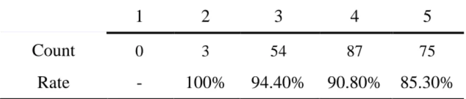
The data demonstrates a correlation between healing status and baseline PAI score (Table 1). As the initial PAI score increases, the healing rate declines by nearly 5%. In spite of this, the highest PAI score yielded an 85% success rate. Factors such as tooth type, arch, and location are listed in table 2. Our inability to demonstrate statistical significance may be explained by our sample size or randomness.
Of the 242 patients, 23 were lost to follow-up. To improve the recall rate, attempts to contact these patient's by phone/letter were documented. These patients were not accounted for, because we could not obtain post-operative radiographs. It is uncertain if these cases healed or not. With a follow-up rate of 90.5% and high sample size, accounting for these patients would have made a negligible difference in the results reported in this study. The remaining 219 participants completed endodontic treatment with an annual follow-up examination.
We determined treatment failure to be solely based on persistent apical lesions. None of the teeth included in this study failed from a fracture. All of the diseased cases had
radiolucencies that remained the same in size. Endodontic surgery or extraction were the
due to the presence of resistant bacteria, such as enterococcus faecalis (16, 63); a gram-positive pathogen found in root filled teeth and non-filled teeth (56-60, 89-91). Although its role in post-treatment disease is unclear (92), studies have found this microbe to be resistant to calcium hydroxide, but susceptible to chlorhexidine (64-66). Chlorhexidine was not used in this study as an adjunctive medication for lesions that did not demonstrate signs of healing.
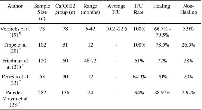
All of the studies assessing the outcomes of teeth with pulpal necrosis and apical
periodontitis are listed in table 3. The success rate of cases with evidence of apical periodontitis ranged from 67 to 88.97% (25-29). Trope et al (26) reported an 81% healing rate of cases treated in 2-visits with calcium hydroxide. Friedman et al (27) assessed 4- to 6-year outcomes of
endodontic treatment of teeth with apical periodontitis and reported a low healing rate of 74%. Penesis et al (28) evaluated the influence of a calcium hydroxide and chlorhexidine gel
combination for the cases that were treated in 2-visits. Although studies have found no added benefit in combining these two medicaments to improve healing (65, 93, 94), they reported a 70% healing rate. A randomized clinical trial conducted by Paredes Vierya et al (29), determined an 88.97% success rate of calcium hydroxide treated teeth. The commonality between all of these reports is a low sample size. None of them showed a statistically significant difference between 1- and 2-visit treatments with the use of calcium hydroxide. This implies that
To our knowledge, there are no clinical studies available to validate the relationship between root fractures and long term exposure of calcium hydroxide. The incidence of fractured teeth from calcium hydroxide use stems from a commonly cited clinical study by Cvek et al (95). In his study, he found the stage of root development to be related to the incidence of fracture more so than the use of calcium hydroxide. On the other hand, there are speculations that
calcium hydroxide exposure alters the mechanical properties of dentin that may predispose teeth to fracture (22, 69, 71). This theory has been controversial and yet, not fully understood. In previous studies, one of the most common mechanical properties evaluated was fracture strength or resistance. Several authors (22, 69, 70) hypothesize that the alkaline pH of calcium hydroxide may disrupt the link between hydroxyapatite crystals and the collagen network by neutralization, dissolution, or denaturation of dentinal collagen, proteoglycans and acid proteins which may ultimately increase fracture susceptibility. In contrast, some authors report no association between fracture strength reduction and calcium hydroxide exposure (11, 96). In addition to fracture resistance, the mechanical properties of dentin can be assessed by measurement of microhardness, modulus of elasticity, and flexural strength. In regards to microhardness, Yoldas et al (97) found calcium hydroxide to soften the dentin to the degree of having influence on physical and chemical properties. As for elastic modulus and flexural strength, Grigaratos et al (98) demonstrated reduction in the flexural strength of dentin when saturated in calcium
hydroxide, whereas the elastic modulus remains unaffected. The influence of any of these properties on clinical outcomes has not been studied.
(101) and Ng et al (102). Moreover, none of the cases demonstrated signs or clear evidence of a fracture during the inter-visit phase or at the one year follow-up. According to a systematic review by Yassen & Platt (99), the data on whether calcium hydroxide exposure for 1 month or shorter has a negative effect on the mechanical properties of radicular dentin is inconsistent, but most studies reported reduction in mechanical properties for exposures longer than 5 weeks. To eliminate bacteria that survived biomechanical instrumentation, calcium hydroxide should be present for at least one week (20). Andreasen et al (22) suggested that calcium hydroxide use should be limited to 30 days and if used for an extended period of time, the root dentin structure is weakened by 50% in one year. Hatibovic-Kofman et al (100) reported that fracture strength reduces in teeth treated with calcium hydroxide between 2 and 12 months. The results from these studies should be interpreted with caution for they are ex-vivo by design and the methodologies fail to accurately reflect clinical scenarios involving human subjects (22, 69-71, 96-98, 100). Without comprehension of the relationship between altered mechanical properties of dentin and clinical outcomes, it is unfair to downplay the usefulness of calcium hydroxide or claim it to be the cause of root fractures. Therefore, the evidence to support the notion of alteration of the root dentin structure from calcium hydroxide exposure is inconclusive.
objective of endodontic therapy and favorable outcomes from this study, the biological effects from calcium hydroxide therapy can lend towards resolution of apical periodontitis.
VI. CONCLUSION
TABLES
Table 1. Healing rates according to PAI score by latest follow-up
1 2 3 4 5
Count 0 3 54 87 75
Rate - 100% 94.40% 90.80% 85.30%
Table 2. Distribution of teeth by tooth type, location, arch position and PAI score at baseline
Group PAI 1 PAI 2 PAI 3 PAI 4 PAI 5 Total
Incisors 0 1 13 26 19 59
Premolars 0 1 12 26 10 49
Molars 0 1 29 35 46 111
Anterior 0 1 13 26 19 59
Posterior 0 2 41 61 56 160
Maxillary 0 2 32 43 18 95
Table 3. Summary of previous studies Author Sample Size (n) Ca(OH)2 group (n) Range (months) Average F/U F/U Rate Healing Non-Healing
Vernieks et al (19) ¥
78 78 6-42 10.2 -22.5 100% 66.7% -
79.5%
3.9% Trope et al
(20) *
102 31 12 - 100% 73.5% 26.5%
Friedman et al (21) *
120 60 48-72 - 51% 72% 28%
Penesis et al (22) *
63 30 12 - 64.9% 70% 20%
Paredes-Vieyra et al
(23)*
282 136 24 - 94% 88.97% 2.94%
F/U: Follow-Up
¥For complete healing between 6-12 months, average of 10.2 months is required; Between 3-42 months, average of 15 months required; Between 6-42 months, average of 22.5 months required.
*No averages reported by authors
Table 4: Case Series Data
CASE # Start Ca(OH)2
F/U Post-op f/u date TRUE PRE-SCORE TRUE POST-SCORE Healed Diseased
1 5/14/2007 9/13/2007 10/15/2007 5 2 1 0
2 4/30/2007 8/22/2007 10/16/2007 5 1 1 0
3 4/19/2005 10/26/2005 1/9/2007 5 2 1 0
4 10/23/2007 10/7/2008 12/9/2009 4 1 1 0
5 6/5/2008 8/29/2008 6/13/2016 4 2 1 0
6 7/12/2005 11/1/2005 1/10/2007 5 2 1 0
7 2/1/2005 11/23/2005 11/23/2005 5 2 1 0
8 2/28/2006 10/23/2006 11/22/2006 5 2 1 0
9 10/26/2004 5/16/2005 9/13/2005 5 1 1 0
11 8/20/2007 11/26/2007 11/26/2007 4 2 1 0
12 12/15/2004 3/15/2005 4/20/2005 4 2 1 0
case #13 excluded
14 3/14/2007 11/27/2007 2/19/2008 3 1 1 0
15 2/16/2005 8/22/2005 8/22/2005 4 1 1 0
16 10/3/2007 1/14/2008 2/25/2008 5 2 1 0
17 3/28/2011 11/23/2011 11/23/2011 5 3 0 1
18 3/29/2005 8/31/2005 9/1/2005 5 2 1 0
19 2/12/2008 5/20/2008 6/24/2009 4 1 1 0
20 3/29/2005 10/3/2005 11/8/2005 5 1 1 0
21 10/29/2007 4/14/2008 4/16/2008 4 2 1 0
22 9/1/2009 3/16/2010 3/22/2010 5 3 0 1
23 6/16/2005 9/19/2005 9/19/2005 3 1 1 0
24 8/8/2006 11/22/2006 9/22/2016 5 1 1 0
Case #25 excluded
26 10/21/2009 2/10/2010 3/2/2010 4 2 1 0
27 10/21/2009 2/10/2010 3/2/2010 4 2 1 0
28 2/17/2005 11/22/2005 11/22/2005 4 1 1 0
Case #29 excluded
30 11/17/2008 3/8/2009 3/10/2009 5 1 1 0
31 10/23/2007 10/7/2008 12/9/2009 5 1 1 0
32 4/14/2005 12/13/2005 12/13/2005 3 1 1 0
33 2/13/2006 6/13/2007 10/1/2007 5 2 1 0
Case #34 excluded
35 9/26/2005 12/21/2005 8/15/2006 5 2 1 0
Case #37 excluded
38 3/24/2009 7/22/2009 5/10/2010 5 3 0 1
39 6/15/2010 10/25/2010 12/1/2010 4 2 1 0
40 5/26/2005 3/6/2006 3/6/2006 5 1 1 0
41 11/23/2005 3/12/2007 3/12/2007 3 1 1 0
42 7/18/2005 6/25/2007 6/25/2007 4 2 1 0
43 9/6/2006 3/14/2007 3/14/2007 4 1 1 0
44 5/14/2009 7/22/2009 9/30/2009 5 1 1 0
45 6/8/2005 2/23/2006 2/23/2006 3 2 1 0
46 3/23/2009 4/25/2009 7/20/2009 4 1 1 0
47 11/25/2009 4/26/2010 4/26/2010 3 2 1 0
48 10/11/2005 4/3/2006 4/25/2006 5 1 1 0
49 11/16/2006 4/4/2007 1/13/2009 5 1 1 0
50 8/7/2006 12/8/2009 9/19/2011 2 1 1 0
51 7/18/2005 3/14/2006 5/10/2006 5 1 1 0
52 1/4/2007 4/24/2007 4/24/2007 4 2 1 0
53 9/17/2009 12/21/2009 1/12/2010 5 2 1 0
54 11/29/2006 5/15/2007 5/15/2007 4 2 1 0
55 1/28/2009 9/16/2009 9/23/2009 5 2 1 0
56 4/26/2005 4/7/2008 4/7/2008 4 1 1 0
57 4/4/2007 9/12/2007 10/15/2007 5 1 1 0
58 6/3/2009 9/1/2009 9/23/2009 4 2 1 0
59 1/29/2008 4/12/2010 4/26/2010 4 2 1 0
Case #60 excluded
61 12/6/2006 5/15/2007 5/28/2007 3 1 1 0
62 6/11/2008 4/29/2009 4/26/2010 4 1 1 0
63 10/11/2005 4/3/2006 4/25/2006 5 1 1 0
65 2/21/2006 5/29/2006 5/29/2006 4 1 1 0
66 1/28/2009 4/29/2009 4/29/2009 4 2 1 0
67 4/11/2006 10/11/2006 11/14/2006 5 2 1 0
68 12/11/2006 7/3/2007 7/4/2007 3 1 1 0
69 11/25/2009 3/22/2010 4/4/2012 4 2 1 0
70 3/19/2007 7/18/2007 7/26/2007 5 2 1 0
71 7/12/2006 10/24/2006 12/6/2006 4 2 1 0
72 5/6/2008 8/11/2008 9/18/2008 4 1 1 0
73 5/11/2005 10/12/2005 1/10/2016 3 1 1 0
74 7/17/2007 10/9/2007 10/9/2007 5 3 0 1
75 2/10/2010 7/12/2010 8/31/2010 4 2 1 0
76 8/24/2010 5/3/2011 5/3/2011 3 2 1 0
77 8/24/2010 5/3/2011 5/3/2011 3 1 1 0
78 8/15/2011 12/5/2011 12/5/2011 4 2 1 0
79 4/4/2011 9/6/2011 9/6/2011 3 3 0 1
80 11/2/2009 12/9/2009 6/21/2010 3 1 1 0
81 7/10/2006 8/7/2006 1/30/2008 4 2 1 0
82 9/14/2010 11/23/2010 11/24/2010 4 2 1 0
83 1/5/2009 4/6/2009 4/7/2009 3 1 1 0
84 10/22/2007 1/8/2008 1/9/2008 4 3 0 1
85 5/2/2007 8/15/2007 8/20/2007 3 2 1 0
86 7/13/2005 4/19/2006 6/28/2006 5 2 1 0
SX 87
7/13/2011 11/28/2011 11/29/2011
4 4 0 1
88 6/10/2008 9/2/2008 11/3/2008 4 1 1 0
89 10/19/2011 2/13/2012 2/13/2012 4 2 1 0
90 5/21/2008 10/2/2008 10/2/2008 4 2 1 0
91 5/28/2008 8/26/2008 8/26/2008 4 2 1 0
93 10/26/2011 2/14/2013 2/14/2013 4 2 1 0
94 1/11/2011 4/4/2011 5/16/2011 4 2 1 0
95 10/14/2010 12/13/2010 12/13/2010 4 4 0 1
96 2/16/2010 8/16/2010 11/23/2010 4 2 1 0
97 3/31/2010 9/1/2010 9/20/2016 5 1 1 0
98 3/29/2011 12/13/2011 12/13/2011 5 2 1 0
99 6/22/2009 10/8/2009 10/8/2009 4 2 1 0
SX 100
10/20/2010 12/20/2010 12/20/2010
4 4 0 1
101 12/21/2009 5/24/2010 5/25/2010 5 1 1 0
102 4/28/2010 7/14/2010 9/7/2010 3 1 1 0
103 4/1/2009 7/7/2009 7/7/2009 5 2 1 0
104 4/1/2009 7/7/2009 7/7/2009 5 2 1 0
105 8/25/2010 11/22/2010 11/22/2010 2 1 1 0
106 8/15/2006 11/29/2006 12/5/2006 5 2 1 0
107 11/28/2006 1/2/2007 1/2/2007 4 2 1 0
108 5/31/2006 6/27/2006 10/25/2006 5 1 1 0
109 5/1/2007 9/24/2007 10/30/2007 4 2 1 0
110 2/1/2010 7/14/2010 9/20/2010 3 1 1 0
111 6/26/2007 11/13/2007 12/18/2007 5 2 1 0
112 2/24/2011 5/30/2011 5/30/2011 4 2 1 0
113 6/22/2009 11/2/2009 11/2/2009 3 1 1 0
114 7/7/2008 8/27/2009 8/27/2009 5 2 1 0
115 10/30/2006 2/19/2007 5/5/2008 5 2 1 0
116 6/26/2007 10/1/2007 7/15/2009 3 1 1 0
117 10/16/2011 11/4/2011 12/7/2011 5 1 1 0
118 10/20/2010 8/16/2011 8/16/2011 3 2 1 0
120 5/6/2008 6/25/2008 9/24/2008 4 2 1 0
121 6/8/2011 8/24/2011 8/24/2011 3 1 1 0
122 9/8/2009 3/1/2010 3/1/2010 4 2 1 0
123 6/9/2010 8/24/2010 8/24/2010 4 1 1 0
Case # 124 excluded
125 10/26/2010 3/15/2011 3/15/2011 5 2 1 0
126 5/3/2010 8/30/2010 9/16/2010 3 1 1 0
127 9/12/2006 12/20/2006 12/20/2006 4 2 1 0
128 11/25/2009 4/26/2010 4/26/2010 2 2 1 0
129 11/19/2007 2/12/2008 2/12/2008 4 2 1 0
130 7/14/2010 2/15/2011 2/15/2011 3 1 1 0
131 5/5/2010 7/22/2010 7/22/2010 3 3 0 1
132 5/5/2010 9/8/2010 10/27/2010 5 2 1 0
133 12/6/2005 8/14/2006 8/14/2006 5 2 1 0
134 3/31/2010 7/6/2010 7/6/2010 3 1 1 0
135 1/6/2010 4/27/2010 4/27/2010 4 1 1 0
136 5/31/2011 10/17/2011 10/17/2011 3 2 1 0
137 10/9/2007 1/21/2008 2/4/2008 3 1 1 0
138 1/4/2010 2/1/2010 2/1/2010 4 2 1 0
139 5/28/2007 8/24/2010 9/2/2010 5 2 1 0
140 11/29/2006 3/12/2007 3/12/2007 5 2 1 0
141 11/1/2005 5/9/2006 5/29/2006 5 2 1 0
142 3/28/2007 7/10/2007 7/11/2007 5 2 1 0
143 12/8/2010 6/21/2011 6/21/2011 4 2 1 0
144 3/30/2010 11/19/2013 11/22/2013 5 1 1 0
145 3/30/2010 11/19/2013 11/22/2013 5 2 1 0
146 3/27/2007 6/27/2007 6/27/2007 3 2 1 0
148 6/20/2010 2/16/2011 2/16/2011 3 1 1 0
149 9/12/2011 1/31/2012 7/3/2012 4 2 1 0
150 4/30/2007 8/22/2007 10/16/2007 4 1 1 0
151 11/4/2009 3/17/2010 3/17/2010 3 1 1 0
152 5/22/2007 9/3/2008 10/9/2008 4 1 1 0
153 10/5/2011 12/13/2011 12/13/2011 4 2 1 0
154 11/8/2006 2/13/2007 5/8/2007 5 2 1 0
155 5/19/2010 10/14/2010 10/14/2010 4 2 1 0
156 7/4/2008 1/14/2009 1/14/2009 5 3 0 1
157 9/15/2010 4/26/2011 4/26/2011 4 1 1 0
158 6/8/2010 5/3/2011 7/5/2011 4 2 1 0
159 6/8/2010 10/4/2011 10/4/2011 3 1 1 0
160 2/6/2007 5/21/2007 5/21/2007 5 2 1 0
161 7/16/2007 2/19/2008 1/18/2010 5 2 1 0
162 11/7/2006 4/24/2007 4/24/2007 3 2 1 0
163 2/6/2006 5/17/2006 6/29/2006 4 2 1 0
164 8/15/2011 10/17/2011 10/17/2011 3 2 1 0
165 3/16/2010 4/5/2011 4/5/2011 5 2 1 0
166 12/14/2011 2/15/2012 2/15/2012 4 3 0 1
167 4/27/2010 9/1/2010 10/12/2010 3 1 1 0
168 2/8/2010 5/18/2010 5/18/2010 3 2 1 0
169 5/8/2007 8/13/2007 8/13/2007 4 2 1 0
170 7/12/2006 1/23/2007 3/12/2007 4 2 1 0
SX 171
1/10/2010 2/18/2010 2/14/2011
5 3 0 1
172 6/15/2010 8/17/2010 8/17/2010 3 2 1 0
173 8/25/2011 12/19/2011 3/25/2013 3 2 1 0
174 3/20/2007 8/14/2007 8/14/2007 3 2 1 0
176 2/23/2010 5/5/2010 5/5/2010 3 2 1 0
177 10/25/2010 5/30/2011 5/30/2011 4 2 1 0
178 1/12/2010 4/19/2010 5/3/2010 4 2 1 0
179 6/21/2010 9/8/2010 9/8/2010 5 3 0 1
180 8/18/2011 1/16/2012 2/8/2012 3 2 1 0
SX 181
1/9/2006 10/26/2006 1/9/2008
5 4 0 1
182 1/19/2010 4/27/2010 12/12/2012 4 1 1 0
183 11/15/2010 7/20/2011 10/1/2012 5 2 1 0
184 9/21/2011 11/7/2011 11/7/2011 4 2 1 0
185 8/27/2007 10/17/2007 10/17/2007 3 2 1 0
186 9/1/2009 3/17/2010 3/17/2010 3 2 1 0
187 10/14/2009 12/17/2009 12/17/2009 5 1 1 0
188 7/12/2010 10/19/2010 12/14/2011 4 2 1 0
189 6/18/2008 9/2/2008 9/2/2008 4 2 1 0
190 7/7/2009 10/20/2009 11/18/2009 3 2 1 0
191 6/14/2005 9/19/2005 10/17/2005 5 2 1 0
192 6/7/2009 9/9/2009 9/23/2009 4 3 0 1
193 10/21/2009 2/23/2010 3/2/2010 4 1 1 0
194 4/27/2011 8/24/2011 8/24/2011 3 1 1 0
SX 195
9/8/2009 1/19/2010 1/19/2010
5 4 0 1
196 2/23/2010 5/10/2010 5/10/2010 5 2 1 0
197 6/8/2011 11/17/2011 5/6/2013 5 1 1 0
SX 198
6/30/2010 1/4/2011 1/4/2011
5 4 0 1
199 5/9/2011 8/22/2011 11/2/2011 4 1 1 0
200 2/23/2009 5/2/2009 5/2/2009 3 2 1 0
201 1/19/2010 5/5/2010 3/9/2011 5 1 1 0
203 5/11/2006 6/1/2006 7/24/2007 3 2 1 0
204 5/10/2010 6/14/2010 6/14/2010 4 2 1 0
205 3/11/2008 10/14/2008 10/14/2008 5 3 0 1
206 6/3/2009 9/9/2009 9/9/2009 4 1 1 0
207 6/14/2010 12/1/2010 11/3/2011 4 1 1 0
208 1/21/2008 1/12/2009 1/12/2009 5 2 1 0
SX 209
8/26/2009 12/1/2009 11/15/2010
4 3 0 1
210 7/13/2010 3/8/2011 5/14/2012 3 1 1 0
211 3/4/2009 5/19/2009 5/19/2009 3 2 1 0
212 9/8/2010 12/8/2010 12/8/2010 4 2 1 0
213 6/8/2004 9/14/2004 9/14/2004 4 3 0 1
214 5/13/2008 9/22/2008 9/22/2008 4 2 1 0
215 2/17/2010 7/7/2010 7/25/2012 4 1 1 0
216 6/29/2011 9/19/2011 9/19/2011 5 2 1 0
217 9/14/2011 6/11/2012 6/11/2012 3 1 1 0
218 12/1/2010 8/15/2011 8/15/2011 4 2 1 0
219 5/11/2011 9/6/2011 9/6/2011 3 1 1 0
SX 220
9/8/2009 4/6/2010 4/6/2010
3 3 0 1
221 6/1/2011 10/18/2011 10/20/2011 4 1 1 0
222 2/27/2006 9/25/2006 10/16/2006 4 1 1 0
223 2/15/2010 4/12/2010 4/12/2010 5 2 1 0
224 11/20/2007 3/31/2008 6/8/2016 4 1 1 0
225 1/12/2011 6/6/2011 7/6/2011 4 2 1 0
226 2/1/2005 11/23/2005 11/23/2005 5 2 1 0
Legend
Ca(OH)2: Calcium Hydroxide F/U: Follow-up
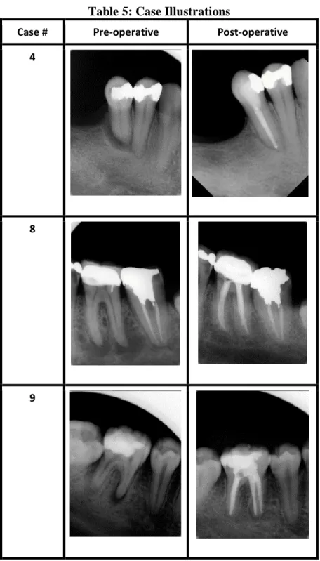
Table 5: Case Illustrations
Case # Pre-operative Post-operative
4
8
24
28
30
44
48
51
64
67
97
115
144
201
FIGURES
Fig. 1. Periapical Index (PAI) Score Reference by Dag Orstavik (6) describing the radiographic healing status of the periapical tissues correlated to histology.
Fig. 4. Periradicular Healing Demonstration- From top to bottom: Pre-operative radiograph with baseline periapical index (PAI) score, Follow-up radiograph with calcium hydroxide, and Post-operative periapical radiograph with PAI score. Left: Case #144; Middle: Case #51; Right
REFERENCES
1. Kakehashi S, Stanley H, Fitzgerald R. The effect of surgical exposures of dental pulps in germ-free and conventional laboratory rats. Oral Surg Oral Med Oral Pathol, 1965 Sep; 20(3): 340–349.
2. Strindberg L. The dependence of the results of pulp therapy on certain factors. An
analytical study based on radiographic and clinical follow-up examinations. Acta Odontol Scand 1956; 14: Supplement 21.
3. Seltzer S, Bender IB, Turkenkopf S. Factors affecting successful repair after root canal therapy. J Am Dent Assoc 1963 Nov; 67(5): 651–662.
4. Kerekes K, Tronstad L. Long-term results of endodontic treatment performed with a standardized technique. J Endod 1979 Mar; 5(3): 83–89.
5. Sjögren U, Hagglund B, Sundqvist G, Wing K. Factors affecting the long-term results of endodontic treatment. J Endod 1990 Oct; 16(10): 498–504.
6. Figini L, Lodi G, Gorni F, Gagliani M. Single versus multiple visits for endodontic treatment of permanent teeth: a Cochrane systematic review. J Endod. 2008 Sep; 34(9):1041-1047.
7. Mohammadi Z, and Dummer PMH. Properties and Applications of Calcium Hydroxide in
Endodontics and Dental Traumatology. Int Endod J 2011 May; 55: 697–730.
8. Farhad A, Mohammadi Z. Calcium hydroxide: a review. Int Dent J 2005 Oct; 55(5): 293-330.
9. Hermann BW. Calcium hydroxid als Mittelzurn, Behandeln und Fullen von Wurzelkanalen [Thesis] Wurzburg 1920.
10. Rivera EM, Williams K. Placement of Calcium Hydroxide in Simulated Canals:
11. Hawkins JJ. Effect of Three Calcium Hydroxide Formulations on Fracture Resistance of Dentin over Time. Dent Traumatol 2015 Apr; 31(5): 380–384.
12. Athanassiadis B, Abbott PV, Walsh SJ. The Use of Calcium Hydroxide, Antibiotics and
Biocides as Antimicrobial Medicaments in Endodontics. Austr Dent J 2007 Mar; 52: 64-82.
13. Khan, AA, Sun, X, Hargreaves KM. Effect of Calcium Hydroxide on Proinflammatory
Cytokines and Neuropeptides. J Endod 2008; 34:1360–1363.
14. Safavi KE, Nichols FC. Alteration of biological properties of bacterial lipopolysaccharide
by calcium hydroxide treatment. J Endod 1994 Mar; 20 (3):127–129.
15. Freeman K, Lundington JR, Svec T, Pinero GJ, Hoover J. Continuously Infused Calcium
Hydroxide: Its Influence on Hard Tissue Repair. J Endod 1994 Jun; 20 (6): 272–275.
16. Evans M, Davies JK, Sundqvist G, Figdor, D. Mechanisms involved in the resistance of Enterococcus faecalis to calcium hydroxide. Int Endod J 2002 Mar; 35: 221-228.
17. Walton RE, Holton IF, Michelich R. Calcium Hydroxide as an Intracanal Medication: Effect on Posttreatment Pain, J Endod 2003 Oct; 29 (10):627-629.
18. Heithersay, GS. Periapical Repair Following Conservative Endodontic Therapy. Austr Dent J 1970 Dec; 15 (6): 511–518.
19. Tronstad L. Root resorption-etiology terminology and clinical manifestations. Endod Dent Traumatol 1988 Dec; 4(6):241-252.
20. Sjögren U., Figdor D, Spångberg L, Sundqvist G. The antimicrobial effect of calcium
hydroxide as a short-term intracanal dressing. Int Endod J 1991 May; 24(3):119–125.
21. Bystrom A, Claesson R, Sundqvist G. The antibacterial effect of camphorated
22. Andreasen JO, Farik B, Munksgaard EC. Long-term calcium hydroxide as a root canal dressing may increase risk of root fracture. Dent Traumatol 2002 Jun; 18(3):134–137.
23. Grigoratos D, Knowles J, Ng YL, Gulabivala K. Effect of exposing dentine to sodium hypochlorite and calcium hydroxide on its flexural strength and elastic modulus. Int
Endod J 2001 Mar; 34(2):113–119.
24. Yoldaş O, Doğan C, Seydaoğlu G. The effect of two different calcium hydroxide combinations on root dentine microhardness. Int Endod J 2004 Dec; 37(12), 828–31.
25. Vernieks AA, Messer LB. Calcium hydroxide induced healing of periapical lesions: a
study of 78 non-vital teeth. Int Endod J 1978 Jul; 11(2):61–69.
26. Trope M, Delano EO, Ørstavik D. Endodontic treatment of teeth with apical periodontitis: single vs. multivisit treatment. J Endod 1999 May; 25(5):345–350.
27. Friedman S, Abitbol S, Lawrence H. Treatment Outcome in Endodontics: The Toronto
Study. Phase 1: Initial Treatment. J Endod 2003 Dec; 29(12):787–793.
28. Penesis VA, Fitzgerald PI, Fayad MI, Wenckus CS, Begole EA, Johnson BR. Outcome
of One-visit and Two-visit Endodontic Treatment of Necrotic Teeth with Apical
Periodontitis: A Randomized Controlled Trial with One-year Evaluation. J Endod 2008 Mar; 34(3): 251–257.
29. Paredes-Vieyra J, Enriquez FJJ. Success Rate of Single- versus Two-visit Root Canal
Treatment of Teeth with Apical Periodontitis: A Randomized Controlled Trial. J Endod 2012 Sep; 38(9):1164–1169.
30. Alliet P, Vande Voorde H (1988) Le roÃ1e de l'hydroxyde de calcium en Endodontie. Revue Belge de Medicine Dentaire 43, 24-39.

![(E,E) 1 [2 (4 Nitrophenyl)ethenyl] 4 [2 (2,4,6 trimethoxyphenyl)ethenyl]benzene](data:image/gif;base64,R0lGODlhAQABAIAAAP///wAAACH5BAEAAAAALAAAAAABAAEAAAICRAEAOw==)