
The structure and life history of some minute prosobranchs of rock pools: Skeneopsis planorbis (Frabicius), Omalogyra atomus (Philippi), Rissoella diaphana (Alder) and Rissoella opalina (Jefffreys)
38
0
0
Full text
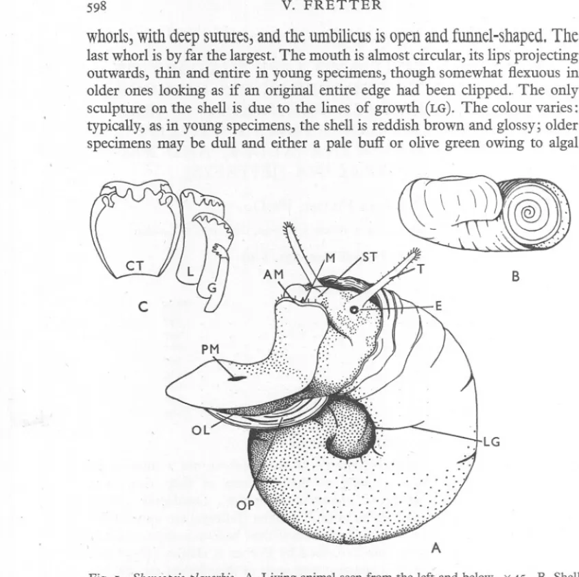
(2) V. FRETTER. 598. whorls, with deep sutures, and the umbilicus is open and fW1llel-shaped. The last whorl is by far the largest. The mouth is almost circular, its lips projecting outwards, thin and entire in young specimens, though somewhat flexuous in older ones looking as if an original entire edge had been clipped.. The only sculpture on the shell is due to the lines of growth (LG). The colour varies: typically, as in young specimens, the shell is reddish brown and glossy; older specimens may be dull and either a pale buff or olive green owing to algal. B. c. ... LG. A Fig. I. Skeneopsisplanorbis. A. Living animal seen from the left and below. x45. B. Shell from the side. x 30. C. Half row of radula teeth. x 230. AM,opening of anterior pedal gland; CT,central tooth of radula; E, eye; G,marginal tooth of radula; L, lateral tooth of radula; LG,puckered lines of growth; M,mouth; OL,opercular lobe offoot; OP,operculum; . PM,opening of posterior pedal gland; ST,snout; T, tentacle.. . growth. As the mollusc grows to maturity algal spores gain a holdfast on the surface of the shell. These germinate and the small plants become embedded in a thick layer of mucus which they themselves secrete, and the layer may be added to by diatoms which are scattered in large nmpbers amongst the algal filaments. Thus in older specimens the shell is completely encased in a mucous covering from which arises a thick woolly coat of algal filaments. When Skeneopsis crawls over weed or rock the shell is not carried erect but tilted sideways and may be rocked from side to side. The exposed parts of the.
(3) MINUTE. PROSOBRANCHS. OF ROCK. POOLS. 599. body have a ground colour of greyish white. On the metapodial (opercular) lobes (Fig. I and PI. IV, fig. I, OL) there is a mottling of deep grey or black, and patches of yellow occur on the body, all due to epithelial pigment granules. Two yellow patches lie between the eyes, each over a similarly pigmented region of the buccal cavity, with another of smaller size behind each eye,. and a streak underlies the operculum. The two dorsal patches which lie close together are contiguous with a black streak extending for some distance beneath the shell. The head bears a pair of long cylindrical and widely diverging tentacles which may be waved slowly in the water as the mollusc creeps. Each is scantily covered with motionless cilia (Fig. I, T), and the eye is situated on the outer side of the swollen base (E). The snout (ST)is broad with a mid-dorsal groove dividiD.git into two rounded lobes, between which lies ventrally the mouth (M). The foot is relatively short, truncated in front and tapering along the posterior third to a blunt point. Around the anterior edge a deep groove marks the opening of the anterior pedal mucous gland (AM), and a row of stiff cilia arises from the upper lip of the groove. The anterior pedal gland comprises three groups of mucous and mucoid cells set in a transverse row in the anterior tissues of the foot (PI. IV,fig. I, MG);the middle group is the most extensive, and the whole appears as a semicircular opaque white mass in the living animal. Gland cells of a similar nature are embedded in the tissues of the sole (GD)and open singly between the cells of the ciliated columnar epithelium. Yet another and still greater supply of mucus comes from a posterior pedal gland which opens as a mid-ventral longitudinal slit' about a third of the pedal length from the posterior end (Fig. I, PM). The opening leads dorsally into a ciliated duct (PI. IV, fig. I, D),which soon bifurcates into left and right branches, and each becomes surrounded by glandular tissue which extends into the haemocoel. On each side, at about the level of the pedal ganglia, this tissue then splits into two lobes, pne passing dorsally (AL) and the other running back alongside the oesophagus and immediately above the columellar muscle (PL). The animal is fond of crawling on the surface film of water and may suspend itself from this by means of a viscid thread issuing from the posterior pedal gland. The free end of the thread floats on the surface, and, as the animal slowly descends, it plays out the mucous rope: from the opening of the gland the secretion is directed into a longitudinal groove along the sole, where, in contact with the sea water, it hardens before leaving the posterior end of the foot. In such a manner Skeneopsis may finally reach the bottom of a shallow pool, or, if the thread be short, the mollusc may even reverse its course and climb along it to reach the surface again. The mantle cavity extends along the whole length of the body whorl. On the left side is the well-developed, bipectinate osphradium, and dorsal to this the monopectinate gill, which has only nine finger-like filaments (PI. IV, fig. I, F) projecting downwards from a somewhat elongated axis. The hypobranchial 39-".
(4) 600,. V. FRETTER. gland runs along the right wail of the cavity anteriorly. It passes back parallel to, and just above, the rectum as a single layer of mucous and mucoid cells, hut near the level of the posterior end of the ctenidium the gland thickens, occupies a more dorsal position and the mucus is replaced by another type of protein. Posteriorly, the mantle cavitY is much reduced in size, and, covered by the hypobranchial gland, pushes back over the kidney (K), which therefore opens on its floor. In the female the large glandular genital duct projects from the right wall of the mantle and partly obliterates the cavity on this side, the rectum running along its median side to open on a prominent papilla anterior to the genital opening. In the male the genital duct is far less conspicuous, and the anal papilla smaller. The penis (PE), which arises behind the right tentacle, is slightly compressed laterally and when fully developed extends the whole length of the mantle cavity. The Alimentary Canal The mouth leads dorsally into a short though very dilatable tube, a ventral extension of the buccal cavity which bears a cuticle, and this also spreads on to the floor of the main part of the cavity in which the odontophore lies. At the inner end of the buccal tube the cuticle is thickened to form two jaws laterally placed, each consisting of six transverse rows of teeth with about twenty in each row (PI. IV, fig. I, J); each tooth is secreted by a single epithelial cell. When the animal is feeding, the radula (R) works over the surface of the weed or other material to rasp off diatoms and algal cells, the jaws meanwhile gripping the substratuni. The radula (Fig. I C) is made up of numerous EXPLANATION OF PLATE IV Fig. I. Skeneopsis planorbis. Right lateral view of whole animal seen as a transparent object. x 80. A, anus; AH, anterior region of stomach; AL, anterior lobe of posterior pedal gland; AM,opening of anterior pedal gland; BU,buccal ganglion; c, columellar muscle; CD,ciliated duct leading from vas deferens to mantle cavity; CE,cerebral ganglion; D, duct of posterior pedal gland; DI, digestive gland; DP, penial duct; E, eye; EX,spherules in excretory cells of digestive gland; F, gill filament; GA,parapedal ganglion; GD, mucous glands opening on sole of foot; H, heart; T,intestine; J, jaw; K, kidney; L, opening of digestive gland into stomach; LI, left salivary gland; MA, mantle; MG, anterior pedal gland; OE, oesophagus; OL, opercular lobe of foot; OP, operculum; P, prostate; PE, penis; PG, pedal ganglion; PH, posterior region of stomach; PL, posterior lobe of posterior pedal gland; PR, pleural ganglion; R, radula; RA, radular sac; RI, right salivary gland; ST, snout; T, tentacle; TE, testis; TO, statocyst; v, vesicula seminalis. Fig. 2. Omalogyra atomus. Right lateral view of whole animal seen as a transparent object. x 120. AB, albumen gland; AG, anterior lobe of digestive gland; B, buccal cavity; BC, bursa copulatrix; CG, capsule gland; DA, duct of albumen gland; FC, fertilization chamber; GA, genital aperture; HD, hermaphrodite duct; HL, head lobe; KM, large glands opening into mantle cavity near anus; MG,anterior pedal gland; MU, mucous gland; MT, muscular tube; 0, ovarian duct; OD, muscles of odontophore; OM, opening of albumen gland into mucous gland; OPE,opening of sperm sac into prostate; OV,egg in ovary; PD, pallial vas deferens; PM, opening of posterior pedal gland; po, pallial oviduct; SP, sperm sac; ST, stomach. Other letters as in fig. I above..
(5) °z °jJH. or 'jJH. AI ELLV'ld. IIAXX. '~ossV ''Tors: 'avw. 'Naflo[.
(6) JOURN. MAR. BlOL. Assoc.. PLATE. XXVII. F. IV. DP. v. PH. L. Fig. 1. OPE. KM. 0. L. Fig. 2..
(7) MINUTE. PROSOBRANCHS. OF ROCK. POOLS. 601. transverse rows of sharply cusped teeth, each row comprising a large central tooth (CT) bordered on each side by one lateral (L) and two marginals (G). Along the mid-dorsal wall of the buccal cavity is a deep ciliated channel along which the food is directed to the oesophagus. The salivary glands (PI. IV, fig. I, RI and u) open at the extreme anterior end of this channel, one on either side. Their secretion lubricates the action of the radula and entangles the food particles which are drawn in by it. The glands rtin parallel with the oesophagus as far as the nerve ring, the one on the left side being slightly longer than that on the right. Mucous cells provide the greater part of the saliva. There is, however, a second type of secreting cell filled with protein spherules which swell and dissolve when expelled, probably liberating an enzyme. The oesophagus (OE)is a straight, narrow, ciliated tube of about the same diameter throughout. From its origin at the posterior end of the buccal cavity it is directed ventrally and slightly to the left of the median line, and traverses the length of the body whorl. There are no glandular pouches along its course, as are common amongst herbivorous prosobranchs, and the only secreting cells are epithelial mucous glands. The stomach is spacious and lies at the posterior end of the body whorl, at right angles to which it is elongated. It is divided by a transverse constriction into two regions: an anterior (AH), which is dome-shaped and lined by a columnar ciliated epithelium, the cilia being thick and closely set; and a more posterior one (PH), the wall of which is, in the main, cuticularized. The upper, anterior, chamber corresponds to the style sac of style-bearing prosobranchs and the lower, posterior, chamber to the part containing the gastric shield. The anterior wall of the posterior chamber is ciliated where it receives the opening of the oesophagus at the level of the transverse constriction. At the extreme posterior end is the opening of the single lobe of the digestive gland (L). The stomach, especially its anterior compartment, is conspicuous in the living animal since there is a heavy deposit of black spherules in the: epithelial cells. The intestine (I) arises from the dorsal wall of the anterior chamber and bends abruptly to pass forward on the right side. For a short distance along its initial length can be traced a longitudinal groove bordered by a fold of epithelium. This gutter, with its accompanying fold, arises on the wall of the duct from the digestive gland, passes forwards to the anterior chamber of the stomach and so reaches the intestine. It affords a path by which waste from the duct may be conveyed to the intestine. On histological grounds the intestine may be divided into four regions. The initial part, in which the columnar ciliated cells are heavily pigmented with black spherules, and along which the channel from the digestive gland can be traced, passes to a slightly wider and more muscular tube in which mucous cells alternate with ciliated cells. Here the faecal pellets are moulded: the particulate waste from the stomach, which consists of diatom cases, algal cells and detritus, is agglutinated by means of mucus and com\Jacted into oval rods..
(8) 602. V. FRETTER. These are still further elaboratedin the third sectionof the intestine,the longest of the four, which takes a somewhat sinuous course as it accompanies the genital duct along the right wall of the mantle cavity. In this section the mucous cells are replaced by another type of gland which is scattered somewhat sparingly amongst the ciliated cells-it is conical in shape, with a broad base, and tapering distally, and towards the base is a large round nucleus, with a prominent nucleolus. The cytoplasm is dense and contains small secretion spherules which swell in the lumen of the gut and presumably help to harden the outer rind of the pellet. In the terminal or rectal region the cilia are longer than elsewhere and mucous cells are scattered in the epithelium, increasing in number towards the anus. The digestive gland (m), together with the gonad (TE), constitute the visceral mass which occupies the smaller coils of the shell. The gonad, frequently the smaller of the two glands, lies on the right side. The digestive gland has a segmented appearance, since it is made up of a series of lobes which open into one another and increase in size towards the stomach, into which they ultimately open. The epithelium of the gland consists of three types of cells: the most frequent, the digestive cell, is narrow at the base and gradually broadens towards a rounded distal end where there is a dense layer of protoplasm containing innumerable small spherules. These have been seen to be discharged and are probably enzymatic. The rest of the cytoplasm, except around the nucleus, is filled with vacuoles which vary in size, and contain during life either a clear refringent liquid or agglomerations of fine particulate matter .sometimes embedded in a mucoid substance. No diatoms, algal cells or- any such particulate food has ever been found within the lumen of the digestive tubules. The stomach, however, may often be seen filled with such objects, and since only empty cases of diatoms and the walls of broken cells are to be found in the intestine, it may be assumed that the bulk of the digestive process occurs in the stomach and that only the resulting solution, with perhaps minute particles, enters the liver, and is taken up by the digestive cells for further treatment. The two other types of cell, neither of which attains the height of that already described, occur in the crypts of the tubules. In one, which appears to be excretory in function, the protoplasm may be highly vacuolated and an irregular clump of brown or black granules be contained in each vacuole. A more frequent appearance is when the cell contains only one large vacuole filled with a single spherical mass of such granules. These can be seen through the shell of a living animal (EX).They have never been found in the lumen of the gland. Somewhat similar cells of undoubted excretory function are described in the digestive gland of tectibranchs (Fretter, 1939), and it is probable that, as in that group, waste matter is absorbed from the blood through the broad bases of the cells. This, however, has not been tested owing to the difficulty of injecting such small molluscs..
(9) MINUTE. PROSOBRANCHS. OF ROCK. POOLS. 603. The third type of cell may be grouped in small numbers around the excretory cell, though sometimes it occurs alone in larger numbers. In longitudinal section it is triangular in outline with a broad base resting on the basement membrane and a tapering distal end which hardly reaches the surface of the epithelium. The spherical nucleus lies towards the base and has a large nucleolus. The protoplasm stains deeply with iron haematoxylin and is vacuolated. Each vacuole contains a colourless refringent spherule which gives a positive reaction for lime. The probable function of these cells is either to act as a storage of lime for shell formation, or for the purpose of controlling the reaction of the secretion from the digestive gland. The Reproductive System The male. The testis (Pi. IV, fig. I, TE),situated on the right side of the visceral mass, consists of a linear series of lobes which open broadly into one another. and lead finally to the gonadial duct. This acts as a vesicula seminalis (v) which is dilated with sperm from the onset of sexual maturity. It coils on the right side of the stomach and then leads into a short narrow tube with a straight course, conducting the sperm from the vesicula seminalis to the prostate (p). This tube, the renal vas deferens, is surrounded by a layer .of circular muscles which closes the lumen except when spermatozoa are actually passing through it during copulation. It opens into the prostate gland on a small papilla. Alongside this is a ciliated duct (CD) which leaves the prostate and runs dorsally to open into the mantle cavity. Such an outlet to the mantle cavity has been described in certain Stenoglossa and in Lamellaria, and provides an escape for unwanted sperm (Frettet, 1941). For the rest of its course the male duct runs straight along the right side of the mantle cavity to the penis (PE)-this section comprises the pallial vas deferens and along the posterior half the epithelium is tall and gl~dular and constitutes the prostate (p). The gland cells, which rest upon a basement membrane, are broad ,at the base and each tapers slightly towards the distal end. Wedged between the distal ends are ciliated cells which drive the prostatic secretion forward along the duct. The prostate is surrounded by a thin layer of connective tissue in which both circular and longitudinal muscle fibres occur. Anteriorly it ends abruptly, the duct narrows and is lined only by a ciliated columnar epithelium. The musculature, however, is greatly increased? for the circular muscle forms a layer which in thickness exceeds twice the height of the epithelial cells. Here, and along the penial duct, the histology of which is similar, the spermatozoa are conducted by peristaltic action. The penis is somewhat compressed laterally and ~apers to a point, the duct through it (DP) running near its ventral surface to open at the tip. Externally it is covered by a slightly cuticularized epithelium, except along the narrow dorsal and ventral walls where longitudinal strips of epithelial mucous glands occur, the secretion from which assists the passage of the pep.is through the. ,.
(10) \. 604. pallial. v. FRETTER. grooveof the female to reach the opening of the receptaculum seminis. (Fig. 2, RE). Beneath the epithelium is a layer of circular and longitudinal muscle fibres,. and through the thickness of the organ oblique and dorsoventral fibres separate numerous blood spaces. These, when gorged with blood during copulation, so enlarge the penis that if the copulating partners be separated it is only after some minutes that the penis can regain its position of rest along the length of the mantle cavity. . The female. The ovary, on the right side of the visceral mass, occupies a position similar to that of the testis and extends as far as the posterior chamber of the stomach. From here, a narrow ovarian duct (Fig. 2, 0) passes towards the posterior end of the mantle cavity where it opens into the fertilization chamber (FC),placed at the posterior end of the pallial oviduct, and receiving a duct from the receptaculum seminis (DR), and another from the albumen gland (DA). Its walls are ciliated and muscular.. Fig. 2. ,Skeneopsis planorbis. Diagrammatic reconstruction of female genital duct. x 100. AB,albumen gland; CG,capsule gland; CH,channel along outer surface of mantle; DA,duct of albumen gland; DR, duct from receptaculum semip.is; ET, entrance to receptaculum seminis; FC,fertilization chamber; GA, genital aperture; MU, mucous gland; 0, ovarian duct; PO, lateral pouch of capsule gland; RE,receptaculumseminis; u, muscular sac.. The albumen gland is of considerable size (AB),and completely surrounds the fertilizationthamber and the various ducts leading to it. The walls are thrown into a:few deep folds, so that the gland has a lobed appearance, and they are lined throughout bya single type of celLin which the cytoplasm, except towards the base, takes up stains specific for mucus and contains somewhat large and irregularly shaped secretion masses of a protein nature, presumably albumen. .. The oviduct extends ah?ng the right side of the maIltle cavity. At first it is narrow and lined by ciliated cells, and, a short distance from the fertilization chamber, a muscular sac (u) opens into its dorsal wall. This sac is in the usual position for a receptaculum seminis, and might be its homologue, but it no longer functions as such and its use is linknown. The only point of interest.
(11) .. MINUTE. PROSOBRANCHS. ,OF ROCK. POOLS. 605. concerning it is the enormous length .of the cilia on tl}.ewalls-at least five times the height of the cells. The pallial oviduct then thickens, and, for the rest of its course, except for a narrow strip along the ventral wall, the plan of the epithelium is essentially the same: gland cells alternating with ciliated cells. .. The initial part of. this glandular section is narrow and both mucous and. mucoid cells occur. It then opens into the ventral wall of a mucous gland (MU) which produces the inner mucous layer of the wall of the egg capsule, and here the lumen is elongated dorso-ventrally, the lateral walls being thick and the dorsal and ventral ones moderately thin. The ventral is composed of a low ciliated epithelium with occasional mucous cells, and a similar thin ventral wall extends as far as the genital aperture; it corresponds to the ventral channel of the pallial oviduct of other prosobranchs. Anteriorly, the mucous gland leads into the final and largest section of the pallial oviduct, the capsule gland (CG),where the egg capsule is retained whilst the main thickness of its wall is deposited. Here the lumen on each side is extended laterally to form a pouch of considerable dimensions (po), though towards the genital aperture the duct gradually narrows. Three types of gland cells occur in the epithelium~ Around the opening from the mucous gland there is a ring of cells in which the cytoplasm is filled with small protein spherules of uniform size. Mucous cells occur at the posterior limit of each lateral pouch and a considerable number lie around the genital aperture (GA). Elsewhere, that is, over the main area of the wall, the cytoplasm of the gland cells produces a fluid which has the appearance and consistency of conchiolin. Within the cells the secretion droplets stain lightly with iron haematoxylin and pale blue with azan. Perhaps the most interesting point in the female genital system is the structure of the receptaculum seminis (RE). Between the pallial oviduct and the underlying columellar muscle the outer surface of the mantle-that is, directly under the inner surface of the shell-is folded to give a fairly deep longitudinal groove, the mouth of which faces outwards. The anterior end of the. groove is about one-third of the length of the pallial quct behind the genital aperture. As it passes back the groove deepens arid is s1.).bdivided longitudinally by a fold of epithelium to give an upper channel (CH) which leads to the receptaculum seminis, and a lower or ventral one which eventually narrows and disappears. The upper .br dorsal channel is lined by tall columnar ciliated cells, the lower .one by a;,'squamous epithelium. Tl}e former passes postero-dorsally up the side of the mantle, and therefore parallel to and alongside the genital duct, and, meanwhile, twists through 90° so that it now lies more towards the dorsal side of the duct, the mouth of the groove pointing in that direction too. At the level of the middle of the mucous lobe of the pallial duct the lips of the groove fuse to form the entrance (ET) to a ciliated tube which leaves the surface of the body and passes deeper to enter the receptaculum seminis (RE).The receptaculum is an elongated pouch on the right of the genital duct and its posterior end is surrounded, except on the median side,. -. '--.
(12) 606. V. FRETTER. by the albumen gland..Within it, the spermatozoa become orientated with their heads embedded in the epithelium. A narrow muscular duct (DR)leads from the posterior end of the receptaculum and takes a somewhat circuitous course to reach the fertilization chamber. The duct is surrounded bya thick layer of circular muscle, and only on rare occasions has sperm been seen in it. It would appear that during copulation the penis of the male is inserted between the shell and the mantle of the female on the right side, and directed along the. course of the longitudinal groove. Its passage would be facilitated by the slight flattening of the penis and by the secretion from the mucous glands situated on its dorsal and ventral walls. The tip of the penis would then reach to the opening of the duct leading down to the receptaculum seminis. Sections of a female fixed immediately after copulation showed the receptaculum and the duct leading to it filled with spermatozoa. Such a specialized method of copulation is not known elsewhere in the Mollusca. Reproduction and Life History The egg capsule of Skeneopsis is described by Linke (1933). It is approximately spherical or ovate (0'45 mm. long, 0'35 mm. broad), though flattened along the surface by which it is attached to an ~lgal filament. It contains one or two heavily yolked eggs which pass through a veliger stage within the capsule and hatch as miniatures of the adult. Each egg is surrounded by a layer of albumen which gradually diminishes as development proceeds, and the young molluscs come to occupy most of the space within the capsule. The wall consists of two layers, a fairly thin mucous lining, and a much thicker fibrous coat. The former is secreted by the posterior mucous lobe of the capsule gland, the latter by the anterior larger section of the pallial duct. Linke (1933) states that the full development takes from 3 to 4 weeks in sea water at a temperature of 12-15° C. At a slightly higher temperature (approximately 14'5-17'5° C.) eggs which were laid on weed in a finger bowl developed in 2i weeks. The young rasp their way-through the wall of the capsule by means of the radula. Lebour (1937) states that Skeneopsisplanorbisbreeds throughout the year at Plymouth. This statement, however, requires amplification. From an examination of animals in rock pools around Cawsand, Plymouth, it is found that the majority are spawning during spring. These adult individuals measure at their broadest diameter 1'55 mm. on the average, which is also about the maximum size to which they grow in this locality. By the first week in June innumerable young are present, exceeding, in mpst rock pools, the number of adults, and the average breadth of these is 0'48 mm. Egg laying continues throughout the summer months, though by September it is decreasing and only a few animals are spawning as late as the end of October. By the new year the typical individual found in the rock pools has a diameter of 0'85 mm. and is sexually immature. A few perhaps exceed a millimetre. The older generation has by this time died out-only one or two individuals have ever been found..
(13) MINUTE. PROSOBRANCHS. OF ROCK. POOLS. 607. so that it is in the main an immature population which tides over the winter,. a population produced by the late spawners.. '. It would thus appear that Skeneopsis is an 'annual' with a normal breeding season during the spring or summer. This, however, 'may be extended in two directions: (I) to the earlier months of the year if the weather be suitable, by precocious ripening of the snails which have survived from the previous year; or (2) it may be prolonged into late autumn, again if the weather is kind, by the arrival at maturity of young which were hatched during the summer months and which would otherwise have to wait for the coming of the next spnng. OMALOGYRA ATOMUS (PHILIPPI) Omalogyra is one of the most minute of British molluscs, as is suggested by its speci:6.cname. The shell (Fig. 3), which resembles that of a miniature Planorbis, measures only about I mm. at its broadest diameter. It is reddish brown in colour, coiled in a plane spiral, and, since the umbilicus is widely open, the interior of the spire is exposed. The shell is thus concave on both sides and has a bilateral symmetry about the sagittal plane. There are three whorls, compactly coiled, with the outer one, the largest, enwrapping'the others which are exceedingly small and diminish in size towards the apex. Each is rounded on the outside, but somewhat flattened on the inside; the sutures are strongly impressed and deep. The mouth, approximately circular in outline, projects slightly outwards and has a sharp and even edge. The surface of the shell is glossy and smooth except for fine striae marking the lines of growth. The operculum (PI. IV, fig. 2, and Fig. 3, oP) is white, circular and flat, with a slightly thickened spiral line which coils outwards from the centre to give three or four turns of increasing diameter. The body is a uniform yellowish white, though little is exposed as the animal creeps along; the shell is then held erect with the mouth parallel to the substratum. The mollusc has a steady gait and the shell is rarely tossed to and fro as in Skeneopsis. Projecting anteriorly from the mouth of the shell is the broad, flat, flexible snout which is notched medially so as to form on each side a flat se~circular lobe (HL); the two lobes are joined by a straight and thin intermediate membrane beneath which is the mouth. Around the outer edge of each lobe and along the intermediate membrane is a row of stiff cilia which are presumably sensory in function: when the animal moves about it frequently waves the lobes in the water or presses them against the weed or rock as though sensing its environment. The lateral and posterior margin of each lobe is thickly ciliated. On the upper surface towards the posterior extremity is the eye (PI. IV, fig. 2, E),which is large for the size of the snail; it is only just exposed when the animal creeps. No tentacles are present; functionally they are replaced by the lateral head lobes..
(14) (. .608. V. FRETTER. The foot, with sides approximately parallel, is rounded or slightly bilobed in front and rounded posteriorly. It is short in proportion to the length of the shell, beyond the posterior end of which it does not project, and rarely is it seen to protrude beyond the snout anteriorly. Its epithelium is thickly ciliated except for a cuticularized band at the j1J11.ctionof the foot with the body. .Projecting from this cuticular surface, immediately beneath the head lobe on each side, and ~n a level with the eye, is a small tuft of tall ciliated cells. Fibres from the pedal nerves pass to them, and they may therefore be epipodial sense organs. There are two pedal glands. The anterior one (MG)opens on the upper surface of the propodium, where, directly in front of the mouth, there is a median ciliated depression (AM).The gland consists of unicellular mucous cells which are embedded in the substance of the foot beneath this depression and open singly between the ciliated cells. The posterior gland, which is much more extensive, has a longitudinal slit-like opening at about a third of the pedal length from the posterior end of the foot (PM). The ciliated duct (D), which passes dorsally from the opening, soon bifurcates into left and right branches and each of thesel as in Skeneopsis, drains the secretion from two main glandular masses: one lies in front of the nerve ring and extends forwards between the eyes (AL),whilst the other (PL), lateral and more posterior, runs back beneath the genital duct on the right side, and, on the left, extends up the side of the odontophore. The posterior part of the gland is composed entirely of mucous cells, except for a few large cells at its extreme posterior end. These are filled with spherules which are readily dissolved on fixation leaving an inconsiderable amount of vacuolated cytoplasm. . In the anterior part of the gland a different type of secreting cell accompanies the mucous cell, with spherules staining lightJy with iron haematoxylin and deep red with azan. All the cells of the posterior pedal gland are arranged in groups, and each group has its own individual duct which leads to one main branch of the collecting duct. The gland is a useful possession for animals inhabiting rock pools; for Omalogyra often creeps on the surface film of water and can employ a thread of secretion to lower itself gradually into the water should it be disturbed, in exactly the same .way as Skeneopsis. Also, when the mollusc is creeping over weed, or occasionally over rocks, its path is lubricated by the viscid secretion, and this gives the firm anchorage which is so necessary on a wave-washed shore, and which in the larger, rock",clinging gastropods maybe given by the .more muscular type of foot.' . . . The mantle cavity extends to the posterior end of the body whorl. Anteriorly, for a short distance, it completely surrounds the body, as the left and right lobes of the mantle fuse with one another beneath the columellar muscle. Farther back, near the anterior end of the odontophore, and between this and the anus (A), it is restricted mainly to the right side, but also.extends over the dorsal surface of the body. On the left, where in other prosobranchs the ctenidium hangs down from the roof of the cavity, the body wall is fused with the over-.
(15) MINUTE. PROSOBRANCHS. OF ROCK. POOLS. 609'. lying mantle: in Omalogyra there is neither ctenidium nor osphfadium. Posterior to the anus the presence of the genital ducts, and of the kidney, confines the mantle cavity to the left of the median line so that it comes to overlie the oesophagus. The anus opens far back on the right side, a position which is probably secondary. From it, two longitudinal strips of ciliated columnar cells, extensions of the rectal epithelium, pass forwards to the mouth of the mantle cavity (omitted, for simplicity, from PI. IV, fig. 2). The dorsal strip,' which is the longer, runs along the roof to its edge, the ventral one along the body wall, ending on a level with the nerve ring. This appears to constitute a tract along which faecal matter is led to the mouth of the mantle cavity. In a living animal faecal matter can be seen through the transparent shell to leave the anus and to be directed forwards in this way, apparently rotating on its" route. Below each strip lie mucous glands which discharge into the mantle cavity, some between the ciliated cells. The dorsal layer of glands is broader than the ventral and extends towards the median line; it represents the hypo",,branchial gland. At its posterior limit, in the vicinity of the anus, the mucous cells are replaced by a group of about a dozen enormous cells of another type, the openings of which lie close together (KM). It is these large glands which have been mistaken for eggs by Jeffreys (1867) and Lebour (1937). Each is broadly elliptical"in longitudinal section, with the nucleus, near the base, surrounded by a dense layer of cytoplasm. Elsewhere the cytoplasm is vacuolated. There may be one vacuole which fills the greater part of the cell or several smaller ones; in living material each containsacolourle,ss fluid. The secretion of these glands appears to be concerned as much in the elaboration of faecal pellets as in lubrication, and thus compensates for the absence of glands in the intestine. The pellets are rod-shaped, often with rounded ends, and are not so compacted as in Skeneopsis. The kidney (K), which lies in the mantle, extends back from the level of the anus to the heart, which is directed obliquely across its posterior wall, with the auricle in front of the ventricle. The kidney is a simple vesicle in which the epithelium is not folded, as is typical of other molluscs, to increase the excretory area. Along its left wall runs the main pallial vein which branches anteriorly over the mantle and leads posteriorly to the auricle. The inner wall of the mantle is covered for the most part by squamous epithelium which separates the blood vessels from the flow of water in the underlying cavity. This water current is presumably set up by the cilia on the longitudinal strips of epithelium passing forwards from the anus, which are constantly beating towards the mouth of the mantle cavity. The gaseous exchange which occurs through the wall of the mantle is apparently adequate for the respiratory needs of a gastropod of such small size, and so explains the absence of any specialized respiratory organ. ..
(16) V. FRETTER. 6ro .The Alimentary. .. Canal. The lateral walls of the extreme anterior end of the buccal cavity are thickened by an epithelium of considerable height bearing a cuticle. These thickenings act as jaws which grasp the weed as the radula plays over its s,:\rface. The odontophore is far removed from the mouth owing to the elongation of the buccal cavity in an anterior direction to form a proboscis-like tube. This tube is very distensible and is also cuticularized. Omalogyra is a pendulum feeder (Ankel, 1938) with Vlva as its favourite food: as it creeps slowly over the surface of a thallus the head is swayed to and fro whilst the surface cells of the alga are rasped by the radular teeth. The animal thus leaves behind it a zig-zag feeding trail (Fig. 3; FT). During the spring, summer and autumn months the stomach, and especially the cells of the digestive gland, are coloured green with chlorophyll from the weed. The contents of the plant cells which are rasped by the radula, and perhaps some severed pieces of alga, are mixed with mucus from. the anterior pedal gland, sucked up into the buccal cavity and passed along the dorsal. ciliated channel which lies above the odontophore. At the extreme posterior end of this channel the salivary ducts open, one on each side; they are extremely narrow Fig. 3. Omalogyra atomus. Living tubes and each passes back to a compact animal feeding on Ulva. Two egg capsules are attached to the weed. group of a few large secretory cells. The x 45. EM, embryo at time of hatchdorsal channel leads imperceptibly into the ing; FT, feeding trail; HL, head lobe; OP, operculum; v, veliger; w, wall oesophagus (PI. IV, fig. 2 OE)which is about of egg capsule. the same diameter throughout its length. In animals which are collected during the summer, when food is plentiful, the oesophagus is broader than in the winter and spring. Moreover, there is a histological difference between them: in Omalogyra which live through the winter the oesophagus is ciliated at its extreme anterior end, along the whole length of its dorsal wall and for a narrow longitudinal strip along the mid-ventral wall. Laterally, however, in a region comparable to the oesophageal pouches of other prosobranchs, it is lined by digestive epithelium which is identical with the digestive cells of the liver, and also comprises the lining of the stomach. In animals which develop and breed during the summer, the longitudinal glandular tracts of the oesophagus are not present, though the stomach has the lining of digestive cells, reminiscent of the embryonic condition of gastropods in which the liver cells cover the wall of the stomach only to be constricted off at a later stage. In these forms, too, the salivary.
(17) MINUTE. PROSOBRANCHS. OF ROCK. POOLS. 6II. glands are relatively larger. The unusual distribution of the digestive epithelium in the adult is correlated with the type of food: the principal, perhaps exclusive, food is plant sap, and since this needs no mechanical treatment prior to digestion, the stomach is no longer necessary for this and can act as an extension of the digestive gland. Presumably in winter, when food is most scarce and when the animals are immature and undergoing slow development, the spreading of the digestive epithelium along the oesophagus enables a more thorough absorption of any food which may be obtained, or if the cells are capable of secreting enzymes it may permit a fuller utilization of the material. As the oesophagus passes back through the body whorl it is directed to the left and consequently gives ample space for the development of the voluminous genital glands on the right side; it opens at the extremeleft of the antero-dorsal wall of the stomach. The stomach (ST),in proportion to the animal's size, is smaller than in Skeneopsis. The anterior wall not only receives the oesophagus, but the intestine (1)and anterior lobe of the digestivegland (AG)also open here. The intestinal aperture is median to that of the oesophagus, and a strip of ciliated epithelium, continuous with that of the oesophagus, runs across the stomach wall from oesophagus to intestine. The small anterior lobe of the digestive gland (AG)enters the stomach to the right of the intestinal opening. This lobe is not present in Skeneopsis,but only the much larger posterior one (m), which leads from the posterior wall of the stomach and occupies the coils of the visceral mass. It would correspond with the left lobe of the digestivegland of other gastropods. This is alwaysthe larger of the two, since the right lobeanterior in Omalogyra-is reduced as a result of torsion,. and may be lost entirely. Each lobe in Omalogyra is a blind tube, constricted at regular intervals along its length, and the epithelium does not form the ramifications which are typical of the more solid construction of the liver of other gastropods. The digestive epithelium consists of two types of cells-digestive cells and rather infrequent lime cells. In the digestivecellsthe cytoplasmis vacuolated and the vacuolesseem to contain ingested food; no particulate matter has ever been found within them~The lime cells arise from a broad base and taper at the distal end; the cytoplasm is filled with large colourless spherules of calcareous matter. . The intestine is a short, straight tube which runs along the right side of the genital duct. Rather unusually there are no gland cells along its course. The cilia of the epithelium are so long that their tips meet across the tube and beneath the epithelium are a few circular and longitudinal muscle fibres embedded in a thin layer of connective tissue. . The Reproductive System The gonad (PI. IV, fig. 2, ov, TE)spreads over the surface of the digestive gland on the right side of the visceral mass. It is composed.oftwo lobes lying alongside.
(18) 61'2 one another,. V. FRETTER one a testisand the other an ovary.. collected in the.spring,and which. I.. have developed. In animals which slowly from. have. been.. late summer. or. autumn eggsthe genital system is in the male condition; the female organs are developed, but are not as yet functional. The anatomy of such an individual is shown in PI. IV, fig. 2, and its reproductive system will now be described. The testicular duct (v) ads as a vesicula seminalis. It leads, by a somewhat sinuous course, to the posterior end of the body whorl, on approaching which it narrows and is surrounded by a sphincter. Thequct opens into the fertilization chamber (FC),a muscular pouch lined by ciliated epithelium, and into this there also passes (i) the ovarian duct (0) which at this stage is retarded in its development, (ii) the duct of the albumen gland (DA)and (iii) the pallial vas deferens (PD).The pallial vas deferens passes along the right side of the mantle cavity and is initially narrow and ciliated. Soon, however, it broadens and is surrounded, except on its ventral side, by numerous gland cells. The outer ends of these pass between the circular muscle fibres beneath the epithelium and form a thick sU\,>epitheliallayer. In seCtions stained with iron haematoxylin and counterstained with mucicarmin~, the differences between the epithelial and subepithelial parts of the cell are most pronounced. In the latter the cytoplasm stains rather deeply with iron haematoxylin, and spherules in the cytoplasm may be either a very pale grey, and slightly affected with mucicarmine, or a few small ones may be stained black. In the distal region the cytoplasm stains pink and the spherules may be either a deep pink or black. The latter occu~ towards the free tips of the cells and probably represent the final stage in the elaooration of the secretion. These glands constitute the prostate (p) and surr.oundthe pallial duct for about a third of its course. Normally, in the mesogastropods, the pallial vas deferens leads to. a penis which lies on the right side of the head, a muscular organ which is distended with blood during copulation. Omalogyra, however, has no such penis, and its method of copulation would appear to be unique amongst the molluscs. Lying in the lumen of the prostate, and projecting back from an anterior origin, is a muscular tube (MT)which is open at its distal end to the prostatic cavity. Around the opening the walls are ciliated. Elsewhere, inside and out, the epithelium of the 'tube is covered by a thin cuticle ; beneath the outer epithelium is a layer of circular muscles, and a layer of longitudinal muscles underlies the inner epithelium. The tube passes forwards beyond the anterior limit of the prostate, where the genital duct narrows, and it comes to occupy the greater part of the lumen of this part of the duct. Here the epithelium of the genital ductis also. cuticularized and surro.unded by circular muscles of considerable thickness. At some distance from the genital aperture the tube originates from the, wall of the vas deferens. Closer investigation of this region shows that it actually passes through the dorsal wall of the duct, and enlarges into a muscular sac (BC)which lies in the haemocoel, and spreads over the right wall of the odontophore on to its dorsal surface. It is as though the lips of the. ,.
(19) MINUTE. PROSOBRANCHS. OF ROCK. POOLS. 613. apening af this sac had been pulled aut to. farm a lang tube which lies in the duct to. which the pauch cannects, painting tawards the visceral mass. The histalagy af the sa.c is similar to. that af the tube except that it lacks an auter epithelium. It is hamalagaus with the bursa capulatrix af ather farms. Strictly, the arigin af the bursa is fram an hermaphradite duct (HD), since pasteriar to.that paint the pallial aviduct (po) jains the vas deferens by way af an extremely minute apening surraunded by a sphincter. But abviausly at this stage the aviduct is nat functianal and the main channel is, in effect, a male ane. The hermaphradite duct, which is extremely muscUlar, appears as a farward cantinuatian af the vas deferens and apens anteriarly an the right side af the head (GA), just. within the shelter af the mantle. It is lined by. a slightly cuticUlarized epithelium in which there are a few mucaus glands. One ather structure which is assaciated with the male genital system and which, like the elangated neck afthe bursa, is anly present in animals callected during the spring manths, has yet to.be described. It is a sac which lies against the left wall of the avary (sp), and is cannected to. the pasteriar end af the prostate (OPE)by a narraw ciliated duct. The duct passes antero-darsally an the left side af the stamach, and aver the pasteriar end af the aesaphagus to. reach the thickly ciliated ventral wall af the prastate. The sac, which will be referred to. as the sperm sac, is lined by an epithelium in which the prataplasm is highly vacualated, and the nuclei are large with praminent nucleali. In fixed material many vacuales appear to.cantain a laase caagUlum, and in the living state a samewhat watery fluid. The sac, and sametimes also.the duct, may be distended with sperm and a mucaid secretian, presumably derived fram the prastate. The suggested made af functianing af these male repraductive ducts is as fallaws. iinimals callected in spring shaw the vas deferens as the principal pallial genital duct, and at this stage the apening af the pallial aviduct into. the hermaphradite duct is far taa small to. allaw the passage af an egg capsUle-in ather wards, the animal is purely male. Since the tube which extends fram the apening af the bursa is an extremely muscular argan, and blacks the passage thraugh the anteriar end af the vas deferens, and since the bursa itself is also. at this time extremely distensible, it may be assumed that, with the absence af the narmal type af penis, these structures are cancerned in capUlatian. The anly means by which this seems passible is far the tube to. suck up, by peristaltic actian, sperm liberated fram the vesicula seminalis, and prastatic secretian. These wauld then fill the bursa. The directian af the penial tube might then be reversed and it caUld be pratruded thraugh the muscular hermaphradite duct so.as to.project fram the genital aperture and pass into. the duct af the capulating partner. Into this the sperm fram the bursa waUld then be passed by muscUlar actian. Hawever, during the male phase no.cantents have been faund in the bursa ar in the penis. I do. nat knaw what part af the genital system receives spermatazaa, and have been unable to.discaver whethe-r there is a mutual crass-fertilization ar nat. JOURN. Mt\R. BIOL. ASSOC. vol.. XXVII. 40. "'".
(20) V. FRETTER. 614. The functiIDnof the sperm s~c, which, it will be recalled, is found in the male phase only, islprobably to cleatt:-the pallial vas deferens of sperm and prostatic secretion which have failed tJ enter the copulatory organ. Sections suggest that here thelspermatozoa un~ergo disintegration: they appear to be digested and the final products absorb~d by the vacuolated epithelial cells. For the in~estigation of the Ifemale genital system it is necessary to examine animals collected in the month~ June to September. The eggs (PI. IV, fig. 2, ov) in the ovary arelrelatively enormpus even before the female system is functional, and appear to be out of all proportion to the size of the ovarian duct (0) along which they Pass to the fertil~zation chamber (FC). From there the eggs are carried to th~ albumen glan~ (AB),which is divided approximately into two lobes lying ~ide by side and communicating with one another along their adjacent wallt The short ciliated duct (DA)from the fertilization chamber opens into the antebor end of the right lobe, whilst at the anterior end of the left a narrow thdugh muscular passage (OM)leads into the mucous gland (MU). Along this rorite the eggs are conducted. The epithelium of the albumen gland is composed ~f a single type of secreting cell. The mucous gland is of a rather irregular shape owing to the folding of its walls; the epithelium is high and the gland cells alternate with ciliated cells which are wedged between their distal ends. The capsu1e gland (CG)is the next and last section of the pallial duct, and receives the eggs from the mucous gland; it lies on the right side of the pallial vas deferens ~d not above it as in the male phase (PI. IV, fig. 2). Its secreting cells have a mucoid cytoplasm which is vacuolated and in the vacuoles are irregularly shaped protein spherules. Wedge-shaped cells, which lie between the distal ends of the secretory cells, are covered with closely set short cilia. The basement membrane is surrounded by a layer of circular muscles which thickens at the entrance to the hermaphrodite duct. In all animals which have been collected in June and the succeeding months of the summer, that is, during the height of the breeding season, the reproductive system in all stages of maturity shows certain fundamental differences from the male phase: no tube extends from the lips of the bursa and no sperm sac is present; the capsule gland, now more voluminous, lies on the right side of the vas deferens; and not above it, and is broadly open to the hermaphrodite duct anteriorly; the pallial vas deferens is glandular along its whole length and its junction with the left wall of the oviduct, to form the hermaphrodite duct, is by way of a minute aperture surrounded by a sphincter. Certain changes have occurred in the structure of the bursa. It now opens by way of a muscular ciliated duct which is of considerable length, so that the sac itself appears to occupy a more posterior positibn and often lies behind the odontophore, between the oesophagus on the left and the male duct on the right. Its musculature is insignificant-in fact it is only with difficulty that any muscle fibres can be seen at all-and its epithelium is nb longer cuticularized. The ..
(21) MINUTE. PROSOBRANCHS. OF ROCK. POOLS. 615. cells are larger, with vacuolated protoplasm and the nuclei are spherical and have prominent nucleoli. The histology of the bursa in the female phase resembles that of the sperm sac in the male phase, and the function of these two structures appears to be similar, for within the bursa waste secretion from the genital ducts may accumulate and later be disposed of; the accumulation. is greatest after an egg capsulehas been deposited. On histologicalgrounds the . hermaphrodite duct may be divided into two distinct regions. The first is lined by a ciliated and glandular epithelium in which mucous cells alternate with ciliated cells, and there are a few subepithelial mucous glands. In the second, which leads to the genital aperture, the epithelium is low, cuticularized and thrown into slight longitudinal folds. Below it lies a thick coat of circular and longitudinal muscles, and a sphincter surrounds the genital opening. Originally it was thought that every animal passed through a functional male phase with the genital structure as shown in PI. IV, fig. 2, and later the reproductive system underwent certain changes associated with the adoption of the female pbase-this would account for the differences noted above. This may indeed be true for the animals which survive the winter and are the early spring spawners. Although the transition has not been followed in any detail, some individuals collected in mid-April do show the capsule gland enJarging around the right wall of the vas deferens, and a reduction in the size of the sperm sac and of the penial tube. It is now known, however, that mdividuals which hatch from eggs of summer spawners fail to develop the full anatomical characteristics of the male, and pass directly into a state anatomically comparable with the completely feminized individual. This anatomical femaleness, however, does not prevent the formation of apparently ripe spermatozoa in the testis and their passage into such parts of the male system as are present, and although the lack of a penis and associated structures would appear inevitably to precl)lde copulation and cross-fertilization, it may not be incompatible with successful self-fertilization. Reproduction and Life History No description of the egg capsule of Omalogyra is known. Jeffreys (1867) states that capsules' are occasionally found in the upper cavity of the last whorl in dried specimens', and Lebour (1937) regarded the large glands which open near the anus as egg capsules, and assumed, from the very young crawling stages she observed, that direct development occurred. The capsules are laid during the spring, summer and early autumn. They are irregularly spherical or ovoid (Fig. 3), and only slightly flattened along the surface which anchors them to weed (Ulva and Enteromorpha). Each measures o'?- mm. in diameter, or 0'17 x 0'20 mm., and contains one or occasionally two eggs surrounded by albumen and encased.in a wall (w) which consists of two layers. The inner is a mucous layer, and the outer is thick, rather adhesive and 40-2.
(22) 6r6. v. FRETTER. of g compositetexture which gives a fibrousappearance anda somewhat wrinkled surface. . The. eggs are fertilizedeither in the fertilizationchamber. gland: in a ripe female spermatozoa albumen. gland the egg and albuminous. secretion pass into the mucous. and here the inner layer of the capsule wall is added. The factured. by the capsule. transferred, and. where. gland, to which it is detained. occasions a capsule has been abnormal. proportions. The. or in the albumen. have been found in both of these. From. found. the egg and. for a much. the. gland,. outer wall is manuits coverings. longer. period.. is next On. two. here, distending the duct to apparently. capsule is finallyfixed to the weed,. on which. the. adults are living, by pressure applied by the foot. The. development. the temperature. of the egg was studied during the month. of the sea water was high (15.0-18.5°. A typical veliger (v) occurs, though. (EM)before they emerge.. the young. develop. of August. when. C. day temperatures). to the crawling stage. At this stage the individual practicallyfillsthe entire. space within the wall of the capsule, the albumen being apparently used as food, and often it may be seen moving around the confined space with difficulty. Pressure thus exerted against the wall, together with the action of the radula, may assist hatching. The development is very rapid and is completed in about 10 days at the above temperature. On hatching, the small active molluscs, which measure about 0.16 mnl. in diameter, begin to feed on Uiva and Enteromorpha, and appear to feed continuously. Their growth rate is rapid. In a week the size is doubled, after 17 days the average length is about 0.55 mm., and by the end of 5! weeks the reproductive organs appear to be functional. Throughout the summer numbers of individuals of all sizes are abundant; during the autumn the numbers fall until in December and January it is frequently difficult to find more than half a dozen specimens in a rock pool which in summer contained hundreds. The specimens which have been collected during the winter months are immature, of an average length of 0.57 mm., and are apparently hatched from the eggs of late spawners; by March the average length of the individual is 0.77 mm. This would suggest that, as in Skeneopsis, animals which have bred do not survive the winter. There is no indication that they migrate elsewhere or choose another habitat. Thus immature individuals tide over the winter and their growth rate is exceedingly slow. In spring they attain maturity, copulation occurs, and egg capsules are produced. The rate of reproduction increases with the rise in temperature and prolific growth of weed in the early weeks of summer. From the anatomy of adult individuals in summer it would appear that self-fertilization may then be practised. Some indication of the rate of increase in numbers during August was obtained from sixty individuals which were kept in a finger bowl and supplied with Uiva: after 3 weeks 156 young were produced and there were 21 embryos..
(23) MINUTE. PROSOBRANCHS. OF ROCK. POOLS. 617. RISSOELLA DIAPHANA (ALDER) The two species of Rissoella, R. diaphana (Alder) and R. opalina (Jeffreys) oCCuI in intertidal coralline pools, together with the two genera which have been described. Externally the species are readily distinguishable by the shape of the shell and by the tentacles and snout (Figs. 4 and 6), but on the whole their internal anatomy is similar. The shell of R. diaphana has the appearance of a rather short oblique cone, whitish in colour, smooth except for the faint markings of the lines of growth (Fig. 4, LG). It is extremely thin and transparent so that the cream, brown and orange coloration of the underlying viscera are exposed to view. There are four and a half convex whorls separated by fine though welldefined sutures: the apical one is blunt and rounded, and they gradually enlarge towards the mouth, the last occupying three-fifths of the entire spire. The mouth of the shell is large with an outer lip which is incurved and somewhat expanded below. The inner lip is sinuate-it follows the curve of the pillar of the shell and is slightly reflected. The exposed parts of the body of R. diaphana are OP yellowish white with brown pigment Fig. 4. Rissoelladiaphana.Livin~ an}mal. patches occurring on the opercular x 5°. MA,mantle. Otherlettersas m Fig.I. lobes of the foot (OL),an isolated streak on each side of the foot anterior to these, and on the head, between the eyes, a narrow median longitudinal streak which expands anteriorly to the middle of the upper lip, and posteriorly along the neck. In some specimens the pigmentation is more intense, of a purple-brown colour, and covers all the exposed surfaces of the body. Through the transparent colourless shell, on the dorsal surface of the body whorl, wavy streaks of light brown pigment in the underlying mantle are conspicuous against a general background of deep cream. On the right a deep brown blotch, the outer pigmented covering of a group of glands, marks, approximately, the position of the anus. Through the coils of the spire may be seen the deep brown digestive gland, accompanied on the columellar sid~ by the gonad, which is of an orange colour. The transparency of the shell is so great that even the different types of cells in the digestive gland can be distinguished: the excretory cells as black dots scattered throughout the gland, the lime cells with their colourless refringent spherules and, most frequently, the brown digestive cells. The snout (ST)is bifid and forms two triangular lobes which diverge from one another at an angle of about 45°. These lobes are shorter and stouter than the tentacles (T) which arise one at the base of each, and the tentacles are.
(24) 6r8. v. FRETTER. cylindrical and taper slightly towards a blunt tip. Each is covered,at least around the distal half, by stiff cilia, and these also fringe the margins of the snout. The eyes (E)are small and placed on slight protuberances far back on the neck, so that when the animal is creeping they lie beneath the shell and never emerge from its shelter. The foot is more or less lanceolate,somewhat bilobed in front and rounded or bluntly pointed behind; the opercular lobe on each side projects beyond the lateral margins of the sole, which is covered by short thick cilia. Between the ciliated cells open the ducts of subepithelial mucous and mucoid glands. The pedal mucous gland opens on a ciliated V-shaped depressionon the upper surface of the foot just in front of the mouth, the angle of the V being directed posteriorly. At about half the pedal length from the anterior end of the sole is a median longitudinal slit, the opening of the posterior pedal gland (PM).This leads into a ciliated duct which, as in Skeneopsisand Omalogyra,bifurcates into left and right branches, each of which receives the openings of unicellular mucous glands. From the opening of the gland a temporary longitudinal groove, passing back to the posterior tip of the foot, may be observed in the living animal. Along this groove cilia direct the secretion which is an essential part of the locomotor mechanism of the animal. The general layout of the organs associated with the mantle cavity of Rissoelladiaphana resembles that of Omalogyraatomus. The cavity extends along the body whorl and contains neither gill nor osphradium. Posteriorly it is reduced in size by the massive pallial section of the female duct which -projectsdorsallyfrom the body wall.The anus is far back on the right side, and from it the dorsal and ventral walls of the rectum are produced anteriorly towards the head as two longitudinal strips of columnar ciliated cells-one along the roof and the other along the floor of the mantle cavity. The former extends farther forwards than the latter. Below each is a broad band of secreting cells which open in, and on either side of, the ciliated strip; the dorsal band is the broader and is composedentirely of mucous cells. It represents the hypobranchial gland. The ventral one is composed of mucous' and mucoid cells, the former opening on the medjan and the latter on the lateral . side of the ciliated epithelium. Faecal matter-diatom cases, pieces of algal filaments and detritus-is directed forwards between these ciliated strips and agglutinated by the secretion from the adjacent glands. Broad, oval pellets are thus expelled from the mantle cavity. Near the anus, and on its median side, is a group oflarge gland cells, about six in number, which open into the mantle cavity. They are surrounded by a thin coat of connective tissue in which dark brown pigment granules occur, and they constitute the conspicuous blotch which is always seen through the transparent shell. In each cell the cytoplasm is filled with very large vacuoles which contain secretion spherules: these dissolve rapidly in acid fixatives. On the median side of the ventral longitudinal strip of gland cells is a deep gutter into which the female duct opens at the.
(25) MINUTE. PROSOBRANCHS. OF ROCK. POOLS. 619. summit of a long papilla, and at the anterior end of which is placed the penis. The latter, when at rest, is folded back along the groove so that its tip lies. imniediately in front of the female aperture.. .. As in Omalogyra the kidney spreads forwards into the thickness of the mantle and opens near the anus. It is a simple sac lined by glandular tissue except for the dorsal wall where squamous cells occur. The glandular tissue is supplied by blood capillaries which are separated from the water current in the mantle cavity only by the squamous epithelium of the floor of the mantle. Through these capillaries the blood is filtered on its way to the efferent pallial vessel which passes along the left wall of the kidney. Owing to the absence of a ctenidium, which is normally responsible for the setting up of a water current through the mantle cavity, Rissoella must depend upon other ciliated tracts for this purpose. Of these the longitudinal strips running forwards from the anus are the most powerful and maintain an exhalant stream. On the right side of the mantle is a band of columnar ciliated cells-a vestige, perhaps, of the ctenidium-where the direction of the effective .beat of the cilia has not been determined, but together with a strip of similar epithelium on the underlying body wall they probably produce an inhalant current. Around the inhalant opening of the mantle cavity is a prominent tuft of ciliated cells which beat to the exterior and, as in other prosobranchs, they may expel the largest and heaviest particles which are drawn into the cavity, with the stream of water.. The Alimentary Canal The food of R. diaphana consists of diatoms, detritus and small algal filaments. These are collected by the radula whilst the object on which they occur may be held by the jaws. These comprise numerous teeth on the lateral walls of the buccal cavity, each secreted by a single cell and having a finely serrated edge. Anteriorly the buccal cavity is protected by a cuticle, and in the epithelium are a few mucous cells. The secretion from the anterior pedal gland, which opens immediately beneath the mouth, may assist in lubricating the action of the radula and agglutinating the small food particles. The radula is similar to that of R. opalina (Thiele, 1929) in that there is one central and two intermediate teeth, the lateral or marginals being absent. The dorsal food channel, above the radula, conveys the food into the oesophagus. From its origin the oesophagus curves abruptly ventrally and to the left of the radula sac. Its displacement is due to the enormous growth of the reproductive ducts which fill the haemocoel dorsally; immediately behind the odontophore they cross to the right side to open into the mantle cavity. For the same reason the salivary glands, which open into the posterior end of the buccal cavity, are displaced; they are ventral in position, the left slightly.
(26) V. FRETTER. 620 IUlterior. to the. right.. a comparatively. few. Each. gllUld. large cells which. numbers. There are mucoid enzyme.. The. It passes. beneath. the. tubular. are of two. structure. types. occurring. consisting in about. of. equal. cellsand glands which, perhaps, produce an. is lined throughout. the albumen. gland. to open. its length. by. ventrally. into the anterior. a ciliated epithelium. wall. of. stomach.. The the. oesophagus. is a small. sto~ach,. body. whorl.. intestine, and. approximately Dorsally,. spherical. along. ventrally the opening. ducts, one dorsal, well behind posterior to the oesophageal of the stomach stomach from gastric. The. its anterior. in shape, wall,. lies at the posterior. it receives. of the oesophagus.. the. There. end. opening. of. are two. of the. liver. the origin of the intestine,the other ventral and opening. The. epithelium. lining the anterior half. is ciliated,but posteriorly is an extensive gastric shield. The. usually contains a large quantity of food, apparently mixed. the digestive gland, and. compressed. with fluid. into a bolus by the cuticle of the. shield.. two lobes of the digestive gland are unequal. the dorsal wall of the stomach posterior end of the albumen. is the smaller and. and capsule glands.. in size.That spreads. It corresponds. opening. forwards. into. to the. to the smaller. right lobe of the gland of other prosobranchs. The second lobe, which opens ventrally, constitutes the greater part of the gland and spreads through the smaller coilsof the visceralmass with the hermaphrodite gland. The digestive epithelium consists of three types of cells: one, which may be termed the digestive cell, arises from the basement membrane to a broad club-shaped distal end. The cytoplasm is vacuolated and in the upper half of the cell lie spherules which are probably enzymaticand are frequently seen in the digestive tubules. In the lower half of the cytoplasm are greenish brown masses, circular or irregular in outline, which may be absorbed food. No particulate matter has ever been seen in the tubules of the liver, only the same greenish brown fluid as occurs in the stomach. Another irregularly scattered type of cell of varying shape is excretory in function and contains spherules usually of a deep brown colour. Frequently their baseis broad and the cytoplasmcontains numerous small spherules, those near the base staining moderately intensely with iron haematoxylin, whilst the more distal ones are unaffected. At what appears to be a later stage in their development, these fuse to give a brown homogeneousmass. None of these masseshas been traced in the faecalmatter. It may be that, since the mollusc is an annual, waste matter, extracted from the blood, is rendered harmless within these cells and accumulatesthroughout its life. The third type of cellin the digestivegland is of large size and contains spherules of calcareousmatter. The intestine is a short tube which from its origin passes posteriorly for a short distance and then runs abruptly forwards along the right side of the genital duct to the anus. It is lined by columnar ciliated epithelium in which there appear to be no gland cells..
(27) MINUTE. PROSOBRANCHS. ,OF ROCK. POOLS. 621. The Reproductive System The gonad, which lies on the columellar side of the visceral mass, is a hermaphrodite gland with sperm and ova developed in the same tubules. A single and very distensible gonadial duct leads to the posterior end of the body whorl. Proximally it acts as a vesicula seminalis which contains sperm even when eggs are passing through it. Simultaneous hermaphroditism occurs,. w. EC. PR. OM. Fig. 5. Rissoella diaphana. Section through capsule gland and a reconstruction of the associated parts of the reproductive system. x 150. AB,albumen gland; AN,albumen; CA,gland cells producing a secretion similar to that of the albumen gland; EC, egg covering; GF, glands which thicken the floor of the egg capsule; HD, hermaphrodite duct; MCH, glands producing a mucoid-conchiolin fluid; OM, opening to mantle cavity; PC, posterior lobe of capsule gland; PD, pallial vas deferens; PR, spermatozoa; RE, modified receptaculum seminis; w, outer wall of egg capsule; Y, yolk granules of egg.. though, as is the usual rule, the male system is more precocious in its development than the female. The distal end of the gonadial duct (Fig. 5, HD)is lined by columnar ciliated epithelium, and on reaching the posterior end of the body whorl it divides into two branches which diverge from one another, one of these leading to the pallial vas deferens (PD) and the other to the albumen gland (AB).. The vas deferens passes forward beneath the pallial oviduct, that is, on the left of the median line, and during this part of its course it is ciliated and a few.
(28) 622. v. FRETTER. circular muscles underlie the epithelium. Near the posterior end of the buccal mass the duct becomes glandular and turns at an obtuse angle across the dorsal surface of the body to the right side. It ends in a relatively short tubular penis, which, when at rest, lies folded back along the groove in the body wall. The glandular section of the duct constitutes the prostate. Here ciliated cells alternate with gland cells, one type of gland occurring posteriorly, and another along the anterior part of the duct. In the posterior glands the cytoplasm is vacuolated except towards the base where it is extremely dense. The vacuoles contain large spherules which have a composite structure; after the iron haematoxylin stain the general substance of the spherule is a rather yellowish grey, and embedded in it, or lying over its surface, are deeply staining bodies which may be kidney-shaped, comma-shaped or linear. The second type of gland, which covers a smaller area and extends to the base of the penis, contains small colourless and homogenous secretory spherules. No glands occur in the penis, and a ciliated columnar epithelium lines the tube. The albumen gland is of considerable size, and. is composed of a tall glandular ciliated epithelium of the usual type for this situation. It is probable that fertilization occurs within the gland, for during the breeding season spermatozoa are frequently found there. The gland opens into the posterior lobe of the capsule gland (pc), the ventral and anterior walls of which a~e thin and ciliated, and receive not only the duct of the albumen gland, but also the extremely narrow opening of a pouch which is presumably homologous with the receptaculum seminis (RE). It does not, however, function as such: it appears to receive unwanted secretion, such as albumen and excess shell material, and spermatozoa from the pallial oviduct. After an egg capsule has been laid the pouch is enlarged by enormous quantities of this material, whereas at other times it may be of quite minute dimensions. In addition to the connexion with the capsule gland a second narrow and thin-walled duct leads from the pouch to the mantle cavity (OM),and through this the surplus secretion and spermatozoa are passed to the exterior-there is no indication that the mollusc puts it to any profitable use. The glandular walls of the posterior lobe of the capsule gland (pc) produce a secretion of conchiolin-like consistency impregnated with mucus. Anteriorly the capsule gland leads forwards above the oesophagus and slightly to the right of the mid-dorsal line; it is a comparatively voluminous structure and projects from the dorsal body wall as an opaque white mass. It opens into the right side of the mantle cavity by way of a muscular papilla which is posterior to the root of the penis and well behind the tentacles. Except in the region of the papilla, the walls of the capsule gland are thickened by tall epithelial secreting cells which alternate with wedge-shaped ciliated cells. In transverse section the lumen is approximately semilunar in shape, with the concavity directed ventro-medially. There are thus two longitudinal grooves along the duct: the outer or right one is the more pronounced,.
Figure


Related documents
at the discretion of the agency if circumstances of the fellowship, including its duration, so warranted. *Change in rates or in the list
The pharmacy operations improperly claim that their drugs , which contain hormones such as estrogen, progesterone, and estriol (which is not a component of an FDA approved drug and
The majority of translation segments in Friends describe errors in handling cultural references, that show that the translator didn’t know how to deal with them and left them as
In this work glycerol underwent steam reforming to obtain synthesis gas, using α-alumina as a filling material, and two cylindrical reactors of 5.2 cm of diameter and 30
Research conducted during 2015-2017, in the climatic conditions of sandy soils in Romania, highlights the role of fertilization and plant density on the growth
For all serogroups (A, C, W-135, Y), the persistence of the antibodies elicited by NIMENRIX ® was similar or higher than those induced by the licensed meningococcal vaccines
Besides assessing morphology of skeletal lesions, dynamic contrast-enhanced CT (DCE-CT) allows for early treatment response assessment of anti- angiogenic therapy in an
It also shows the depths of time: Some of the smaller smudges are galaxies so far away that their light has taken more than 12 billion years to reach us.. A major goal of this book
