β-Tricalcium Phosphate in the Surgical Treatment of
Proximal Humeral Fractures
Fosfato β-Tricálcico Monobloco no Tratamento de Fraturas
Proximais do Úmero Cirúrgicas
1. Serviço de Ortopedia. Centro Hospitalar Lisboa Norte. Lisboa. Portugal.
2. Departamento de Ortopedia. Faculdade de Medicina. Universidade de Lisboa. Lisboa. Portugal. Autor correspondente: Marco Sarmento. [email protected]
Recebido: 24 de abril de 2015 - Aceite: 02 de agosto de 2015 | Copyright © Ordem dos Médicos 2016 Marco SARMENTO1,2, Samuel MARTINS1,2, Jacinto MONTEIRO1,2 Acta Med Port 2016 Jan;29(1):41-45
RESUMO
Introdução: As fraturas proximais do úmero são cada vez mais frequentes, com maior tendência para o seu tratamento cirúrgico, predominado a utilização da osteossíntese com placa e parafusos bloqueados. Pela falência mecânica e biológica, apesar da evolução deste tipo de implantes, a utilização de enxertos ósseos sintéticos passaram a ser uma opção.
Material e Métodos: Num período de 96 meses, avaliámos os doentes com fraturas proximais do úmero, tratados cirurgicamente com placa e parafusos bloqueados e nos quais foi usado enxerto ósseo sintético de fosfato β-tricálcico. Avaliaram-se os resultados funcionais pelo arco de mobilidade e pelos exames radiográficos.
Resultados: Nos 19 doentes avaliados, para um follow-up médio de 53 meses, obteve-se um arco de mobilidade com valores médio de abdução de 140º, flexão anterior de 142º, rotação externa de 37º e rotação interna com mão a L3, para um ângulo cefalo-diafisário de 136º.
Discussão: A utilização de enxerto ósseo sintético de fosfato β-tricálcico permite a estabilização da redução após fixação das fraturas proximais do úmero estabilizadas com placa e parafusos bloqueados. Esta redução que se traduz na manutenção do ângulo cefalo-diafisário, permite a obtenção de bons resultados funcionais como o demonstra o arco de mobilidade nos vários planos.
Conclusão: O enxerto ósseo sintético de fosfato β-tricálcico deverá ser encarado como uma terapêutica auxiliar na osteossíntese extramedular das fraturas proximais do úmero, principalmente naquelas com maior dificuldade de manutenção da redução pela maior cominução do calcar medial.
Palavras-chave: Beta-Fosfato Tricálcico; Fixação Interna de Fraturas; Fraturas do Ombro; Substitutos Ósseos. ABSTRACT
Introduction: The proximal humeral fractures are becoming more frequent, with a greater tendency for its surgical treatment by osteosynthesis with plate and locked screws. The mechanical and biological failure in these fractures and devices, despite the evolution of this type of implants, highlighted the synthetic bone grafts became an option.
Material and Methods: Over a period of 96 months, patients considered were those with proximal humeral fractures treated surgically with a plate and locked screws, and in which β-tricalcium phosphate bone graft had been used. Functional results were evaluated by the shoulder range of motion as the radiological results.
Results: In 19 patients, with a medial follow up of 53 months, we obtained an average shoulder range of motion of 140º in abduction, 142º in forward flexion, 37º in external rotation and L3 hand position in internal rotation for a cefalo-diaphyseal angle of 136º.
Discussion: The β-tricalcium phosphate synthetic bone graft allows the maintenance of reduction after fixation of proximal humeral fractures stabilized with plate and locked screws. This reduction which means the maintenance of cefalo-diaphyseal angle is in close relationship with functional results as shown by shoulder range of motion in all planes.
Conclusion: The β-tricalcium phosphate synthetic bone graft should be seen as an adjuvant therapy in extramedullary fixation of proximal humeral fractures, especially those with greater comminution of the medial calcar.
Keywords: Bone Substitutes Beta-Tricalcium Phosphate; Fracture Fixation, Internal; Shoulder Fractures.
INTRODUCTION
Increase in average life expectancy is related to an
increased incidence of proximal humeral fractures.1,2
Surgical approach is becoming the most selected option, ranging from transosseous suture fixation to prosthetic
replacement.3-8 However, fracture reduction and locking
plate osteosynthesis is probably the most frequently used procedure.
Despite a significant improvement in this type of implant, with higher anatomical suitability and the use of low-profile prosthesis, reduction loss and subsequent worst functional outcome is still a reality.9-11
Several additional measures have been adopted in
order to overcome this failure, which is mainly related to
the lack of medial calcar support,12,13 ranging from the
use of an additional plate around the calcar region13 to
the use of autologous or homologous, vascularized or
non-vascularized bone grafting,9,14 synthetic bone graft
substitute15 or bone cement.16
Our case series aimed to assess the authors’ experience in using β-tricalcium phosphate bone graft substitute associated to a locking plate and screw system for the surgical approach to proximal humeral fractures as well as to assess radiological, clinical and functional outcomes.
MATERIAL AND METHODS
All patients presenting with proximal humeral fractures
and undergoing osteosynthesis with Philos® (Depuy/
Synthes) locking plate and screw system over a 96-month period and requiring the use of a β-tricalcium phosphate Chronos® (Fig. 1) (Depuy/Synthes) cylinder of synthetic
bone graft substitute were enrolled.
Causal mechanisms and concomitant traumatic injuries were considered; fractures were classified using the Neer
system.17
The final radiological evaluation allowed for the determination of the cephalo-diaphyseal angle in the antero-posterior plane, the presence of humeral head necrosis and collapse, as well as the presence of screw perforation of the humeral head. The biological behaviour of the synthetic graft was determined through radiological evaluation. Functional evaluation was obtained by the range-of-motion measurement in the coronal, sagittal and transverse planes. Pain evaluation was quantified according to a visual analogue scale (ranging from 0 to 10).
The presence of infection, foreign body reaction to implanted body materials and any failure in osteosynthesis was determined throughout follow-up, as well as the incidence and reasons for re-operation.
Our study complied with the Helsinki declaration.
RESULTS
In total, 19 patients (10 female; average age 60.1; 22-84 range; female patients average age 70; 60-84 range and male 49.1; 22-72 range) presenting with proximal humeral fractures underwent osteosynthesis with a locking plate and screw system associated to the application of β-tricalcium phosphate synthetic bone graft substitute, between July
2005 and July 2013.
Three-part fracture was the most common fracture found (13 patients); 4 patients presented with Neer type 4 fractures, 1 patient with a four-part fracture dislocation and 1 with a Neer type 2 fracture.
Most traumatic events were low-energy simple falls (16 patients) and three patients suffered high-energy injuries related to traffic accidents (two patients) and to falls from heights (one patient). Most patients presented with unifocal injuries; one female patient was admitted with concomitant traumatic brain injury and subarachnoid haemorrhage. Patients were followed up for an average of 53 months (10 months minimum). An average 136º cephalo-diaphyseal angle was obtained in patient’s final evaluation, with 110º maximum varus deformity and 153º maximum valgus deformity.
No osteonecrosis was found in any patient, although three patients presented with intraoperative screw perforation of the humeral head. Osseointegration of synthetic grafts was obtained in all patients.
On average, a score of 1 (0-4) was obtained as regards pain assessment. A 140º (60º-180º) average shoulder range of motion was obtained in abduction, 142º in forward flexion (50-180º), 37º in external rotation with the arm at the side (0º-90º) and dorsum of hand reaching L3 in internal rotation (T7-sacroiliac joint) (Table 1).
Four patients were reoperated; one patient underwent arthroscopic subacromial decompression due to a displaced lesser tuberosity fracture leading to impingement upon its medialization, associated to reduction loss. One patient underwent the same procedure and for the same reason, although using an open technique and associated to osteosynthetic material removal. One patient developed postoperative/post-traumatic stiffness, in need for capsulotomy associated to osteosynthetic material removal. The last patient in this group had the osteosynthetic material removed within the occupational accident insurance with no clinical indication.
No infection or foreign-body reaction related to the implant or graft material were found.
DISCUSSION
The approach to proximal humeral fractures has greatly improved with the use of locking plate and screw systems. However, long-term follow-up of these patients has shown that the complications related with these fractures remained, although less common: humeral head collapse associated to changes in the cephalo-diaphyseal angle (namely with a varus deformity); screw perforation of the humeral head; necrosis of the humeral head; subsequent limited range of motion, mainly above the scapular plane and a potential implant-related subacromial mechanical impingement. These complications, already found in the studies by Hertel et al., are mainly associated to: (i) a structural factor, due to the severity of the medial calcar comminution and to the subsequent interference with the cephalo-diaphyseal angle and (ii) a biological factor due to the vascular
ARTIGO ORIGINAL
impairment of the humeral head.
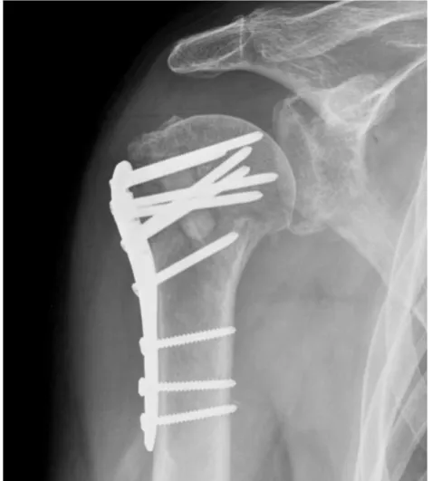
The anatomical reduction with rigid osteosynthesis based on the use of locking plate and screws of proximal humeral fractures with multiple individualized fragments or with severe comminution occurring in young people is usually not feasible, due either to the lack of structural support to the humeral head as to medial calcar comminution or to its short height. For this reason, we have considered the use of synthetic bone graft substitute in fractures with severe medial calcar comminution, with greater difficulty keeping its height or short of bone structure in the humeral head. The 10 mm height β-tricalcium phosphate synthetic bone substitute cylinder was passed through the fracture-related bone window, generally using the fracture line between the greater and the lesser tuberosity upon reduction and before fixation, lodged close to the medial calcar region and aimed to maintain the height of the medial calcar region and subsequently the diaphyseal axis. (Fig. 2)
We were able to maintain fracture reduction in the 19 patients in whom we used this graft, for a follow-up over four years, with a 136º cephalo-diaphyseal angle, similar to the normal average angle. A satisfactory range of motion
was obtained as this angle was effectively recovered, with 140º average shoulder range of motion in abduction and forward flexion, 35º in external rotation and dorsum of hand reaching L3 in internal rotation.
The worst functional outcomes occurred in patients with fracture consolidation in varus (<130º), due to the technical inability associated to the severity of medial calcar comminution or to the calcar region height loss. In these two patients, with final cephalo-diaphyseal angles between 110 and 120º, shoulder ranges of 100 and 60º in abduction, 100 and 60º in forward flexion, 15º in external rotation and dorsum of hand reaching L3 and sacroiliac region were respectively obtained.
Screw perforation of the humeral head was associated to the consolidation in varus of the humeral head in the same two patients. This also occurred in the only patient with a fracture consolidation in valgus (>150º).
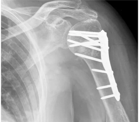
Non-integration, higher risk of infection and foreign-body reaction are the major complications associated to the use of a synthetic graft, although these did not occur in our group of patients (Fig. 3). At the same time, its use seemed to favour the maintenance of vascular viability of
ARTIGO ORIGINAL
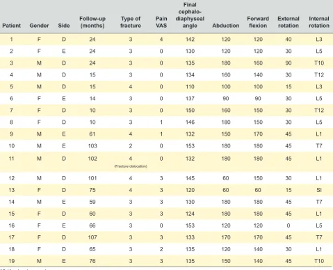
Table 1 - Epidemiological data
Patient Gender Side Follow-up (months) fractureType of PainVAS
Final cephalo-diaphyseal
angle Abduction Forward flexion External rotation Internal rotation
1 F D 24 3 4 142 120 120 40 L3
2 F E 24 3 0 130 120 120 30 L5
3 M D 24 3 0 135 180 160 90 T10
4 M D 15 3 0 134 160 140 30 T12
5 M D 15 4 0 110 100 100 15 L3
6 F E 14 3 0 137 90 90 30 L5
7 F D 10 3 0 150 160 150 30 T12
8 F D 10 3 1 146 180 150 30 L5
9 M E 61 4 1 132 150 170 45 L1
10 M E 103 2 0 153 180 180 45 T7
11 M D 102 4
(Fracture dislocation)
0 132 180 180 45 L1
12 M D 101 4 3 145 60 150 30 L1
13 F D 75 4 3 120 60 60 15 SI
14 M E 59 3 3 130 180 180 45 T7
15 F D 60 3 3 124 180 180 45 L1
16 F E 66 3 0 153 120 120 0 L5
17 F D 107 3 3 133 170 170 45 T7
18 F D 65 3 2 135 120 140 30 L1
19 M E 76 3 3 135 150 140 45 T10
the cephalic region, as no progression to necrosis occurred in our group of patients. Nevertheless, a 20% reoperation rate has still been found with this pathology, even with the use of grafting and the causes remain the same, with higher predominance of subacromial impingement, either due to reduction loss of the tuberosities as to implant-related mechanical impingement. Joint stiffness is the second major cause and correlates to the initial injury or to a more aggressive surgical approach. No direct correlation with the use of synthetic graft seems to exist though.
CONCLUSION
The use of tricalcium phosphate synthetic bone graft emerged as an ancillary therapy for osteosynthesis of proximal humeral fractures and is apparently more useful in fractures with more severe comminution of the medial calcar region, in which humeral head height is more difficult to be maintained either in reduction as upon osteosynthesis. In our group of patients, its correct use allowed satisfactory fracture reductions to be obtained without any humeral head collapse, shown by the maintenance of the
proximal cephalo-diaphyseal angle and obtaining good ranges of motion on the different planes.
The introduction of this technique should be systematic and adds value for cases when reduction and fixation is more difficult due to higher impairment of the medial calcar region.
HUMAN AND ANIMAL PROTECTION
The authors declare that the followed procedures were according to regulations established by the Ethics and Clinical Research Committee and according to the Helsinki Declaration of the World Medical Association.
DATA CONFIDENTIALITY
The authors declare that they have followed the protocols of their work centre on the publication of patient data.
CONFLICTS OF INTEREST
The authors declare that there were no conflicts of interest in writing this manuscript.
FINANCIAL SUPPORT
The authors declare that there was no financial support in writing this manuscript.
ARTIGO ORIGINAL
Figure 2 - Consolidated fracture with osseointegrated graft
Figure 3 - Beta-tricalcium phosphate synthetic graft
REFERENCES
1. Bahrs C, Stojicevic T, Blumenstock G, Brorson S, Badke A, Stockle U, et al. Trends in epidemiology and patho-anatomical pattern of proximal humeral fractures. Int Orthop. 2014;38:1697-704.
2. Court-Brown CM, Garg A, McQueen MM. The epidemiology of proximal humeral fractures. Acta Orthop Scand. 2001;72:365-71.
3. Resch H, Povacz P, Frohlich R, Wambacher M. Percutaneous fixation of three- and four-part fractures of the proximal humerus. J Bone Joint Surg Br. 1997;79:295-300.
4. Lin CL, Hong CK, Jou IM, Lin CJ, Su FC, Su WR. Suture anchor versus screw fixation for greater tuberosity fractures of the humerus - a biomechanical study. J Orthop Res. 2012;30:423-8.
5. Zhu Y, Lu Y, Shen J, Zhang J, Jiang C. Locking intramedullary nails and locking plates in the treatment of two-part proximal humeral surgical neck fractures: a prospective randomized trial with a minimum of three years of follow-up. J Bone Joint Surg Am. 2011;93:159-68.
6. Bastian JD, Hertel R. Osteosynthesis and hemiarthroplasty of fractures of the proximal humerus: outcomes in a consecutive case series. J Shoulder Elbow Surg. 2009;18:216-9.
7. Klein M, Juschka M, Hinkenjann B, Scherger B, Ostermann PA. Treatment of comminuted fractures of the proximal humerus in elderly patients with the Delta III reverse shoulder prosthesis. J Orthop Trauma. 2008;22:698-704.
8. Levy J, Frankle M, Mighell M, Pupello D. The use of the reverse shoulder prosthesis for the treatment of failed hemiarthroplasty for proximal humeral fracture. J Bone Joint Surg Am. 2007;89:292-300. 9. Gardner MJ, Boraiah S, Helfet DL, Lorich DG. Indirect medial reduction
and strut support of proximal humerus fractures using an endosteal implant. J Orthop Trauma. 2008;22:195-200.
10. Brunner F, Sommer C, Bahrs C, Heuwinkel R, Hafner C, Rillmann P, et al. Open reduction and internal fixation of proximal humerus fractures using a proximal humeral locked plate: a prospective multicenter analysis. J Orthop Trauma. 2009;23:163-72.
11. Sproul RC, Iyengar JJ, Devcic Z, Feeley BT. A systematic review of locking plate fixation of proximal humerus fractures. Injury. 2011;42:408-13.
ARTIGO ORIGINAL importance of medial support in locked plating of proximal humerus
fractures. J Orthop Trauma. 2007;21:185-91.
13. Hertel R. Fractures of the proximal humerus in osteoporotic bone. Osteoporosis Int. 2005;16:S65-72.
14. Kim SH, Lee YH, Chung SW, Shin SH, Jang WY, Gong HS, et al. Outcomes for four-part proximal humerus fractures treated with a locking compression plate and an autologous iliac bone impaction graft. Injury. 2012;43:1724-31.
15. Somasundaram K, Huber CP, Babu V, Zadeh H. Proximal humeral
fractures: the role of calcium sulphate augmentation and extended deltoid splitting approach in internal fixation using locking plates. Injury. 2013;44:481-7.
16. Unger S, Erhart S, Kralinger F, Blauth M, Schmoelz W. The effect of in situ augmentation on implant anchorage in proximal humeral head fractures. Injury. 2012;43:1759-63.