489
© 2018 by the Serbian Biological Society How to cite this article: González-Félix ML, Santana-Bejarano EB, Perez-Velazquez M, Villalba-Villalba AG. Partial characterization, quantification and activity of pancreatic lipase in the gastrointestinal tract of Totoaba macdonaldi. Arch Biol Sci. 2018;70(3):489-96.
Partial characterization, quantification and activity of pancreatic lipase in the
gastrointestinal tract of
Totoaba macdonaldi
Mayra L. González-Félix1,*, Edna B. Santana-Bejarano1, Martin Perez-Velazquez1 and Ana G. Villalba-Villalba2
1Department of Scientific and Technological Research, University of Sonora, Edificio 7-G, Blvd. Luis Donaldo Colosio s/n,
e/Sahuaripa y Reforma, Col. Centro, C.P. 83000, Hermosillo, Sonora, Mexico.
2Department of Physics, University of Sonora, Edificio 3-R, Blvd. Luis Encinas y Rosales s/n, Col. Centro, C.P. 83000,
Hermosillo, Sonora, Mexico
*Corresponding author: [email protected]
Received: February 2, 2018; Revised: March 16, 2018; Accepted: March 16, 2018; Published online: March 21, 2018
Abstract: Lipids are one of the main macronutrients that constitute balanced feeds used in aquaculture. Adequate utiliza-tion of dietary lipid is influenced by the activity of pancreatic lipase, one of the enzymes that promotes digesutiliza-tion of dietary lipids in the gastrointestinal tract of fish. The culture of Totoaba macdonaldi is quite recent; its nutritional requirements have been partially established. Knowing the characteristics of pancreatic lipase for this species could help optimize the dietary lipids included in balanced feeds for its culture. Therefore, the aim of this work is to partially characterize and evaluate the enzymatic activity of pancreatic lipase for T. macdonaldi. Biological indices showed that experimental organisms had a good nutritional status. Pancreatic lipase molecular weight was determined by sodium dodecyl sulfate polyacrylamide gel electrophoresis (SDS-PAGE) and its activity was evaluated in crude enzymatic extracts from different gastrointestinal tract regions. The molecular weight of lipase was estimated to be 70.4 kDa; the highest lipolytic activity was observed at 45°C and at a pH optimum of 8.0 in the anterior intestine and pyloric caeca, where the concentration and activity of the enzyme was significantly higher (P=0.004) compared to the distal parts of the intestine. Biochemical characteristics observed for the pancreatic lipase of T. macdonaldi are quite similar to other lipases of fish cultured worldwide; results provided in this study will help understand the role this lipolytic enzyme plays in the digestive process of this species.
Key words:Totoaba macdonaldi; pancreatic lipase; enzymatic activity; gastrointestinal tract; lipids
INTRODUCTION
Understanding the utilization of dietary lipids in ma-rine fish is crucial for understanding their role in ener-gy metabolism [1]. Nevertheless, the knowledge of the biochemical mechanisms that allow marine fish the use of this particular nutrient is rather basic. Pancreatic li-pase (PL) is one of the main digestive lipolytic enzymes in higher vertebrates secreted by the exocrine pancreas that has strong preference for acylglycerols over other lipids, and it may contribute to the in vivo hydrolysis of retinyl esters. It is also known as a colipase-dependent lipase because the coenzyme colipase, also secreted by the pancreas, is required for lipid binding and allows the enzyme to anchor to the water-lipid interface to carry out the hydrolysis of dietary lipid effectively [2-5]. Alternatively, the enzyme carboxyl ester lipase, also
known as bile salt-activated or bile salt-dependent li-pase (BSDL), is another pancreatic lili-pase that possesses the widest range of substrate specificity, being able to hydrolyze mono-, di-, and triacylglycerols, cholesteryl, aryl and wax esters, as well as vitamin A and E esters, phospholipids, lysophospholipids and ceramides. This enzyme requires the presence of primary bile salts to hydrolyze triacylglycerols or phospholipids with me-dium or long-chain fatty acids, but it has basal activ-ity on molecules with short-chain fatty acids in the absence of bile salts [6,7].
Pancreatic lipase has been reported in zebrafish
Danio rerio [8], rainbow trout Oncorhynchus mykiss
O. mykiss, one that predominantly hydrolyzes triacyl-glycerols and the other wax and steryl esters [9,12]. In turbot Scophthalmus maximus, a non-specific lipase with activity in the absence of bile salts was reported [13-15], although it was later confirmed to be a bile salt-dependent lipase [16,17].
Totoaba macdonaldi is a marine sciaenid of the Gulf
of California with great potential for aquaculture, al-though knowledge about its digestive enzyme assem-bly, their activity, and physiology is still rather scarce. Development of aquafeeds for its culture is progressing as quantitative nutritional requirements are established, but lipid levels from 8 to 22% do not seem to affect its growth performance, similarly to red drum, Sciaenops
ocellatus, another sciaenid for which lipase is
appar-ently not stimulated by increasing dietary lipid content. Dietary lipid is one of the crucial macronutrients in balanced feeds for cultured fish species [18]; it plays an important role as a source of energy, as a constituent of cell membranes and tissues, as a supply of essential fatty acids, and as a precursor of molecules that have many important and diverse functions in their metabolism [19]. However, lipid requirement varies according to species, sex, developmental stage and habitat, among other variables [20]. When dietary lipid is provided to cultured fish in excess, it is stored in their bodies, e.g. in the liver, the peritoneal cavity and muscle, and could affect the quality and shelf-life of the marketed fillet [21,22]. Moreover, excess dietary lipid may limit feed consumption resulting in reduced fish growth during culture, as reported for S. ocellatus [23,24] and totoaba [25]. Knowing the type of lipase present in a particular fish species and its functional characteristics contrib-utes to the understanding of the digestive physiology of the organism and could perhaps help develop strate-gies to improve feed formulations. The objective of the present work was to partially characterize, quantify and investigate the activity of pancreatic lipase along the gastrointestinal tract of Totoaba macdonaldi.
MATERIALS AND METHODS Ethics statement
All fish were treated according to the Official Mexican Norm (NOM-062-ZOO-1999) on the technical speci-fications for the production, care and use of laboratory
animals (in Spanish), issued by the Official Journal of the Federation on August 22, 2001, which is approved by the Ethics Committee of the University of Sonora. Experimental fish and biological indices
Forty experimental fish were donated by the Center for Reproduction of Marine Species of the State of So-nora (CREMES) located in Kino Bay, SoSo-nora, Mexico.
T. macdonaldi individuals were killed by immersion in
iced water (≈4°C). Individual weight (IW, g) and total length (IL, cm) were measured to determine the con-dition factor K=(wet body weight×100)/total length3,
cm [26]. Then, fish were eviscerated in situ and the gastrointestinal tract (GIT) was removed, separately stored in sealed plastic bags, and transported in an ice-filled cooler to the Nutrition Laboratory of the Department of Scientific and Technological Research of the University of Sonora (DICTUS). The weights of the liver and viscera were obtained for each individual to determine the hepatosomatic index HSI [%]=(liver weight/total weight)×100, and the viscerosomatic in-dex VSI [%]=(viscera weight/total weight)×100. GIT samples were stored at -82.0°C until further analyses. Enzyme extraction and SDS-PAGE
In order to determine the molecular mass of lipase and to quantify its content in the different sections of the GIT of fish, SDS-PAGE was carried out in a four-gel vertical electrophoresis system (Mini-Protean Tetra Cell, Bio-Rad; CA, USA) using 10% polyacrylamide gels. The enzymes were resolved at a constant voltage of 115 V applied for about 2.5 h at 10°C. Gels were then fixed in a solution of 40% ethanol and 10% acetic acid, rinsed twice with distilled water, and stained overnight in gentle agitation with QC Colloidal Coomassie stain, then destained by rinsing again in distilled water. An internal standard (Precision Plus Protein Standard, Bio-Rad®, Hercules, CA, USA) with proteins of 10-250 kDa, and human pancreatic lipase (BCR-693, Euro-pean Community Bureau of Reference) were used as a molecular weight marker and as a quantitative internal standard, respectively. Lastly, the gels were scanned with a GS-900 Calibrated Densitometer (Bio-Rad®, Hercules, CA, USA) for identification and quantifica-tion of protein bands using the ImageLab 5.0 software (Build 18, Bio-Rad®, Hercules, CA, USA).
Zymography
To confirm the band identified as PL in the SDS-PAGE gels, a zymogram was obtained following a pre-viously described procedure [28]. Briefly, SDS-PAGE was done as previously described. After resolving at a constant voltage of 115 V, the gel was immediately rinsed three times with distilled water and equilibrat-ed in a 50-mM Tris-HCl (pH 8) buffer for 30 min at 25°C. A chromogenic substrate solution was prepared, containing 0.01% phenol red, 1% oleic acid, and 2% agar dissolved in a 10-mM CaCl2 solution with the pH adjusted to 8.2 using 0.1 N NaOH. This solution was warmed to 95°C for 2 min to dissolve the agar and cooled down to 40°C at ambient temperature. The gel was then covered with the chromogenic substrate and allowed to solidify during 30 min, followed by incuba-tion for approximately 3.5 h at 37°C. After incubaincuba-tion, lipase activity was observed as a yellow spot on the fuchsia colored gel.
Activity of pancreatic lipase
Activity of PL within the different sections of the GIT was determined in quadruple samples by the enzymat-ic assay of lipase (EC 3.1.1.3) using olive oil as a
sub-strate, with minor modifications. The homogenates were prepared using three volumes of 200 mM Tris-HCl buffer solution (pH 7.7) and centrifuging twice at 21000 × g and 4°C for 30 min, until the samples were clear. The supernatant was recovered and used to pre-pare a solution of 1 mL 200 mM Tris-HCl buffer (pH 7.7), 2.5 mL tridistilled water, 1 mL of crude extract, and 3 mL olive oil. Then, the samples were mixed vigorously and incubated in a shaker at 35°C and 120 rpm for exactly 30 min. The reaction was stopped by adding 3 mL of 95% ethanol, followed by addition of four drops of 0.1% phenolphthalein, and titration with a solution of 50 mM NaOH to a light pink color. In all blanks, the enzyme crude extract was added after the 30-min incubation period. Each reaction sample and blanks were analyzed in duplicates. One unit of activity of lipase (U) per mL of crude extract was de-fined as the amount of enzyme that will hydrolyze 1.0 microequivalent of fatty acid from a triglyceride in one hour at pH 7.7 and 35°C, using the equation U [mL-1]=[(mL NaOH used) (molarity of NaOH) (1000)
(2) (dilution factor)]/(volume of enzyme used in mL). In this equation, 1000 is the conversion factor from milliequivalent to microequivalent, and 2 is the time conversion factor from 30 min to 1 h (unit definition). Effect of temperature on lipase activity
Enzymatic activity was higher in the anterior intestine and pyloric caeca as compared to the other two sections of the GIT. Thus, these two sections were used to evalu-ate the effect of temperature on enzymatic activity. The analysis was run in triplicate samples for each tempera-ture tested: 20, 25, 30, 35, 40, 45, 50, 55 and 60°C. Each triplicate sample and its blank were run per duplicate, following the same method previously described. Effect of pH on lipase activity
Statistics
Biological indices, the molecular weight and concen-tration of pancreatic lipase determined by electropho-resis were analyzed by descriptive statistics, whereas enzymatic activity in the different sections of the GIT, at different temperatures and at different pH, were subjected to one-way analysis of variance (ANOVA) using a significance level of P≤0.05. Tukey’s HSD test was used as the mean separation procedure where significant differences were observed. All statistical analyses were performed using the Statistical Analysis System (SAS Institute Inc., 2013, Software Release 9.4, Cary, NC, USA) software package.
RESULTS
Biological indices
Individual mean average weight (±standard error of the mean, SEM) was 1477.25±22.98 g. The weight of the viscera, consisting of esophagus, stomach, pyloric caeca and intestine, as well as associated organs like heart, liver, swim bladder, gallbladder, etc., averaged 72.52±1.47 g. Fish total length averaged 52.86±0.31 cm. The condition factor (K) averaged 1.00±0.01, while hepatosomatic and viscerosomatic indices av-eraged 0.91±0.03% and 4.92±0.08%, respectively. Molecular weight, concentration and activity of lipase in the GI
The mean molecular weight (±SEM) of the semi-purified pancreatic lipase of T. macdonaldi deter-mined by SDS-PAGE was 70.4±0.26 kDa. This band was confirmed by zymography (Fig. 1). Coomassie blue-stained polyacrylamide gels also provided an timated content of the enzyme along the GIT. The es-timated content in the anterior, middle and posterior intestine, as well as the pyloric caeca were 11.0, 2.5, 2.4, and 2.7 mg of PL g-1 of tissue, respectively.
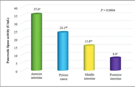
Lipo-lytic activity was significantly higher (P=0.0004) in the anterior intestine, 35.6±5.4 U mL-1 of crude enzymatic
extract, followed by pyloric caeca, 24.1±2.6 U mL-1,
middle intestine, 15.8±2.1 U mL-1, and posterior
in-testine 8.0±1.3 U mL-1, where activity was significantly
lower compared to other sections of the GIT (Fig. 2).
Effect of temperature and pH on lipase activity
The anterior intestine and pyloric caeca were the sec-tions of the GIT of T. macdonaldi where more lipase activity was observed. Thus, the effect of temperature and pH on enzyme activity was evaluated using both segments. Activity was consistently higher in the ante-rior intestine as compared to the pyloric caeca (Table 1), and significantly higher activity (P<0.0001) was observed at 45°C, with 70.3 U mL-1 of crude enzymatic
extract in the anterior intestine compared to the rest of the evaluated temperatures (Fig. 3). In addition, significantly higher enzymatic activity (P<0.0001) was observed at the pH of 8.0 (Table 2), 73.8 U mL-1 in the
anterior intestine, compared to rest of the evaluated values (Fig. 4).
Fig. 2. Activity of pancreatic lipase in the different sections of the GIT of Totoaba macdonaldi at 35°C. Values of activity with differ-ent superindices are significantly differdiffer-ent (P=0.0004).
Fig. 1.A – PAGE gel of the semi-purified crude extract of pan-creatic lipase from different sections of the gastrointestinal tract of Totoaba macdonaldi, with a molecular weight of 70.4 kDa. Std – Precision Plus Protein Standard (Bio-Rad®, Hercules, CA, USA).
B – PAGE gel of pancreatic lipase of T. macdonaldi stained with
QC Colloidal Coomassie. C – Zymogram demonstrating lipase
DISCUSSION
Dietary protein has proven to be readily used as a source of energy in fish; however, dietary lipid and carbohydrate can have a protein-sparing effect, al-lowing protein to be preferentially used for somatic growth [25,29-31], a goal usually set for any cultured species in aquaculture. Juvenile Totoaba macdonaldi
has shown similar growth performance when fed di-etary lipid (provided as sardine fish oil) levels rang-ing from 8 to 22% at a fixed crude protein level of 46% [32], suggesting there is a limit in the capability to use this macronutrient, as has been observed in other species. For instance, in Dicentrarchus labrax, a plateau in enzymatic activity of lipase and phospho-lipase A2 was observed when supplementing more than 15% dietary triglycerides and more than 4.5% dietary phospholipid, respectively [33]. For S. ocella-tus, maximal lipase activity was suggested to occur at a dietary inclusion of neutral lipid close to 11.8% [34]. This is an indication of a limited physiological ability in marine fish species to utilize this nutrient, during either digestion, absorption, transport or storage of dietary lipid. Since T. macdonaldi is a relatively new species in aquaculture, little is known with respect to the set of digestive enzymes present in the gastroin-testinal tract of this species and their characteristics. Knowing these properties for its lipase will not only contribute to understanding the digestive physiology of this marine fish, but could also help improve feed formulations for its culture.
Table 1. Pancreatic lipase activity in the anterior intestine and pyloric caeca of Totoaba macdonaldi at different temperatures.
Anterior intestine
Enzymatic activity U mL-1 of
crude extract
Pyloric caeca
Enzymatic activity U mL-1 of crude
extract
Temperature Mean* SEM Temperature Mean* SEM
20°C 13.2de 3.1 20°C 7.5ef 1.4
25°C 14.8de 2.6 25°C 10.0ef 1.4
30°C 31.0bcde 4.9 30°C 15.6def 1.9
35°C 35.5bcd 4.2 35°C 24.1bcd 2.6
40°C 39.0bc 4.1 40°C 28.8b 1.6
45°C 70.3a 7.8 45°C 43.1ª 2.8
50°C 45.8b 8.1 50°C 26.3bc 3.0
55°C 22.5cde 1.4 55°C 16.9cde 2.1
60°C 9.5e 4.2 60°C 5.0f 1.4
ANOVA Pr>F <0.0001 ANOVA Pr>F <0.0001 *Means in each column with the same letter are not significantly differ-ent (P≤0.05).
Table 2. Pancreatic lipase activity in the anterior intestine and pyloric caeca of Totoaba macdonaldi at different pH.
Anterior intestine
Enzymatic activity U mL-1 of crude
extract
Pyloric caeca
Enzymatic activity U mL-1 of crude
extract
pH Mean* SEM pH Mean* SEM 6 17.5e 0.7 6 8.3d 1.5
7 32.1c 1.1 7 24.2b 2.2
8 73.8a 0.7 8 36.7a 2.5
9 40.4b 1.8 9 18.3bc 2.2
10 24.6d 1.1 10 11.3cd 1.9 ANOVA Pr>F <0.0001 ANOVA Pr>F <0.0001
*Means in each column with the same letter are not significantly differ-ent (P≤0.05).
The molecular weights reported for lipases of ma-rine fish vary according to species [35], as can be ex-pected, and although evidence suggests the presence of bile salt-dependent lipase is more common, there is evidence of a PL-colipase system as well [19]. Molecular weights reported for BSDL of different marine fish spe-cies include those of 64 kDa in Pagrus major [36] and 60 kDa in Gadus morhua [37]. In the pyloric caeca of
Oncorhynchus tshawytscha, two bands of 79.6 and 54.9
kDa were reported as possibly the uncleaved and the fi-nal form of its BSDL, respectively [38]. Other molecular weights reported for lipases include a 74 kDa hepatic lipase of Cyprinus carpio [35], and a 57.4 kDa pancre-atic lipase in the intestine of Cynoscion othonopterus
[18]. In this study, the molecular weight determined for pancreatic lipase of T. macdonaldi was 70.4 kDa, which is within the range of values previously mentioned for other fish species. For this sciaenid, there is a strong possibility that the pancreatic lipase described in the current study is not a BSDL because the activity of the enzyme was detected without supplementation of bile salts, and its activity did not increase in the presence of bile salts or natural bile extracts [39], although this inference requires further confirmation.
From the quantification of PL in the different regions of the gastrointestinal tract of T. macdonal-di, it was clear that the enzyme was more abundant within the anterior section of the intestine and the pyloric caeca. Moreover, the activity of PL was also significantly higher (P=0.0004) in the aforementioned sections. This observation coincides with the higher concentrations and activity of lipase also reported in the pancreas, anterior intestine and pyloric caeca of several fish species, like S. aurata [40,41], Thunnus
orientalis [42], Glyptosternum maculatum [43] and the
hybrid Oreochromis niloticus × O. aureus [44], among others. This is not surprising because pancreatic en-zymes are secreted within the upper intestine of fish [45], thus, their concentration and activity would be more conspicuous in this area and would decrease towards the distal sections of the GIT. However, the activity of lipases and other enzymes is subjected to modifications by the influence of many factors, such as age, the source and quality of the dietary lipid, the prandial status, and the species [46].
It was established that the BSDL of S. ocellatus
had the highest activity at a pH close to 8.0 and 50°C
[47], the same temperature value reported for optimal lipolytic activity in S. aurata [41]. The maximum li-polytic activity from pancreatic crude extracts of T.
orientalis, T. macdonaldi and Morone saxatilis was
re-ported at pH 8.0 and temperatures ranging from 35 to 45°C [39]. Optimal lipolytic activity was observed at 40°C for Sardinops sagax caerulea [48], while the highest lipolytic activity was observed at 35°C and at a pH of 8-8.5 for O. tshawytscha and Macruronus
novaezelandiae [38]. In this study, the highest lipolytic
activity was observed at 45°C and a pH optimum of 8.0 in both anterior intestine and pyloric caeca of T.
macdonaldi, confirming the observations provided
previously for this species [39], and agreeing with the general observations of related sciaenids and marine fish. As expected, pancreatic enzymes, including PL and BSDL, would be more active in the alkaline pH range, between 7.0 and 9.0, values that may very well be expected in the intestine of marine teleosts. How-ever, unlike pH, it is often observed that tempera-tures for optimal activities of some enzymes are higher than the physiological temperature of the organisms where they are found [49]; when temperature rises, the kinetic energy of the molecules increases, also in-creasing the reaction rate, but at higher temperature denaturation of the enzyme is more likely to occur [50], which explains to some extent why optimal en-zymatic activities differ from optimal physiological temperatures.
CONCLUSIONS
A pancreatic lipase of 70.4 kDa was identified in the GIT of Totoaba macdonaldi and confirmed through the colored trace of enzymatic activity observed in a zymogram. The concentration and activity of the en-zyme were significantly higher in the anterior intestine and pyloric caeca, decreasing towards the middle and posterior sections of the digestive tract. Higher lipo-lytic activity was confirmed to be at 45°C and at a pH optimum of 8.0 in both anterior intestine and pyloric caeca. There is a strong possibility that the pancre-atic lipase described for T. macdonaldi in this study is not a BSDL because the activity of the enzyme was detected without supplementation of bile salts, and its activity did not increase in the presence of bile salts or natural bile extracts as previously reported, although additional studies are required to confirm this infer-ence, and to further characterize this enzyme. Fully understanding the enzymatic action and potential of pancreatic lipase will contribute to understanding the catabolism of lipid in vivo for this species and pro-mote better formulations of balanced feeds, and may perhaps allow its utilization in industrial applications.
Acknowledgments: Funding for Mrs. Santana-Bejarano was pro-vided by the Consejo Nacional de Ciencia y Tecnología (CONA-CYT-México). The authors would like to thank the Center for Reproduction of Marine Species of Sonora State (C.R.E.M.E.S.), Kino Bay, Sonora, Mexico, for donating the experimental fish. The mention of trademarks or proprietary products does not constitute an endorsement of the product and does not imply its approval to the exclusion of other products that may also be suitable.
Author contributions: Santana-Bejarano, M.Sc. contributed to all of the analytical determinations and data analysis. Dr. Perez-Velazquez procured the experimental organisms and contributed to their processing, estimation of biological indices, the editing of this manuscript and with financial support. Dr. Villalba-Villalba participated in the analytical determination of enzymatic activity. Dr. González-Félix participated in the SDS-PAGE analysis, the analysis of data, with the financial support, and with the writing of this manuscript.
Conflict of interest disclosure: The authors have no conflict of interest related to this work.
REFERENCES
1. Patton JS, Nevenzel JC, Benson AA. Specificity of digestive lipases in hydrolysis of wax esters and triglycerides studied in anchovy and other selected fish. Lipids. 1975;10:575-83.
2. Lowe ME. Structure and function of pancreatic lipase and colipase. Annu Rev Nutr. 1997;17:141-58.
3. Lowe ME. The triglyceride lipases of the pancreas. J Lipid Res. 2002;43:2007-16.
4. Van Tilbeurgh H, Bezzine S, Cambillau C, Verger R, Carrière F. Colipase: structure and interaction with pancreatic lipase. Biochim Biophys Acta. 1999;1441:173-84.
5. Crenon I, Foglizzo E, Kerfelec B, Verine A, Pignol D, Her-moso J, Bonicel J, Chapus C. Pancreatic lipase-related pro-tein type I: A specialized lipase or an inactive enzyme. Pro-tein Eng. 1998;11:135-42.
6. Terzyan S, Wang Ch-S, Downs D, Hunter B, Zhang X. Crys-tal structure of the caCrys-talytic domain of human bile salt acti-vated lipase. Protein Sci. 2000;9:1783-90.
7. Kurtovic I, Marshall SN, Zhao X, Simpson BK. Lipases from mammals and fishes. Rev Fish Sci. 2009;17:18-40.
8. Holmes RS, Cox LA. Bioinformatics and evolution of verte-brate pancreatic lipase and related proteins and genes. J Data Mining in Genom Proteomics. 2012;3:1-10.
9. Leger C. Digestion, absorption and transport of lipids. In: Cowey CB, Mackie AM, Bell JG, editors. Nutrition and feed-ing of fish. Academic Press, London, UK; 1985. p. 299-331. 10. Patton JS, Haswell MS, Moon TW. Aspects of lipid synthesis,
hydrolysis, and transport studied in selected Amazon fish. Can J Zool. 1978;56:787-92.
11. Mukundan MK, Gopakumar K, Nair MR. Purification of a lipase from the hepatopancreas of oil sardine (Sardinella longiceps Linnaeus) and its characteristics and properties. J Sci Food Agric. 1985;36:191-203.
12. Tocher DR, Sargent JR. Studies on triacylglycerol, wax ester and sterol ester hydrolases in intestinal caeca of rainbow trout (Salmo gairdneri, L.) fed diets rich in triacylglycerols and was esters. Comp Biochem Physiol. 1984;77:561-71. 13. Izquierdo MS, Henderson RJ. The determination of lipase
and phospholipase activities in gut contents of turbot (Scophthalmus maximus) by fluorescence-based assays. Fish Physiol Biochem. 1998;19:153-62.
14. Koven WM, Henderson RJ, Sargent JR. Lipid digestion in turbot (Scophthalmus maximus) I: Lipid class and fatty acid composition of digesta from different segments of the diges-tive tract. Fish Physiol Biochem. 1994;13:69-79.
15. Koven WM, Henderson RJ, Sargent JR. Lipid digestion in turbot (Scophthalmus maximus) II: Lipolysis in vitro of 14C-labelled triacylglycerol, cholesterol ester and phospha-tidylcholine by digesta from different segments of the diges-tive tract. Fish Physiol Biochem. 1994;13:275-83.
16. Hoehne-Reitan K, Kjørsvik E, Gjellesvik DR. Development of bile salt-dependent lipase in larval turbot. J Fish Biol. 2001;58:737-45.
17. Hoehne-Reitan K, Kjørsvik E, Reitan KI. Bile salt-dependent lipase in larval turbot, as influenced by density and lipid content of prey. J Fish Biol. 2001;58:746-54.
18. González-Félix ML, Minjarez-Osorio C, Perez-Velazquez M, Urquidez-Bejarano P. Influence of dietary lipid on growth performance and body composition of the Gulf corvina, Cynoscion othonopterus. Aquaculture. 2015;448:401-9. 19. Tocher DR. Metabolism and functions of lipids and fatty
20. Halver JE, Hardy RW. Fish Nutrition. Elsevier Sciences. 3er edition. Orlando, Florida, USA; 2002. 353 p.
21. Bromley PJ. Effect of dietary protein, lipid and energy con-tent on the growth of turbot Scopthalmus maximus. Aqua-culture. 1980;19:359-69.
22. Hillestad M, Johnsen F. High-energy⁄low-protein diets for Atlantic salmon: effects on growth, nutrient retention and slaughter quality. Aquaculture. 1994;124:109-16.
23. Ellis SC, Reigh RC. Effects of dietary lipid and carbohydrate levels on growth composition of juvenile red drum Sciaenops ocellatus. Aquaculture. 1991;97:383-94.
24. McGoogan BB, Gatlin DM III. Dietary manipulations affect-ing growth and nitrogenous waste production of red drum Sciaenops ocellatus. I. Effects of dietary protein and energy levels. Aquaculture. 1999;178:333-48.
25. Rueda-López S, Lazo JP, Correa-Reyes G, Viana MT. Effect of dietary protein and energy levels on growth, survival and body composition of juvenile Totoaba macdonaldi. Aquacul-ture. 2011;319:385-90.
26. Ricker WE. Computation and interpretation of biologi-cal statistics of fish populations. B Fish Res Board Can. 1975;191:1-382.
27. Association of Official Analytical Chemists (AOAC). Offi-cial Methods of Analysis. Association of Analytical Chem-ists, Arlington, VA, USA; 2005.
28. Singh R, Gupta VK, Goswami VK. A simple activity staining protocol for lipases and esterases. App Microbiol Biotechnol. 2006;70:679-82.
29. Shiau SY, Lan CW. Optimum dietary protein level and pro-tein to energy ratio for growth of grouper (Epinephelus mala-baricus). Aquaculture. 1996;145:259-66.
30. Thoman ES, Davis DA, Arnold CR. Evaluation of growout diets with varying protein and energy levels for red drum (Sciaenops ocellatus). Aquaculture. 1999;176:343-53. 31. Hixson SE. Fish nutrition and current issues in
aquacul-ture: The balance in providing safe and nutrious seafood, in an environmentally sustainable manner. J Aquac Res Dev. 2014;5:1-10.
32. Perez-Velazquez M., Minjarez-Osorio C., González-Félix ML. Effect of dietary lipid level on growth performance, feed utilization, and body composition of totoaba, Totoaba macdonaldi (Gilbert, 1890). Aquacult Res. 2017;48:2607-17. 33. Zambonino-Infante JL, Cahu CL. High dietary lipid levels
enhance digestive tract maturation and improve Dicentrar-chus labrax larval development. J Nutr. 1999;129:1195-200. 34. Buchet V, Zambonino-Infante JL, Cahu CL. Effect of lipid
level in a compound diet on the development of red drum (Sciaenops ocellatus) larvae. Aquaculture. 2000;184:339-47. 35. Görgün S, Akpinar MA. Purification and characterization
of lipase from the liver of carp, Cyprinus carpio L. (1758), living in Lake Tödürge (Sivas, Türkiye). Turk J Fish Aquat Sci. 2012;12:207-15.
36. Iijima N, Tanaka S, Ota Y. Purification and characterization of bile salt-activated lipase from the hepatopancreas of red sea bream, Pagrus major. Fish Physiol Biochem. 1998;18:59-69. 37. Gjellesvik DR, Lombardo D, Walther BT. Pancreatic bile salt
dependent lipase from cod (Gadus morhua): purification and properties. Biochim Biophys Acta. 1992;1124:123-34.
38. Kurtovic I, Marshall SN, Zhao X. Purification and properties of digestive lipases from Chinook salmon (Oncorhynchus tshawytscha) and zeland hoki (Macruronus novaezelandiae). Fish Physiol Biochem. 2010;36:1041-60.
39. Rueda-López S, Martínez-Montaño E, Viana MT. Biochemi-cal characterization and comparison of pancreatic lipases from the Pacific bluefin tuna, Thunnus orientalis; totoaba, Totoaba macdonaldi; and striped bass, Morone saxatilis. J World Aquacult Soc. 2017;48:156-65.
40. Deguara S, Jauncey K, Agius C. Enzyme activities and pH variations in the digestive tract of gilthead sea bream. J Fish Biol. 2003;62:1033-43.
41. Nolasco H, Moyano LFJ, Vega-Villasante F. Partial charac-terization of pyloric-duodenal lipase of gilthead seabream (Sparus aurata). Fish Physiol Biochem. 2011;37:43-52. 42. Matus de la Parra A, Rosas A, Lazo JP, Viana MT. Partial
characterization of the digestive enzymes of Pacific blue-fin tuna Thunnus orientalis under culture conditions. Fish Physiol Biochem. 2007;33:223-31.
43. Xiong DM, Xie CX, Zhang HJ, Liu HP. Digestive enzymes along digestive tract of a carnivorous fish Glyptosternum maculatum (Sisoridae, Siluriformes). J Anim Physiol Anim Nutr. 2011;95:56-64.
44. Jun-Sheng L, Jian-Lin L, Ting-Ting W. Ontogeny of protease, amylase and lipase in the alimentary tract of hybrid juvenile tilapia (Oreochromis niloticus × Oreochromis aureus). Fish Physiol Biochem. 2006;32:295-303.
45. Rust MB. Nutritional Physiology. In: Halver JE and Hardy RW, editors. Fish Nutrition. Academic Press, New York, USA; 2002. p. 368-446.
46. Morais S, Conceição LEC, Rønnestad I, Koven W, Cahu C, Zambonino-Infante JL, Dinis MT. Dietary neutral lipid level and source in marine fish larvae: effects on digestive physiol-ogy and food intake. Aquaculture. 2007;268:106-22. 47. Lazo JP, Mendoza R, Holt GJ, Aguilera C, Arnold CR.
Characterization of digestive enzymes during larval devel-opment of red drum (Sciaenops ocellatus). Aquaculture. 2007;265:194-205.
48. Noriega-Rodriguez JA, Gámez-Meza N, Alanis-Villa A, Medina-Juárez LA, Tejeda-Mansir A, Angulo-Guerrero O, García HS. Extraction and fractionation of lipolytic enzyme from viscera of Monterey sardine (Sardinops sagax caerulea). Int J Food Sci Technol. 2009;44:1223-28.
49. Grogan G. Practical biotransformations. A beginner’s guide. Wiley and Sons, Ltd. Cornwall, UK; 2009. 344 p.
50. Murray RK, Mayes PA, Granner DK, Rodwell VW. Enzyme kinetics. In: Rodwell, VW, editor. Harpers Biochemistry, Mc Graw Hill Inc., Mexico City, Mexico; 1990. p. 68-81. 51. Minjarez-Osorio C., González-Félix ML, Perez-Velazquez M.
Biological performance of Totoaba macdonaldi in response to dietary protein level. Aquaculture. 2012;362-363:50-4. 52. Lizama MAP, Ambrosio AM. Condition factor in nine
spe-cies of fish of the Characidae family in the upper Parana River Floodplain, Brazil. Braz J Biol. 2002;62:113-24. 53. Chaturvedi, J, Saksena DN. Diet composition, feeding