Address for correspondence
Dr. Ali Mohamed Gargoom MBChB, MSc, MD. Dermatology Department, Faculty of Medicine Benghazi University, Benghazi-Libya
Ph: : 00218 92 511 3849
Original Article
Ablation of xanthelasma palpebrarum by ultrapulsed
carbon dioxide laser
Introduction
Xanthelasma palpebrarum (XP) is a form of plane xanthoma and it is the most common clinical presentation of all types of xanthomas. XP is seen in females more frequently than males.1 Although xanthelasma is a benign condition but significant hypercholesterolemia is detected in about 60% of cases.2 It is esthetically annoying because of its shape and site. Therefore it represents frequent referral among the dermatology and ophthalmology outpatient departments. Surgical excision of XP was the ‘classical’ management option for many years. Nevertheless, this effective
therapeutic procedure is associated with a significant risks including; persistent erythema, infection, pigmentary and textural changes. Furthermore, giant xanthelasma; a xanthelasma that occupies the upper and lower palpebrarum may not be operable at all.2-4 Alternatives to surgical excision are; electrofulguration, chemical cautery and cryotherapy. However, these procedures bear mixed result and considerable risks of side effects. Xanthelasma is considered as an ideal target for laser therapy because of its superficial location.5 Therefore, different ablative and non-ablative lasers have been tried in photoablation of xanthelasmas includes; CO2 laser,1 erbium:yttrium-aluminum-garnet (Er:YAG) laser,6 Q-switched neodymium:yttrium-aluminum-garnet
(Nd:YAG) laser,7 pulsed dye laser,8 and diode laser.9 Recently, carbon dioxide laser was Ali Mohamed Gargoom, Gamal Ahmed Duweb
Dermatology Department, Faculty of Medicine, Benghazi University, Benghazi, Libya
Abstract
Objective To investigate the efficacy and safety of ultrapulsed carbon dioxide (UCO2) laser ablation in xanthelasma palpebrarum (XP) management.Methods Between January 2015 and December 2015, eleven Libyan female patients with 21 xanthelasma lesions were treated with one laser session using UCO2 laser. All patients were followed up for 6 months period.
Results The 21 xanthelasma lesions were completely removed after one UCO2 laser session. During post-laser treatment follow-up period, 2 (9.5%) xanthelasma lesions showed transient hyperpigmentation for four months, which faded thereafter. None of the studied patients showed scarring and all patents were very satisfied with the end result of the ultrapulsed CO2 laser treatment.
Conclusion UCO2 laser is an authentic therapeutic option and represents a convenient option to the conventional methods of xanthelasma management.
Key words
commonly reported laser modality in xanthelasma laser management and showed promising esthetic results in dealing with xanthelasma palpebrarum.10
Methods
Between January 2015 and December 2015, eleven Libyan females patients aged between 32 years and 56 years (mean age 46.2 years) with 21 XP lesions were enrolled in the present study. Lesions ≥1cm over upper or lower eyelid were counted as a single lesion and those less than 1cm were not included. Blood sample after 12-hours fasting was analyzed for lipid profile from every patient. The laser procedure and the expected complications and the outcome were explained to every patient in a simple language prior to laser procedure. After informed consent, the lesions were cleaned with disinfecting solution then infiltrated with 1% lignocaine via insulin syringe attached to 29-G needle. Lesions were treated with one laser session with UCO2 laser (Fraxis from Ilooda; 10,600 nm; 100-200 Hz; 200-400 μs) with fixed spot-size handpiece (spot diameter 1.0 mm). The other eye was
covered with opaque goggle and the
photoablated eye was protected by aid of wooden tongue depressor held by an assistant. The photoablated tissue was gently removed with a cotton swap soaked in normal saline to expose the rest of underlying fatty tissue. Laser
photoablation and gentle removal of
photoablated tissue was repeated for 4-5 passes until the appearance of underlying pink tissue which was considered as the endpoint of laser photoablation. Post-operatively, patients were instructed to apply fusidic acid cream till crust formation and to keep the area clean and dry for the same duration. Photographic documentation of the lesion before laser treatment and immediately afterward and then reviewed at day
3, day 7, 1 month, 2 months and finally at 6 months. Before and after photographs were taken with Canon XUS 220 HS camera. The results rated from good to excellent; excellent meant the skin of the lesion returned normal as the rest of the surrounding skin, very good meant the skin of the lesion returned near normal as the rest of the surrounding skin, and good meant the skin of the lesion had acceptable color.
Results
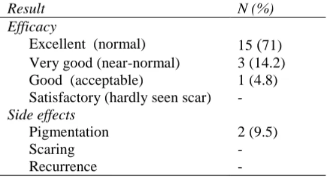
All of the studied patients were females with mean age 46.2 year and all were of skin type IV. Sixteen (76%) of the 21 XP lesions involved the upper eyelid while 5 (24%) lesions involved the lower eyelid. The results of lipid profile of all patients were within normal range. Xanthelasma laser ablation procedure was convenient and well tolerated by all treated patients. All 21 XP lesions of the 11 enrolled patients were completely removed after one UCO2 laser session (Figures 1a-4b). All patents were satisfied with the end result of the UCO2 laser session (Table 1). Only two patients (9.5%) showed transient hyperpigmentation for four months, which faded thereafter. Neither scarring nor recurrence developed during the follow-up period of 6 months.
Table 1 Details of the results (efficacy and side effects) of the study (n=21 lesions).
Result N (%)
Efficacy
Excellent (normal) 15 (71) Very good (near-normal) 3 (14.2) Good (acceptable) 1 (4.8) Satisfactory (hardly seen scar) - Side effects
Pigmentation 2 (9.5)
Scaring -
(a) (b)
Figure 1 Small-sized xanthelasma palpebrarum in 32-year-old women at left upper eyelid (a) and (b) 6 months later following one ultrapulsed CO2 laser session showing excellent outcome without any pigmentary or textural changes.
(a) (b)
Figure 2 Medium-sized xanthelasma palpebrarum in 42-year-old women on right upper eyelid (a) and (b) 2 months later following one ultrapulsed CO2 laser session.
(a) (b)
Figure 3 Medium-sized xanthelasma palpebrarum in 54-year-old women at left eyelid (a) and (b) 6-months later following one ultrapulsed CO2 laser session showing excellent outcome without any pigmentary or textural changes
(a) (b)
Figure 4 Multiple sized xanthelasma palpebrarum in 54-Years-old women on left eyelid (a) and (b) 6-months later following one ultrapulsed CO2 laser session showing very good outcome (near-normal).
Discussion
Xanthelasma is a type of plane xanthoma with a considerable esthetic concern.10 Clinically XP lesions appear as yellowish, flat plaques or nodules and overlying most commonly the inner canthus of the eye on the superior and/or the inferior palpebrarum. XP may be seen as an isolated clinical condition with normal serum lipid level. Nevertheless, in many patients it is
associated with significant
hypercholesterolaemia.2,11
Histologically, XP can be distinguished from the rest of xanthomas by its position in the upper part of the dermis allowing it as an ideal target for laser therapy.5 Different laser modalities were used in the treatment of xanthelasma including CO2 laser, Er:YAG laser, Q-switched Nd:YAG laser, pulsed dye laser, and diode laser. Nguyen et al. who recently reviewed 21 published studies on different laser modality in xanthelasma treatment concluded that both pigment-specific and ablative lasers will offer a reasonable to marked improvement in xanthelasma appearance with mild undesired consequence.10
with its wavelength which is selectively absorbed by water.3 Since approximately 77% of skin consists of water, this absorption was the optimal condition for the ablation effect.1,3 CO2 laser in the continuous mode is effective but associated with considerable possibility of texture changes and post-therapeutic skin color alterations because of its uncontrollable penetration depth. This is in contrast to ultrapulsed CO2 laser mode that emits a considerably very high energy in extremely short pulses. Consequently the pulse duration lies outside the thermal relaxation time of skin hence minimizing the collateral destruction.1,3 Since the publication of Raulin et al.1 on the advantages of ultrapulsed CO2 in dealing with xanthelasma many similar studies were conducted to evaluate this tool in the management of xanthelasma.1,3,4,10 Moreover, according to Nguyen et al.10 review, the majority of xanthelasma laser management was conducted with CO2 laser.
duration of follow-up and could be attributed to the normolipidemic profile of our studied series. Only one patient developed hyperpigmentation, which was transient and faded after 4 months but more significantly no scars was recorded in our series.
Er:YAG laser, because of its wavelength which is maximally absorbed by water is a feasible option in management of xanthelasma and indeed it was the second most commonly used laser modality in this field.10 In a comparison between CO2 and Er:YAG lasers in xanthelasma laser ablation, Rohrich et al.12 concluded that the CO2 laser offer better hemostasis therefore considered better choice especially for deeper XP lesions.
Conclusion
The UCO2 laser is an excellent therapeutic option and represents a convenient substitute to the more conventional methods of xanthelasma management. The advantages of this therapeutic modality are: a quick out-patient procedure, well tolerable by patient, minimal postoperative care, low risk of visible scarring and low recurrence rate and above all, a positive esthetic outcome. Therefore, we greatly recommend the ultrapulsed carbon dioxide laser to be considered as the management of the first choice in xanthelasma treatment.
References
1. Raulin C, Schoenermark P, Werner S, Greve B. Xanthelasma palpebrarum treatment with ultrapulsed CO2 laser. Lasers Surg Med. 1999;24:122-7.
2. Reddy K, Kunneth T, Lakshminarayana P, Yallappa M, Chandrashekara R, Nanjundaswamy SK. Comparative study to evaluate the efficacy of radiofrequency ablation versus trichloroacetic acid in the treatment of xanthelasma palpebrarum. J Cutan Aesthet Surg. 2016;9:236-40.
3. Pathania V, and Chatterjee M. Ultrapulse carbon dioxide laser ablation of xanthelasma palpebrarum: A case series. J Cutan Aesthet Surg. 2015;8:46-9.
4. Corradino B, Di Lorenzo S, Triolo A, Moschella F. Laser treatment of giant xanthelasma palpebrarum. Lasers Med Sci. 2015;30:2205-7.
5. Abdelkadera M, Alashry SE. Argon laser versus erbium:YAG laser in the treatment of xanthelasma palpebrarum. Saudi J Ophthalmol. 2015;29:116-20.
6. Borelli C, Kaudewitz P. Xanthelasma palpebrarum: Treatment with the Erbium: YAG laser. Lasers Surg Med. 2001;29 :260-4.
7. Fusade T. Treatment of xanthelasma palpebrarum by 1064-nm Q-switched Nd: YAG laser: A study of 11 cases. Br J Dermatol. 2008;158:84-7.
8. Karsai S, Czarnecka A, Raulin C. Treatment of xanthelasma palpebrarum using a pulsed dye laser: A prospective clinical trial in 38 cases. Dermatol Surg. 2010;36:610-7. 9. Park EJ, Youn SH, Cho EB, Lee GS, Hann
SK, Kim KH et al. Xanthelasma palpebrarum treatment with a 1,450-nm-diode laser. Dermatol Surg. 2011;37:791-6. 10. Nguyen AH, Vaudreuil AM, Huerter CJ.
Systematic review of laser therapy in xanthelasma palpebrarum. Int J Dermatol. 2017;56:47-55.
11. Nair PA, Patel CR, Ganjiwale JD, Diwan NG, Jivani NB. Xanthelasma palpebrarum with arcus cornea: A clinical and biochemical study. Indian J Dermatol. 2016;61:295-300.