The Effect of Changes in Postural Position Angle Degree
on Central Venous Pressure Measurement
Hendy Lesmana1), Maria Imaculata Ose1), Rahmatuz Zulfia1), Kurniaty Ika Sari Tobing2)
1 )Nursing Department, Faculty of Health, Universitas Borneo Tarakan 2)Intensive Care Unit of Tarakan Hospital, North Kalimantan
ABSTRACT
Background: Central venous pressure is often used in intensive care, especially in patients who experience impaired fluid balance, heart failure, evaluation of therapeutic response and media for therapy or hypertonic fluid. The patient's hemodynamic condition during treatment in the intensive care unit (ICU) is constantly changing (unstable), therefore, serial monitoring of central venous pressure is needed and the patient's position must be constant. Changing the position of the patient in a place is sometimes something that cannot be avoided when the patient is in intensive room. This study aimed to examine the effect of changing the position of patients in bed at 00, 150,
300, and 450 on central venous pressure (CVP) values.
Subjects and Method: This was quasi-experimental study, using a post test without control group with repeated measures. This study was conducted in the ICU/ICCU Room at Tarakan Hospital, North Kalimantan, from May to June 2019. A total of 30 patients was selected by acci-dental sampling. The dependent variable was central venous pressure. The independent variable was the position of the patient when a central venous pressure examination was carried out, with the degree of positions which were 00, 150, 300, and 450. The data was obtained from observation
sheet. CVP was measured by water manometer. The data were analyzed by Anova.
Results: The lowest mean CVP was achieved at 0º (Mean=15.13; SD= 5.79). The highest mean CVP was achieved at 45º (Mean=18.18; SD=5.35). The different mean between 0º and 45º was sta-tistically significant (p=0.001). The mean CVD at 15º was mean=16.35; SD=5.73. The mean CVD at 30º was mean=17.07; SD=5.42). The different mean between 15º and 30º was statistically signifi-cant (p=0.047).
Conclusion: The best position for perform central venous pressure is 45o.
Keywords: central vein pressure, intensive care, patient position
Correspondence:
Hendy Lesmana. Nursing Department, Faculty of Health, Universitas Borneo Tarakan. Email: [email protected].
BACKGROUND
Central Venous Pressure (CVP) or central venous pressure is an invasive method of hemodynamic monitoring. CVP is often used in intensive care, especially in patients who experience impaired fluid balance, he-art failure, evaluation of therapeutic res-ponse and media for therapy or hypertonic fluid. In the UK around 200,000 central ve-nous catheters are inserted (inserted) every year (Jevon and Ewens, 2009), as well as in
in assessing heart function, circulating blo-od volume, vascular tone and patient res-ponse to therapy. However, measurement of central venous pressure can be influen-ced by a number of factors that can refract the measurement results, including the use of vasopressor drugs, gravity (patient posi-tion), device factors (clogged catheters and improper catheter tip locations), measure-ment error factors (incorrect calibration, inconsistent measurement procedures and respiratory oscillations) and in patients who have ventilators installed (especially the Positive End Expiratory Pressure mode).
The hemodynamic condition of patients during treatment in the ICU was always changing (unstable), so serial moni-toring of central venous pressure is needed and in the patient's position, it must be constant and must use the same reference point (mid-axilla) (Jevon and Ewens, 2009). The obstacle faced by nurses in the ICU treatment room was that the patient's position sometimes cannot be constant. This was because the condition of the pa-tient sometimes changes and must change the position (degree) of the bed from the patient.
Changes in the position of these pati-ents have an impact on changes in the results and meanings of the central venous pressure. Based on the theoretical descrip-tion and facts of the problem that the author found in the ICU related to the mea-surement of central venous pressure, the authors were interested in conducting the study entitled "Analysis of changes in the patients position of 00, 15º, 30º, and 45º on the results of central venous pressure measurements”
SUBJECTS AND METHOD 1. Study Design
a post test without control group design method with repeated measures. This study was conducted in the ICU/ICCU Room at Tarakan Hospital North Kalimantan Pro-vince during May to June 2019.
2.Population and Sample
Population in this study was patients who were installed in the central veins catheter which required monitoring of central veno-us pressure in the ICU/ICCU room. Total 30 patients were selected for this study by accidental sampling.
The study subject consisted of 1 group with 3 treatments, at first, the patient was at 0 degrees after 5 minutes of CVP measu-rement, then the patient was positioned 15 degrees and CVP measurements were per-formed (after 5 minutes), the patient was positioned in 30 degrees (after 5 minutes) CVP measurements were carried out, and finally the patients were positioned in 45 degrees (after 5 minutes) CVP measure-ments were taken.
3.Study Variables
The dependent variable was central venous pressure. The independent variable was the position of the patient when a central ve-nous pressure examination was carried out, with the degree of positions which were 00,
150, 300, and 450.
4.Study Instrument
The study instrument used observation sheet changes in position with the results of CVP measurements, CVP measurements with water manometer techniques so that the unit of measurement used cmH2O.
5.Data Analysis
The data were analyzed by Anova. 6.Research Ethics
RESULTS
Based on table 1, the results of repeated ANOVA tests to four positions showed the value of p = 0.001, therefore, there was an effect of changes in positions of 00, 150, 300,
and 450 on CVP measurements.
Refering to the change in the mean and standard deviation of each change in the position of the patient on the bed, the higher the change in the position of the patient in the bed, the higher the central venous pressure value.
1. The effect of change of position 00, 150, 300, and 450 on CVP value Table 1. The effect of change of position 00, 150, 300, and 450 on CVP value
Patients Position in Bed Mean+SD p
00 Position 15.13 + 5.79 0.001
150 Position 16.35 + 5.73 0.001
300 Position 17.07 + 5.42 0.001
450 Position 18.18 + 5.35 0.001
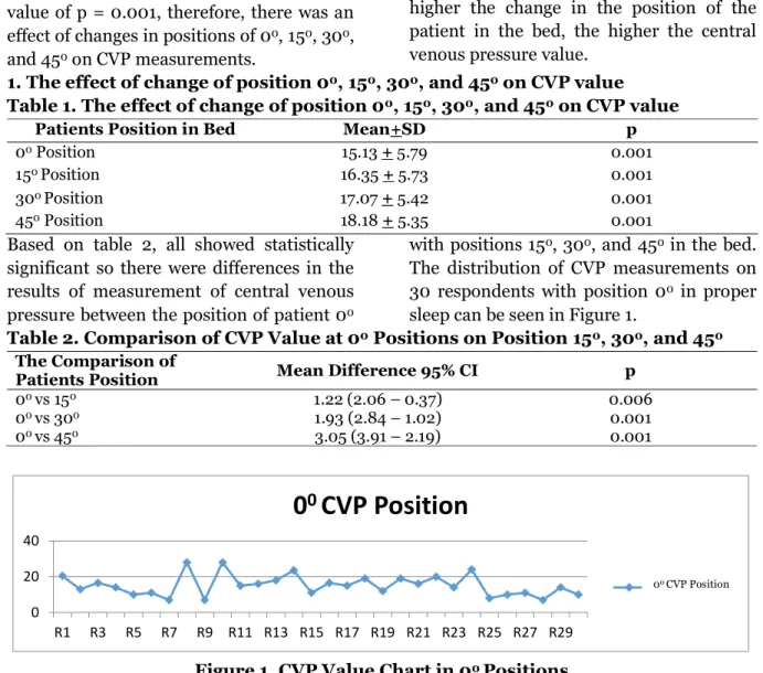
Based on table 2, all showed statistically significant so there were differences in the results of measurement of central venous pressure between the position of patient 00
with positions 150, 300, and 450 in the bed.
The distribution of CVP measurements on 30 respondents with position 00 in proper
sleep can be seen in Figure 1.
Table 2. Comparison of CVP Value at 00 Positions on Position 150, 300, and 450
The Comparison of
Patients Position Mean Difference 95% CI p
00 vs 150 1.22 (2.06 – 0.37) 0.006
00 vs 300 1.93 (2.84 – 1.02) 0.001
00 vs 450 3.05 (3.91 – 2.19) 0.001
0 20 40
R1 R3 R5 R7 R9 R11 R13 R15 R17 R19 R21 R23 R25 R27 R29
0
0CVP Position
CVP 0 derajat
Figure 1. CVP Value Chart in 00 Positions
Table 3. Comparison of CVP Value at 150 Positions on Position 00, 300, and 450
The Comparison of
Patients Position Mean Difference P
150 vs 00 1.22 (0.37 – 2.06) 0.006
150 vs 300 0.72 (1.42 – 0.01) 0.047
150 vs 450 1.83 (2.76 – 0.91) 0.001
Table 3 showed a significant difference. This was shown by the three comparisons of these positions statiscally significant. Among the three comparisons, the most significant difference was in the ratio of
position 150 vs position 450, this was
indicated by the value of p = 0.001. The distribution of CVP values to respondents with position 150 can be seen in figure 2
below.
0 50
R1 R3 R5 R7 R9 R11 R13 R15 R17 R19 R21 R23 R25 R27 R29
15
0CVP Position
CVP Posisi 15 Derajat
Figure 2. CVP Value Chart in 150 Positions
Table 4. Comparison of CVP Value at 300 Positions on Position 00, 150, and 450
The Comparison of
Patients Position Mean Difference 95% CI p
300 vs 00 1.93 (1.02 – 2.84) 0.001
300 vs 150 0.72 (0.01 – 1.42) 0.047
300 vs 450 1.12 (1.60 – 0.63) 0.001
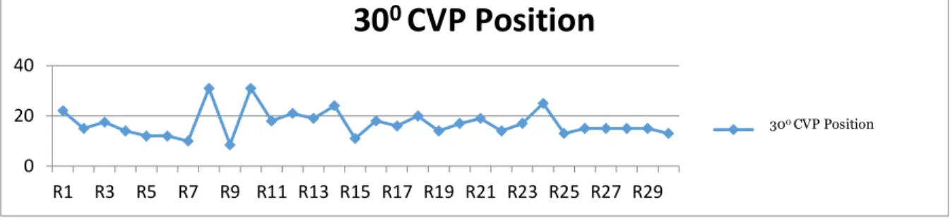
Table 4 showed that the three comparisons of these positions showed significant diffe-rences. Three comparisons of these
posi-tions were statistically significant. The dis-tribution of CVP values to respondents with position 300 can be seen in Figure 3.
0 20 40
R1 R3 R5 R7 R9 R11 R13 R15 R17 R19 R21 R23 R25 R27 R29
30
0CVP Position
CVP Posisi 30 Derajat
Figure 3. CVP Value Chart in 300 Positions
Table 5. Comparison of CVP Value at 450 Positions on Position 00, 150, and 300
The Comparison of
Patients Position Mean Difference P
450 vs 00 3,05 (2.19 – 3.90) 0.001
450 vs 150 1.83 (0.91 – 2.76) 0.001
450 vs 300 1.12 (0.63 – 1.60) 0.001
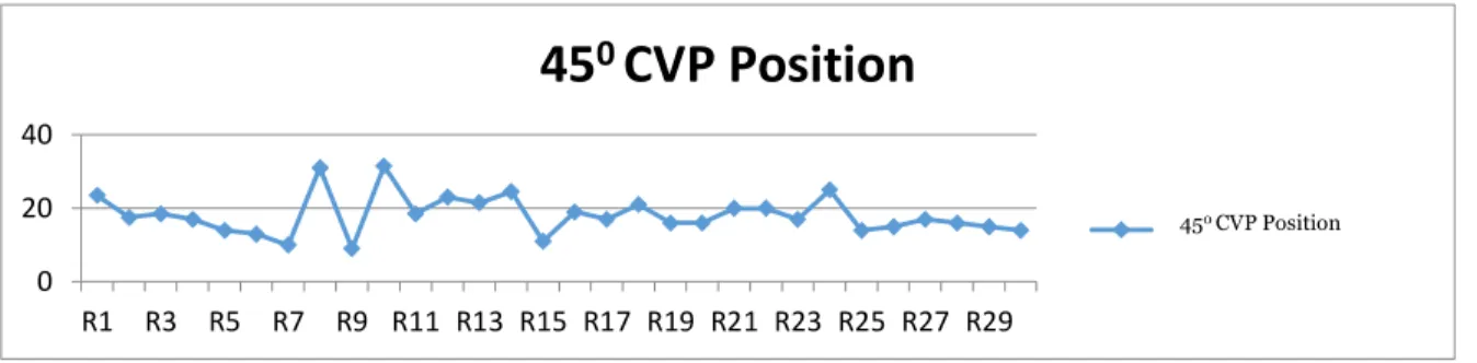
Table 5 showed that three comparisons showed were statistically significant, there-fore, there was a difference in CVP values in the ratio of 450 vs. 00 position, 450 vs 150
position, and 450 vs. 300 position. This was
different from the comparison of the
posi-tion of patients in bed previously, in the pa-tient with 450 position compared with 00,
150, and 300, the distribution of CVP values
to respondents with position of 450 can be seen in Figure 1.4 below.
150 CVP Position
0 20 40
R1 R3 R5 R7 R9 R11 R13 R15 R17 R19 R21 R23 R25 R27 R29
45
0CVP Position
CVP Posisi 45 Derajat
Figure 4. CVP Value Chart in 450 Position
DISCUSSIONS
Monitoring of central venous pressure can be useful in assessing heart function, circu-lating blood volume, vascular tone, and patient response to therapy. CPV can be measured precisely with the patient in the supine position and the head elevated by 45º (Magerman, 2010). The position of the patient's body when measuring the central venous pressure has an effect on the mea-surement results. The research conducted by Nakao, et al. quoted by Arthur, et al. (2008) stated that body position has been shown to affect the diameter, extent and shape of the inferior vena cava and this can indirectly affect central venous pressure. Study by Arthur, et al. (2008) aimed to prove the effect of inferior vena cava dia-meter on central venous pressure where it was found that there was a correlation between central venous pressure and infe-rior vena cava diameter in patients with normal ejection fraction.
Based on the results of the study, it can be concluded that the position of the client at 0º, the CVP value tend to be lower compared to the respondents at positions of 15º, 30º, and 45º. Likewise vice versa on the position of 45º, CVP values tend to be higher compared to 0º, 15º and 30º positions.
Siegenthaler et al. (2014), stated that factors affecting the measurement of cen-tral venous pressure, venous return/cardiac
output, total blood volume, regional vascu-lar tone. This was in line with Morton et al. (2014), who stated that the supine position is 0º, the gravitational force affects the whole body evenly or equally. In the lying position supination the effects of gravity on the body would decrease which made more blood flow back to the heart through the blood vessels. If the blood returned more to the heart, then the body can pump more blood every beat. This mean that the heart rate was needed a minute to fulfill blood needs, oxygen and nutrients would be less.
In the lying position, the blood was back against the force of gravity. It can be seen that while working in a standing posi-tion, the contents of the stroke increased linearly and reached the highest score of 40% to 60% VO2 maximum. VO2 Max was the maximum volume of O2 that was pro-cessed by the human body during intensive activities. In the lying position, in a state of rest the key content approaches the maxi-mum value, while there was only a slight in-crease in work. Wong (2000) stated that any increase of 100 from the head would cause a decrease in intracranial pressure of 1 mmHg. This increase in head position would increase the back flow of blood from the brain towards the heart through the superior vena cava, so that there was an increase in the amount of blood returning to the heart which increased volume and also affected central venous pressure.
The value of lying in a resting position was almost the same as the maximum value obtained at work with a standing position. The number of buds in male adults has a variation between 70 to 100 ml. The greater the work intensity (exceeding the 80% limit of work capacity), the smaller the contents, this was due to shortening of diastolic fil-ling time due to increased heart rate (if it reached 180/minute, then 1 cycle of heart rate lasted only 0.3 seconds and diastole fil-ling was part of 0.3 seconds (Guyton, 2002).
Changes in body posture from the su-pine position to the upright position change caused mechanical changes due to the effects of gravity on the circulatory system and changes caused by the nerve reflex res-ponse produced. Changes in body posture from 300 and 600 and 00 Head Up Till.
Cardiovascular response to changes in position 300 showed a lower Cardiac Index
value compared to 0 positions, a baro-reseptor mechanism of peripheral arteries as peripheral vasoconstriction which main-tained pressure from decline. However, the change in position to 30◦ resulted in a reduced stroke volume due to reduced venous return and reduced preload. Asso-ciated with changes in position with respect to circulation, the change in position 30◦ also raised circulation values on the peri-phery, where the value was measured by using the CAVI value (Cardiac Ankel Vas-cular Index) which functioned as an arterial stiffness index to estimate afterload. In addition, in the phase of influence of ortho-static stress, an increase in receptor gain contributed to the hyperactivity of sym-pathetic nerves and ultimately increased peripheral resistance (Zwain et al., 2013).
45◦ angle position was a position that increased cardiac and ventricular out-put and facilitated elimination of vecal and voiding, in this position the bed was raised
by 45º and the client's knees were slightly elevated so that there were no circulatory barriers to extremity (Potter and Perry, 2005). The supination position of 45º tends to be more stable, this was because the sympathetic vasoconstrictor system was stimulated and the signals were transmitted simultaneously through the skeletal nerves to the skeletal muscles of the body, espe-cially the abdominal muscles. This situation would increase basic tone, the muscles suppress the entire abdominal vein to help expel blood from the vascular abdominal reserve of the heart. This made the amount of blood available to the heart to pump up to increase. The entire response was called the abdominal compression reflex (Guyton and Hall, 2002).
below the heart), was 180 mmHg. The simi-lar thing applied to venous pressure. So it can be concluded that with changes in posi-tion there was a change in central venous pressure.
Another thing that needed to be con-sidered in nursing if the patient was changed from using a CVP water monitor-ing system to a pressure transducer system, the nurse must remember that the normal values in the two systems were different. CVP measured by water manometers which normally range from 5 to 8 cm H20 and CVP measured by transducer pressure nor-mally ranges from 0 to 6 mmHg. The value was different because merquri was heavier than water. To change the value in cmH02 to mmHg, the value in cmH20 was divided by 1.36. The tendency of the most signifi-cant value was shown when it was associa-ted with the patient's cardiovascular dys-function and the response to intervention.
Decreased CVP values indicated hypo-volemic status that often required adminis-tration of fluids. Responses anticipation of fluid therapy was an increase in CVP. In the same way, diuretic therapy decreased intra-vascular volume and was associated with a decrease in CVP value. Low CVP values or decreased CVP values can also be asso-ciated with vasodilatation due to sepsis or vasodilatation drugs, both of which caused relative hypovolemic because blood volume did not change. More precisely, the intra-vascular space became bigger according to the patient's blood volume (Convertino et al., 2001)
The enhancement of CVP value of pa-tients can be caused by a number of com-plex and interconnected factors, each of which required precision. Right ventricular failure and mechanical ventilation were two common causes of increasing CVP values. Excessive intravascular volume and hyper-volaemia rarely cause an increase in CVP
values. Mechanical ventilation increased intra-thoracic pressure, which was passed on to the vascularization of the lungs, heart and large blood vessels. The CVP value can be directly affected by this pressure. The CVP value can also increase because intra-thoracic pressure suppresses large blood vessels. The CVP value can be directly affec-ted by intrathoracic pressure pressing on the pulmonary arteries resulting in resist-ance to the right ventricular back pulmo-nary blood flow, right atrium and vena cava. In severe cases, increased intrathora-cic pressure caused by mechanical ventila-tion resulted in severe right ventricular dys-function, increased intrathoracic pressure caused by increased mechanical ventiation due to a decrease in blood flow up and blood supply and pressure in the right atri-um and vena cava (Randazzo et al., 2003).
examinations/observations, such as aus-cultation of breath sounds, heart rate and respiratory frequency, EKG examination results, distention of the jugular vein and urine output. Patients who experienced sepsis may have low CPV values due to an increase in heart rate but there were no other clinical symptoms. The value of CPV did not have an accurate meaning, but when it was used in conjunction with other clinical data, the value of CVP would be very useful in the management of predic-ting the clinical course of the patient's dise-ase.
In this study, it showed that the gravi-ty factor (patient position) in the measure-ment showed a difference in good position from 0º, 15º, 30º and 45º position, there was an increase in the value of the measure-ment of central vein. The huge change in the position of the respondent from the ini-tial position would cause a large change in the results of the CVP measurement. The biggest change in position was found in changes in 0º vs 45º and the smallest cha-nge occurred from 15º vs 30º. But overall, the change in the position of the patient in bed from 00 position to 15º, 30º, and 45º
positions, changes from 15º to 0º, 30º, and 45º positions, changes in 30º to 0º, 15º, and 45º positions, and changes in 45º to 0º, 15º, and 30º positions caused changes in the measurement results of CVP.
AUTHORS CONTRIBUTION Hendy Lesmana collected data and proces-sed data analysis. Maria Imaculata Ose exa-mined the conceptual framework and sug-gested the methodology. Rahmatuz Zulfia and Kurniaty Ika Sari Tobing interpreted the results of data analysis.
FUNDING AND SPONSORSHIP This study was funded by the Head of the Institute for Research and Service to the
Borneo Tarakan University community.
ACKNOWLEDGEMENT
The author would like to say thank you to the Head of the Institute for Research and Service to the people of Borneo Tarakan University who had supported the funds so that this study can be finished.
CONFLICT OF INTEREST There was no conflict of interest.
REFERENCE
Arthur ME, Landolfo C, Wade M, Castre-sana (2009). Inferior vena cava dia-meter (IVCD) measured with trans-esophageal echocardiography (TEE) can be used to derive the central ve-nous pressure (CVP) in anesthetized mechanically ventilated patients. Journal of CV Ultrasound & Allied Tech.
Convertino VA, Ludwig DA, Elliott JJ, Wade CE, Assistance T, Owens R (2001). Evidence for central venous pressure resetting during initial expo-sure to microgravity, 2021–2028. Guyton AC, Hall JE (2014). Buku Ajar
Fisiologi Kedokteran (Textbook of Medical Physiolog). Edisi 12. Jakarta. EGC.
Jevon P, Ewens B, Pooni JS (2009). Pe-mantauan Pasien Kritis (Critical Pati-ent Monitoring). Edisi ke II. Erlangga Medikal Series. Jakarta.
Magerman Y (2010). Central Venous Pres-sure. Lecturer Notes, 16.
Morton PG (2014). Keperawatan Kritis Pendekatan Asuhan Holistik (Critical Nursing Holistic Care Approach). Potter PA, Perry AG (2005). Buku Ajar
Process-es, and Practices). 4(2). Jakarta. EGC Randazzo MR, Snoey ER, Levitt MA, Binder
K (2003). Accuracy of Emergency Physician Assessment of Left Ventri-cular Ejection Fraction and Central Venous Pressure Using Echocardio-graphy, 10(9). https://doi.org/10.11-97/S1069-6563(03)00317-8.
Siegenthaler N, Giraud R, Saxer T, Cour-vosier DS, Romand J, Bendjelid K (2014). Haemodynamic Monitoring in the Intensive Care Unit: Results from
a Web-Based Swiss Survey, 2014. https://doi.org/10.1155/2014/129593 Wong F (2000). Prevention of Secondary
Brain Injury. Critical Care Nurse. 20(5): 18-27.