Combined HIV-1 Envelope Systemic and Mucosal Immunization of
Lactating Rhesus Monkeys Induces a Robust Immunoglobulin A
Isotype B Cell Response in Breast Milk
Cody S. Nelson,aJustin Pollara,aErika L. Kunz,aThomas L. Jeffries, Jr.,aRyan Duffy,aCharles Beck,aLisa Stamper,aMinyue Wang,a Xiaoying Shen,aDavid J. Pickup,a Herman F. Staats,aMichael G. Hudgens,bThomas B. Kepler,cDavid C. Montefiori,a
M. Anthony Moody,aGeorgia D. Tomaras,aHua-Xin Liao,aBarton F. Haynes,aGuido Ferrari,aGenevieve G. A. Fouda,a Sallie R. Permara
Human Vaccine Institute, Duke University Medical Center, Durham, North Carolina, USAa
; Department of Biostatistics, University of North Carolina, Chapel Hill, North Carolina, USAb
; Department of Microbiology, Boston University School of Medicine, Boston, Massachusetts, USAc
ABSTRACT
Maternal vaccination to induce anti-HIV immune factors in breast milk is a potential intervention to prevent postnatal HIV-1
mother-to-child transmission (MTCT). We previously demonstrated that immunization of lactating rhesus monkeys with a
modified vaccinia Ankara (MVA) prime/intramuscular (i.m.) protein boost regimen induced functional IgG responses in milk,
while MVA prime/intranasal (i.n.) boost induced robust milk Env-specific IgA responses. Yet, recent studies have suggested that
prevention of postnatal MTCT may require both Env-specific IgA and functional IgG responses in milk. Thus, to investigate
whether both responses could be elicited by a combined systemic/mucosal immunization strategy, animals previously
immu-nized with the MVA prime/i.n. boost regimen received an i.n./i.m. combined C.1086 gp120 boost. Remarkably, high-magnitude
Env-specific IgA responses were observed in milk, surpassing those in plasma. Furthermore, 29% of vaccine-elicited Env-specific
B cells isolated from breast milk were IgA isotype, in stark contrast to the overwhelming predominance of IgG isotype
Env-spe-cific B cells in breast milk of chronically HIV-infected women. A clonal relationship was identified between Env-speEnv-spe-cific blood
and breast milk B cells, suggesting trafficking of that cell population between the two compartments. Furthermore, IgA and IgG
monoclonal antibodies isolated from Env-specific breast milk B cells demonstrated diverse Env epitope specificities and multiple
effector functions, including tier 1 neutralization, antibody-dependent cellular cytotoxicity (ADCC), infected cell binding, and
inhibition of viral attachment to epithelial cells. Thus, maternal i.n./i.m. combined immunization is a novel strategy to enhance
protective Env-specific IgA in milk, which is subsequently transferred to the infant via breastfeeding.
IMPORTANCE
Efforts to increase the availability of antiretroviral therapy to pregnant and breastfeeding women in resource-limited areas have
proven remarkably successful at reducing HIV vertical transmission rates. However, more than 200,000 children are infected
annually due to failures in therapy implementation, monitoring, and adherence, nearly half by postnatal HIV exposure via
ma-ternal breast milk. Intriguingly, in the absence of antiretroviral therapy, only 10% of breastfed infants born to HIV-infected
mothers acquire the virus, suggesting the existence of naturally protective immune factors in milk. Enhancement of these
protec-tive immune factors through maternal vaccination will be a critical strategy to reduce the global pediatric AIDS epidemic. We
have previously demonstrated that a high magnitude of HIV Env-specific IgA in milk correlates with reduced risk of infant HIV
acquisition. In this study, we describe a novel HIV vaccine regimen that induces potent IgA responses in milk and therefore
could potentially protect against breast milk HIV MTCT.
M
ore than 200,000 new pediatric human immunodeficiency
virus (HIV) infections occur annually via mother-to-child
transmission (MTCT), nearly half through breastfeeding (1).
An-tiretroviral (ARV) drugs can dramatically reduce the rate of
MTCT, but in areas of high HIV prevalence, acute HIV infection
in pregnant and postpartum women as well as poor access and
adherence to ARV treatment throughout the breastfeeding period
has limited progress in the prevention of breast milk transmission
(2). According to UNAIDS in 2014, only 68% of HIV-infected
pregnant women in low- and middle-income countries received
ARV therapy during pregnancy, and only 61% of those women
continued this therapy postpartum (3). Despite the risk of HIV
acquisition, breastfeeding is necessary for infant survival in many
regions of the world, as breastfed infants have lower rates of
diar-rheal and respiratory infections (4). It is well established that
an-tibodies are transferred to infants via the placenta and through
breast milk consumption (5); thus, maternal immunization could
Received22 February 2016 Accepted24 February 2016
Accepted manuscript posted online2 March 2016
CitationNelson CS, Pollara J, Kunz EL, Jeffries TL, Jr, Duffy R, Beck C, Stamper L, Wang M, Shen X, Pickup DJ, Staats HF, Hudgens MG, Kepler TB, Montefiori DC, Moody MA, Tomaras GD, Liao H-X, Haynes BF, Ferrari G, Fouda GGA, Permar SR. 2016. Combined HIV-1 envelope systemic and mucosal immunization of lactating rhesus monkeys induces a robust immunoglobulin A isotype B cell response in breast milk. J Virol 90:4951–4965.doi:10.1128/JVI.00335-16.
Editor:G. Silvestri
Address correspondence to Sallie R. Permar, [email protected].
be an important alternative strategy to allow safe breastfeeding in
areas of high HIV prevalence.
The specificity and function of antibodies important to prevent
mucosal HIV transmission remain unclear. Despite the high level
of total IgA in breast milk, the predominant envelope
(Env)-spe-cific antibody response in the breast milk of both HIV-infected
women and simian immunodeficiency virus (SIV)-infected
rhe-sus monkeys is IgG (6,
7). The presence of high levels of functional
IgG in breast milk, particularly IgG mediating
antibody-depen-dent cellular cytotoxicity (ADCC), has been linked to reduced
incidence of MTCT (8). Env-specific IgG in breast milk is likely
due to transudate from the systemic compartment, since HIV
Env-specific IgG responses in breast milk correlate well with those
in plasma, though they are lower by two orders of magnitude (6).
Interestingly, the majority of Env-specific B cells in the breast milk
of chronically HIV-infected women produce IgG and not IgA (9,
10). Indeed, it has traditionally proven to be exceedingly difficult
to induce Env-specific IgA or IgA isotype B cells in the mucosal
compartment via vaccination (11), and thus the potential role of
IgA in preventing viral transmission at the mucosal surface is
poorly understood.
The RV144 ALVAC/AIDSVAX vaccine efficacy trial in
Thai-land, which demonstrated 31.2% efficacy, suggested a deleterious
role of plasma Env-specific IgA responses of certain specificities in
protection against sexually transmitted HIV infections (12).
Fur-ther analysis revealed an inverse correlation between ADCC
activ-ity and acquisition risk in vaccinees with low IgA responses (12). It
was hypothesized that IgA blocks ADCC by competing for the
same binding sites as ADCC-mediating IgG antibodies. Indeed,
vaccine-elicited IgA specific for conformational epitopes in the C1
region of Env can block ADCC activity in clinical samples (13).
Nevertheless, there is solid evidence that mucosal IgA could
con-tribute to the prevention of HIV transmission. In a recent study,
vaccine-elicited mucosal IgA responses were associated with
pro-tection against vaginal simian-human immunodeficiency virus
(SHIV) challenge (14), and multiple investigations have
demon-strated that passive rectal immunization with neutralizing dimeric
IgA can protect from subsequent SHIV exposure (15,
16).
Simi-larly, we recently reported an association between a high
magni-tude of Env-specific IgA and low rates of breast milk HIV
trans-mission (17). Thus, Env-specific IgA responses in the systemic and
mucosal compartments seem to play dramatically different
phys-iologic roles.
In previous studies, we demonstrated that HIV vaccination can
elicit strong Env-specific antibody responses in milk of rhesus
monkeys (18). Hormonally induced lactating animals were
im-munized with the transmitted/founder Env C.1086 using either a
gp140 DNA or modified vaccinia Ankara (MVA) prime followed
by two C.1086 gp120 protein boosts delivered either systemically
(intramuscularly [i.m.]) or mucosally (intranasally [i.n.]). The
systemic and mucosal vaccine regimens elicited comparable levels
of plasma Env-binding IgG, yet mucosal immunization induced
significantly higher Env-binding IgA responses in breast milk.
Furthermore, systemic vaccination induced a much more potent
functional IgG response (tier 1 neutralization, ADCC) in breast
milk than mucosal vaccination. An optimal vaccine regimen for
the prevention of breast milk transmission may need to elicit both
strong Env-binding and functional antibody responses in milk
(19).
Thus, we sought to design a vaccine strategy that could
com-bine the potent functional antibody response observed in the
sys-temically vaccinated animals with the robust Env-specific IgA
re-sponse in the mucosally vaccinated animals. Animals previously
immunized with the C.1086 MVA prime/i.n. boost regimen
re-ceived an additional simultaneous i.n./i.m. protein boost.
Env-binding and functional responses were then assessed in both
plasma and breast milk. To examine the antibody responses in
more detail, B cells from both blood and milk were isolated, V
H/V
Lgenes sequenced, and IgA and IgG monoclonal antibodies (MAbs)
analyzed for specificity and associated function. This study
pro-vides a detailed understanding of the antibody-mediated immune
responses elicited by combined systemic and mucosal vaccination,
enabling critical assessment of this maternal vaccination strategy
for the prevention of HIV-1 breast milk transmission.
MATERIALS AND METHODS
Animals and vaccine regimen.Recombinant MVA expressing the C.1086
Env gp140 gene and recombinant Env gp120 glycoproteins was generated as described previously (18). Lactation was induced in 4 female Indian rhesus monkeys by depot medroxyprogesterone and estradiol injections followed by oral dopamine antagonist administration as described previ-ously (20). The lactating monkeys were initially primed with 109PFU of
recombinant MVA (rMVA) expressing the HIV C.1086 Env gene and boosted twice intranasally (i.n.) at weeks 12 and 16 with 200g of HIV C.1086 gp120 protein (100g in each nostril) adjuvanted with the Toll-like receptor 7/8 (TLR7/8) agonist R848 (500g/animal) (Fig. 1). The i.n. boosted animals were immunized a third time with a combined i.n./i.m. boost technique at week 50 following the original MVA prime. The i.n. component of the combined boost was identical to the previous i.n. boosts (200g HIV Env C.1086 gp120 plus 500g R848). The intramuscular (i.m.) component of the combined boost consisted of 100g HIV Env C.1086 gp120 and 250l of the adjuvant STR8S-C (squalene-containing STS base adjuvant plus R848 plus oCpGs) (21). i.n. protein boosts were administered to anesthetized monkeys placed on their backs in 30-l aliquots at 30-s intervals for a total volume of 150l per nostril (22). Between aliquot administrations, the nares were held closed. The i.m. protein boost was administered at a single injection site in the quadriceps of anesthetized monkeys. Blood samples were collected weekly for 4 weeks following immunization and then bimonthly for an additional 1 to 2
FIG 1Combined i.n./i.m. HIV-1 Env vaccine regimen. Lactation was
months. Milk was collected twice weekly by manual collection (20) and then separated into cellular, supernatant, and lipid fractions by centrifu-gation (7,23). Animals were maintained according to theGuide for the Care and Use of Laboratory Animals(grants.nih.gov), and the animal pro-tocol was approved by the Duke University Animal Care and Use Com-mittee.
Plasma, milk, and MAb HIV-1 Env recombinant protein ELISA. Env-binding IgG and IgA were assessed for plasma/milk (18) and for monoclonal antibodies (MAbs) (24). For plasma/milk assays, 384-well enzyme-linked immunosorbent assay (ELISA) plates were coated over-night with HIV C.1086 gp120 (30 ng/well) and then blocked with the assay diluent (phosphate-buffered saline [PBS] containing 4% whey, 15% nor-mal goat serum, and 0.5% Tween 20). Dilutions of plasma or milk were then added to the plates. Antibodies were detected with a horseradish peroxidase (HRP)-conjugated polyclonal goat anti-monkey IgG (Rock-land, Gilbertsville, PA) or IgA (Rock(Rock-land, Gilbertsville, PA) and the addi-tion of the ABTS-2 peroxidase substrate system (KPL, Gaithersburg, MD).Macaca mulattapurified IgG and IgA (Nonhuman Primate Reagent Resource) were used to develop standard curves, and the concentration of IgG or IgA antibody was calculated relative to the standard using a 5-pa-rameter fit curve (WorkOut 2.5; PerkinElmer, Waltham, MA). For re-combinant MAb ELISA, 384-well ELISA plates (Corning Life Sciences, Lowell, MA) were coated with each antigen at 30 ng/well. Antigens in-cluded C.1086 gp120 D7 and A1.con env03 gp140 (12), B.con env03 gp140 (12), C.con env03 gp140 (12), AE.A244 gD gp120 (12), C.1086 V1V2, C.1086 V2, C.1086 V1V2 N156Q, and gp70 B.CaseA2 V1V2 (18), gp70 B.CaseA2 V1V2/169K (18), C.con env03.V3 linear peptide and BioRV144 C1 peptide (13), C.YU2 core, C.YU2 core D368R, RSC3 (25), RSC3 d371/P363N (25), and C.1086 D7 gp120 K160N, ConC gp120, and ConC gp120 N332A. Serial dilutions of monoclonal antibody were dis-tributed to the plates after blocking, and the MAbs were detected using a peroxidase-conjugated goat anti-monkey IgG (Rockland, Gilbertsville, PA) and the SureBlue Reserve tetramethylbenzidine (TMB) peroxidase substrate (KPL, Gaithersburg, MD). For monoclonal antibodies, the 50% effective concentration (EC50) was calculated by the concentration of
an-tibody which resulted in a 50% reduction in optical density (OD) from the maximum value.
Quantitative SIgA ELISA.ELISA plates (Corning Life Sciences,
Low-ell, MA) were coated overnight at 4°C with Con6 gp120 B. Dilutions of breast milk were added to the plate following blocking. The antibodies were detected using anti-secretory-chain IgA Ab SC 9H7 CL3 (a mouse-derived monoclonal IgG2A kappa antibody which cross-reacts with rhe-sus and human secretory components [SCs] though does not react with dimeric IgA alone) (kindly provided by Barton Haynes), followed by an HRP-conjugated polyclonal anti-mouse IgG (Promega, Madison, WI) and then TMB peroxidase substrate. The standard used was b12 secretory IgA (SIgA) prepared by complexing b12 dimeric IgA (dIgA) (NHP re-agent program), with rhesus SC followed by an overnight incubation, resulting in a 1:1 molar ratio of dIgA to SC. The concentration of HIV-1 Env-specific SIgA antibody was calculated relative to the standard curve using a 5-parameter fit curve (WorkOut 2.5; PerkinElmer, Waltham, MA). A positive result was defined as the mean OD of preimmune milk samples plus 3 standard deviations (SD). The limit of detection (LOD) in this assay was determined to be 100 ng/ml based upon the lowest concen-tration of the dIgA-SC standard with a corresponding OD that exceeded 3 times that of the blank wells.
sCD4 and A32 blocking ELISA.Assessment of antibody specificity for
the CD4bs and C1 conformational epitopes was done as described previ-ously (24). Briefly, 384-well plates were coated overnight with C.1086 gp120 (30 ng/well) and then blocked before addition of serially diluted MAbs. For the soluble CD4 (sCD4) blocking ELISA, sCD4 (a gift from Bing Chen) was next added to the plates (6.4 ng/well), followed by addi-tion of a biotinylated anti-CD4 (0.3 ng/well). For the A32 blocking ELISA, biotinylated A32 antibody was added (0.625 ng/well). In both assays, HRP-conjugated streptavidin (at a 1:30,000 dilution) and SureBlue
Re-serve TMB peroxidase substrate were used to detect the biotinylated an-tibodies. Background was normalized using negative-control wells that received only assay diluent in place of primary monoclonal antibody. Percent inhibition was calculated as follows: 100⫺(serum triplicate mean/no-inhibition control mean)⫻100. A reduction of absorbance by
⬎50% by a MAb indicated blocking of sCD4 binding to gp120 or blocking of A32 MAb binding to the C1 region (26).
TNC ELISA.A 384-well plate was coated with 2g/ml of anti-tenascin
C (anti-TNC) antibody rabbit polyclonal IgG (H-300; Santa Cruz Bio-technology) and incubated at 4°C overnight. Wells were washed with PBS plus 0.1% Tween 20 and blocked with 7.5% bovine serum albumin (BSA) (Gibco) at room temperature for 1 h. Milk samples delipidized by centrif-ugation at 21,000⫻gwere diluted in 7.5% BSA, and a commercially available purified TNC (Millipore) was used as the protein standard, rang-ing from 5g/ml to 5 ng/ml. The standards and samples were added to the plate and incubated for 1 h at room temperature. Plates were washed 2 times, and 1g/ml of tenascin C mouse monoclonal antibody (T2H5; Fisher) was added and incubated for 1 h at room temperature. Plates were washed 2 times, and a 1:10,000 dilution of goat anti-mouse HRP-conju-gated antibody (Promega) was added to each well and incubated for 1 h at room temperature. Plates were washed 4 times, and SureBlue Reserve TMB microwell substrate (KPL) was added and incubated in the dark at room temperature for 5 min. TMB stop solution (KPL) was added, and plates were read at 450 nm.
Peptide array serum specificity mapping.Serum epitope mapping of
heterologous strains was performed as described previously with minor modifications (27,28). In short, a peptide library of overlapping peptides (15-mers overlapping by 12), covering 7 full-length HIV-1 gp160 Env consensus sequences (clades A, B, C, and D, group M, CRF1, and CRF2) and 6 vaccine and laboratory strain gp120 sequences (A244_1, TH023_1, MN_B, 1086_C, TV1_C, and ZM651_C), was printed onto epoxy glass slides (provided by JPT Peptide Technologies GmbH [Germany]). Se-quences of all peptides in the library have been previously reported (29). Microarray binding was performed using the HS4800 Pro Hybridization Station (Tecan, Männedorf, Switzerland). All arrays were blocked with blocking buffer (PBS, 1% milk, 5% normal goat serum [NGS], 0.05% Tween 20) for 1 h at 30°C, followed by a 2-h incubation at 30°C with serum diluted 1:50 in blocking buffer. Arrays were subsequently incu-bated for 45 min at 30°C with goat anti-human IgG conjugated with AF647 (Jackson ImmunoResearch, PA) (0.75g/ml final concentration) diluted with blocking buffer. Washes between all steps were with PBS containing 0.1% Tween 20. Arrays were scanned at a wavelength of 635 nm using an Axon Genepix 4300 scanner (Molecular Devices, Sunnyvale, CA, USA) at a photomultiplier tube (PMT) setting of 580 and 100% laser power. Images were analyzed using Genepix Pro 7 software (Molecular Devices). The intensity of binding of the postimmunization serum to each peptide was corrected with its own background value, which was defined as the median signal intensity of the prebleed serum for that peptide plus 3 times the standard errors among the 3 subarray replicates present on each slide.
Neutralization.Neutralization of clade C tier 1 (C.MW965) and
au-tologous tier 2 (C.1086) HIV-1 pseudovirus variants by plasma, milk, and isolated MAbs was measured in TZM-bl cells via a reduction in luciferase reporter gene expression (30). In brief, dilutions of plasma, milk, or MAbs were incubated with an optimized amount of virus for 45 min at 37°C in a 96-well plate, and then freshly trypsinized TZM-bl cells in growth me-dium were added to each well. Following a subsequent 48-h incubation, the culture medium was removed and replaced with a luciferase reagent (Bright-Glo; Promega), causing cell lysis and luminescence proportional to the amount of infection. The luminescence was measured using a Vic-tor 2 luminometer (PerkinElmer). The 50% inhibiVic-tory dilution (ID50)
MAbs that mediated tier 1 virus neutralization were further screened against a multiclade panel of tier 1 viruses B.BaL.26, B.MN.3, and B.SF162.
ADCC.The ADCC activity of plasma, delipidized breast milk, purified
IgG, and MAbs was assessed using the GranToxiLux (GTL) assay (32). Briefly, CEM.NKRCCR5cells (NIH AIDS Reagent Program, Division of AIDS, NIAID, NIH; from Alexandra Trkola) (33) were used as targets after coating with recombinant C.1086 gp120 protein (5g/ml). Cryo-preserved human peripheral blood mononuclear cells (PBMC) from an HIV-seronegative donor with the heterozygous 158F/V genotype for Fc-gamma receptor IIIa were used as the source of the effector cells (34). Serial dilutions of plasma, milk, purified IgG, and MAbs were tested. The maximum percent granzyme B (GzB) activity was defined as the peak proportion of cells positive for proteolytically active GzB out of the total viable target cell population. The final results are expressed after subtrac-tion of the background percent GzB activity observed in wells containing effector and target cells in the absence of antibodies. ADCC endpoint titers and concentrations were determined by interpolating the dilutions of plasma and breast milk, or the concentrations of purified antibodies, that intersect the positive cutoff using GraphPad Prism software version 6.0f (GraphPad Software, Inc., La Jolla, CA).
ADCC inhibition.For studies with spiked breast milk (seeFig. 8), a
1:16 dilution of breast milk was spiked with previously determined opti-mal ADCC concentrations of either DH532 (10g/ml) or a mixture of ADCC-mediating antibodies (A32, 2G12, 7B2, and CH44 at 15 ng/ml). The results are reported as ADCC activity (percent granzyme B activity) and percent ADCC inhibition, calculated as the percentage of ADCC ac-tivity reduction based on the ADCC mediated by the IgG MAb or MAb mixture in prevaccination milk.
Isolation of C.1086 Env-specific B cells.The C.1086 Env-specific B
cell population was identified in blood, breast milk, and gastrointestinal (GI) tissue of all 4 monkeys via flow cytometry. Data were collected using the FACSAria2 instrument (BD Biosciences) with the FACSDiVa software and were analyzed by manual gating with FlowJo software. The complete gating strategy for the total B cell population was defined as CD3⫺CD14⫺ CD16⫺CD20⫹. The total numbers of identified B cells from blood and breast milk of each monkey were as follows: A6E030, 81,191 and 1,383; A6E042, 73,258 and 97; A6E088, 107,300 and 248; and AU71S, 34,859 and 14. Env-specific B cells were obtained from B cell population by conjuga-tion of C.1086 Env to dyes AF647 and Pac-Blue (Invitrogen) and selecconjuga-tion for double-positive B cells.
MAb genetic characterization and small-scale testing.Following B
cell sorting, the expressed Ig VHand VLgenes were amplified by reverse
transcription and nested PCR as described previously (35–37). The PCR products were purified and sequenced. Ig isotype was determined by se-quence homology. Somatic hypermutation frequencies, inferred V(D)J rearrangement, and CDR3 length were determined from Clonalyst (40). Overlapping PCR was performed to coexpress the variable-region PCR products with full-length IgG1 (heavy chain) and kappa or lambda (light chain) cassettes, and the PCR products were transiently transfected in 293T cells without the need for a cloning step. The supernatants of trans-fected cells were screened for reactivity against the following HIV Env proteins by small-scale ELISA: C.1086 gp140, C.10868 gp120, M.ConS gp120, B.MN gp120, A.9004SS gp120, M.ConS gp140, A.92RW020 gp140, B.BxB/Bal gp140, and C.CH505TFqODwV3. Six IgA and six IgG antibod-ies were selected for large-scale production based upon (i) the strength of binding to the immunogen gp120 region (those with higher OD against gp140 construct were excluded) and (ii) broad clade binding specificity. Heavy and light chains for the 12 MAbs selected were cloned into the pcDNA3.1⫹mammalian expression vector. The heavy chain for all anti-bodies, including IgA, included an IgG backbone. Plasmids were tran-siently transfected into 293F cells, and the antibody was purified using protein A beads as described previously (35).
Epithelial cell binding.The ability of breast milk Env-specific MAbs
to impede infectious virus binding to colonic epithelial cells was assessed
(24). In short, colonic HT-29 cells (ATCC) were grown to confluence on a 96-well flat-bottom plate in modified McCoy’s 5A medium supple-mented with 10% FBS and antibiotics. The HT-29 cells were washed once with serum-free medium and treated with 100l of 50g/ml mitomycin C for 1 h to prevent further division, followed by two washes. The isolated MAbs then were diluted in serum-free medium, incubated with 10⫻ con-centrated HIV pseudovirus MW965 for 1 h at 37°C, added in quadrupli-cate to the colonic epithelial cell monolayer, and then finally incubated at 37°C for 4 h. Monolayers were washed twice with PBS to remove free virus, and 104TZM-bl reporter cells were added to each well of the
virus-bound monolayer. After 48 h, luciferase reagent (Bright-Glo; Promega) was added to the well, and the RLU were measured. Percent inhibition was calculated by dividing the RLU of each well by the median RLU of epithe-lium-bound virus that was not preincubated with isolated rhesus Abs. The IgG isoform of the influenza virus MAb CH65 was used as a negative control, whereas the IgG isoform of broadly neutralizing CD4 binding site MAb VRC01 was used as a positive control. The cutoff value was deter-mined by the mean plus 2 SD of CH65 IgG relative to no antibody.
Binding of MAbs to infected cells.The ability of MAbs to recognize
CEM.NKRCCR5infected with a C.1086 infectious molecular clone (IMC)
was assessed (32). CEM.NKRCCR5cells were infected with a C.1086 IMC
that encodes theRenillaluciferase reporter gene and preserves all HIV-1 open reading frames (38) using dextran-DEAE as described previously (32). At 48 h postinfection, the ability of MAbs to recognize the HIV-infected cells was measured by indirect surface immunofluorescence anal-ysis. The infected CEM.NKRCCR5cells were incubated with 10g/ml of
the rhesus-derived MAbs in R10 medium for 2 h at 37°C and then stained with a vital dye (Live/Dead Fixable Aqua Dead Cell Stain; Invitrogen) to exclude nonviable cells from subsequent analyses. Primary Ab binding was detected by secondary labeling with fluorescein isothiocyanate (FITC)-conjugated goat anti-rhesus IgG (SouthernBiotech Inc., Birming-ham, AL), and HIV-1-infected cells were identified by staining for intra-cellular expression of p24 (KC57-RD1; Beckman Coulter) using standard methods. Infected cell binding was evaluated by gating on live, p24⫹ events using FlowJo version 9.8 software (TreeStar, Inc., Ashland, OR).
IgG purification and concentration.IgG was isolated from plasma
and breast milk using protein G columns (6) (protein G resin prepacked into 96-well depletion plates [GE Healthcare]). Plasma was diluted 2-fold with Tris-buffered saline (TBS) (pH 7.5), and 200l of the diluted sample was added per well. For breast milk, 300l of undiluted sample was added to each well. The plates were incubated at room temperature, with shak-ing, for 1 h. The unbound fractions were removed by centrifugation at 700⫻gfor 3 min. Wells were then washed 3 times with 400l of TBS to remove loosely bound material. The IgG bound to the resin was eluted with 200l of 2.5% glacial acetic acid (pH 2.51) and immediately neu-tralized with 120l of 1 M Tris-HCl (pH 9.0). The eluted IgG fractions were concentrated using Amicon Ultra centrifugal filters (Millipore) with a 30,000-molecular-weight cutoff. The sample volume was reduced to 50
l by centrifugation at 14,000⫻gin a microcentrifuge precooled to 4°C. A buffer exchange was then performed using 2.0 volumes of PBS, pH 7.5. The concentrated IgG was assayed for protein concentration using a NanoDrop 8000 spectrophotometer (Thermo Fisher Scientific) using the IgG reference setting and then diluted to 1 mg/ml with PBS.
Statistical methods. Exact Wilcoxon signed rank tests were
per-formed for all outcome and time point comparisons described in the text. R software version 2.141.1 was used for calculations (R Foundation for Statistical Computing, Vienna, Austria). Note that 0.13 is the lowestP value attainable with 4 experimental animals.
RESULTS
Combined i.n./i.m. Env immunization increases Env-specific
IgA responses in breast milk.
In an attempt to elicit both
combined i.n./i.m. approach 50 weeks after the original MVA
prime (Fig. 1). Lactation was hormonally induced in the rhesus
monkeys prior to immunization (20). In both plasma and milk we
observed an increase in Env-specific IgA over the response
follow-ing i.n. Env boost alone. This increase was more pronounced in
milk yet was not statistically significant (median milk IgA after
boosts 2 and 3, 1,712 ng/ml and 7,641 ng/ml, respectively;
P
⫽
0.13) (Fig. 2). The magnitude of the Env-specific IgA response
elicited in breast milk following i.n./i.m. boost is remarkable and
distinct from that found in chronically HIV-infected individuals,
as median breast milk Env-specific IgA levels exceeded those in
plasma (median milk IgA
⫽
7,641 ng/ml and plasma IgA
⫽
6,200
ng/ml;
P
⫽
0.13). Interestingly, the vaccine-elicited antibody
re-sponses were quite durable over the 34 weeks prior to i.n./i.m.
boost 3 (week 50), with high-magnitude Env-specific IgG and IgA
measured in the breast milk of vaccinated monkeys prior to the
final boost. We next investigated what proportion of the
vaccine-elicited Env-specific IgA response was secretory (SIgA) by
assess-ing the amount of secretory component in milk (Fig. 2B). Due to
sample limitation after the third protein boost, SIgA in the breast
milk was measured only preimmunization and following boosts 1
and 2. Env-specific SIgA was detected in 3 of 4 animals following
i.n. boost 2, comprising only 9% of the total Env-specific IgA in
breast milk (median IgA
⫽
1,712 ng/ml and SIgA
⫽
152 ng/ml;
P
⫽
0.13). This finding suggests that the majority of
vaccine-elic-ited Env-specific IgA did not traffic to the breast milk
compart-ment via poly-Ig receptor interaction. Thus, the predominance of
vaccine-elicited Env-specific IgA antibodies in milk was likely
either passively transcytosed through mammary epithelia or
produced locally. The vaccine-elicited IgA responses in other
mucosal compartments were also assessed. Following i.n./i.m.
im-munization, Env-binding IgA increased in the vaginal
compart-ment, surpassing levels previously attained following i.n. Env
boosts (18). There was no detectable IgA in rectal secretions (Fig.
2C
and
D).
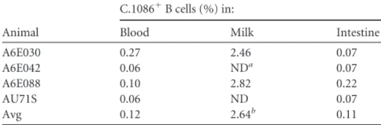
High proportion of Env-specific IgA isotype memory B cells
isolated from breast milk of i.n./i.m. Env-immunized lactating
monkey A6E030.
Env-specific B cells were isolated from blood,
FIG 2The combined i.n./i.m. HIV-1 Env boost enhances IgA responses in mammary and vaginal compartments over those elicited by i.n. boost alone. The
milk, and intestinal tissue using a fluorescently labeled C.1086 Env
probe (data not shown). Populations of Env-specific B cells were
identified in the blood and intestines of all four animals,
compris-ing an average of 0.12% and 0.11% of all B cells isolated in each
respective compartment. Two animals had low B cell counts in
breast milk (
⬍
100 total CD20
⫹cells). However, intriguingly, in
the remaining two animals with a significant population of breast
milk B cells, 2.64% on average were Env specific (Table 1). Based
upon the large number of breast milk B cells in monkey A6E030,
this animal was selected for investigation of the vaccine-elicited
Env-specific breast milk B cell repertoire. In total, 33 Env-specific
B cells were isolated from blood and 28 from breast milk. The
isotype distribution of the blood Env-specific MAbs was 97% IgG
and 3% IgA (Fig. 3E). In contrast, 29% of Env-specific MAbs in
the breast milk compartment were IgA, whereas 68% were IgG
and 3% IgM (Fig. 3F). This relatively high proportion of
IgA-producing B cells in milk suggests that i.n./i.m. vaccination may
induce localization of Env-specific IgA isotype B cells to the
mam-mary compartment. A similar analysis was conducted for breast
milk B cells isolated from animal A6E088; however, only 5
Env-specific B cells were isolated from blood and 5 from breast milk.
Though it is difficult to draw conclusions from the small number
of MAbs isolated from A6E088, 20% (1 of 5) of Env-specific B cells
were IgA isotype in both blood and breast milk
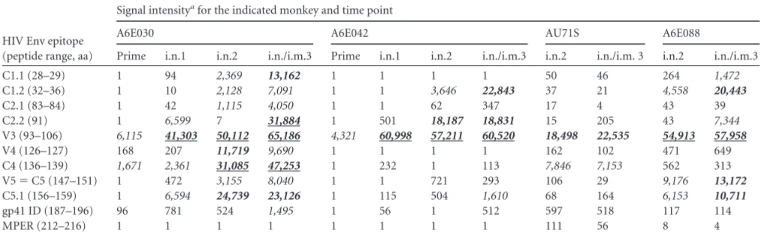
The i.n./i.m. Env vaccine regimen increases both the
magni-tude and breadth of the systemic Env epitope-specific IgG
re-sponse.
To determine the impact of our immunization regimen
on the fine specificity of the plasma IgG response, we used a
pep-tide array assay to assess binding to overlapping linear peppep-tides
covering 7 consensus strains and 6 vaccine strains of Env gp160.
The magnitude and breadth of the IgG response against Env
pep-tides increased following each vaccination (Table 2). In all
ani-mals, anti-V3 binding was the dominant response observed;
how-ever, all vaccinated animals also developed binding responses to
other linear regions, including C1, C2, V3, C4, and C5. The animal
with the highest-magnitude IgG response (A6E030) developed
notable epitope breadth with binding responses against all these
regions. The greatest increase in gp120 peptide binding
magni-tude was seen following the first i.n. Env boost, as evidenced in the
two animals for which the preimmune and i.n. boost 1 time points
TABLE 1Percentages of Env-specific memory B cells isolated from
blood, milk, and intestines of four i.n./i.m. Env-vaccinated lactating monkeys
Animal
C.1086⫹B cells (%) in:
Blood Milk Intestine
A6E030 0.27 2.46 0.07
A6E042 0.06 NDa 0.07
A6E088 0.10 2.82 0.22
AU71S 0.06 ND 0.07
Avg 0.12 2.64b 0.11
a
ND, none detected (CD20⫹total cell count of⬍100).
bAverage excluding values not detected due to low total CD20⫹cell count.
FIG 3Isolation of C.1086 Env-specific IgA-producing B cells from breast milk of animal A6E030. (A and B) Env-specific B cells were isolated by selection of cells
were measured. However, the i.n./i.m. Env boost introduced
ro-bust epitope expansion in all animals, with the exception of
mon-key AU71S.
Combined i.n./i.m. Env boost increases neutralizing activity
in breast milk but does not elicit ADCC responses.
We
previ-ously observed that systemic (i.m.) Env immunization, but not
mucosal (i.n.) immunization, was able to elicit neutralizing and
ADCC-mediating responses in the breast milk of lactating rhesus
macaques (18). Thus, we measured these functional antibody
re-sponses following combined i.n./i.m. Env immunization in both
TABLE 2i.n./i.m. vaccine regimen increases both linear epitope magnitude and breadth of systemic gp120-specific IgG responses
HIV Env epitope (peptide range, aa)
Signal intensityafor the indicated monkey and time point
A6E030 A6E042 AU71S A6E088
Prime i.n.1 i.n.2 i.n./i.m.3 Prime i.n.1 i.n.2 i.n./i.m.3 i.n.2 i.n./i.m. 3 i.n.2 i.n./i.m.3
C1.1 (28–29) 1 94 2,369 13,162 1 1 1 1 50 46 264 1,472
C1.2 (32–36) 1 10 2,128 7,091 1 1 3,646 22,843 37 21 4,558 20,443
C2.1 (83–84) 1 42 1,115 4,050 1 1 62 347 17 4 43 39
C2.2 (91) 1 6,599 7 31,884 1 501 18,187 18,831 15 205 43 7,344 V3 (93–106) 6,115 41,303 50,112 65,186 4,321 60,998 57,211 60,520 18,498 22,535 54,913 57,958
V4 (126–127) 168 207 11,719 9,690 1 1 1 1 162 102 471 649
C4 (136–139) 1,671 2,361 31,085 47,253 1 232 1 113 7,846 7,153 562 313 V5⫽C5 (147–151) 1 472 3,155 8,040 1 1 721 293 106 29 9,176 13,172
C5.1 (156–159) 1 6,594 24,739 23,126 1 115 504 1,610 68 164 6,153 10,711
gp41 ID (187–196) 96 781 524 1,495 1 56 1 512 597 518 117 114
MPER (212–216) 1 1 1 1 1 1 1 1 111 56 8 4
aMaximum binding (signal intensity) to a single peptide within each identified epitope. Italic, signal intensity of 1,000 to 10,000; bold italic, signal intensity of 10,000 to 30,000;
underlined bold italic, signal intensity of⬎30,000. Plasma samples for the postprime and post-i.n. boost 1 time points were not available for animals AU71S and A6E088 due to
sample limitation.
FIG 4Functional HIV-1 Env-specific antibody responses elicited in milk and plasma following combined i.n./i.m. Env immunization. (A and B) Neutralization
plasma and breast milk (Fig. 4). In each vaccinated, lactating
mon-key, the neutralization response against a tier 1 clade C variant,
MW965, was augmented in milk following the combined i.n./i.m.
Env boost (median ID
50s after boosts 2 and 3, 226 and 702,
respec-tively;
P
⫽
0.13) (Fig. 4B). Surprisingly one animal (A6E030) had
comparable neutralization activity in plasma and breast milk
(ID
50s of 5,974 and 4,796, respectively). The plasma of all animals
demonstrated ADCC antibody responses, which is consistent with
results seen in the MVA prime/i.n. boost group prior to
adminis-tration of the i.n./i.m. combined boost (18). ADCC responses
were enhanced in plasma following the i.n./i.m. boost (median
titers after boots 2 and 3, 5,994 and 81,498, respectively;
P
⫽
0.13)
(Fig. 4C). However, no ADCC activity was detectable in breast
milk following the i.n./i.m. boost.
The concentration of TNC does not increase in response to
vaccination.
Tenascin C (TNC) is an innate protein found in
breast milk, which we have previously described to have HIV-1
neutralizing function (39). Thus, we investigated whether an
in-crease in innate factors like tenascin C might play a role in the
enhanced neutralization titers following each immunization
ob-served in this study. However, measurement of breast milk TNC
concentration across time points of the immunization schedule
revealed that while TNC was detectable in rhesus monkey milk, it
was comparable between all 4 animals and 4 time points (Table 3).
We have previously found that the concentration of TNC in
hu-man breast milk ranges from 2.2 to 671
g/ml and that TNC has a
neutralization IC
50of 82 to 158
g/ml (39). Therefore, the low
level of tenascin C detected in the vaccinated monkey breast milk
across all time points (0.53 to 3.94
g/ml) suggests that the
se-quential increase in neutralization titers throughout the vaccine
schedule is not due to a change in the composition of innate breast
milk factors.
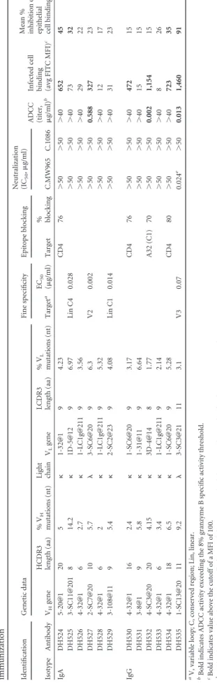
Genetic characteristics of IgA and IgG MAbs produced from
breast milk and blood Env-specific memory B cells.
Genetic
analysis of paired heavy and light chains from Env-specific blood
and breast milk B cells was conducted using Cloanalyst to
deter-mine the utilized V
Hand V
Lgenes, complementarity-determining
region 3 (CDR3) length, and mutation frequency (40).
Interest-ingly, we identified a B cell clonal lineage spanning the systemic
and mammary compartments (Fig. 5). This clonal lineage was
comprised of 8 IgG MAbs, including 6 from blood and 2 from
breast milk, which strongly suggests trafficking of vaccine-elicited
Env-specific IgG isotype B cells between the two compartments.
We found that the V
H4 gene family was predominant in blood and
breast milk compartments (66% of all MAbs). The median
heavy-chain CDR3 (HCDR3) length and mean percent V
Hmutation
frequency were 15 amino acids (aa) and 5% in blood and 13 aa and
5% in breast milk. Among the 28 MAbs isolated from A6E030
breast milk B cells, the median HCDR3 length and mean percent
V
Hmutation frequency were 9 aa and 5% for IgA but 15 aa and 5%
for IgG (Table 4). From these 28 milk MAbs, 6 IgA and 6 IgG
MAbs were selected for subsequent study based on the magnitude
and breadth of the Env-binding profile following transient
trans-fection. The HCDR3 length and percent V
Hmutation frequency
of the 6 IgA and 6 IgG MAbs selected were representative of the
total breast milk MAb population (Table 5).
i.n./i.m. Env vaccine-elicited IgA and IgG MAbs isolated
from breast milk B cells have variable Env epitope specificity.
First, the Env-binding breadth of the 6 IgA and 6 IgG antibodies
chosen for large-scale production was examined by ELISA using
TABLE 3The concentration of innate HIV-neutralizing factor tenascin
C in breast milk does not significantly change during the vaccination schedule
Animal
Tenascin C concn (g/ml) at time point:
Postprime Post-boost 1 Post-boost 2 Pre-boost 3
A6E030 0.53 1.47 1.37 2.47
A6E042 0.91 1.92 0.90 3.94
A6E088 1.30 0.66 2.69 2.22
AU71S 3.62 2.81 3.19 NAa
a
NA, sample not available.
FIG 5Clonal relationship of vaccine-elicited Env-specific blood and breast milk IgG isotype B cells. (A) A clonal lineage comprising monoclonal antibodies
consensus clade A, B, C, and AE gp140 proteins. As anticipated,
the vaccine-elicited breast milk MAbs showed highest binding
strength to clade C Env proteins; however, 8 of the 12 MAbs
bound Env proteins of multiple clades (Fig. 6). Next, epitope
spec-ificity of the MAbs was evaluated against a panel of Env peptides.
Individual IgA MAbs showed strong binding to the V2 (DH527,
EC
50⫽
0.002
g/ml), C1 (DH529, EC
50⫽
0.014
g/ml), and C4
(DH525, EC
50⫽
0.028
g/ml) regions. Additionally, one IgG
MAb was specific for V3 linear peptide (DH535, EC
50⫽
0.07
g/ml) (Fig. 6). To determine whether any of the breast milk
MAbs were specific for the CD4-binding site, binding to YU2 core
gp120 alongside the CD4-binding site-specific mutant (YU2
D368R) was assessed. One IgA and two IgG vaccine-elicited MAbs
demonstrated strong binding to the wild-type YU2 core but not to
the YU2 D368R mutant (DH524, DH530, and DH534) (Fig. 6). In
addition, these three MAbs were able to block binding of soluble
CD4 (sCD4) to Env protein in a competitive ELISA (Table 5).
Glycan dependence was assessed by measuring binding to gp120
proteins with and without mutations at well-described
glycosyla-tion sites (C.1086 gp120/C.1086 gp120 K160N and ConC gp120/
ConC gp120 N332A). None of the vaccine-elicited MAbs
ap-peared to depend on these glycosylation sites for Env binding (Fig.
6). Finally, MAbs were tested for the ability to block Env binding
of A32, an ADCC-mediating MAb directed against a C1
confor-mational epitope. One IgG MAb (DH532) demonstrated 70%
blocking, suggesting conformational C1 specificity (Table 5). Two
IgA and two IgG vaccine-elicited breast milk MAbs were unable to
be epitope mapped and presumably bind to conformational
epitopes (Fig. 6;
Table 5).
i.n./i.m. Env vaccine-elicited MAbs isolated from breast milk
B cells have diverse antiviral function.
The neutralization
po-tency of the isolated milk Env-specific MAbs was assessed in
TZM-bl cells against four tier 1 viruses (C.MW965, B.SF162,
B.BaL.26, and B.MN.3), as well as the autologous tier 2 virus
C.1086. One V3-specific IgG MAb, DH535, neutralized all the tier
1 viruses tested but not the tier 2 autologous virus (Table 5). The
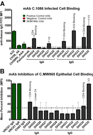
ability of MAbs to bind CEM.NKR
CCR5cells infected with C.1086
autologous virus was measured by immunofluorescence. Six
MAbs (2 IgA and 4 IgG) were observed to bind the surface of
C.1086-infected cells: DH524, DH530, DH534 (sCD4 blocking),
DH527 (V2), DH532 (A32 blocking), and DH535 (V3) (Table 5;
Fig. 7). Three of these MAbs also demonstrated ADCC activity
against CEM.NKR
CCR5cells coated with C.1086 gp120: DH527
(V2), DH532 (A32 blocking), and DH535 (V3) (Table 5;
Fig. 8A).
Finally, the ability of the MAbs to inhibit binding of HIV
C.MW965 virions to epithelial cells was assessed (41). Four MAbs
(2 IgA and 2 IgG) blocked epithelial cell binding: DH524 and
DH534 (sCD4 blocking), DH525 (linear C4), and DH535 (V3)
(Table 5;
Fig. 7). Thus, vaccine-elicited breast milk MAbs had a
wide array of antiviral functionalities which could contribute to
blocking viral infection at mucosal surfaces.
Postimmunization breast milk has a moderate inhibitory
ef-fect on MAb ADCC function.
Since vaccine-elicited IgA in plasma
has previously been observed to interfere with the IgG effector
function of antibodies with the same specificity (13), we next
sought to determine whether the robust i.n./i.m.-elicited IgA
re-sponses in breast milk could inhibit ADCC activity by milk IgG.
Though ADCC was not observed in the unfractionated breast
milk of i.n./i.m. immunized animals, IgG purified from the same
breast milk was found to mediate a low level of ADCC activity
TABLE 5 Genetic characterization, specificity, and function of Env-specific monoclonal antibodies isolated from C.1086 Env-specific blood and milk B cells following combined i.n./i.m. Env immunization Identification Genetic data Fine specificity Epitope blocking Neutralization (IC 50 , g/ml)
ADCC (titer, g/ml)
(approximate endpoint concentrations of 0.2
g/ml for A6E042
and 0.1
g/ml for A6E088) (Fig. 8B). This result suggests a
poten-tial ADCC-blocking effect of the high-magnitude Env-specific IgA
response, but another possibility is the presence of an unidentified
blocking factor in milk. To further investigate this phenomenon
of ADCC inhibition, pre- and post-i.n./i.m. vaccination breast
milk was spiked with 10
g/ml of DH532 (the A32-blocking,
vac-cine-elicited milk MAb that mediates robust ADCC) or 15 ng/ml
of an ADCC MAb mix consisting of A32, 2G12, 7B2, and CH44
(Fig. 8C). Partial inhibition of ADCC effector function was
ob-served in the post-i.n./i.m. vaccination milk spiked with both
DH532 (29% inhibition) and the ADCC MAb mix (71%
inhibi-tion) but not in the pre-i.n./i.m. milk sample (Fig. 8D). Thus, the
high-magnitude IgA response or unidentified factors in post-i.n./
i.m. vaccination milk may account for the inability to detect
ADCC activity in unfractionated milk of i.n./i.m. Env-immunized
monkeys.
DISCUSSION
An inherent challenge in the prevention of breast milk HIV MTCT
is elicitation of robust immune responses more potent than those
following natural infection. Breast milk from HIV-infected
women contains protective Env-specific antibodies (6,
8,
17,
42),
the presence of which makes development of a maternal HIV
vac-cine to enhance these preexisting responses an attractive strategy
to prevent breastfeeding transmission of HIV. In this study, we
gained further understanding of the isotypes, specificities, and
functions of HIV-specific breast milk antibody responses induced
by i.n./i.m. immunization and then compared them to previously
described responses that may be important to impede postnatal
transmission.
Although breast milk from both HIV-infected humans and
SIV-infected rhesus monkeys contains higher-magnitude
Env-specific IgG than IgA, in our recent analysis of breast milk
anti-body responses of women from the Breastfeeding Antiretrovirals
and Nutrition (BAN) study, only breast milk HIV envelope
gp140-specific IgA was associated with reduced risk of postnatal
HIV-1 acquisition (6,
7,
17). These results suggest that a maternal
vaccine with the ability to elicit robust IgA responses in milk could
potentially be protective. It has traditionally proven very difficult
to induce Env-specific IgA in mucosal compartments by HIV
vac-cination (11). Yet, the combined systemic and mucosal
vaccina-tion regimen (i.n./i.m.) used in this study elicited remarkably
high-magnitude Env-specific IgA responses in breast milk, with a
concentration exceeding that in plasma. This provides proof of
principle that a vaccine can selectively elicit strong IgA responses
in the mammary compartments. It is intriguing that although
se-cretory IgA typically constitutes the majority of IgA in mucosal
secretions (43), following i.n. boost 2, SIgA represented only
ap-proximately 9% of the total Env-specific IgA in breast milk. This
finding indicates that the majority of Env-specific breast milk IgA
was either produced locally or passively transudated across
epithe-lial cells rather than secreted across mammary epithelia. However,
despite the observed differences in antibody response magnitude,
it is important to note that these differences were not statistically
significant given the small number of experimental animals.
In addition to the increased IgA-binding responses, i.n./i.m.
immunization also enhanced neutralization titers in breast milk.
Many innate factors in breast milk, such as tenascin C (TNC) and
lactoferrin, have been described to have an HIV-1-neutralizing
function (39,
44). To investigate whether an increased production
of these innate factors following immunization could contribute
to the observed increase in neutralization titers, we determined
the concentrations of tenascin C at multiple time points during
the immunization schedule. We observed that the concentrations
were quite low compared to human levels, far below TNC
neutral-ization IC
50levels previously observed (39), and were similar
be-tween all animals and across all samples. These findings suggest
that the increase in neutralizing titer following immunization is
the result of an enhancement in Env-specific IgG and IgA
re-sponses rather than other innate factors. Interestingly, this
inves-tigation is the first to report the presence of TNC in breast milk of
rhesus monkeys.
Concordant with the high magnitude of Env-specific IgA in
FIG 6Binding profile and determined specificities for 6 IgA and 6 IgG vaccine-elicited Env-specific MAbs isolated from breast milk B cells and selected for
functional characterization. EC50s (ing/ml) obtained by ELISA are shown. Tested antigens are grouped into categories (Env glycoproteins, Env regional
milk, a relatively high proportion of Env-specific breast milk B
cells were found to be IgA isotype. Previous studies have indicated
that IgA isotype B cells comprise 75 to 80% of all mammary
gland-associated B cells (45), and there is a body of evidence which
sup-ports the existence of a gut-mammary axis in primates by which
gut IgA-producing plasmablasts home to lactating mammary
tis-sue (46,
47). Quite counterintuitively, we and others have
ob-served that in the setting of chronic HIV infection, HIV
Env-specific memory B cells in breast milk are overwhelmingly IgG
isotype (9,
10). Therefore, the relatively high proportion (29%) of
Env-specific IgA-producing B cells induced by i.n./i.m.
vaccina-tion in breast milk is a novel finding and suggests that this vaccine
regimen led to trafficking of clonally proliferating Env-specific B
cells to the mammary compartment. The immune function of
these B cells in breast milk once ingested by the infant is poorly
understood. Intriguingly, activated mononuclear cells have been
observed to persist in the mammalian neonatal gut, bind to the
brush border, and infiltrate neonatal tissues (48,
49). This
obser-vation leaves open the possibility that ingested Env-specific
mem-ory B cells could serve as immune sentinels or further differentiate
into antibody-producing plasma cells in the infant GI tract.
Among our limited panel of 12 breast milk MAbs (6 IgA and 6
IgG), there was no clear association of antibody isotype with
epitope specificity, breadth, or function. As these MAbs were
iso-lated from a single immunized animal, it is unknown whether our
results are representative of all immunized animals. Nevertheless,
our results clearly demonstrate that i.n./i.m. HIV vaccination can
induce IgA isotype memory B cells of diverse specificities in the
mucosal compartment and that vaccine-elicited IgA MAbs are
capable of mediating functions such as infected cell binding and
inhibition of viral binding to epithelial cells. Importantly, this
study is the first to isolate and characterize vaccine-elicited IgA
from mucosal Env-specific memory B cells. The significant
epitope and functional variability observed among our MAb panel
suggests that breast milk B cells are not restricted in their epitope
specificity. Given our previous discovery that a higher proportion
of breast milk B cells are gp120 directed in breast milk than in
plasma during chronic HIV-1 infection (9), we anticipate that
gp120 i.n./i.m. immunization of a chronically infected woman
could further enrich the proportion of gp120-specific B cells.
Interestingly, one of the breast milk IgG MAbs (DH535) was
able to mediate multiple effector functions. This V3-specific MAb
was determined to have broad tier 1 neutralization function, bind
to the surface of C.1086 infected cells, mediate ADCC, and inhibit
viral binding to epithelial cells. In a cohort of 248 HIV-infected
mothers, the magnitude of the V3-specific IgG in maternal plasma
was recently associated with a decreased risk of perinatal MTCT
(50). The role of a V3-binding antibody in the prevention of
post-natal transmission has not clearly been established, yet it is
possi-ble that the polyfunctionality demonstrated by DH535 is required
to block mucosal HIV transmission. Indeed, a comparison
be-tween two HIV clinical trials (RV144 and VAX003) demonstrated
that the ability of a vaccine to induce multiple, coordinated
anti-viral responses is associated with lower rates of HIV transmission
(51,
52).
The role of ADCC in protection against breast milk HIV
trans-mission remains unclear. While Mabuka et al. reported an
associ-ation between ADCC-mediating antibodies in milk and a reduced
rate of transmission (8), in our analysis of the BAN cohort, ADCC
responses in milk were not associated with protection (17).
Im-portant differences between the cohorts of HIV-infected women
could explain these discordant results, including sample size, the
virus clade, and the maternal immune status. Surprisingly,
com-bined systemic/mucosal immunization did not elicit ADCC
activ-ity in milk, though multiple ADCC-mediating MAbs were isolated
from breast milk B cells. As IgA blocking of ADCC effector
func-tion was demonstrated in the RV144 vaccine trial analysis, we
sought to determine if the lack of detectable ADCC activity in milk
was due to IgA interference. Purified IgG from post-i.n./i.m.
vcination breast milk was able to mediate low levels of ADCC
ac-tivity, suggesting a possible IgA-blocking effect. Furthermore,
when post-i.n./i.m. vaccination breast milk was spiked with
ADCC-mediating antibodies, there was a reduction in the potency
of ADCC effector activity. Therefore, the high concentration of
Env-specific IgA in breast milk could be an important factor
af-fecting ADCC-mediated antibody function after i.n./i.m.
vaccina-tion. Nevertheless, the biological relevance of this ADCC blocking
for virus transmission remains to be determined.
FIG 7Vaccine-elicited Env-specific MAbs from breast milk B cells bind
in-fected cells and prevent viral binding to epithelial cells. (A) 6 MAbs (2 IgA and 4 IgG) demonstrated binding to the surface of C.1086 HIV-infected CEM. NKRCCR5cells. (B) 4 MAbs (2 IgA, 2 IgG) prevented C.MW965 from binding
Since Env-specific IgA and ADCC activities have been
associ-ated with a reduced risk of postnatal HIV transmission, these
re-sponses are the benchmarks we have to judge the success or failure
of maternal HIV vaccination strategies to induce potentially
pro-tective immune responses in breast milk. It is remarkable that the
i.n./i.m. vaccination regimen both elicited a high level of
Env-specific breast milk IgA and induced local IgA isotype breast milk
B cells, which is a quite distinct immune response from that
fol-lowing natural infection. The ability of these robust
vaccine-elic-ited Env-specific IgA responses to prevent breast milk HIV
trans-mission should be further investigated in a nonhuman primate
model through infant oral SHIV challenge in the setting of
expo-sure to breast milk of a vaccinated dam. These studies would
ad-dress the potential
in vivo
protection of high-magnitude
Env-spe-cific IgA responses in breast milk and also interrogate any impact
of these IgA responses on ADCC functionality. Furthermore, a
nonhuman primate model has the potential to address the
feasi-bility of administering this proposed immunization regimen
dur-ing pregnancy, as well as the safety of an MVA prime for a
devel-oping fetus. If the procedure is proven efficacious and safe, such
studies would enable identification of novel immune correlates of
protection for postnatal HIV MTCT and would suggest that a
maternal vaccination strategy such as i.n./i.m. combined
immu-nization given during pregnancy has potential to curb the ongoing
pediatric HIV epidemic.
ACKNOWLEDGMENTS
The content is solely the responsibility of the authors and does not neces-sarily represent the official views of the National Institutes of Health.
The authors have no commercial affiliations or financial conflicts of interest to disclose.
We acknowledge the following individuals for their technical contri-butions and support: Josh Amos, Brooke Liebl, Carrie Ho, R. Glenn Over-man, Robert Parks, Krissey Lloyd, Ashley Trama, Dawn Marshall, Ashley Allen, Lawrence Armand, David Martinez, and Norman Letvin.
FUNDING INFORMATION
This work, including the efforts of Sallie R. Permar, was funded by HHS | NIH | National Institute of Allergy and Infectious Diseases (NIAID) (5R01AI106380, 1P01A117915-01, 5P30AI064518). This work, including the efforts of Barton Haynes, was funded by HHS | NIH | National Institute of Allergy and Infectious Diseases (NIAID) (UM1-AI100645-01).
The funders had no role in study design, data collection and interpreta-tion, decision to publish, or the preparation of the manuscript.
REFERENCES
1.Kourtis AP, Butera S, Ibegbu C, Belec L, Duerr A.2003. Breast milk and
HIV-1: vector of transmission or vehicle of protection? Lancet Infect Dis
3:786 –793.http://dx.doi.org/10.1016/S1473-3099(03)00832-6.
2.Shapiro RL, Hughes MD, Ogwu A, Kitch D, Lockman S, Moffat C,
Makhema J, Moyo S, Thior I, McIntosh K, van Widenfelt E, Leidner J, Powis K, Asmelash A, Tumbare E, Zwerski S, Sharma U, Han-delsman E, Mburu K, Jayeoba O, Moko E, Souda S, Lubega E, Akhtar M, Wester C, Tuomola R, Snowden W, Martinez-Tristani M,
Mazhani L, Essex M.2010. Antiretroviral regimens in pregnancy and
breast-feeding in Botswana. N Engl J Med362:2282–2294.http://dx .doi.org/10.1056/NEJMoa0907736.
3.WHO, UNICEF, UNAIDS.2014. Global HIV/AIDS response. Epidemic
update and health sector progress towards universal access. Progress re-port 2014. World Health Organization, Geneva, Switzerland.
4.American Academy of Pediatrics Work Group on Breastfeeding.1997.
Breastfeeding and the use of human milk. Pediatrics100:1035–1039.http: //dx.doi.org/10.1542/peds.100.6.1035.
5.Munoz FM, Englund, JA.2000. A step ahead. Infant protection through
maternal immunzation. Pediatr Clin North Am47:449 – 463.
6.Fouda GG, Yates NL, Pollara J, Shen X, Overman GR, Mahlokozera T,
Wilks AB, Kang HH, Salazar-Gonzalez JF, Salazar MG, Kalilani L, Meshnick SR, Hahn BH, Shaw GM, Lovingood RV, Denny TN, Haynes B, Letvin NL, Ferrari G, Montefiori DC, Tomaras GD, Permar SR,
Center for HIV/AIDS Vaccine Immunology.2011. HIV-specific
func-tional antibody responses in breast milk mirror those in plasma and are primarily mediated by IgG antibodies. J Virol85:9555–9567.http://dx.doi .org/10.1128/JVI.05174-11.
7.Permar SR, Wilks AB, Ehlinger EP, Kang HH, Mahlokozera T, Coffey
RT, Carville A, Letvin NL, Seaman MS.2010. Limited contribution of
mucosal IgA to simian immunodeficiency virus (SIV)-specific
neutraliz-FIG 8The ADCC effector function of MAbs is partially inhibited in milk following i.n./i.m. immunization. (A) Three MAbs were determined to mediate ADCC.