CHARACTERIZING THE ROLE OF ACTIN BINDING, BUNDLING, AND TYROSINE PHOSPHORYLATION IN MODULATING VINCULIN FUNCTION
Caitlin Elizabeth Tolbert
A dissertation submitted to the faculty at the University of North Carolina at Chapel Hill in partial fulfillment of the requirements for the degree Doctorate of Philosophy in the Department
of Developmental and Cell Biology
Chapel Hill 2013
Approved by: Keith Burridge Sharon L. Campbell James Bear
ii © 2013
iii ABSTRACT
CAITLIN E. TOLBERT: Characterizing the Role of Actin Binding, Bundling, and Tyrosine Phosphorylation in Modulating Vinculin Function
(Under the direction of Dr. Keith Burridge and Dr. Sharon Campbell) Vinculin is an essential adhesion protein involved in controlling motility and force transduction, in part by coupling the actin cytoskeleton to the extracellular matrix. Vinculin is regulated by intramolecular interactions between the N-terminal head domain (Vh) and the C-terminal tail domain (Vt) which are linked by a proline-rich region. Upon binding to F-actin, Vtundergoes a conformational change that exposes a cryptic dimerization site that enables it to self-associate and bundleF-actin.Previous publications have investigated vinculin variants that are deficient in F-actin binding and have found a number of cellular defects; however, these constructs either lack Vt or contain large deletions that significantly destabilize Vt, and it is likely that the observed phenotypes are due to a number of disrupted interactions.
In an effort to improvethe vinculin/F-actin model using select mutations with little structural consequences, we have identified residues that selectively bind and/or bundle F-actin. Deletion of the C-terminal hairpin (VtΔC5) maintains a normal structure and binding to other interaction partnersin comparison to wildtype (WT) Vt. However, VtΔC5 is unable to bundle F-actin and forms a non-functional dimeric species. To disrupt the vinculin/F-F-actin interaction, we characterized two variants: I997A and V1001A. When these variants are expressed in cells, cells are smaller and have fewer with larger adhesions when allowed to spread on FN. Cells
iv
These data suggest that vinculin-mediated F-actin binding and bundling plays a role in cell spreading and cellular reinforcement.
The C-terminal hairpin contains the only tyrosine phosphorylation site in Vt (Y1065). However, the role of tyrosine phosphorylation in Vt and its full impact on regulating vinculin have not been elucidated. I provide evidence that Src-mediated phosphorylation of Vt modulates F-actin bundling. I have structurally characterized both phosphorylatedVt and Vt variants that either mimic or prevent phosphorylation to understand its impact on vinculin function.
v
ACKNOWLEDGMENTS
vi
TABLE OF CONTENTS
LIST OF TABLES ... xii
LIST OF FIGURES ... xiii
LIST OF ABBREIVATIONS ... xv
I. Introduction ... 1
A. The Role of Vinculin during Development, Cancer, and Cardiovascular Function ... 2
B. The Role of Vinculin during Cell Spreading, Migration, and Mechanotransduction ... 4
C. Vinculin‟s Binding Partners and its Auto-Inhibited Structure ... 7
D. Vinculin Activation ... 8
E. The Vinculin Tail Domain and its Binding Partners ... 10
1. The Structure of the Vinculin Tail Domain ... 10
2.Interactions with PIP2 ... 12
3. Interactions with F-actin ... 13
4. Vinculin Tail Oligomerization ... 15
i. Vinculin's Non-Physiological Dimer ... 15
vii
5. Phosphorylation of Vinculin ... 18
II. The Vinculin C-terminal Hairpin Mediates F-actin Bundle Formation, Focal Adhesion, and Cell Mechanical Properties ... 27
A. Overview ... 27
B. Introduction ... 28
C. Materials and Methods ... 31
1. Vinculin tail protein expression and purification ... 31
2. Actin co-sedimentation assay ... 32
3. Fluorescence microscopy of F-actin bundles ... 33
4. NMR spectroscopy... 34
5. Chemical cross-linking of Vt proteins ... 34
6. Cell culture ... 35
7. DNA constructs and transfection ... 35
8. Adhesion site analysis ... 35
9. Force microscopy ... 36
D. Results ... 37
1. Deletion of the Vt strap does not affect actin binding and bundling. ... 38
2. Vt CT deletion mutants are impaired in actin bundling but not actin binding. ... 39
viii
4. Vt association with F-actin promotes Vt dimerization. ... 43
5. Vinculin CT deletion affects cell adhesion. ... 46
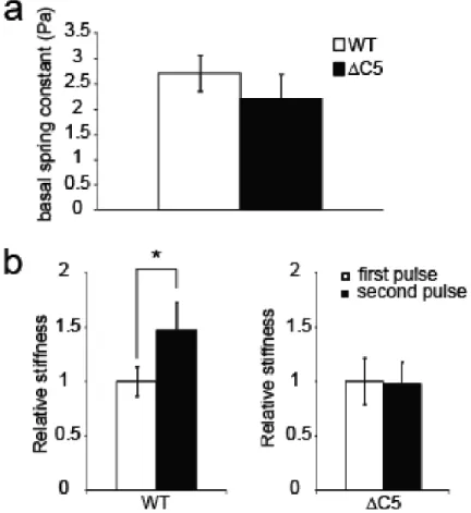
6. The vinculin CT hairpin is necessary for the mechanical response to force on integrins. ... 47
E. Discussion and Conclusions ... 48
III: Identification of a New Actin Binding Surface on Vinculin that Mediates Cellular Mechanical and Focal Adhesion Properties ... 69
A. Overview ... 69
B. Introduction ... 69
C. Materials and Methods ... 71
1. Vinculin expression and purification ... 71
2. Actin co-sedimentation assays ... 73
3. Lipid co-sedimentation assays ... 73
4. EM sample preparation and analysis ... 74
5. DMD model generation ... 75
6. Cell culture ... 76
7. DNA constructs and transfection ... 76
8. Cell Resuspension and Spreading Assay ... 76
9. Adhesion site analysis ... 77
ix
D. Supplementary Methods ... 77
1. IpaA peptide ... 77
2. Circular dichroism (CD) spectroscopy ... 78
3. NMR spectroscopy... 78
4. Calculation of ΔΔGbind ... 79
E. Results ... 79
1. Identification of Vt variants deficient in actin binding ... 79
2. Deficiencies in Actin Binding by Vinculin Alter Cellular Properties ... 82
3. Identification of an alternative actin binding surface ... 85
F. Discussion and Conclusions ... 87
IV: Phosphorylation at Y1065 in Vinculin Mediates Actin-Induced Vinculin Dimer Formation, Cell Spreading, and Mechanical Responses to Force ... 108
A. Overview ... 108
B. Introduction ... 108
C. Materials and Methods ... 111
1. Expression and purification of proteins ... 111
2. Generation of phosphorylated Vt ... 113
3. Circular Dichroism (CD) ... 113
x
5. NMR Spectroscopy ... 114
6. Lipid co-sedimentation assays ... 115
7. Actin co-sedimentation assays ... 115
8. Fluorescence microscopy of F-actin bundles ... 116
9. Vinculin Head-Tail Pulldowns ... 116
10. Cell culture ... 117
11. DNA constructs and transfection ... 117
12. RTCA and Spreading ... 117
13. Force Microscopy ... 118
14. DMD model generation ... 118
D. Results ... 119
1. Mutation and Modification to Y1065 Reveals Subtle Structural Alterations ... 119
2. Y1065 Mutants and Phosphorylated Vt Maintain Lipid Interactions ... 121
3. Phosphorylation or mutation at Y1065 does not affect actin binding, but alters actin bundling... 122
4. Phosphorylation and Mutations at Y1065F Alter Vinculin Head-Tail Interactions ... 124
5. Mutation at Y1065 Affects Cell Spreading ... 125
xi
7. Identification of an actin-induced Vt dimer interface ... 127
E. Discussion and Conclusions ... 128
V: Conclusions and Future Directions ... 147
A. Overview ... 147
B. The Vinculin C-terminal Hairpin Mediates F-actin Bundle Formation, Focal Adhesion, and Cell Mechanical Properties ... 148
C. Identification of a New Actin Binding Surface on Vinculin that Mediates Cellular Mechanical and Focal Adhesion Properties ... 149
D. Phosphorylation at Y1065 in Vinculin Mediates Actin-Induced Vinculin Dimer Formation, Cell Spreading, and Mechanical Responses to Force ... 149
E. Future Directions ... 150
1. Examining actin-binding deficient mutants in cells... 150
2. Identification of other residues involved in the formation of the F-actin-induced Vt dimer ... 151
3. Identify if the residues in the C-terminal hairpin or the F-actin-induced vinculin dimer also serve a scaffolding function in cells. ... 153
4. Investigate FA dynamics in the presence of Y1065 mutants ... 154
F. Conclusions ... 157
xii
LIST OF TABLES Table
1. Summary of F-actin binding by Vt variants.. ... 101
2. Summary of F-actin binding and bundling by Vt Y1065 variants... 146
3. Summary of crosslinking studies with Y1065 mutants.. ... 160
4. Results of FRAP experiments when cells are plated on two FN concentrations.. ... 161
5. Loss of intracellular tension increases the exchange for all the Y1065 mutants.. ... 162
xiii
LIST OF FIGURES
Figure 1.1- Crystal Structure of Full-Length Vinculin.. ... 22
Figure 1.2 - The Vinculin Tail Domain ... 23
Figure 1.3 - Previous F-actin binding model proposed by Janssen et al. ... 25
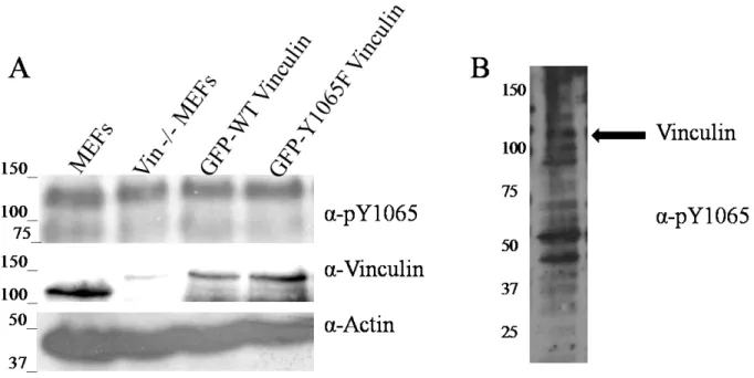
Figure 1.4 - Lack of specificity with pY1065 antibody. ... 26
Figure 2.1- Vt NT deletions within the strap do not affect actin binding and bundling. ... 55
Figure 2.2 - Vinculin CT hairpin is critical for bundling F-actin, but not for binding F-actin ... 57
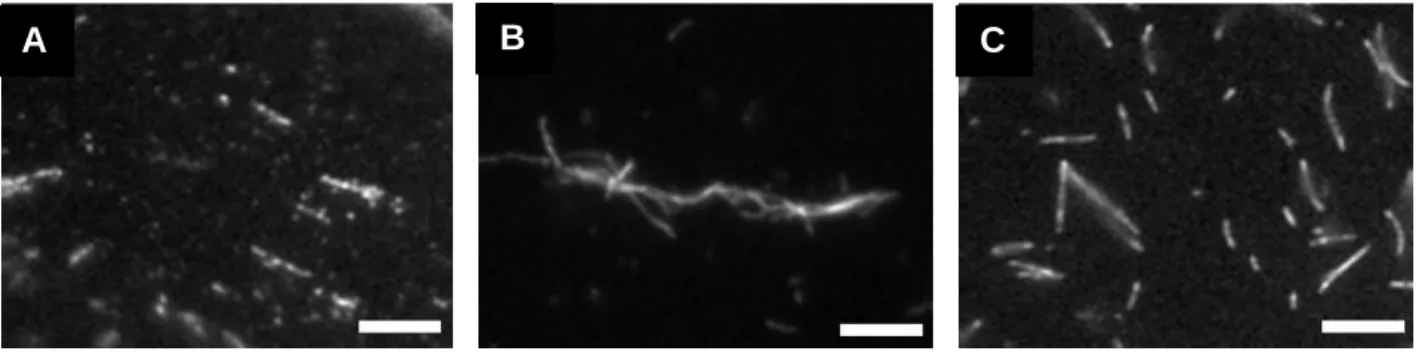
Figure 2.3 - Visualization of actin bundles in the presence and absence of WT Vt and VtΔC5 using fluorescence microscopy ... 58
Figure 2.4 - Removal of residues within the Vt C-terminal hairpin does not alter Vt conformation. ... 59
Figure 2.5 - Vt forms a distinct dimer (~45 kDa) in the presence of F-actin ... 61
Figure 2.6- Vt CT hairpin deletion affects cell adhesion.. ... 62
Figure 2.7 - The Vt CT hairpin is necessary for the mechanical response to force on integrins. . 63
Figure 2.8 - Model for Actin-Induced Vinculin Tail Oligomerization and Bundling of F-Actin.. ... 64
Supplemental Figure 2.1 - The crosslinking agent, BS3, does not enhance F-actin bundling. .... 65
Supplemental Figure 2.2- Effects of increased actin concentration on the ability of Vt to bind and bundle F-actin. ... 66
Supplemental Figure 2.3 - Vt forms a distinct dimer (~45 kDa) in the presence of F-actin ... 67
Figure 3.1 - VtI997A and VtV1001A are deficient in F-actin binding and bundling yet retain specificity and association with PIP2.. ... 93
xiv
Figure 3.3- Actin binding to vinculin is necessary for the mechanical response
to force on integrins.. ... 96
Figure 3.4- The proposed actin binding surface on Vt is consistent with EM reconstruction. ... 97
Figure 3.5 - The proposed binding surface is not accounted for in the J-model... 100
Supplemental Figure 3.1 - Binding of F-actin by full-length vinculinWT, vinculinI997A, and vinculinI948A.. ... 102
Supplemental Figure 3.2 - CD analyses of Vt variants. ... 103
Supplemental Figure 3.3 - 2D NMR 1H-15N HSQC spectral overlay of VtWT and Vt variants. ... 104
Supplemental Figure 3.4 - Evaluation of the DMD model.. ... 106
Figure 4.1 - CD and fQCR of pY-Vt and Y1065 variants.. ... 134
Figure 4.2 - 2D NMR 1H-15N HSQC spectral overlay of WT Vt and Vt variants... 136
Figure 4.3 - Vinculin Y1065 variants and pY-Vt do not affect PIP2 association.. ... 137
Figure 4.4 - Actin binding and bundling with pY-Vt and Vt Y1065 mutants.. ... 138
Figure 4.5 - Phosphorylation of Y1065 and select Y1065 mutants affect vinculin head-tail interactions. ... 139
Figure 4.6 - Y1065 mutants in full-length vinculin alter actin bundling but not actin binding.. 140
Figure 4.7 Vinculin variants at Y1065 are able to localize to FAs, affect cell spreading and FAs... 141
Figure 4.8 The phosphorylation state of Y1065 regulates the mechanical response applied to integrins. ... 143
Figure 4.9 Proposed interface of the actin-induced Vt dimer.. ... 144
Supplemental Figure 4.1 - FTICR-MS verification of Vt 1065 phosphorylation by Src (83.8%). ... 145
xv
LIST OF ABBREIVATIONS 3DFM Three-dimensional force microscopy
ΔC Vinculin C-terminal deletion (lacks residues 1052-1066) ΔC1 Vinculin C-terminal deletion (lacks residue 1066)
ΔC2 Vinculin C-terminal hairpin deletion (lacks residues 1065-1066) ΔC5 Vinculin C-terminal hairpin deletion (lacks residues 1061-1066) ΔN5 Vinculin N-terminal strap deletion (lacks residues 879-883) Δstrap Vinculin N-terminal strap deletion (lacks residues 879-892)
A/V Actin/Vinculin Tail Ratio
BME β-mercaptoethanol
BS3 Bis[sulfosuccinimidyl] suberate CAP Cbl-associated protein
CD Circular dichroism
CI Cell index
CT C-terminus (residues 1051-1066)
DMD Discrete molecular dynamics
DMEM Dulbecco‟s modified Eagle‟s medium
DSS Disuccinimidyl suberate
DTT Dithiothreitol
ECM Extracellular matrix
EM Electron microscopy
xvi
ERK Extracellular signal-regulated kinase F-actin Filamentous actin
FA Focal adhesion
FAK Focal adhesion kinase
FBS Fetal bovine serum
FCX Focal Complex
FN Fibronectin
FRAP Fluorescence recovery after photobleaching FRET Fluorescence resonance energy transfer
G-actin Monomeric actin
HIC 4-methyl histidine
HIS Canonical histidine
HSQC Heteronuclear Single Quantum Coherence
KO Knockout
LD Lipid-deficient
NMR Nuclear magnetic resonance
NT N-terminus (residues 879-892)
PC Phosphatidylcholine
PE Phosphatidylethanolamine
PIP2 Phosphatidylinositol (4,5)-bisphosphate PKCα Protein Kinase C alpha
PS Phosphatidylserine
xvii SUVs Small unilamellar vesicles
TIRF Total internal reflection fluorescence VASP Vasodilator-stimulated phosphoprotein VBS Vinculin-binding sites
Vin -/- MEFs Vinculin knockout murine embryo fibroblasts VT Vinculin Tail (residues 879-1066)
VH Vinculin Head (residues 1-855)
1
I. Introduction
Vinculin is a widely expressed, 116 kDa protein that is recruited to both adherens junctions and cell-extracellular (ECM) junctions, better known as focal adhesions (FAs). At adhesions, vinculin helps couple the actin cytoskeleton via transmembrane receptors and mediates the recruitment of numerous proteins to these maturating complexes. It is through proteins located in adhesions, such as vinculin, that are necessary to regulate cell adhesion and cytoskeletal changes in order to control cell migration. Efficient control of cell migration is critical during embryonic development or wound healing and under pathological conditions such as cancer progression via invasion(1-3). In support of vinculin's role in disease, vinculin
knockout (KO) studies reveal defects leading to death during development, cardiovascular dysfunction, and promotion of cancer progression(2, 4-6). Furthermore, it is through these
adhesion sites that vinculin mediates the transmission of mechanical tension into a global cellular response.1
1
The text, figures, and figure legends in Chapter I are reprinted and/or modified with permission from the following:
Tolbert CE, Burridge K, Campbell SL. Vinculin regulation of F-actin bundle formation: what does it mean for the cell? Cell AdhMigr 7: 219-225, 2013
2
A. The Role of Vinculin during Development, Cancer, and Cardiovascular Function During development, areas of adhesion, whether through adherens junctions or FAs, need to be tightly controlled in order for organisms to develop. These adhesions sites mediate signals that transduce messages from between cells and from external stimuli. Vinculin plays a crucial role in regulating a number of processes at both FAs and adherens junctions, and as a result, impacts a number of cellular processes. The importance of vinculin has been
previously demonstrated through mice KO studies where embryos die by day E10.5 and have defects in heart and neural tube formation (2). Additionally, studies that KO vinculin in C. elegans and zebrafish found defects in muscle and cardiac development, with the zebrafish displaying a dilated cardiomyopathy phenotype that includes impairments in blood congestion, pericardial edema, and cardiac contractility (7, 8). Upon isolation of fibroblasts from the KO mice embryos, numerous defects were found including difficulties in cell adhesion, spreading, increased motility, higher levels of focal adhesion kinase (FAK) and paxillin signaling that lead to increased extracellular signal-regulated kinase (ERK) signaling, and resistance to apoptosis from a number of stimuli as well as resistance to anoikis(2-4, 9). Because of these defects, vinculin is considered to be a tumor suppressor. To further support vinculin's tumor suppressor properties, recent studies have shown that by activating vinculin, melanoma cells become more sensitive to chemotherapeutic treatments (10). Moreover, studies show that vinculin protein levels are correlated with very invasive cancers and upon re-expression of vinculin in these cell lines, the cells are less tumorigenic(11). Vinculin mislocalization has also been shown to impact the loss of adherens junctions during initial stages of tumor formation (12-16).
3
additional 68-amino acid insertion within the tail domain. In muscles, both vinculin and
metavinculin are enriched in both the I-band and in costameres, structures that link sacromeres to the membrane(17-19). While metavinculin serves a very similar purpose as vinculin in muscles, it has some distinct properties that are attributed to the 68-amino acid insertion. For instance, vinculin itself is very conserved between species while metavinculin displays greater sequence variety within the 68-amino acid insertion among different species (20). Furthermore,
metavinculin displays altered affinities for binding partners to the tail domain in comparison to the vinculin tail domain (Vt). For instance, metavinculin has a higher affinity for raver1 and less affinity for PIP2(21, 22). Interestingly, some of the most striking differences between vinculin and metavinculin come from their differences in their interaction with F-actin. While Vt normally binds and crosslinks F-actin into parallel filaments, metavinculin tail domain is also able to bind F-actin and create an alternative F-actin structure that looks “web-like”(18). Due to these differences in F-actin associations, metavinculin displays a specialized role in mechanical tension when external forces are applied to cells(23). For instance, metavinculin varies in expression levels when forces are applied to cells that appear to be muscle-type dependent(17, 19, 22,24-26). When examining common mutations in metavinculin that occur within the 68-amino acid insert region (A934V, ΔL954, and R975W), these mutations are believed to be involved in metavinculin‟s mechanosensitive properties due to altered interactions with F-actin and with the metavinculin head domain, and are found in cardiomyopathies(27-29). These diseases occur due to the improper generation of force and lead to stress-induction on the heart.
While most mutations that lead to cardiomyopathies are found in metavinculin within the specific 68-amino acid insert region, there are select mutations that occur in vinculin. For
4
cardiomyopathy (30). A recent report highlighted a pedigree containing a missense mutation in vinculin (K815R) was associated with familial dilated cardiomyopathies and congenital heart abnormalities (31).
B. The Role of Vinculin during Cell Spreading, Migration, and Mechanotransduction Following activation, vinculin is recruited to two main sites of adhesion: adherens junctions and FAs. In both sites, vinculin mediates a similar function: to recruit additional
interaction partners to these sites as the adhesion matures and to help link the transmembrane receptors to the actin cytoskeleton(32). While less is known about vinculin in adherens junctions since the most focus in the field has examined vinculin is FAs, recent reports have highlighted the importance of vinculin in adherens junctions and have suggested vinculin serves three functions: vinculin acts downstream of myosin VI, a minus end-directed motor that is necessary for E-cadherin dependent border-cell migration; regulates the expression of E-cadherin on the surface of cells; vinculin plays a mechanosensitive role in response to force applied to E-cadherin (33).
As implicated by fibroblasts isolated vinculin KO mice, cells have a number of defects that are directly mediated through their FAs. These defects include deficiencies in cell adhesion, spreading, migration, responses to force as well as resistance to anoikis(2-4, 9). Once activated at FAs, vinculin facilitates recruitment of additional proteins and links to F-actin, thereby enabling vinculin-mediated regulation of FA dynamics, efficient cell spreading, migration, or respond to external forces.
5
adhesions. Only through the interplay of these two components can effective migration and spreading be achieved. Following integrin engagement and recruitment of key cytoskeletal proteins, including talin and vinculin that establishes the link between active integrins and F-actin, actin flow slows and transmits force on the ECM to enable cells to move(34) and allow adhesions to mature(35). In studies examining motility and spreading in the absence of vinculin, spreading is severely impaired but the cells are able to migrate at a higher rate (36). These data suggest that vinculin is needed to stabilize adhesions in order to spread instead of impacting rapid actin polymerization and depolymerization that is necessary for effective migration. Recent studies have suggested that, in addition to bundling F-actin, vinculin can promote actin
polymerization and cap filaments (37-39). However, it has been hard to observe the result of these different functions in cells due to the use of large and destabilizing vinculin C-terminal deletion variants. Humphries et al. showed that expression of vinculin tail domain alone was sufficient for vinculin to be recruited to areas of high contractility that were under mechanical tension, such as FAs, but not to the lamellipodia(40). An actin deficient deletion mutant lacking helix 2 and 3 within Vt displayed not only larger and denser FAs, but also prevented an invasive phenotype (41), indicating that the F-actin/vinculin interaction may regulate cellular functions that organize the actin network during migration. However, given the size of this deletion and likelihood that Vt is destabilized, this variant likely possesses multiple defects. A recent report that utilizes a more specific actin-binding deficient variant, vinculinI997A, implicates the role of the F-actin/vinculin interaction in cell migration by regulating the flow velocity of F-actin at the leading edge of cells and this in turn modulates FA maturation (42).
6
cells to sense changes in stiffness is believed to be primarily mediated through proteins at FAs, including vinculin. In addition its ability to sense external forces, vinculin has been reported to regulate how applied mechanical stress alters adhesion composition in order to induce cell morphological changes (45-47). Recent data indicates that vinculin is crucial for transmission of mechanical forces resulting from either actomyosin or cell-generated forces, since the
7
C. Vinculin’s Binding Partners and its Auto-Inhibited Structure
Vinculin consists of three main domains: a large multi-helical head domain (Vh), a flexible proline-rich linker, and a smaller helical tail domain (Vt). Each of these regions has their own distinct set of binding partners. Vh associates with talin, α-actinin, α/β-catenin, MAPK, and IpaA from Shigellaflexneri(52-57). The proline-rich linker region binds to vasodilator-stimulated phosphoprotein (VASP), Cbl-associated protein (CAP)/Ponsin, nArgBP2, vinexin-α/β, p130CAS and the Arp2/3 complex(58-63). Finally, Vt interacts with protein kinase C-α (PKCα), F-actin, paxillin, Hic-5, PIP2, raver1 and α-synemin(21, 64-70).
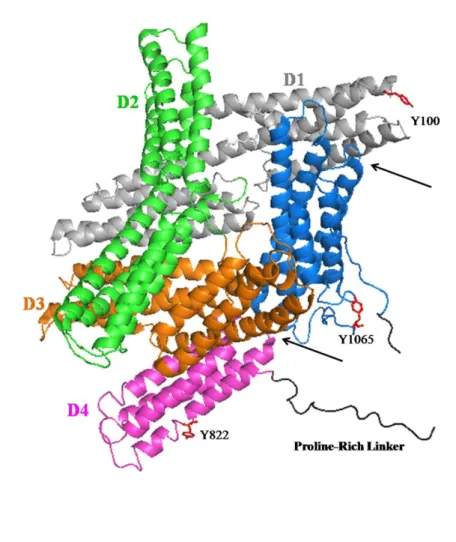
Through x-ray crystallography, many structures of vinculin examining the full-length molecule, isolated domains, and domains bound to ligands have been solved. Bakolista et al., showed the full-length structure in the inactive, or auto-inhibited, conformation of vinculin, revealing the multi-helical regions of the protein (71). As shown in Figure 1.1, Vh (D1-D4) consists of multiple tandem pairs with multiple helices. From D4, there is a flexible, proline-rich region that connects to Vt, a five-helix bundle. It has been shown that isolated Vt shares a
number of structural similarities to Vt within the full-length protein(71, 72). In the inactive state, there are a number of intramolecular interactions that are responsible for the auto-inhibited conformation. A number of these interactions occur between Vt-D1 while a few additional contacts occur between Vt-D3 and Vt-D4 (Figure 1.1). Through these multiple intramolecular interactions, the proposed Kd for inactive vinculin is ~ 1 nM.
8
(10-5 M) which supports the idea that other regions within Vh have additional contacts to Vt in order to mediate the inactive conformation(73). It was determined that contacts within D4 also interact with Vt and upon mutation of residues within D4 (N773/E775A), head-tail interactions were reduced about 100-fold and talin binding was still permitted. In order to create a
constitutively active vinculin mutant, the T12 mutant (D974/K975/R976/R978A) was utilized to disrupt head-tail interactions. Upon expression of the activating-vinculin mutant in cells, size and residency of vinculin in FAs were both increased (73). While a third site was identified in the x-ray crystal structure of auto-inhibited vinculin between Vt and D3, this interaction has not been verified (33).
D. Vinculin Activation
When it is in the auto-inhibitory conformation, vinculin has tight intramolecular interactions between Vh and Vt at multiple distinct sites. Once these intramolecular interactions are released, vinculin transitions into its 'active' state, or rather the exposure of additional binding sites for its interaction partners. How these intramolecular interactions are relaxed that enables for vinculin activation have not been fully elucidated. Vinculin activation can occur through two main mechanisms: application of external mechanical forces or through interaction of multiple binding partners to both the Vh and Vt. It has been found that when external forces are applied to cells, there is a fast and robust recruitment of vinculin to sites of integrin engagement(74).
9
of vinculin activation is solely integrin-dependent or if external forces to cadherins can elucidate a similar response.
In the better characterized model for vinculin activation, the combinatorial model, suggests that multiple binding partners relax the interactions between Vh and Vt in order to activate the molecule. The specific binding partners that aid in vinculin activation are
presumably dependent on their location; for vinculin activation at FAs, these binding partners are typically talin and F-actin. It has been shown that talin alone is sufficient to disrupt interactions between isolated Vt and D1 domains. However, talin is not able to break all the intramolecular interactions to enable for vinculin activation in the context of the full-length protein. Only in the presence of both talin and F-actin does vinculin activate in a dose-dependent manner (75).
Currently, there is no single protein that is endogenous to the cell that can activate vinculin. The only protein that can activate vinculin by itself is the virulent protein, IpaA, from Shigellaflexneri(76). Within IpaA, there are two vinculin-binding sites (VBS) and the binding site between IpaA and Vh seem to mimic VBS from both talin and α-actinin(77). Interestingly, the VBS from IpaA has reported having a much higher affinity (10-fold) for D1 of Vh when compared to other VBS from talin(77). This accounts for why IpaA can solely activate vinculin. However, when F-actin was added to the vinculin fluorescence resonance energy transfer (FRET)-activation biosensor in the presence of IpaA, vinculin's activation was increased,
10
While the majority of these studies have been performed with purified proteins and have significantly improved our understanding of how vinculin can become activated, studies from cells have implicated that vinculin activation is much more complicated than anticipated. Using a FRET-based vinculin activation biosensor, the conformation of vinculin can be monitored in live cells. While it was no surprise that vinculin is found to be active and in an open conformation when located at FAs and cytoplasmic vinculin was inactive, there are distinct pools of vinculin within FAs that are inactive when vinculin is initially recruited to FAs and as adhesions turnover (74). These results suggest that there are additional signaling events controlling the activation state of vinculin, and it is tightly regulated by unknown ligands prior to FA turnover.
Additionally, these data suggest that vinculin recruitment is decoupled from its activation, which is likely due to the lack of additional binding partners available to activate vinculin upon
immediate localization to FAs. For instance, while talin can recruit vinculin, an additional binding partner such as F-actin is not necessarily in the same vicinity in order to bind and activate vinculin. This hypothesis is supported by the distances between talin and F-actin according to recent high-resolution microscopy studies (78). These data could account for the multiple pools of vinculin in different conformations at FAs.
E. The Vinculin Tail Domain and its Binding Partners
1. The Structure of the Vinculin Tail Domain
11
the context of the full-length molecule. However, the isolated domain was solved as a dimer; a non-physiological dimer that has been previously observed by rotary-shadowing electron microscopy (EM) that occurs when Vt is in abundance beyond ~360 µM, a requirement for forming crystals. The interface where this non-physiological occurs is the same interface where Vt interacts with D1 in the auto-inhibited conformation and where F-actin interacts with Vt.
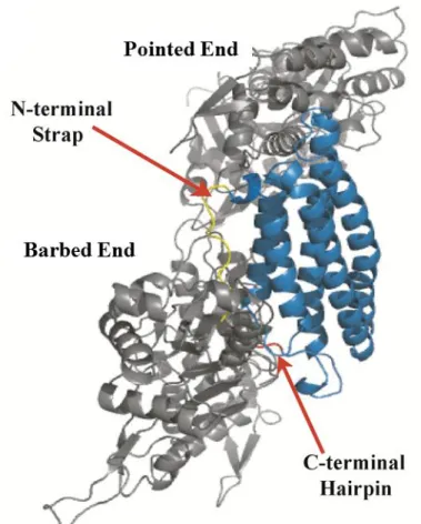
The N-terminal strap (residues 879-893) is a flexible area of the protein as suggested by the multiple conformations that are observed for this region. When the N-terminal strap is more ordered, it packsnext to helix 1 and 2 where it forms multiple contacts. Specifically, residues D882 interacts with S914 and K924 that are located in the loop between helix 1 and 2 and along helix 2, respectively. Additionally, residues K1061 and Y1065, which are located in the
C-terminal hairpin, are the major residues that enable the N- and C-C-terminal interactions. Moreover, residue F885 within the strap packs into a hydrophobic patch between helix 1 and 2 in order to make contacts with H906 (Figure 1.2A) (71, 72).
There are two distinct basic regions on Vt that have been proposed to play a role in lipid interactions and are termed the basic ladder and the basic collar (Figure 1.2B). The basic ladder consists of basic, exposed residues along helix 3 and within the loop that serves as the base for the helical bundle (residues K944, R945, K956, R963, K966, K970, R978, R1008, and R1049). The basic collar consists of exposed, basic residues from helix 1, 5, and the C-terminus (residues R910, K911, R1039, R1060, and K1061).
C-12
terminal hairpin, also referred to as the hydrophobic hairpin, consists of the last five amino acids (TPWYQ) and has contacts with the N-terminal strap.
2.Interactions with PIP2
Multiple groups have shown Vt's propensity for binding to acidic phospholipids,
specifically phosphatidylinositol 4,5-bisphosphate (PIP2)(67, 71, 72, 80-83). PIP2 itself has been noted as a potent molecule that helps mediates a number of signaling events such as regulating cell morphology and actin dynamics(84, 85). Due to the role of PIP2 in these cell signaling events, it could help explain vinculin's role in these processes, moreover the specific role of Vt. The interaction between PIP2 and Vt has been shown to impact the binding between Vt's other interaction partners such as F-actin and PKCα (65, 71, 72,86). The PIP2/Vt interaction has also been proposed to help disrupt head-tail interactions leading to vinculin‟sactivation(65, 71, 80, 83,86). However, these studies were performed with purified components and excess lipids. Additionally, recent reports have shown that the affinity for PIP2 is significantly reduced in the context of the full-length protein and is unable to activate vinculin(80). Furthermore, it has beenproposed that the purpose of the PIP2/vinculin interaction is to control adhesion formation,FA turnover and force transduction (81, 87).
13
Palmer et al. showed that deletion of R1060 and K1061 cause the significant structural changes as these residues play a crucialrole in maintaining the tertiary structure of Vt (88). Furthermore, the authors also showed that deletion of the C-terminal hairpin (Vt ΔC5) did not disrupt binding to PIP2. This is in contrast with other recent reports that implicate the C-terminal hairpin in membrane insertion as shown by a 23-mer peptide of the C-terminus in the excess of lipids(89). However, using a peptide in place of Vt or the full-length protein is not a true representation of the interaction and additional studies would be needed to verify if the C-terminal hairpin inserts itself into the membrane, an unlikely result given previous reports.
Another group identified a lipid-deficient (LD) variant that introduced a number of mutations within the basic collar, ladder and C-terminus (residues
K952/K956/R963/K966/R1060/K1061Q)(81). While these mutants showed a significant disruption in binding to PIP2, no structural studies were performed. Given recent evidence that showed deletion of R1060 and K1061 significantly destabilized Vt, it is likely that the LD mutant is also destabilizing (88). Palmer et al. were able to show that removal of the N-terminal strap greatly enhanced binding to PIP2, which points to residues in the basic collar as the most likely candidates for mediating PIP2 binding (88). This is further supported by other studies that have highlighted residues K911 and K924,residues within the basic collar, as sites that mediate PIP2 interactions (72, 79).
3. Interactions with F-actin
One of vinculin's main functions is to bind F-actin in order to help link transmembrane receptors to the actin cytoskeleton(40). Vt contains an F-actin binding site that is partially
14
additional binding partners, such as IpaA or talin(76, 90). When Vt is further broken down into two fragments, both fragments can bind to F-actin indicating that there are two binding sites on Vt, although these sites do not look like typical actin-binding sites(66). Studies that examined variations intryptophan fluorescence and protease sensitivity when bound to F-actin indicate that there are additional conformational changes in Vt when it binds to F-actin (72). In addition to binding F-actin, Vthas been shown to self-associate through a cryptic dimerization site that enables actin filaments to be crosslinked (Figure 1.3) (90).
Janssen et al. combined negative stain EM, computational docking and mutagenesis approaches and proposed a structural model for the Vt/F-actin complex (Janssen model) (91). In the Janssen model, F-actin interacts with Vt at two sites; the top of helix 2 and 3 (upper site) as well as the bottom of helix 3 and helix 5 (lower site) leading into the C-terminal hairpin (Fig. 1.3) (91). They also proposed that deletion of the amino (N)-terminal strap impaired actin bundling while deletion of the C-terminus enhanced the bundling efficiency of Vt(91).However, taking into consideration a number of factors including the resolution used to dock Vt (50 Å), computational modeling revealing a number of conformational clashes occur on both Vt and actin, and a lack in supporting mutagenesis data, suggest that the current model needs additional refinement.
15
impacted cell morphology and promoted an invasive phenotype, indicating that the
F-actin/vinculin interaction may mediate a number of cellular processes (41). However, given the size of this deletion, Vt is likely to be destabilized, and this could attribute the number of defects observed.
4. Vinculin Tail Oligomerization
i. Vinculin's Non-Physiological Dimer
One of the most intriguing properties of Vt is its propensity to oligomerize under varying conditions and with different binding partners. Vt dimerizes at high concentrations in the absence of any other ligands (23, 72,93). Atphysiological protein concentrations, it can also
homodimerizein the presence of actin (66, 72, 91), and oligomerize (dimer and trimer) in the presence of PIP2 (72, 88, 94). Furthermore, vinculin not only homodimerizes via Vt, but can also heterodimerize with the tail domain of metavinculin (22).
16
asymmetric interaction between helices 4 and 5 of both Vt molecules (72, 93,96), with the two molecules rotated 90 with respect to each other. There are no large-scale conformational
changes associated with this dimer. The propensity of Vt to self-associate into this structure often leads to difficulty in experimental design. The percentage of Vt in solution that exists in dimeric form must be accounted for when interpreting results, especially for experiments requiring high protein concentrations greater than 100 µM. Also, in the presence of crosslinking reagents, Vt can form multiple high molecular weight bands indicating a number of different oligomers may be present (39).
The self-association dimer is the most-understood Vt oligomer from a structural
perspective, but is also the least relevant. In the cell, an abundance of Vt ligands and the presence of Vh mean the concentration of “free” Vt will be very low. When coupled with the weak Kd of the self-association dimer, it is unlikely that this dimer is physiologically relevant. Rather, the
hydrophobic interface may be critical for interaction of Vt with its ligands.
ii. Actin-Induced Vinculin Dimer
17
larger constructs containing the entire protein (97) or residues 154–1066 (99), suggesting that either the vinculin head is distant enough or the proline-rich linker is flexible enough to avoid interference of the vinculin head with the actin bundle.
While it is now known that Vt crosslinks F-actin through formation of a Vt dimer, the structure of this dimer is unknown. Complicating the matter is that this dimer is not formed in the absence of F-actin (90). It is currently believed that Vt undergoes a conformational change upon
binding to F-actin that allows for dimer formation. Previous studies have supported this hypothesis with proteolytic cleavage(72), fluorescence (72), FRET (74), and crosslinking experiments (39, 90). The proteolysis studies showed an increase in susceptibility in the strap and the H1-H2 loop to proteases in the presence of F-actin (72), while the FRET experiment predicts an increase in the distance between the strap and the C-terminal hairpin (74). It was previously hypothesized that the helix bundle “unfurled” upon binding F-actin, resulting in the exposure of the “cryptic” dimerization site (90). However, using negative-stain EM, Janssen et al. show that there is no significant unfurling of the helix bundle (91). Instead, clashes between the N-terminal strap and C-terminus indicate that one or both of these termini change conformation upon association of Vt with actin(91). As presented in Chapter II, the C-terminal hairpin has recently been shown to be required for actin bundling (39), suggesting that it may form a new interaction with the opposite Vt monomer. Moreover, removal of the N-terminal strap slightly increases the F-actin bundling of Vt, suggesting that it must change conformation to expose a site important for dimer
formation (39). Together, these studies point to the existence of an actin-dependent
18
While a structure for the actin-induced dimer is lacking, the current data provide a good starting point for model generation. The increase in proteolytic susceptibility (72), the FRET studies (74), and the differences in the structure of the tail in metavinculin (29) suggest that the interactions between the strap and C-terminus are weakened upon binding of the tail to F-actin, resulting in the release of the strap from the helix bundle. This frees the C-terminal hairpin, essential for formation of the dimer (39), to interact with the other Vt molecule. The
hydrophobicity of the hairpin and the relative orientation of the two tails (91) suggests that the hairpin inserts itself into the interior helix bundle of the other tail. This hydrophobic interaction mediated through the C-terminal hairpin is likely the main force behind dimerization, as removal of these residues eliminates actin bundling by Vt(39). The specific orientation of the dimerand the resulting F-actin geometry are likely influenced by other interactions, probably involving the helix bundle.
While recent evidence points to the surface where the F-actin/Vt interaction occurs, little is known on how binding to F-actin causes this conformational change to expose the cryptic dimer. A possible scenario is that the conformational change that occurs in Vt upon binding to F-actin is allosteric (72, 74,90). Addressing these holes in our understanding of vinculin‟s ability to bundle F-actin will allow us to better identify the biological consequences associated with
vinculin-mediated F-actin bundling and may further clarify the role vinculin in cells and disease.
5. Phosphorylation of Vinculin
Vinculin was one of the first substrates found to be phosphorylated by the tyrosine kinase, Src (100), and has also been found to be phosphorylated by PKCα(65, 101). While it has been long known which kinases and at what sites they phosphorylate vinculin, how
19
There have been three tyrosine residues identified in vinculin that can be phosphorylated. Y100, a residue exposed in Vh, Y822 in D4, and Y1065 in Vt that resides within the C-terminal hairpin and is buried by the proline-rich linker in the full-length protein (Figure 1.1). While it has been proposed that all three residues can be phosphorylated by Src, only Y100 and Y1065 have been shown to be directly phosphorylated by Src and the kinase that phosphorylates Y822 has yet to be identified(102). Recent studies have started to investigate the purpose of these phosphorylation sites. Y822 has been shown to modulate the interaction between FAK and paxillin which in turn regulates apoptosis; although a non-phosphorylatable variant, Y822F, does not restore the sensitivity to apoptosis in vinculin KO cells (9). Cells expressing
non-phosphorylatable variants, Y100F and Y1065F, display defects in cell spreading, but only in the presence of the double mutation(102). Y1065F has additionally been shown to exert lower cell traction forces and slower exchange rates from FAs. While the phospho-mimetic, Y1065E has a higher exchange rate from FAs(103). Phosphorylation at Y1065 has also been implicated in helping to anchoring vinculin to the membrane(87), although exactly how this occurs is
20
In addition to its possible role in FA dynamics, phosphorylation of Vt reduces its affinity for Vh as shown by pulldown assays(102). Phosphorylation at Y1065 also implicates its role in regulating the binding of the Arp2/3 complex as prevention of phosphorylation using the Y1065F variant, loses its ability to bind to Arp2/3 complex(105). The ability of Y1065 to regulate the binding status of Arp2/3 is supported by the vinculin crystal structure where Y1065 has hydrogen bonds with P878, the specific binding site identified that allows for Arp2/3 binding to vinculin(62). The studies that showed Y1065F can disrupt binding to the Arp2/3 complex also showed similar results are observed in the presence of the Y100F mutant; the reason behind this remains to be understood(105). However if the phosphorylation state of Y1065 regulates the binding of the Arp2/3 complex, this could explain the phenotypes observed (decreased cell spreading and lower traction forces exerted) when phosphorylation is prevented. Although some observations made in vitro, specifically the impact of phosphorylation of Vh/Vt interactions, are a little harder to explain given what has been observed in cells. If phosphorylation at Y1065 was needed for vinculin activation, then a higher proportion of phosphorylated vinculin would be observed in FAs; however, in transformed cells overexpressing v-Src, only 2% of the total vinculin is phosphorylated even though there is a significantly high proportion of active vinculin in the cells(100, 102). It could be that Y1065 helps in activation since vinculin has shown to be in multiple activation states in FAs(74).
Using molecular dynamic simulations, Goljiet al. have proposed a mechanism for
21
22
23
Figure 1.2. The Vinculin Tail Domain.(A) The tail domain (residues 879-1066; PDB ID IST6) is a bundle of five helices, with a flexible N-terminal strap (yellow) and C-terminal hairpin (red).
A
24
25
26
27
II. The Vinculin C-terminal Hairpin Mediates F-actin Bundle Formation, Focal Adhesion, and Cell Mechanical Properties
A. Overview
Vinculin is an essential and highly conserved cell adhesion protein, found at both focal adhesions and adherens junctions, where it couples integrins or cadherins to the actin
cytoskeleton. Vinculin is involved in controlling cell shape, motility and cell survival, and has more recently been shown to play a role in force transduction. The tail domain of vinculin (Vt) contains determinants necessary for binding and bundling of actin filaments. Actin binding to Vt has been proposed to induce formation of a Vt dimer that is necessary for crosslinking actin filaments. Results from this study provide additional support for actin-induced Vt
28 B. Introduction
The ability of cells to sense and respond to environmental cues such as mechanical forces is critical for multiple cellular processes including embryogenesis and wound healing (107-109). To transmit forces across the cell membrane in both directions, the actin cytoskeleton couples transmembrane receptors (integrin or cadherin), through points of cell adhesion consisting of multiple protein complexes that form cell - extracellular matrix (focal adhesions) and cell-cell (adherens junctions) contacts (109, 110). Vinculin is an abundant protein found in both focal adhesions and adherens junctions, and plays a key role in regulating cell morphology, cell motility and force transduction (69, 108). Vinculin is essential during development as vinculin knockout (KO) mouse embryos fail to survive beyond day E10 with extensive defects in myocardial and endocardial structures (2). Consistent with a role in muscle structure, vinculin KO mice are predisposed to stress-induced cardiomyopathy (6). Moreover, mutations and deletions in the tail domain of metavinculin, a splice isoform of vinculin, are associated with dilated cardiomyopathy (27, 111,112). Vinculin also possesses tumor suppressor properties as vinculin KO cells are less adherent, have a rounded morphology, reduced lamellipodial stability, increased motility (2, 4), and are resistant to apoptosis and anoikis (9).
Vinculin is a highly conserved cytoskeletal protein which has 1066 residues and contains a globular head, a flexible proline-rich linker and a tail domain (71). Each discrete vinculin domain recognizes multiple binding partners: the vinculin head (Vh) binds to talin, α-actinin and IpaA (113-115); the proline-rich linker interacts with VASP, CAP/ponsin, vinexin and the Arp2/3 complex (58, 59, 61, 62); the vinculin tail (Vt) associates with filamentous actin (F-actin), phosphatidylinositol (4,5)-bisphosphate (PIP2) and paxillin (64, 66, 67). In the
29
binding to most of its ligands (71). Vinculin becomes activated upon release of autoinhibitory Vh/Vt interactions, leading to recruitment and formation of multiprotein adhesion complexes (73, 76, 83,116). When activated at focal adhesions (FAs) (75), vinculin serves as a
mechanotransducer because it helps establish the link between membrane associated integrins and the cytoskeleton through its association with talin and F-actin. Vinculin interacts with a number of cell adhesion proteins, acidic phospholipids and can be phosphorylated by both tyrosine and serine/threonine kinases. However, it is currently unclear how these interactions and covalent modifications, contribute to its role in regulating cell morphology, motility, and cell survival.
30
believed to be a good model of Vt/ligand interactions that occur when Vt is freed from Vh upon activation.
Models for how Vt interacts and bundles F-actin have been proposed. For example, Johnson et al. proposed that actin association with Vt promotes a conformational change in Vt that exposes a cryptic dimerization site that promotes actin filament bundling (90). In a more recent study combining electron microscopy, computational docking and mutagenesis
approaches, Janssen et al. proposed a model for the Vt/F-actin complex (91) in which F-actin associates through two distinct binding sites on Vt, with site 1 containing residues in helices 2 and 3 of Vt and site 2 comprising a binding interface from helices 3, 4 and the CT. Moreover, results obtained from deletion mutagenesis studies indicate that the NT and CT of Vt have opposing roles in actin bundling. While removal of the NT strap impaired actin bundling, deletion of the CT enhanced the ability of Vt to bundle actin filaments (91). However, large deletions within globular proteins can affect their structural integrity. In fact, the CT of vinculin forms contacts with both the strap and the helix bundle, and it has been previously shown that a Vt variant (Vt ΔC, 1052-1066) containing a fifteen amino acid CT deletion alters NMR spectral properties, reduces stability and makes Vt ΔC more susceptible to proteolytic degradation (79, 88).
31
Further, we investigated the effect of vinculin CT hairpin deletion on FA morphology and mechanical properties in vinculin KO murine embryonic fibroblasts (Vin -/- MEFs). Our results demonstrate that deletion of the CT hairpin affects the number of focal adhesions and alters its response to mechanical forces (118).
C. Materials and Methods
1. Vinculin tail protein expression and purification
The tail domain of chicken vinculin (Vt) containing residues 879-1066 was cloned into a pET15b vector (Novagen, Madison, WI) (88). Several deletion variants were generated from this Vt construct, including two Vt NT strap deletions (a five amino acid strap deletion (884-1066 (ΔN5)) and a full strap deletion (893-1066 (Δstrap)), and three CT deletion variants (879-1061 (ΔC5), 879-1064 (ΔC2) and 879-1065 (ΔC1)). All Vt variants were generated using QuikChange site-directed mutagenesis (Stratagene, La Jolla, CA), with sequences verified by DNA
sequencing. Vinculin protein expression and purification have been reported previously (88). Briefly, Vt plasmids were transformed into E. Coli strain BL21(DE3)RIPL. Cells were grown either in Lysogeny Broth rich media or M9 minimal media with 15NH4Cl as the sole nitrogen source at 37 C to an OD600 of 0.6. The cell cultures were cooled to 18 C before adding isopropyl β-D-1-thiogalactopyranoside to a final concentration of 0.5 mM to induce Vt
expression overnight. Cells were then pelleted by centrifugation at 5,800 X g for 30 min and re-suspended in the lysis buffer (20 mM Tris, 150 mM NaCl, 5 mM imidazole, 2 mM
32
Germantown, MD) and washed twice with an Ni wash buffer (20 mM Tris, 150 mM NaCl, 60 mM Imidazole, 5 mM BME, pH 7.5). Histidine (his)-tagged Vt protein was then eluted with a Ni elution buffer (20 mM Tris, 150 mM NaCl, 500 mM imidazole, 5 mM BME, pH 7.5). The his-tag was removed by addition of thrombin (Sigma, St. Louis, MO), and further purified by cation-exchange (Hiprep SP XL 16/10) fast protein liquid chromatography (GE Healthcare, Piscataway, NJ). All Vt variants were examined by SDS-PAGE gel to assess purity and protein integrity before being employed for biochemical assays.
2. Actin co-sedimentation assay
The actin binding and bundling properties of the Vt variants were assessed using an adapted actin co-sedimentation assay (119). Briefly, 4.6 mg/ml monomeric actin (G-actin) purified from rabbit muscle acetone powder (Pel-Freez Biologicals, Rogers, AR) was polymerized with an equal volume of 2X actin polymerization buffer (20 mM Tris, 200 mM KCl, 5 mM MgCl2, 4 mM dithiothreitol (DTT), pH 7.5) at room temperature for 30 min. To assess binding of the Vt variants with F-actin, a 100 µl sample in 1X actin polymerization buffer containing actin at concentrations ranging from 0-30 µM and 10 µM Vt protein was incubated at room temperature for 1 h. It should be noted that the A/V ratio was determined based on the G-actin concentration, given difficulties associated with quantifying F-G-actin concentration due to the heterogeneity of F-actin polymers. The samples were centrifuged at high speed (184,200 X g) on a Beckman-Coulter TLA 100 rotor for 30 min at 25 °C. For bundling assays, a 100 µl sample in 1X actin polymerization buffer containing 20 µM actin and 10 µM Vt protein was incubated at room temperature for 1 h. Samples containing F-actin bundles were obtained by careful
33
Actin and Vt protein contained in both the supernatant and solubilized pellet were separated using 15% SDS-PAGE gels. Actin and Vt proteins in both samples were quantified using the densitometry software package Alpha EaseFC (Alpha Innotech, San Leandro, CA). Actin binding properties of Vt variants are reported in percentage
%𝑉𝑡 𝑏𝑖𝑛𝑑𝑖𝑛𝑔 = 𝑉𝑡𝑑𝑒𝑛𝑠𝑜 ,𝑝𝑒𝑙𝑙𝑒𝑡
𝑉𝑡𝑑𝑒𝑛𝑠𝑜 ,𝑝𝑒𝑙𝑙𝑒𝑡 + 𝑉𝑡𝑑𝑒𝑛𝑠𝑜 ,𝑠𝑢𝑝𝑒𝑟𝑛𝑎𝑡𝑎𝑛𝑡
Where 𝑉𝑡𝑑𝑒𝑛𝑠𝑜 ,𝑝𝑒𝑙𝑙𝑒𝑡 is the densitometry reading of Vt pelleted at 184,200 X g
while𝑉𝑡𝑑𝑒𝑛𝑠𝑜 ,𝑠𝑢𝑝𝑒𝑟𝑛𝑎𝑡𝑎𝑛𝑡 is the densitometry reading of Vt retained in the supernatant. Actin bundling properties of Vt variants are calculated as follows:
%𝐹 𝑎𝑐𝑡𝑖𝑛 𝑏𝑢𝑛𝑑𝑙𝑒𝑑 = 𝐴𝑐𝑡𝑖𝑛𝑑𝑒𝑛𝑠𝑜 ,𝑝𝑒𝑙𝑙𝑒𝑡
𝐴𝑐𝑡𝑖𝑛𝑑𝑒𝑛𝑠𝑜 ,𝑝𝑒𝑙𝑙𝑒𝑡+𝐴𝑐𝑡𝑖𝑛𝑑𝑒𝑛𝑠𝑜 ,𝑠𝑢𝑝𝑒𝑟𝑛𝑎𝑡𝑎𝑛𝑡 Where 𝐴𝑐𝑡𝑖𝑛𝑑𝑒𝑛𝑠𝑜 ,𝑝𝑒𝑙𝑙𝑒𝑡 is the densitometry reading of actin pelleted at 5,000 X g while 𝐴𝑐𝑡𝑖𝑛𝑑𝑒𝑛𝑠𝑜 ,𝑠𝑢𝑝𝑒𝑟𝑛𝑎𝑡𝑎𝑛𝑡 is the
densitometry reading of actin retained in the supernatant.
3. Fluorescence microscopy of F-actin bundles
34
images were acquired on a Zeiss axiovert 200M microscope equipped with a 60 X objective lens and a Hamamatsu ORCA-ERAG digital camera.
4. NMR spectroscopy 15
N-enriched Vt samples were exchanged into NMR buffer (10 mM potassium phosphate, 50 mM NaCl, 0.1 % NaN3, 2 mM DTT, pH 5.5) and concentrated to 0.3 mM with 10 % D2O added. 2D 1H-15N Heteronuclear Single Quantum Coherence (HSQC) NMR spectra were acquired on a Varian INOVA 700 MHz spectrometer at 37 °C (120). NMR data were processed with NMRPipe (121) and analyzed using NMRViewJ (122).
5. Chemical cross-linking of Vt proteins
Chemical cross-linking of Vt in the presence and absence of actin was carried out using a procedure similar to that described by Johnson et al(90). However, instead of using
anti-35
chicken Vt antibody (90), a gift from Dr. Susan Craig (John Hopkins University), and HRP-conjugated anti-rabbit IgG (Promega, Madison, WI). Actin containing bands were detected using a rabbit anti-actin polyclonal antibody (Sigma Life Science, St. Louis, MO).
6. Cell culture
Vinculin-null murine embryo fibroblasts (Vin -/-) (6) were obtained from Dr. Eileen Adamson (Burnham Institute, La Jolla, CA) and were grown in Dulbecco‟s modified Eagle‟s medium (DMEM; Invitrogen) supplemented with 10 % fetal bovine serum (Sigma) and antibiotic-antimycotic solution (Sigma).
7. DNA constructs and transfection
GFP-tagged full length chicken vinculin (1-1066) plasmid in pGZ21XdZ vector was obtained from Dr. Kenneth Yamada (NIH). The ΔC5 variant of GFP vinculin was generated using a QuikChange site-directed mutagenesis kit (Stratagene) and verified by DNA sequencing. Cells were transfected with vinculin expression constructs using Lipofectamine (Invitrogen) and Plus Reagent (Invitrogen) according to manufacturer‟s protocol and examined 48 hours
following transfection.
8. Adhesion site analysis
36
Metamorph workstation (Universal Imaging Corp.). A previously described method was adapted to identify and quantify the properties of the vinculin stained adhesions (123). This method applies a high pass filter to the images and applies a user-specified threshold to identify adhesions. We found that the watershed-based segmentation method proposed in (123) was unnecessary for our application, instead objects connected in a four-pixel neighborhood were assigned unique adhesion labels. Adhesions touching the edge of the field of view were removed and any holes in individual adhesions were filled. Finally, adhesion sizes and the number of adhesions per image were counted.
9. Force microscopy
Three dimensional force microscopy (3DFM) (118)was used for applying controlled and precise 60-100 pN local force to the magnetic beads. Tosyl-activated magnetic dynabeads (2.8 µm, Invitrogen) were washed with phosphate buffer and incubated for 24 h with FN at 37 °C. After three washes with PBS, the beads were sonicated and incubated with cells for 40 min. Upon force application, bead displacements were recorded with high speed video camera (Jai Pulnix, San Jose, CA) and tracked using Video Spot Tracker software developed by the Center for Computer Integrated Systems for Microscopy and Manipulation at the University of North Carolina – Chapel Hill (http://cismm.cs.unc.edu). The 3DFM system was calibrated prior to experiments using a fluid of known viscosity. Beads that showed displacements less than 10 nm (detection resolution of the 3DFM) were not selected for analysis. Custom Matlab scripts were used to calculate the creep compliance Jmax, also referred to as deformability, which is defined as the average time dependent deformation normalized by the constant stress applied:
a F r J 6 max
37
wherermax is the displacement of the bead due to an applied force, F and a is the radius of the bead. Each compliance curve was then fit to a Jeffrey‟s (modified Kelvin-Vogit) model for viscoelastic materials (124, 125). Stiffness was reported as the value of k in Pa. Subsequent pulses were fit in the same manner and the average k for each cell type and pulse number was obtained and reported as mean ± standard error of the mean (SEM). All statistical analyses, including two-tailed Student‟s t-test for p values, were performed in Microsoft Excel.
D. Results
The structure of the chicken Vt domain has been solved by X-ray crystallography (72), and is comprised of a 5 helix bundle fold containing both NT and CT extensions that form contacts with the helix bundle. The NT extension (residues 879-892), termed „strap‟, packs against an interface formed between Vt helices 1, 2 and the CT hairpin. Johnson et al.
(90)performed chemical cross-linking studies on Vt in the absence and presence of F-actin using a Vt construct with a partial deletion in the strap (884-1066), and proposed a model in which actin binding to Vt induces a conformational change in Vt that promotes Vt dimerization and F-actin bundling. Moreover, results obtained by Janssen et al. indicated that the strap region of Vt was important for F-actin induced Vt dimerization, as removal of the strap (893-1066)
co-38
workers focused on large deletions (13-15 amino acid) of the NT and CT of Vt that could potentially alter the structural integrity of the protein, we conducted actin binding and bundling analyses with smaller deletion variants of Vt.
1. Deletion of the Vt strap does not affect actin binding and bundling.
We first compared actin bundling properties of WT Vt (879-1066) with the Vt variant that lacks the strap (Vt Δstrap, 893-1066) as well as a smaller five amino acid NT deletion mutant (Vt ΔN5, 884-1066). While actin binding and bundling assays on vinculin have been reported with different actin/vinculin (A/V) molar ratios (90, 91, 119, 126), this ratio is an important consideration, since a low A/V ratio will limit the amount of the Vt dimer induced in the presence of F-actin and consequently the amount of actin bundling. Therefore, we have employed an A/V ratio in our actin bundling experiments which results in saturation or close to saturation binding of Vt to actin. According to the structure based model of the Vt/actin
39
To assess the role of the Vt NT strap in actin binding, we conducted actin
co-sedimentation experiments at actin concentrations of 0-30 µM and a Vt concentration of 10 µM, and compared actin binding properties of WT Vt with two NT deletion mutants, Vt ΔN5 and Vt Δstrap, with the results shown in Figure 2.1. Results from these studies (Figure 2.1B) indicate that both the partial (ΔN5) and full (Δstrap) strap deletion variants show slightly enhanced association with actin compared with WT Vt. We also examined actin bundling properties of the two NT deletion variants, at an A/V ratio of 2. As shown in Figure 2.1C, we observe similar actin bundling efficiency for both VtΔN5 and Vt Δstrapcompared to WT Vt. Importantly, previous work by our lab suggested that deletion of five NT amino acids (Vt ΔN5) from Vt does not affect the structural integrity of the Vt helix bundle (88). Taken together, these results suggest that neither a small deletion of the N-terminus or removal of the entire strap impairs actin binding or bundling, in contrast to previous findings (91).
2. Vt CT deletion mutants are impaired in actin bundling but not actin binding.
40
examined the effect of smaller deletions within the CT hairpin (1061-1066) on the ability of Vt to bind and bundle F-actin.
As shown in Figure 2.2A, the CT deletion variants, Vt ΔC5 1061), Vt ΔC2 (879-1064) and Vt ΔC1 (879-1065), showed similar actin binding relative to WT Vt, over an A/V ratio range of 0.5 to 3. These results indicate that smaller (five amino acids or less) amino acid deletions of the Vt C-terminus do not alter the actin binding to Vt. We then compared the ability of these deletion variants to bundle actin filaments. These analyses were conducted at an A/V ratio of 2 where WT Vt and the deletion variants show close to maximum actin binding. Our findings indicate that all three C-terminal deletion variants impair the ability of Vt to form actin bundles. As shown in Figure 2.2B, removal of one residue (Q1066) from the Vt C-terminus significantly reduces the percentage of bundled F-actin from 75% down to 55%. Removal of a second amino acid, Y1065, from Vt results in an even more dramatic reduction in actin bundling to 17%, which corresponds to a 77% drop in bundling relative to WT Vt. Further deletion of the entire hairpin (VtΔC5) leads to an additional decrease in bundling F-actin to 14%. In the absence of Vt, we observe approximately 10% of actin is bundled in our control samples (Figure 2.2B). Quantification of Vt variants associated with F-actin bundles reveals that the amount of Vt variants (Figure 2.2C) correlates with the amount of actin bundles observed in Figure 2.2B. For example, deletion of CT hairpin (VtΔC5) leads to ~78% reduction in Vt associated with F-actin bundles whereas the amount of F-actin bundles decreases by 81%. As removal of the CT hairpin severely attenuates actin bundling properties of Vt but does not affect actin binding, the Vt CT hairpin appears to play a critical role in bundling actin.
41
in the absence of WT Vt. However, in the presence of WTVt (Figure 2.3B), most actin filaments are incorporated into thick actin bundles at the A/V ratio of 2. These observations agree well with our actin bundling assay results, indicating that approximately 80% of the actin filaments form bundles. However, removal of the CT hairpin precludes the ability of the mutant to bundle actin filaments in comparison to WTVt (Figure 2.3C). Only randomly oriented thin F-actin fragments are observed, corresponding to a small percentage of actin bundles (~ 14%) (Figure 2.2B). Both actin co-sedimentation assay results and the fluorescence micrograph analyses suggest that the Vt CT hairpin plays an indispensable role in bundling F-actin.
3. Vt CT deletion within the hairpin does not alter Vt conformation.
We previously conducted NMR and CD analyses on a CT deletion variant (VtΔC5) of Vt that lacks the hairpin (88). Results from these studies indicated that loss of five amino acids from the Vt CT, does not alter the structural integrity of the helix bundle. However, results shown in Figure 2.2B indicate that amino acids in CT hairpin play an important role in the ability of Vt to bundle F-actin filaments, as Vt ΔC1, Vt ΔC2 and Vt ΔC5 have significantly impaired actin bundling abilities compared to WT Vt, with Vt ΔC2 possessing similar bundling efficiency to Vt ΔC5. Although our previous NMR studies suggested that VtΔC5 doesn‟t alter the structural integrity of Vt (88), given the dramatic drop in actin bundling properties associated with both the Vt ΔC2 and Vt ΔC5 variants, we wanted to further compare NMR spectra of Vt ΔC2 and Vt ΔC5 with that of WT Vt. Two-dimensional 1H-15N HSQC NMR spectra are often employed to
42
with the exception of proline contains a backbone amide that can be used as a site specific probe. Perturbation of a backbone amide‟s chemical environment will alter its chemical shift as
43
To corroborate our NMR data, we also acquired CD spectra on both WTVt and Vt ΔC2 (Figure 2.4B). Both far- and near- UV spectra of WTVt and Vt ΔC2 overlay very well,
suggesting that neither the secondary structure nor the tertiary structure of Vt ΔC2 is altered. Among the three tryptophan residues (W912, W1058, and W1065) in Vt, W912 located in the loop between helix 1 and 2, packs against W1058 in the C-terminus, forming a tertiary
interaction (pdb:1ST6). The near- UV CD spectrum is likely dominated by Vt tryptophan residues and does not change upon deletion of the last two residues within the CT hairpin, suggesting that this tertiary interaction is unaffected. Therefore, both the NMR and CD data indicate CT deletion within the hairpin does not alter the Vt helix bundle fold.
4. Vt association with F-actin promotes Vt dimerization.
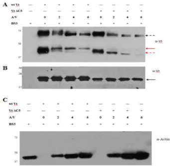
Vt has a weak propensity to dimerize at sub-millimolar concentrations (37). However, Johnson et al. detected formation of a Vt dimer in the presence of F-actin, at concentrations where the native dimer was not observed. They proposed that actin binding to Vt induces a conformational change that exposes a cryptic dimerization site, with dimer formation important for bundling of F-actin filaments (90). These earlier chemical cross-linking studies were
44
to DSS, BS3 forms chemical crosslinks with lysine side chains. We first added BS3 to purified WTVt and incubated the mixture at room temperature for 40 min in the absence of actin. The reaction products were monitored by western blot analysis using a Vt antibody. In the presence of BS3 (Figure 2.5A), Vt bands were observed at positions on the gel that correspond to both monomeric and dimeric forms of Vt. Densitometry quantification indicated that the dimer band accounts for ~ 22 % of total Vt in the sample. This result was not too surprising, as Vt can dimerize in solution (37, 72), however, with a weak affinity (Kd ~ 360 µM)(93). In fact, a similar dimer contact, which is located at the helix 4/5 interface, was reported (71). Our previous NMR studies revealed that the dimer observed by X-ray crystallography was similar to that observed by us in solution (93), suggesting that the Vt dimer observed in the presence of BS3 corresponds to the dimer observed by both X-ray crystallography and NMR (71, 93, 120). To evaluate whether actin bundling is altered by the presence of BS3, we conducted actin co-sedimentation experiments on WT Vt, Vt ΔC2, and Vt ΔC5, in the absence or presence of a water soluble crosslinker BS3. As shown in supplemental Figure 2.S1, the amount of bundled actin is essentially the same in the absence or presence of BS3, indicating that actin bundling is not artificially enhanced by the use of the cross-linking agent.