TAM RTK Inhibition Causes Different Effects in Myeloid-Derived Suppressor Cells and Dendritic Cells
By William Harris
Senior Honors Thesis Biology Department
University of North Carolina at Chapel Hill
March 20, 2017
Approved:
___________________________ Dr. Shelton Earp, Thesis Advisor (Approved)
___________________________ Dr. Mark Peifer, Reader (Approved)
Abstract:
While cancer immunotherapy presents the possibility of safe and effective treatments, a variety of factors may reduce the effectiveness of existing immunotherapies. Of particular note is the ability of cancer to activate cellular signaling pathways that ultimately suppress inflammatory responses. Two cell types thought to mediate cancer-related immunosuppression are
myeloid-derived suppressor cells (MDSCs) and dendritic cells (DCs). The TAM family of receptor tyrosine kinases (Tyro3, Axl, and Mer) is implicated in regulation of the immune system and the clearance of apoptotic cells. We hypothesize that TAM RTK signaling, particularly MerTK signaling, contributes to cancer-related immunosuppression by regulating the ability of MDSCs and DCs to suppress or stimulate the adaptive immune response. To determine the role of TAM RTKs in MDSC and DC function, we measured the expression level of various RNAs and proteins associated with the regulation of the immune system in MDSCs extracted from Mer-/-mice and DCs extracted from Tyro3-/-, Axl-/-, and Mer-/- mice. MDSCs were also treated
with TAM RTK inhibitors UNC 4198 and UNC 2371 developed by the Earp Lab. We also compare the ability of TAM knock-out and control DCs to stimulate T-cell proliferation. Due to the known role of nitric oxide signaling and arginase activity in MDSC-mediated suppression of the immune response, the immunosuppressive capabilities of MDSCs of both wildtype and TAM RTK knockout genotypes were assessed with enzymatic assays for nitric oxide synthase (iNOS), reactive oxygen species (ROS), and arginase activity. We found that TAM RTK knockout MDSCs exhibited significant changes in RNA and protein expression and increased iNOS and arginase activity, indicating they have a reduced immunosuppressive capacity. Analysis of TAM RTK knockout DCs yielded mixed results, though results consistent with literature supported the immunosuppressive role of TAM RTK signaling in DCs as well, though some contradictory results in T-cell proliferation and mRNA expression will require further study. Thus, the role of TAM RTK signaling in immune responses may be more complex than previously believed, but inhibition of TAM RTKs holds promise as a means of bolstering the immune response to cancer and providing treatment to individuals with the disease.
Introduction:
Myeloid Derived Suppressor Cells and Dendritic Cells: As scientists and clinicians continue to
place a greater emphasis on the use of immunotherapy in cancer treatment, it is important to better define the role of different cell types in the immune response against cancer.
Myeloid-derived suppressor cells (MDSCs) are a set of immature immune cells noted for their ability to regulate the inflammatory response in the context of cancer and other diseases. MDSCs consist of both granulocytic and monocytic subsets, which eventually develop into mature
granulocytes and monocytes, respectively. Both subsets express high levels of CD11b, and can be distinguished from each other by the expression of the markers Ly6G and Ly6C, with granulocytic MDSCs having the phenotype Cd11b+Ly6G+Ly6Clow and monocytic MDSCs
activity through the production of nitric oxide by inducible nitric oxide synthase (iNOS), which occurs primarily in monocytic MDSCs;the depletion of arginine in the local environment via arginase activity, which is more prominent in granulocytic MDSCs; and the secretion of reactive oxygen species (ROS) by both subsets.1 All of these functions are associated with
downregulation of the adaptive and innate immune system in the tumor microenvironment, with ROS and NO react to form radicals capable of nitrosylating, and thus inhibiting, T-cell receptors, and arginase activity depleting arginine necessary for cytotoxic T-cell function 234. Dendritic
cells (DCs), another component of the immune system, function as the primary
antigen-presenting cells of the immune system, utilizing both the MHCI and MHCII systems of signaling proteins to activate the adaptive immune response.5 DCs stimulate the CD8+ T-cell
response to cancer cells, but they are also capable of tolerizing the immune system to certain antigens, which can reduce ability of T-cells to fight the disease.5 Both MDSCs and DCs act on
the immune system, and are controlled by other cells through different extracellular signaling pathways.
TAM Family Receptor Tyrosine Kinases: The TAM-family of receptor tyrosine kinases (TAM
RTKs) is one of these networks; the binding of the ligands Gas6, which binds to Tyro3, Axl and Mer TK, and Protein S, which binds to the extracellular domains of Tyro3 and MerTK forms a bridge between the receptors and exposed phosphatidyl serine on apoptotic cell membranes, leading to the activation of intracellular signaling cascades such as the JAK/STAT3 and
MAPK/ERK pathways associated with increased cell metabolism and proliferation, and altered function in many cell types expressing the receptors. Because these proteins are associated with the suppression of the immune response 6 and are expressed in many immune cells, including
monocytes, granulocytes (which develop from bone marrow precursors) and DCs,7 8 TAM RTKs
likely are involved in regulating their response to cancer.
The immunosuppressive nature of the TAM RTKs makes them suitable targets for small molecule inhibition. To this extent, our collaborators in the UNC Eshelman School of Pharmacy Center for Integrative Chemical Biology and Drug Discovery, have created a panel of TAM RTK specific small molecule inhibitors. The inhibitors are designed to preferentially bind to one or more TAM receptor. For our studies, we have used UNC2371 and UNC4198. UNC2371 is a MerTK specific inhibitor (IC50 MerTK= 1.3 nM, Axl= 15 nM, Tyro3=7 nM) while UNC4198 is a MerTK/Tyro3-inhibitor (IC50 MerTK= 0.98 nM, Axl= 5.5 nM, Tyro3= 0.56 nM). While inhibition of TAM RTK signaling holds promise as a means of treating cancer by bolstering the immune response, the effects of TAM RTK inhibition must be characterized before inhibitors can be used in medicine.
Preliminary results indicate that inhibition of TAM RTK signaling reduces the ability of MDSCs to produce nitric oxide and arginase, but the underlying mechanisms behind this activity remain unknown (Holtzhausen, unpublished data). Since cancer cells are capable of upregulating the expression of TAM RTK ligands Gas6 and Protein S,9 10 11 TAM RTK signaling is believed to
could, therefore, reduce this capacity of cancer to reduce the inflammatory response, improving the ability of the immune system to fight the disease. DCs also express TAM RTKs in relatively high quantities, so signaling through these receptors likely affects many cell processes, possibly including the ability of DCs to present antigens to CD8+ T-cells to stimulate a response.6
Cross-presentation, or the ability of DCs to present exogenous antigens to cells of the adaptive immune system through the MHCI system instead of the usual MHCII system, is important in activating the anti-cancer CD8+ T-cell response.12 On the other hand, tolerogenic DCs reduce the
inflammatory response through mechanisms involving production of inhibitory cytokines coincident with activation of CD4+ T-regulatory cells.13 In cancer, DCs continually take up
antigen from apoptotic tumor cells and present these antigens to T-cells to trigger an anti-cancer immune response. However, signaling molecules produced by the cancer cells are capable of modifying the signals from DCs to CD8+ T-cells, causing the DCs to inhibit cytotoxic T-cell
activity towards antigens associated with the cancer cells instead of promoting it. This shift is accompanied by a change in the production of cytokines by dendritic cells, changing from the production of signals such as interferon-γ, which is associated with increased cross-presentation and greater CD8+ T-cell activity, to inhibitory cytokines such as IL-10 and IL-12. These cells
suppress CD8+ T-cells and reduce the immune response,14 so potential cancer treatments would
focus on reducing the tolerogenic activity of DCs while promoting cross-presentation and activation of CD8+ T-cells.
TAM RTK Inhibition and Cancer Treatment: Based on the preliminary data, inhibition or
knockout of TAM RTKs in MDSCs reduces the ability of these cells to suppress the immune response in the context of cancer. Detectable changes in expression and phosphorylation of intracellular signaling proteins were predicted to alter the immunosuppressive functions of MDSCs with TAM RTK inhibition. Assuming that in DCs, TAM RTK signaling also suppresses the immune response to cancer, knockout of the one or more TAM RTKs was expected to increase the ability of DCs to stimulate CD8+ T-cells and reduce the tolerizing mechanism. DCs
with a Tyro3-/-, Axl-/-, or Mer-/- genotype would also show changes in the expression of RNA and
proteins associated with antigen-presenting and immunostimulatory activity.
The capacity of MDSCs to inhibit other immune cells depends on the production of NO and ROS, along with the depletion of arginine caused by arginase activity; direct measurement of these functions through enzymatic assays would have indicated a reduction in all three if TAM RTK signaling does contribute to their immunosuppressive capability. Changes in the expression and phosphorylation of proteins associated with MDSC activity, including P38/p-P38 and NFκB/ p-NFκB would also have evinced the reduction of MDSC-mediated immunosuppression
associated with knockout of TAM RTKs. We used western blots to detect these changes in expression and illustrate the underlying mechanism regulating suppressive activity in MDSCs.
Because the contribution of DCs to the anti-cancer immune response primarily comes from their ability to induce the activity of CD8+ and CD4+ T-cells, proliferation assays were used
T-cell proliferation, along with decreased proliferation of CD4+ T-cells was observed. Moreover,
TAM RTK knockout DCs were expected to demonstrate a decreased production of inhibitory cytokines along with an upregulation in the expression of proteins associated with DC-mediated activation of the inflammatory response via cross-presentation.1516171819 202122232425 These
changes were detected via real-time quantitative PCR (for measuring changes in RNA expression) and by Western blot (for measuring changes in protein expression).
Methods:
WT and Mer-/- MDSCs: Mice from both WT and Mer-/- strains of C57BL/6 mice
(Jackson Labs) were injected with syngeneic B16 melanoma cells, and kept until tumor diameter reached 15 mm. The spleens of the mice were then harvested and splenocytes were extracted by mechanical disaggregation of spleens followed by lysing of erythrocytes in the single-cell suspension. CD11b+ cells were isolated from the resulting suspensions using CD11b-conjugated
magnetic beads (Miltenyi Biotec), and M-MDSCs and G-MDSCs were separated from CD11b+
myeloid cells using fluorescence activated cell sorting based on the phenotypic expression of markers Cd11b+Lys6G-/-Lys6Chigh (M-MDSC) and Cd11b+Lys6GhighLys6Clow (G-MDSC). The cell
sorting (Alisha Holtzhausen) was performed using commercially-available antibodies (BD Pharmingen) and a FACSAria III cell sorter (BD Biosciences), purifying populations of interest to 97% purity.
WT and TAM RTK-null DCs: Dendritic cells were obtained from the spleens of WT,
Tyro3-/-, Axl-/- , and Mer-/- strains of C57/B6 mice (Jackson Labs). Cells were purified using a
CD11c magnetic bead positive selection kit (Stem Cell Technologies), and isolated DCs were incubated in 96-well plates for three days before use in experiments. Purity of the cells was checked using flow cytometry to assess expression of CD11b and MHCII.
T-Cell Proliferation Assay: Murine CD11c+DCs were purified as described above using
an Easy Sep magnetic bead positive selection kit (Stem Cell Technologies). OT-1 CD4+T-cells
and CD8+ T-cells extracted from the spleens of OT-1 mice (Jackson Labs) were isolated using an
Easy Sep kit to select for the expression of the respective protein markers (Stem Cell
Technologies). B16 cells were treated with 10 micromolar staurosporine overnight to induce apoptosis, and CD11c+DCs were incubated with these apoptotic cells in the presence of OVA
peptide for two days prior to co-culture with OT-1 CD8+T-cells. As a control for
antigen-presenting activity, a sample of CD11c+DCs was co-cultured with both OVA peptide
and OT-1 CD8+T-cells simultaneously and allowed to incubate for the same amount of time.
DCs were incubated with CD8+ T-cells in both 40,000:200,000 and 40,000:240,000 DC:T-Cell
ratios, and DCs were incubated with CD4+ T-cells in a 40,000:240,000 ratio. CD4+ T-cell
activity was assessed by culturing CD4+ cells with CD11c+ DCs for two days. A CyQuant T-cell
qRT-PCR: DCs obtained from both WT and TAM RTK knockout mice using the method described above were processed using a Qiagen RNeasy Mini Kit to obtain RNA. qRT-PCR was performed using standard concentrations 1 𝜇mol RNA from DCs with a Taqman RNA-to-Ct 1-Step Kit (ThermoFisher Scientific) and commercially obtained probes for Tyro3, Axl, Mer, Gas6, and ProS (Applied Bioscience). For these genes, RNA standards varying in concentration (1,666-21 fg) were used as controls. Expression of other genes (Akt1, Ccl2, MyD88 , IFNγ, IL-10, IL-12, Traf6, Vegf, Ido, PD-L1, PGE-2) was quantified by using a Power SYBR Green Master Mix protocol to compare expression of these genes to GAPDH as a control (ThermoFisher Scientific). A 𝛥𝛥Ct method was used to analyze the results and compare TAM RTK knockout cells to WT. All PCR was performed with controls and samples in triplicate.
Arginase Assay: MDSCs obtained using the method described above were lysed using
buffer containing MnCl2 in a 100:1 water-Tris solution with a Roche complete mini protease inhibitor cocktail tablet (Roche Pharmaceuticals) for 10 minutes. After lysing, lysates were incubated with 0.5M L-arginine solution at 37oC for 120 minutes. The arginase reaction, which
produces a colored urea product, was stopped with the addition of a 1:3:7 H2SO4 (96%):H3PO4 (85%): H2O solution to each well containing lysed cells. Urea concentration was then measured using a colorimetric urea assay measuring at a wavelength of 430 nm. A blank (lysis buffer) and 1 mM urea solution were used as negative and positive controls, respectively, for the assay.
iNOSAssay (NO production): Activity of iNOS was measured in MDSCs using a
Measure-iT High-Sensitivity Nitrite Assay Kit (Thermo Fisher Scientific). Cell lysates were plated onto conditioned media in 96-well plates and Measure-iT nitrite quantitation reagent was added, with media being incubated in reagent for 5 hours at room temperature. The reaction was then halted with the addition of 2.8 M NaOH solution, and the assay was read using fluorescence spectroscopy with a 365/450 nm excitation/emission wavelength. Concentrations of nitrite species produced were determined through comparison with a comparison with sodium nitrite standard solutions of eight different concentrations (0-55 μM).
ROS Assay: MDSCs collected in 96-well plates were stained with dichlorofluorescin
diacetate (DCFDA), which, when oxidized, yields fluorescent dichlorofluorescin (DCF). Cells were incubated with DCFDA at 37oC for 120 minutes before DCF fluorescence was measured
using 485/535 nm excitation/emission wavelength.
Genotyping: To ensure that all mice used in experiments were correctly labeled
according to their genotype (WT or Mer-/-), PCR and gel electrophoresis were performed on cells
extracted from clipped mouse tails using a TaKaRa La Taq Protocol (Takara) with primers specific for WT and knockout Mer alleles along with a neomycin-resistance cassette in order to differentiate between heterozygous WT and Mer-/- mice (see appendix). PCR was performed to
Western Blotting: Gel electrophoresis was performed on cell lysates from WT and Mer
-/-DC subsets with bands being resolved using SDS-PAGE gels. After semi-dry transfer to
nitrocellulose paper, blots were incubated using protein-specific antibodies overnight according to manufacturer’s specifications (Cell Signaling Technology). The following day, blots were incubated in secondary anti-rabbit or anti-mouse antibodies, washed, and imaged using
SuperSignal West Femto Maximum Sensitivity Substrate (Thermo Fisher Scientific). A BioRad Kaleidoscope protein ladder was used as a reference for identifying bands (BioRad).
Results:
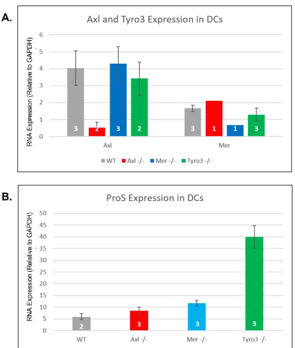
Based on the known role of TAM RTKs in reducing the inflammatory response, the expression of proteins associated with antigen presentation and immunosuppression were assessed with qRT-PCR and Western blot. Given the key roles the members of the TAM RTK family play in many immunosuppressive processes, knocking out these proteins should reduce the antigen presenting capability of these cells. First, the genotype of the knockout dendritic cells was confirmed using RT-qPCR by showing that Mer expression is reduced in Mer-/- and that
Tyro3 expression is reduced in Tyro3-/- DCs (Fig. 1A). The production of RNA for TAM RTK
ligand Protein S was also assessed, as knockout of receptor proteins is often associated with a compensatory overexpression of their ligands. This revealed an increase in ProS expression in Tyro3-/- DCs (Fig. 1B) compared to Axl-/-, Mer-/-, and WT DCs.
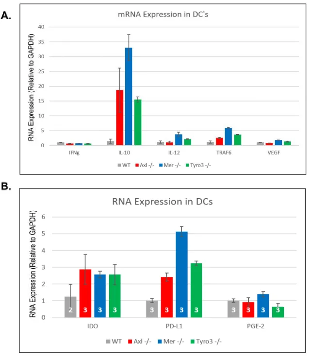
Following the assumption that reduction in TAM RTK signaling in DCs would reduce antigen-presenting capacity and promote a tolerizing phenotype, the expression of genes associated with both processes in DCs were measured in Tyro3-/-, Axl-/- ,Mer-/-, WT DCs using
qRT-PCR to quantify mRNA levels. mRNA for IL-10, TRAF6, IDO, and PD-L1 was higher in TAM RTK-knockout DCs than WT counterparts (Fig. 2), indicating that TAM RTK signaling reduces the expression of the corresponding genes. Conversely, IFNɣ mRNA expression was reduced in all three knockout genotypes (Fig. 2), implying that TAM RTK signaling facilitates greater expression of this protein.
In addition to the observed differences in mRNAs in TAM RTK knockout DCs, the expression of intracellular signaling proteins associated with the regulation of DC
antigen-presenting activity was determined using Western blotting. The knockout of TAM-RTKs in DCs promoted an increase in the expression of STAT3, along with a concomitant increase in its phosphorylated form, pSTAT3. A similar effect was demonstrated for the protein complex SMAD 2/3 and its phosphorylated form, though the increase was only observed in Mer-/- and
Tyro3-/- DCs. Although the expression of NF-κB was equal between WT and TAM-RTK
knockout DCs, the expression of the protein’s phosphorylated form, pNF-κB, was virtually non-existent in WT DCs, but substantially increased in all three knockout genotypes (Fig. 3B). Together these results indicate that while TAM RTK signaling affects the expression of
affect the expression of different proteins, and TAM RTK signaling plays a role in both the expression and post-translational modification of proteins.
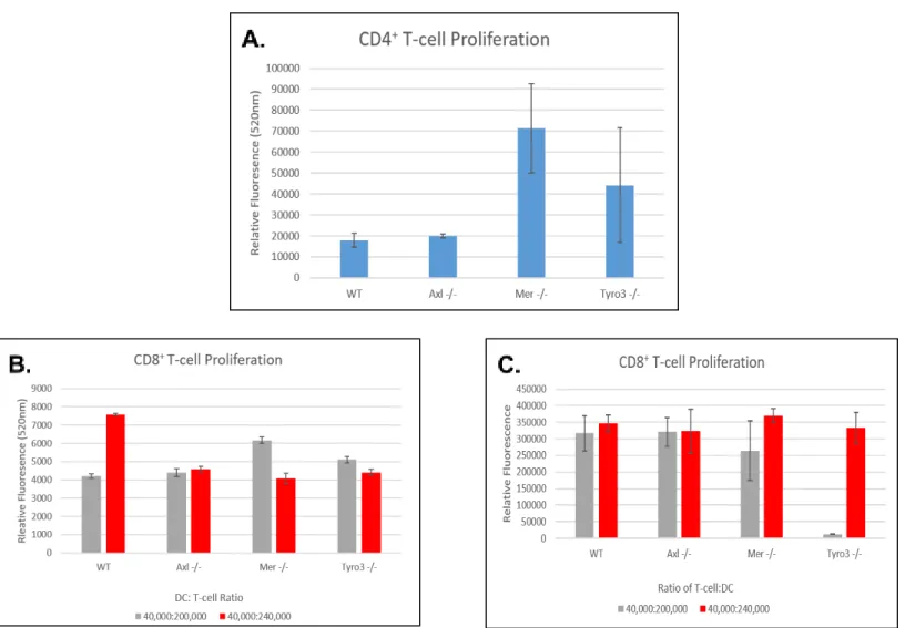
While the differences in the expression of mRNAs and proteins associated with antigen presentation and tolerogenesis by DCs were notable, the combined effect of these changes on the ability of DCs to induce a T-cell response remained unknown. A CyQuant T-cell Proliferation Assay Kit was used to assess the ability of DCs of various genotypes to stimulate the activity of both CD8+ T-cells and CD4+ T-regulatory cells. Incubation of DCs with CD4+ cells resulted in a
clear increase in proliferation of these cells in the presence of Mer-/- and Tyro3-/- DCs (Fig. 4A).
Furthermore, incubation of CD8+ T-cells with DCs following a period of antigen uptake reduced
the proliferation of the T-cells when cultured with a sufficient number of DCs (Fig. 4B). However, when CD8+ T-cells were co-cultured with DCs that did not have sufficient time for
antigen uptake and the CD8+ T cells were processed no differences in T-cell proliferation were observed (Fig. 4C), and result consistent with the use of the non-uptake condition as a control. A significant decrease in proliferation was observed in the 40,000:200,000 condition in Tyro3
-/-condition, however this outlier is likely due to plate reader error or loss of T-cells through washing rather than a phenotyping difference in the ability of DCs to induce T-cell proliferation. Overall, these results imply that TAM RTK signaling promotes a DC-mediated inflammatory response, while knockout of the TAM RTKs reduces the ability of DCs to stimulate CD8+ T-cell
activity and instead results in a tolerogenic response to antigens.
As MDSCs primarily suppress the inflammatory response through the production of nitric oxide and reactive oxygen species (ROS), and through depletion of arginine in the local environment, these functions were measured in MDSCs of both WT and TAM RTK knockout genotypes using enzymatic assays. ROS production in both monocytic and granulocytic subsets was demonstrably reduced in all three knockout genotypes compared to WT (Fig. 5), with slight variation between Tyro3-/-, Axl-/-, and Mer-/- MDSCs. While Axl-/- iNOS activity, measuring nitric
oxide production, was not significantly different between Axl-/- and WT G-MDSCs, all other
knockout cells demonstrated a reduction in iNOS activity compared to their WT counterparts (Fig. 6A). Furthermore, low dose (10 mg/kg) treatment of mice in vivo with Axl/Mer inhibitor UNC 2371 prior to MDSC harvesting resulted in a noticeable decrease in iNOS activity of MDSCs; however this effect was negated at higher doses (Fig. 6B). Finally, arginase activity, when assessed for the different subsets and genotypes of MDSC did not vary with the presence of TAM-family RTK expression (Fig. 7).
To assess the underlying causes of the observed phenotypic differences in MDSCs, the expression of key intracellular signaling proteins involved with the immunosuppressive activity of MDSCs was measured using Western blots. While no clear differences were observed
between WT and Mer-/- MDSCs of the same subset (Fig. 8A), there was a significant decrease in
phosphorylation of Erk and P38 associated with knockout of Tyro3 in both M-MDSCs and G-MDSCs (Fig. 8B). In Axl-/- MDSCs, there was a significant reduction in STAT3 expression in
MDSCs (Fig. 8C). These observed changes in protein expression support hypothesized role of TAM RTK signaling in regulation of immunosuppressive activity of MDSCs.
Discussion:
Protein and mRNA Expression in DCs: Given the immunosuppressive role TAM RTKs were
known to play in T-cells and macrophages, one would predict that knockout of these proteins in dendritic cells would produce a proinflammatory effect. In dendritic cells the immune response would be increased through heightened stimulation of cytotoxic T-cell activity, production of pro-inflammatory cytokines and other signaling proteins, and reduction in tolerogenic activity. Instead, from the qRT-PCR (Fig. 1A) it is apparent that knockout of TAM RTKs results in an increased expression of mRNAs for IL-10 and TRAF6, and a decreased IFN-ɣ expression. While TRAF6 has been implicated in the maturation of dendritic cells from myeloid precursors, IL-10 is a potent anti-inflammatory signaling protein and IFN-ɣ activity promotes an anti-tumor response from T-cells.5,16,19 Additionally, there was a clear increase in expression of mRNAs for
IDO and PD-L1 in all TAM RTK knock compared to WT (Fig. 1B), phenotypic changes with implications for DC function since IDO has been recognized as a promoter of the tolerogenic phenotype in DCs, and PD-L1 is associated with T-cell inactivation.20,23 Overall, these results
indicate that TAM RTK signaling likely contributes to DC maturation, but that signaling through this pathway facilitates a shift in DCs from a tolerogenic (immunosuppressive) phenotype to an immuno-stimulatory one.
In addition to differences in mRNA expression, TAM RTK DCs showed differences in the expression of a variety of key proteins in the regulation of antigen cross-presentation and CD8+ T-cell activation. The increases in STAT3/pSTAT3 expression and in NF-κB
phosphorylation (Fig. 2) are seemingly paradoxical, as STAT3 activation is associated with the inhibition of antigen-presentation activity in DCs while activated NF-κB is crucial for
antigen-presentation to occur.22,25 Additionally, there was an increase in the expression of both
phosphorylated and unphosphorylated forms of the SMAD2/3 complex in Mer-/- and Tyro3
-/-DCs. Due to the role of SMAD2/3, as a component of the TGF-β signaling pathway, in stimulating DC maturation and pro-inflammatory activity, DCs lacking Mer and Tyro3 likely exhibit greater antigen-presentation capability and potential to activate anti-cancer T-cell responses compared to WT cells. Together, these alterations in DC protein expression, while seemingly contradictory, indicate that the role of TAM RTK signaling in DCs may be more complex than initially believed. A repetition of Western blotting, including quantification using imageJ or similar software, to examine other members of the TGF-β, STAT3, and NF-κB signaling pathways would provide a better picture of how TAM RTK expression influences DC activation against cancer and other diseases.
T-Cell Proliferation Assays: While differences in protein and mRNA expression in DCs may
antigen-presenting cells to induce greater CD8+ T-cell activity, while reducing CD4+
T-regulatory cell activity, directly contributes to the ability of the immune system to destroy cancer cells. The ability of DCs to stimulate CD8+ T-cell proliferation following antigen uptake
and processing is greatly reduced in cells lacking TAM RTKs when T-cells are incubated with a sufficient number of DCs (Fig. 4B). When compared to control (Fig 4C), the proliferation of CD8+ T-cell is greatly increased due to the direct activation of T-cells by the influenza peptide
relative to the activity stimulated by antigen-presentation (Fig 4B). Overall, the lack of significant differences in proliferation in the control indicates that differences observed in the experimental condition are due to differences in the ability of DCs to stimulate proliferation through antigen-presenting activity, rather than release of cytokines or metabolic activity of DCs themselves. Ultimately, the decrease in CD8+ T-cell proliferation in Fig 4B evinces a reduction
in cross-presentation capacity because cross-presentation is critical for the direct activation of CD8+ T-cells by DCs. Inhibiting TAM RTK signaling of DCs in cancer treatment would thus
reduce the cytotoxic T-cell response to the disease, and could make such treatment less
effective.12 Additionally, there was an observed increase in CD4+ T-cell proliferation following
incubation with Mer-/- and Tyro3-/- DCs (Fig. 4A). CD4+ is a marker of both T-regulatory cells,
which reduce the response of cytotoxic T-cells to cancer, and helper T-cells, which contribute to an increased anti-tumor response. The increase in CD4+ T-cell proliferation following Mer TK
knockout is consistent with previous results, which were attributed to a decrease in helper T-cell proliferation and activity.26 Thus, the increase in CD4+ T-cell proliferation likely corresponds to
increased helper T-cell activity with reduced Mer or Tyro3 signaling, meaning that knockout of these proteins would promote a more robust immune response through CD4+ activity. However,
the increase in CD4+ cell could also be attributed to an increase in T-regulatory cell proliferation,
as these cells also express CD4. Such an increase would reduce in the anticancer immune response, so it is necessary to differentiate between the two cell types when assessing the effects of TAM RTK inhibition.13 Future experiments will require proper characterization of the
expanded CD4+ T-cell population using flow cytometry to screen cells for both CD4 and
FOXP3, a marker specific to T-regulatory cells. If in fact the increase in CD4+ cells can be
attributed to T-regulatory cells, the tendency for DCs with fully functional and impaired TAM RTK signaling to induce different types of T-cells would indicate a shift to a more tolerogenic state following a decrease in TAM RTK signaling in DCs. This result would run counter to our initial hypothesis and would imply that inhibition of TAM RTK signaling should be avoided to promote greater stimulation of T-cells by DCs as part of an anti-cancer immune response. On the other hand, if the increase in CD4+ T-cells is due to helper T-cell proliferation, the results of the
CD4+ and CD8+ T-cell proliferation assays would be contradictory, indicating that a reevaluation
of the methods would be necessary to accurately gauge the role of TAM RTK signaling on DC-mediated T-cell proliferation. The procedure used (see Methods) required multiple washes on weakly adherent T-cells in a 96-well plate, so the observed decrease in CD8+ T-cells may
proliferation. A dye-dilution assay employing flow cytometry or 3H-labeled thymidine
incorporation assay would avoid this problem and might provide a more accurate assessment of CD8+ T-cell proliferation by both WT and TAM RTK knockout DCs.
Enzymatic Assays of MDSC Activity: As we initially hypothesized, knockout of each of the
three TAM RTKs in MDSCs produced a significant decrease in the production of reactive oxygen species (ROS) by both monocytic (M-MDSC) and granulocytic (G-MDSC) subsets, with the Tyro3-/- MDSCs of both subsets showing the greatest difference compared to WT (Fig. 5).
Furthermore M-MDSCs lacking each of the TAM RTKs demonstrated a reduced ability to produce nitric oxide. Because ROS and NO produced by MDSCs react with each other in vivo to produce peroxynitrate species capable of deactivating CD8+ T-cells in the tumor
microenvironment by nitrosylating T-cell receptors, decreased TAM RTK signaling may reduce the immunosuppressive ability of MDSCs in a synergistic fashion.3,4 Moreover, the decrease in
constitutive MDSC iNOS activity observed with treatment of mice with 10 mg/kg of UNC 2371 (Fig. 6B) reveals that the phenotypic difference in immunosuppressive activity of MDSCs can be approximated in WT by inhibiting TAM RTK signaling with a drug. The increase in endogenous nitric oxide production associated with higher doses of UNC 2371 is likely due to non-specific binding to other cellular regulators of iNOS, and further refinement of the inhibitors used will increase the specificity of inhibitors for TAM RTKs, improving the ability of the drugs to inhibit MDSC-mediated immunosuppression.
Enzymatic assays also revealed that arginase activity, as measured by the conversion of arginine into urea, is not significantly different in Axl-/- and Tyro3-/- MDSCs. While this result is
inconsistent with our hypothesis due to the role of arginase in reducing cytotoxic T-cell activity in the tumor microenvironment through depletion of arginase,2 this result indicates that the role
of arginase is less important than NO and ROS production on the TAM RTK-mediated
immunosuppressive activity of MDSCs. Overall, the lack of difference in arginase activity would have little effect on the viability of TAM RTK inhibition as a means of increasing the anticancer immune response by reducing the activity of MDSCs due to the reduction in the other
immunosuppressive mechanisms of those cells.
Protein Expression in MDSCs: To explain the underlying mechanisms behind the effect of
TAM RTK signaling on MDSC activity, protein expression was assessed with Western blotting. While the results of Mer TK knockout were difficult to discern using this method, the clear reduction in phosphorylation of Erk and P38 in Tyro3-/- MDSCs supports the hypothesis that
TAM RTK signaling facilitates the immunosuppressive function of MDSCs, since intracellular signaling pathways involving Erk and P38 are associated with greater MDSC proliferation in tumor-bearing mice.26 In Axl-/- MDSCs, the decrease in expression of STAT3 (both
Ultimately, while the role of TAM RTK signaling in DCs requires further evaluation in the context of cancer treatment, an inhibition of TAM RTK signaling likely contributes to greater immunosuppression by MDSCs. TAM RTK inhibitors may promote anticancer immune
Figures:
Figure 1- Expression of TAM RTKs and Ligands in DCs Confirms TAM RTK Knockout Data were obtained using qPCR to quantitate mRNA expression in murine CD11c+DCs of
indicated phenotypes. Splenic DCs were harvested from mice, and RT-PCR was performed on extracted RNA to generate cDNA for use in qPCR. mRNA expression was quantified by
Figure 2- Expression of Immune Cell Signaling Proteins Varies with Genotype
Data were obtained using qPCR to quantitate mRNA expression in murine CD11c+DCs of
Figure 3- TAM RTK Signaling Alters Expression of Intracellular Signaling Proteins Western blotting was performed on murine CD11c+DCs of indicated phenotypes, using
Figure 4- Suppression of TAM-RTK Signaling Promotes a Tolerizing Phenotype in DCs The extent of T-cell proliferation was assessed using a CyQuant T-cell proliferation assay. A. OT-1 CD4+ T-cells harvested from mice were incubated with DCs of the indicated phenotype
and influenza peptide in a 240,000:40,000 ratio for three days before analysis. B. Dendritic cells were incubated with influenza peptide for a two day period and media was replaced. CD8+
T-cells were then added to DCs in 240,000:40,000 and a control (200,000:40,000) ratio, and cells were incubated for three more days. C. To control for direct stimulation of OT-1 CD8+T-cells
Figure 6- iNOS Activity Declines with Knockout of Mer TK, Tyro3 TK
Figure 7-Arginase Activity Patterns Vary between MDSC Genotypes
Figure 8-Expression of Intracellular Signaling Proteins is Affected by TAM RTK Signaling
Western blots were used to assess levels of expression of proteins critical for immunosuppressive activity and maturation in MDSCs of various genotypes. A. WT and Mer-/- MDSCs of both
monocytic and granulocytic subsets were compared with a focus on the phosphorylation and expression of STAT3, ERK, and P38. Actin expression was used to ensure equal loading of wells. B. Levels of ERK/p-ERK and P38/p-P38 were assessed in WT and Tyro3-/-MDSC subsets
C. The expression of various cell signaling proteins was compared in WT and Axl-/- MDSCs, and
Appendix:
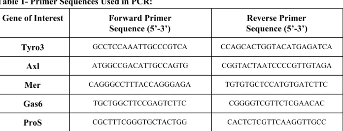
Table 1- Primer Sequences Used in PCR:
Gene of Interest Forward Primer
Sequence (5’-3’)
Reverse Primer Sequence (5’-3’)
Tyro3 GCCTCCAAATTGCCCGTCA CCAGCACTGGTACATGAGATCA
Axl ATGGCCGACATTGCCAGTG CGGTACTAATCCCCGTTGTAGA
Mer CAGGGCCTTTACCAGGGAGA TGTGTGCTCCATGTGATCTTC
Gas6 TGCTGGCTTCCGAGTCTTC CGGGGTCGTTCTCGAACAC
ProS CGCTTTCGGGTGCTACTGG CACTCTCGTTCAAGGTTGCC
References:
1. Gabrilovich, D. I., & Nagaraj, S. (2009). Myeloid-derived suppressor cells as regulators of the immune system. Nat Rev Immunol, 9(3), 162–174.
2. Munder, M. (2009). Arginase: an emerging key player in the mammalian immune system. British Journal of Pharmacology, 158(3), 638–651.
3. Yang, Y., Bazhin, A. V, Werner, J., & Karakhanova, S. (2013). Reactive Oxygen Species in the Immune System. International Reviews of Immunology, 32(3), 249–270.
4. Bogdan, C. (2001). Nitric oxide and the immune response. Nat Immunol, 2(10), 907–916. 5. Palucka, K., & Banchereau, J. (2012). Cancer immunotherapy via dendritic cells. Nat Rev
Cancer, 12(4), 265–277.
6. Linger, R. M. A., Keating, A. K., Earp, H. S., & Graham, D. K. (2008). TAM Receptor Tyrosine Kinases: Biologic Functions, Signaling, and Potential Therapeutic Targeting in Human Cancer. Advances in Cancer Research, 100, 35–83.
7. Graham, D. K., DeRyckere, D., Davies, K. D., & Earp, H. S. (2014). The TAM family: phosphatidylserine-sensing receptor tyrosine kinases gone awry in cancer. Nat Rev Cancer, 14(12), 769–785.
8. Rothlin, C.V., Ghosh, S., Zuniga, E. I., Oldstone, M. B. A., & Lemke, G. (2007). TAM Receptors Are Pleiotropic Inhibitors of the Innate Immune Response. Cell, 131(6), 1124–1136.
9. Battistelli, S., Vittoria, A., Cappelli, R., Stefanoni, M., & Roviello, F. (2005). Protein S in cancer patients with non-metastatic solid tumours. European Journal of Surgical
Oncology (EJSO), 31(7), 798–802.
Carcinoma Reflecting Tumor Advancement and Survival. Clinical Cancer Research, 15(14), 4742 LP-4749.
11. Sun, W., Fujimoto, J., & Tamaya, T. (2004). Coexpression of Gas6/Axl in Human Ovarian Cancers. Oncology, 66(6), 450–457.
12. Huang, A. Y., Golumbek, P., Ahmadzadeh, M., Jaffee, E., Pardoll, D., & Levitsky, H. (1994). Role of bone marrow-derived cells in presenting MHC class I-restricted tumor antigens. Science, 264(5161), 961 LP-965.
13. Maldonado, R. A., & von Andrian, U. H. (2010). How tolerogenic dendritic cells induce regulatory T cells. Advances in Immunology, 108, 111–165.
14. Levings, M. K., Sangregorio, R., & Roncarolo, M. G. (2001). Human CD25+ CD4+ T regulatory cells suppress naive and memory T cell proliferation and can be expanded in vitro without loss of function. Journal of Experimental Medicine, 193(11), 1295-1302. 15. Trinchieri, G., Pflanz, S., & Kastelein, R. A. (2003). The IL-12 Family of Heterodimeric
Cytokines: New Players in the Regulation of T Cell Responses. Immunity, 19(5), 641–644.
16. Moore, K. W., de Waal Malefyt, R., Coffman, R. L., & O'Garra, A. (2001).
Interleukin-10 and the interleukin-10 receptor. Annual review of immunology, 19(1), 683-765.
17. Gabrilovich, D. I., Chen, H. L., Girgis, K. R., Cunningham, H. T., Meny, G. M., Nadaf, S., … Carbone, D. P. (1996). Production of vascular endothelial growth factor by human tumors inhibits the functional maturation of dendritic cells. Nat Med, 2(10), 1096–1103. 18. Heath, W. R., & Carbone, F. R. (2001). Cross-presentation, dendritic cells, tolerance and
immunity. Annual review of immunology, 19(1), 47-64.
19. Schoenborn, J. R., & Wilson, C. B. (2007). Regulation of Interferon-γ During Innate and Adaptive Immune Responses. In B. T.-A. in Immunology (Ed.) (Vol. Volume 96, pp. 41–101). Academic Press.
20. Mellor, A. L., & Munn, D. H. (2004). Ido expression by dendritic cells: tolerance and tryptophan catabolism. Nat Rev Immunol, 4(10), 762–774.
21. Anderson, D. M., Maraskovsky, E., Billingsley, W. L., Dougall, W. C., Tometsko, M. E., Roux, E. R., … Galibert, L. (1997). A homologue of the TNF receptor and its ligand enhance T-cell growth and dendritic-cell function. Nature, 390(6656), 175–179. 22. Nefedova, Y., Huang, M., Kusmartsev, S., Bhattacharya, R., Cheng, P., Salup, R., …
Gabrilovich, D. (2003). Hyperactivation of STAT3 Is Involved in Abnormal
Differentiation of Dendritic Cells in Cancer. The Journal of Immunology, 172(1), 464 LP-474.
23. Romieu-Mourez, R., François, M., Boivin, M.-N., Stagg, J., & Galipeau, J. (2007). Regulation of MHC Class II Expression and Antigen Processing in Murine and Human Mesenchymal Stromal Cells by IFN-γ, TGF-β, and Cell Density. The Journal of
24. Yu, X., & Malenka, R. C. (2003). [beta]-catenin is critical for dendritic morphogenesis. Nat Neurosci, 6(11), 1169–1177.
25. Yoshimura, S., Bondeson, J., Foxwell, B. M. J., Brennan, F. M., & Feldmann, M. (2001). Effective antigen presentation by dendritic cells is NF-κB dependent: coordinate
regulation of MHC, co-stimulatory molecules and cytokines. International Immunology, 13(5), 675–683.
26. Cabezón, R., Carrera-Silva, E. A., Flórez-Grau, G., Errasti, A. E., Calderón-Gómez, E., Lozano, J. J., … Benítez-Ribas, D. (2015). MERTK as negative regulator of human T cell activation. Journal of Leukocyte Biology, 97(4), 751–760.
27. Yang, F., Wei, Y., Cai, Z., Yu, L., Jiang, L., Zhang, C., ... & Wang, J. (2015). Activated cytotoxic lymphocytes promote tumor progression by increasing the ability of 3LL tumor cells to mediate MDSC chemoattraction via Fas signaling. Cellular & molecular
immunology, 12(1), 66-76.
Acknowledgements:
● This work would not have been possible without the support of the Earp Lab and its members, special thanks to Dr. Shelton Earp for his support of my research
● Thank you to Dr. Alisha Holtzhausen Hanks for the support and guidance offered for the past two years, and for assisting with flow cytometry and other procedures