TAXANE CONJUGATE NANOPARTICLES FOR IMPROVED NON-SMALL CELL LUNG CANCER TREATMENT IN A NOVEL ORTHOTOPIC MOUSE MODEL
Lei Peng
A dissertation submitted to the faculty at the University of North Carolina at Chapel Hill in partial fulfillment of the requirements for the degree of Doctor of Philosophy in the Division
of Molecular Pharmaceutics in the UNC Eshelman School of Pharmacy.
Chapel Hill 2014
Approved By:
Russell J. Mumper Michael Jay Leaf Huang Sam Lai
©2014 Lei Peng
ABSTRACT
Lei Peng: Taxane Conjugate Nanoparticles for Improved Non-small Cell Lung Cancer Treatment in a Novel Orthotopic Mouse Model
(Under the direction of Russell J. Mumper)
The objectives of these studies were to develop lipid-based nanoparticles (NPs) to deliver taxane conjugates for improved non-small cell lung cancer (NSCLC) treatment. The chemotherapy agents Taxol, Abraxane and Taxotere have been limited by severe side effects, sub-optimal pharmacokinetic profile, and moderate therapeutic efficacy. Therefore,
Paclitaxel (PX) or Docetaxel (DX) was formulated in lipid-based NPs in these studies to achieve improved therapeutic index for NSCLC treatment. The NPs were engineered from oil-in-water microemulsion precursors with Miglyol as the oil core, and polyoxyl 20-stearyl ether (Brij 78) and D-alpha-tocopheryl polyethylene glycol succinate (TPGS) as the
surfactants, and abbreviated as BTM NPs.
A novel orthotopic NSCLC mouse model was established and characterized for in vivo evaluation of the developed formulation. During the effort of developing an orthotopic NSCLC mouse model, three different surgical procedures including intrabronchial injection, chest injection into left lung and dorsal injection into left lung were investigated. The
therapeutic efficacy of 2’-(2-bromohexadecanoyl)-docetaxel (Br-C16-DX) NPs versus Taxotere® was investigated in this model. The results demonstrated longer progression-free survival and median survival of the NP-treated group as compared to the Taxotere group.
For PX delivery, a 2’-(2-bromohexadecanoyl)-paclitaxel (Br-16-PX) conjugate was synthesized and formulated into lipid NPs. The goals were to improve drug entrapment in the drug delivery system and to enhance in vivo pharmacokinetics and conversion to paclitaxel at the tumor site. The developed system was evaluated in luciferase-expressing A549 cells in vitro and in the developed orthotopic NSCLC mouse model. The results demonstrated that the Br-C16-PX NPs had an increased maximum tolerated dose (MTD) and an improved pharmacokinetic profile as compared to Taxol®, which resulted in significantly improved antitumor efficacy for the treatment of advanced NSCLC.
ACKNOWLEDGEMENTS
The journey of my Ph.D. studies could not have been so fruitful and memorable without the support from my mentors, friends, and family. First and foremost I would like to thank my advisor Dr. Russell J. Mumper for his guidance and encouragements which lead me through the completion of this research project. He has taught me the most important critical thinking and problem-solving skills through five years’ invaluable supervision, and
significantly improved my writing and communication skills. I benefited tremendously from his enthusiasm, dedication, extremely efficient working style and great attention to details. His optimistic and always positive attitude cheered me up from frustrations and made the journey more enjoyable. In addition to the scientific supervision, Dr. Mumper has provided me the flexibility to explore different areas including the translational medicine training program and industrial internship. This important exploration process helped to elucidate my research interest and career goals. I am truly thankful to his support and patience.
I would like to thank everyone in the Biological and Biomedical Sciences Program (BBSP) including Dr. Fulton Crews, Dr. Liya Qin and Anna Ballew O’Connell. BBSP provided precious lab rotation opportunities for the students to fully explore their interest. Special thanks go to my committee member Dr. Leaf Huang. He opened the door of School of Pharmacy for me when I came from BBSP with a biology background. He introduced the fascinating world of drug delivery which totally caught my interest and lead to my decision of joining the pharmaceutical sciences program. I have been inspired by his constructive suggestions in committee meetings, his philosophy of doing cutting-edge science that solves fundamental problems, his way of training young scientists as well as his efforts in taking good care of family. I will benefit from this throughout my whole life.
I am very grateful to Dr. E. Claire Dees, my committee member and clinical co-mentor in Graduate Training Program in Translational Medicine. Dr. Dees provided unique clinical guidance for my dissertation research and kindly gave me the opportunity to shadow her in the clinic. Talking with breast cancer patients face-to-face was a touching moment underlining the significance and urgency of pharmaceutical research. This is the initial and primary motivation of my dedication to human healthcare. I thank all faculty and staff in the Howard Hughes Medical Institute-funded Graduate Training Program in Translational Medicine, especially Dr. Patrick Brandt, Dr. Virginia Miller and Dr. Bill Coleman. The extensive human pathology training and monthly seminars greatly improved my
understanding of translational research and its clinical application.
Division of Molecular Pharmaceutics including but not limited to Drs. Philp C. Smith, Paul Dayton, Xiao Xiao, Moo J. Cho, Rudolph Juliano, Anthony Hickey, Shawn Hingtgen, Alexander V. Kabanov and Elena V. Batrakova. I thank all previous and current staff in School of Pharmacy especially Ning Sun, Ain Mason, Holly Maguire and William Vogt for their diligent work.
I would like to thank the wonderful core facilities at University of North Carolina including Animal Studies Core especially Charlene Santos, Mark Ross and Alain Valdivia, Biomedical Research Imaging Center especially Dr. Hong Yuan, Animal Histopathology Core, Microscopy Services Laboratory and Animal Clinical Chemistry Core for their skills and expertise. Without their help the completion of my dissertation project would have been much more difficult and time-consuming.
I would like to acknowledge all current and previous Mumper lab and Jay lab members, with special thanks to Dr. Ping Ma, Dr. Clinton Jones, Dr. Feng Lan, Dr. S. Rahima Benhabbour, Dr. Dongyun Liu, Dr. Katsuhiko Sueda, Dr. Matthew Sadgrove, Dr. Yu-Tsai Yang, Dr. Anekant Jain, Dr. Xiuling Lu, Mianmian Sun, James Huckle, Jing Fu, Dylan Glatt, Yong Zhang and Junghyun Kim. I sincerely value their helpful discussions, generous help and friendship. I am grateful to all my friends including Dr. Hailin Wang, Dr. Changyun Xiong, Dr. Kevin Chu, Dr. Yu-Cheng Tseng, Dr. Yuan Zhang, Dr. Tianxiang Han, Dr. Yang Liu, Lee Mullin, Zhijian He, Dongfen Yuan, Yuhang Jiang and Xiaomeng Wan for accompanying me through all the struggles and good times.
TABLE OF CONTENTS
LIST OF TABLES ... xiv
LIST OF FIGURES ... xv
LIST OF ABBREVIATIONS ... xvi
CHAPTER 1: PACLITAXEL CONJUGATE FOR IMPROVED CANCER THERAPY ... 1
1. Paclitaxel and its limitations ... 1
2. Small Molecule Conjugates ... 3
2.1 Water-soluble small molecule conjugates ... 3
2.2 Lipophilic small molecule conjugates ... 6
3. Polymeric Conjugates ... 10
3.1 Poly(ethylene glycol) ... 10
3.2 Hyaluronic acid ... 14
3.3 N-(2-hydroxypropyl)-methacrylamide (HPMA) ... 15
3.4 Poly(L-glutamic acid) ... 16
4. Dendrimer ... 17
5. Protein conjugates of PX ... 19
5.1 Antibody conjugates ... 19
5.2 Plasma protein conjugates ... 21
6. Summary ... 21
Figure 1.2 PX-peptide conjugates ... 24
Figure 1.3 PX-fatty acid conjugates... 25
Figure 1.4 PX-lipid conjugates ... 26
Figure 1.5 PX-PEG conjugates ... 27
Figure 1.6 PX-2’-hyaluronic acid ... 28
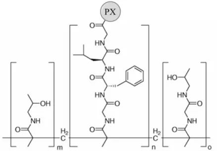
Figure 1.7 PX-2’-HPMA ... 29
Figure 1.8 (A) PX-2’-poliglumex (B) PX-2’-poly(L-γ-glutamyl-glutamine) ... 30
Figure 1.9 PX-dendrimer conjugate showing the cascade release of PX ... 31
Figure 1.10 PX-2’-PAMAM conjugate ... 32
Figure 1.11 Generation 1 and Generation 2 PX-2’-triazine dendrimer ... 33
Figure 1.12 Different types of PX conjugates ... 34
Table 1.1 Advantages and limitations of PX and PX conjugates ... 35
CHAPTER 2: DEVELOPMENT OF A NOVEL ORTHOTOPIC NON-SMALL CELL LUNG CANCER MODEL AND THERAPEUTIC BENEFIT OF 2’-(2-BROMOHEXADECANOYL)-DOCETAXEL CONJUGATE NANOPARTICLES ... 36
1. Summary ... 36
2. Introduction ... 37
3. Materials and Methods ... 39
4. Results ... 44
4.1 Orthotopic lung tumor implantation and survival study ... 44
4.2 Histology characterization of the A549-luc-c8 tumor ... 45
4.3 Micro PET/CT Imaging ... 46
4.4 Preparation and Characterization of BTM NPs Containing Br-C16-DX ... 47
5. Discussion ... 50
Figure 2.1 In vitro bioluminescence imaging of A549-luc-c8 cells ... 55
Figure 2.2 Orthotopic lung tumor implantation and survival study... 56
Figure 2.3 Histology characterization of orthotopic lung tumors. ... 57
Figure 2.4 Micro PET/CT imaging and correlation with ... 58
Figure 2.5 Structure of Br-C16-DX ... 59
Figure 2.6 Efficacy study with Taxotere and Br-C16-DX NPs ... 60
Figure 2.7 Immunohistochemistry quantification of resistance-related proteins... 61
Figure 2.8 Gross images and H&E staining of lungs from surviving mice in two treatment groups ... 62
Figure 2.9 Metastasis in control group of the efficacy study ... 63
Figure 2.10 Comparison of chest wall injection and dorsal side injection ... 64
CHAPTER 3: 2’-(2-BROMOHEXADECANOYL)-PACLITAXEL CONJUGATE NANOPARTICLES FOR THE TREATMENT OF NON-SMALL CELL LUNG CANCER IN AN ORTHOTOPIC XENOGRAFT MOUSE MODEL ... 65
1. Summary ... 65
2. Introduction ... 66
3. Materials and Methods ... 68
4. Results ... 74
4.1 Optimization and characterization of Br-C16-PX NPs... 74
4.2 Tubulin polymerization activity of Br-C16-PX ... 75
4.3 Cytotoxicity of Br-C16-PX NPs ... 76
4.4 In vivo antitumor efficacy studies ... 76
4.5 Pharmacokinetic and biodistribution studies ... 77
4.6 In vivo toxicity of Br-C16-PX NPs ... 79
Figure 3.1 Synthesis scheme of Br-C16-PX ... 86
Figure 3.2 Toxicity study of Br-C16-PX ... 87
Figure 3.3 In vivo efficacy study in an orthotopic NSCLC model ... 88
Figure 3.4 Pharmacokinetic (PK) and biodistribution studies in an orthotopic NSCLC model ... 89
Table 3.1 Formulation optimization and characterization of Br-C16-PX loaded BTM NPs... 90
Table 3.2 Summary of important pharmacokinetic parameters ... 91
Table 3.3 Summary of NP toxicity study... 92
CHAPTER 4: SUMMARY AND FUTURE DIRECTIONS... 93
1. Summary ... 93
2. Future directions ... 95
2.1 Fluorescent labeling of the BTM NPs ... 96
2.2 Targeted delivery to NSCLC ... 97
2.3 Pegylation ... 99
2.4 Conversion of Br-C16-PX to PX ... 101
Figure 4.1 Structure of (A) BODIPYFL C12, (B) Nile Red and (C) 5-FAM-cadaverine... 106
Figure 4.2 (A) F888, (B) NR668 and (C) schematic presentation of a nanodroplet ... 107
Figure 4.3 Zebrafish microangiography ... 108
Figure 4.4 Comparison of CD44 and EGFR expression levels in NSCLC cell lines. ... 109
Figure 4.5 (A) Brij78-NTA-Ni (B) and (C), NP internalization in A549 cells ... 110
Figure 4.6 (A) Synthesis of Brij 78-acid (B) Preparation of CD44-targeted BTM NPs (C) Cell uptake study ... 111
Figure 4.8 L-arginine lauryl ester ... 113
Figure 4.9 GALA-mediated membrane fusion and endosomal escape ... 114
Figure 4.10 PX prodrug with a diglycolic linker ... 115
APPENDIX: STANDARD OPERATING PROCEDURE ... 116
5.1 Cell culture and storage of A549-luc-C8 cells ... 117
5.2 Cell culture of A549-luc-C8 cells for animal studies ... 118
LIST OF TABLES
LIST OF FIGURES
Figure 1.1 Structure of PX ... 23
Figure 1.2 PX-peptide conjugates ... 24
Figure 1.3 PX-fatty acid conjugates... 25
Figure 1.4 PX-lipid conjugates ... 26
Figure 1.5 PX-PEG conjugates ... 27
Figure 1.6 PX-2’-hyaluronic acid ... 28
Figure 1.7 PX-2’-HPMA ... 29
Figure 1.8 (A) PX-2’-poliglumex (B) PX-2’-poly(L-γ-glutamyl-glutamine) ... 30
Figure 1.9 PX-dendrimer conjugate showing the cascade release of PX ... 31
Figure 1.10 PX-2’-PAMAM conjugate ... 32
Figure 1.11 Generation 1 and Generation 2 PX-2’-triazine dendrimer ... 33
Figure 1.12 Different types of PX conjugates ... 34
Figure 2.1 In vitro bioluminescence imaging of A549-luc-c8 cells ... 55
Figure 2.2 Orthotopic lung tumor implantation and survival study... 56
Figure 2.3 Histology characterization of orthotopic lung tumors. ... 57
Figure 2.4 Micro PET/CT imaging and correlation with ... 58
Figure 2.5 Structure of Br-C16-DX ... 59
Figure 2.6 Efficacy study with Taxotere and Br-C16-DX NPs ... 60
Figure 2.7 Immunohistochemistry quantification of resistance-related proteins ... 61
Figure 2.8 Gross images and H&E staining of lungs from surviving mice in two treatment groups ... 62
Figure 2.9 Metastasis in control group of the efficacy study ... 63
Figure 3.1 Synthesis scheme of Br-C16-PX ... 86
Figure 3.2 Toxicity study of Br-C16-PX ... 87
Figure 3.3 In vivo efficacy study in an orthotopic NSCLC model ... 88
Figure 3.4 Pharmacokinetic (PK) and biodistribution studies in an orthotopic NSCLC model ... 89
Figure 4.1 Structure of (A) BODIPYFL C12, (B) Nile Red and (C) 5-FAM-cadaverine... 106
Figure 4.2 (A) F888, (B) NR668 and (C) schematic presentation of a nanodroplet ... 107
Figure 4.3 Zebrafish microangiography ... 108
Figure 4.4 Comparison of CD44 and EGFR expression levels in NSCLC cell lines. ... 109
Figure 4.5 (A) Brij78-NTA-Ni (B) and (C), NP internalization in A549 cells ... 110
Figure 4.6 (A) Synthesis of Brij 78-acid (B) Preparation of CD44-targeted BTM NPs (C) Cell uptake study ... 111
Figure 4.7 Schematic illustrations of PEG configurations on NP surface ... 112
Figure 4.8 L-arginine lauryl ester ... 113
Figure 4.9 GALA-mediated membrane fusion and endosomal escape ... 114
LIST OF ABBREVIATIONS
AL L-arginine lauryl
ALP alkaline phosphatase
ALT alanine aminotransferase ANOVA analysis of variance
ATP adenosine triphosphate
AUC area under the curve
BLI bioluminescence imaging
BMS Bristol-Myers Squibb
Br-C16-DX 2’-(2-bromohexadecanoyl)-docetaxel Br-C16-PX 2’-(2-bromohexadecanoyl)-paclitaxel Brij 78 polyoxyl 20-stearyl ether
BUN blood urea nitrogen
C22-PX 2’-behenoyl-paclitaxel conjugate
Cmax maximum concentration
CT computed tomography
CT-2103 poly(L-glutamic acid)
DHA docosahexaenoic acid
DMAP 4-dimethylaminopyridine DMSO dimethyl sulfoxide
DX docetaxel
EEA early endosome antigen
EPR enhanced permeability and retention
FBS fetal bovine serum
FDG fluorodeoxyglucose
FITC fluorescein isothiocyanate
GALA WEAALAEALAEALAEHLAEALAEALEALAA
GPC gel permeation chromatography GRAS generally regarded as safe GST glutathione S-transferase H&E Hematoxylin & Eosin
HA hyaluronic acid
HER2 human epidermal growth factor receptor 2 HPLC high-performance liquid chromatography HPMA N-(2-hydroxypropyl)-methacrylamide
hr hour
IC50 half maximal inhibitory concentration
ICP-MS inductively coupled plasma mass spectrometry
i.p. intraperitoneal
i.v. intravenous
kg kilogram
LAMP lysosomal-associated membrane protein LC-MS liquid chromatography–mass spectrometry LRP lung resistance-related protein
MEND multifunctional envelope-type nanodevice MDR multiple drug resistance
mg milligram
min minute
ml milliliter
MMP matrix metalloproteinase mPEG methoxy poly(ethylene glycol) MRP multidrug resistant protein
MRT mean residence time
MS mass spectrum
MTD maximum tolerated dose
MTT 3-[4,5-dimethylthiazol-2-yl]-2,5-diphenyl tetrazolium bromide
MW molecular weight
nab-PX albumin-bound paclitaxel
ng nanogram
nm nanometer
NMR nuclear magnetic resonance
NP nanoparticle
NSCLC non-small cell lung cancer PAMAM poly(amidoamine)
PBS phosphate buffered saline
PEG poly(ethylene glycol)
PGG poly(L-γ-glutamyl-glutamine)
Pgp P-glycoprotein
PLA poly(lactic acid)
PLGA poly(lactic-co-glycolic acid)
PK pharmacokinetic
PX paclitaxel
PX-2’-MPA PX-2’-methylpyridinium acetate RES reticuloendothelial system
RHAMM hyaluronan-mediated motility receptor
ROI region of interest
sb-PX solvent-based paclitaxel
s.c. subcutaneous
SEC size exclusion chromatography SUV standardized uptake value
T1/2 half-life
TLC thin layer chromatography
TPGS D-α-tocopheryl polyethylene glycol 1000 succinate
µg microgram
µl microliter
Vd volume of distribution
VEGF vascular endothelial growth factor
CHAPTER 1.
PACLITAXEL CONJUGATE FOR IMPROVED CANCER THERAPY
1. Paclitaxel and its limitations
Paclitaxel (PX) is a microtubule stabilization agent discovered from the bark of Pacific Yew (Taxus brevifolia) in the early 1960s by the U.S. National Cancer Institute. It is a white crystalline powder with a melting point of ~210° (Figure 1.1, C47H51NO14, MW
853.93). PX has been widely used as a chemotherapeutic drug mainly to treat lung, ovarian and breast cancer [1]. It works through promoting tubulin polymerization which leads to formation of abnormally stable microtubules. The lack of microtubule dynamics during mitosis further results in cell arrest at G2-M phase and apoptotic cell death of fast-proliferating cells.
The use of PX has been limited by its low water solubility (~0.4 µ g/ml). The first commercial product Taxol® was approved by the FDA in 1992, in which PX is formulated in organic solvents of polyoxyethylated castor oil (Cremophor EL) and dehydrated ethanol (50/50, v/v). However, the high amount of toxic co-solvents, especially Cremophor EL, leads to serious side effects including anaphylaxis and severe hypersensitivity [2]. Premedication with corticosteroids, diphenhydramine, and H2 antagonist is required prior to Taxol
pharmacokinetics in vivo [3].
Abraxane,an albumin-bound paclitaxel nanoparticle (mean diameter 130 nm), was approved by the FDA in 2005 for the treatment of breast cancer and later in 2012 for the treatment of non-small cell lung cancer (NSCLC). In the phase III trial CA-031, weekly albumin-bound paclitaxel (nab-PX) plus carboplatin was compared with solvent-based paclitaxel (sb-PX) plus carboplatin in advanced NSCLC patients [4]. Although Nab-PX exhibited decreased toxicity, the 10% improvement of progression-free survival (p = 0.21) and overall survival (p = 0.27) were not statistically significant. Moreover, although
Abraxane exhibited linear, thus more predictable, pharmacokinetic profile, the half-life (p = 0.48) and area under the concentration versus time curve (AUC, p = 0.52) of Abraxane at 260 mg/m2 were not significantly increased as compared to Taxol at 175 mg/m2 [5].
Due to the challenges in directly formulating PX, conjugates of PX have been
2. Small Molecule Conjugates
2.1 Water-soluble small molecule conjugates
Because the water solubility of PX is very low, early efforts of developing PX conjugates focused on improving its water solubility in order to minimize the use of toxic co-solvents. Water-soluble PX conjugates including succinate, glutarate, sulfonate, amino acid, carbonate, phosphate, and peptide conjugates were synthesized and evaluated.
2.1.1 Early Development
Investigations of PX conjugates were initiated in late 1980s and were focused on improving water solubility in early development. Major conjugates are PX C-2’ and C-7 esters such as succinate, glutarate, sulfonate and amino acid and carbonate [9-11]. Despite improved water solubility, the C-2’ conjugates were not stable in aqueous medium or human plasma, and the C-7 conjugates readily released the parent drug PX and thus did not usually exhibit prodrug properties, leading to reduced antitumor activity in vivo as compared to PX. Nicolaou et al. synthesized a series of PX 2’-monoester of dicarboxylic acid in 1993 [12]. This study underlines the importance of the electron-withdrawing group in the side chain, wherein increased release of PX was observed with the electron-withdrawing ability of the aryl substitutes or the linking heteroatom. These protaxols were demonstrated to have similar cytotoxicity to PX but only modestly improved water solubility. Later, Nicolaou et al.
basic tumor microenvironment. As more studies have established the relatively acidic conditions in tumor tissue, development of these conjugates were discontinued. One promising category of small molecule conjugates are PX-2’-carbonates [11]. Although 10-fold less toxic than PX in human colon cancer cell line HCT116, the PX-2’-carbonates released PX in a murine lung carcinoma (M109) model and achieved longer survival than PX. However, due to low water solubility, the PX-2’-carbonates were administered i.p. in 10% DMSO or 10% Tween 80.
2.1.2 Phosphate esters
2’-OH and 7-2’-OH, respectively [16, 17]. The resulting compound had 1000-fold greater water solubility than PX and similar antitumor efficacy in M109 murine lung carcinoma model.
2.1.3 Peptide conjugates
Synthetic peptides have the advantages of smaller size, cost-effective production and low immunogenicity as compared to large proteins or antibodies. Several peptide PX
conjugates have been investigated including cell penetrating peptide [18, 19], integrin-targeting RGD peptide [20, 21], and peptides that target HER-2 [22]. However, the majority of the studies reported only in vitro cytotoxicity with limited or untested in vivo efficacy.
One promising approach was the octaarginine transporter which was attached to the C2’ or C7 positions of PX by a cleavable disulfide linker [18]. The C2’ conjugate (Figure 1.2A) was shown to be more effective than the C7 conjugate and free PX in a mouse model with human ovarian carcinoma cell line UCI 101. Importantly, the PX-octaarginine was demonstrated to overcome Pgp and induce cell death in resistant cancer cells. Although the C7 conjugate produced similar effect as free PX in mice implanted with sensitive OVCA-429 cells, it significantly prolonged survival relative to PX in resistant mouse model with OVCA-429T cells.
longer mean survival (42.8 days) in mice bearing intracranial U-87 MG glioma as compared to mice treated with transferrin modified PX loaded micelle (39.5 days), PX loaded micelle (34.8 days) or free PX (33.6 days).
2.2 Lipophilic small molecule conjugates
Drug delivery systems including nanoparticles (NPs) have been developed to deliver PX and to utilize the advantages of nanosystems including EPR (enhanced permeability and retention) effect, prolonged circulation, and active targeting. Despite low water solubility of PX, its solubility in many lipids is also limited, leading to compromised drug entrapment and drug retention in lipid-based NPs. Therefore, more lipophilic PX conjugates were developed to enhance the drug entrapment and drug retention in hydrophobic drug delivery systems.
2.2.1 Fatty acid conjugates
Fatty acid has been investigated extensively to produce lipophilic PX conjugates through forming an ester bond with the 2’-OH group. It has been suggested that some fatty acids may serve as biochemical precursors or energy sources for tumors thus are taken up from arterial blood into tumor cells. Improved pharmacokinetic profile could be achieved when the conjugate is incorporated into nanosystems. Lundberg et al. prepared a lipid emulsion with PX-2’-oleate conjugate (Figure 1.3A) with a particle size of 50 nm [7]. The PX-2’-oleate was much more lipophilic than PX with decreased water solubility from 12.8 µM to 34 nM and increased Ko/w from 311 to 8,074. Pharmacokinetic studies in rabbits
emulsion or free PX in Cremophor EL/ethanol. The delivery vehicle helped to improve the pharmacokinetic profile and the increased lipophilicity of the conjugate as compared to PX enhanced drug retention in the vehicle. Further evaluations of the in vivo efficacy showed moderate advantage of the liposome formulation as compared to commercial PX in B16F10 (murine melanoma) tumor bearing mice [23].
Benita and co-workers synthesized a PX-2’-palmitate formulated in different targeted systems. The PX-palmitate was incorporated in cationic immunoemulsions with anti-HER2 monoclonal antibody (Herceptin) for the treatment of prostate cancer [24]. The drug-loaded immunoemulsions did not activate the complement as compared to the commercial and PX-palmitate hydroalcoholic formulations. Furthermore, the immunoemulsion had significantly improved tumor growth inhibition in vivo as compared to the untargeted cationic emulsion (p < 0.05) and the paclitaxel palmitate hydroalcoholic formulations (p < 0.01). Later, the
conjugate was formulated in pegylated polyester immunonanoparticles which showed significantly increased t1/2, Cmax and AUC values as compared to the PX-palmitate solution
[25]. Moreover, the targeted immunonanoparticles had decreased liver and spleen
accumulation than untargeted NPs. Recently, the same conjugate was formulated in PLGA NPs with an anti-EGFR antibody cetuximab [26]. Significantly greater tumor growth inhibition and increased survival rates were observed in a metastatic lung cancer mouse model as compared to the non-targeted drug solution and non-targeted drug NPs.
susceptible to hydrolysis. In vitro studies in A2780 and MCF-7 cells showed 3 to10-fold and 30 to 90-fold greater IC50 of the succinate-linked conjugates as compared to the
diglycolic-linked conjugates, respectively, presumably due to lack of release of active PX from the more stable succinate bond. Furthermore, NPs containing succinate prodrug were ineffective in vivo in a xenograft mouse model with HT29 human colon carcinoma tumor, while NPs loaded with diglycolate prodrug were demonstrated to inhibit tumor growth. Interestingly, the antitumor activity of the diglycolate prodrug-loaded NPs was correlated with the aliphatic chain length, with the longer chain leading to improved efficacy. This was due to the longer partitioning half-lives of the more lipophilic conjugates from the lipid NPs which lead to sustained release and subsequent activation by esterase to produce PX.
Besides modulating fatty acid chain length and linker chemistry, different research groups have also investigated the inclusion of an electron-withdrawing group on the fatty acid chain to make the ester bond more cleavable. Mayhew and co-workers synthesized a 2’-α-bromohexadecanoyl PX conjugate in which hydrolysis of the prodrug was accelerated by the electron-withdrawing bromine atom at the α position of the acyl chain [28-31]. The bromohexadecanoyl prodrug was first administered dissolved in Cremophor EL and it showed improved in vivo activity as compared to PX in a subcutaneous human ovarian cancer (OVCAR-3) mouse model. Later, Mayhew and co-workers incorporated the prodrug in a liposomal formulation to avoid the use of Cremophor. Liposomes with 2’-α
-bromohexadecanoyl PX were demonstrated to have superior antitumor efficacy in vivo as compared to PX.
[32-38]. DHA is a natural fatty acid found in human milk and is classified as a nutritional
additive by the FDA. The DHA-PX is a prodrug without microtubule stabilization activity. It must be converted to active PX to be effective. It was demonstrated that the AUCs of PX from DHA-PX were 1.7- and 5.8-fold greater than those of PX at equimolar and equitoxic doses in an M109 mouse tumor model. A complete tumor regression was observed at 120 mg/kg of DHA-PX, while no significant tumor suppression was observed for equitoxic PX at 20 mg/kg [32]. Furthermore, DHA-PX was a 4-fold weaker substrate for Pgp as compared to free PX and therefore may be effective in treating resistant tumors. DHA-PX has been investigated in clinical trials for the treatment of melanoma and decreased toxicity compared to conventional therapy was observed (no hypersensitivity, no hair loss, and mild nausea) [35-38]. Weekly administration appeared to be a better dosing regimen as compared to a single dose administered every three weeks for the treatment of solid tumors. However, it should be noted that although DHA-PX was formulated in 80% less Cremophor EL than Taxol, both Cremophor EL and ethanol were still present in the DHA-PX formulation.
2.2.2 Other lipophilic small molecule conjugates
was shown to release PX in bovine plasma with a half-life of 80 min [40]. The conjugate was then incorporated into a liposomal formulation with a size of 2.77 µm for aerosol delivery to treat lung cancer. Moderate cytotoxicity was observed in A549 cells and in vivo evaluation of the system was not performed.
3. Polymeric Conjugates
Linear polymers including poly(ethylene glycol), hyaluronic acid, N-(2-hydroxypropyl)-methacrylamide and Poly(L-glutamic acid) have been investigated to conjugate PX for improved hydrophilicity and/or to be incorporated in NPs.
3.1 Poly(ethylene glycol)
Poly(ethylene glycol) (PEG) is one of the most popular conjugation polymers in prodrug approach due to its high water solubility and biocompatible nature. PEG goes through renal or hepatic elimination in the body and has been approved for human use.
3.1.1 PX-PEG conjugates as injectable solutions
The first series of PX-PEG conjugates (Figure 1.5A) was synthesized by Greenwald et al. in the 1990s, in which PEG polymer was conjugated with PX at the 2’-OH or 7-OH group [41]. The 7-PEG ester had T1/2(hydrolysis) > 400 hr in PBS buffer (pH 7.4) and was
respectively. The conjugates of Greenwald et al. were designed to have an electron-withdrawing group in the α-position for effective hydrolysis [12, 13]. Although all three conjugates had similar in vitro cytotoxicity to P388/O and L1210/O murine leukemia cell line as PX, the PEG chain length proved to be critical for in vivo efficacy. The 40 kDa PX-2’-PEG conjugate showed slightly improved antitumor efficacy in a P388 mouse model while the 5 kDa conjugate showed lower efficacy than PX. The longer PEG chain was believed to be important for prolonged circulation time. Similar observations were made by Li et al., where a 5 kDa EPG was conjugated to the 2' position of PX through a succinyl group spacer [42]. This PX-2’-PEG was dissolved in saline for the treatment of solid breast tumor MCA-4 in vivo. However, at the same dose of 40 mg/kg, PX-treated mice showed slightly longer survival than the conjugate-treated group. Furthermore, mice treated with PX-2’-PEG had a 12% weight loss compared to PX at only 5%, indicating some toxicity of the conjugate formulation. These results demonstrated that the molecular weight of the polymer utilized in the conjugation was crucial for prolonged circulation in vivo which further enabled therapeutic efficacy. It was then being recognized that longer circulation and subsequent increased accumulation in leaky tumor vasculature is an important advantage of nano-therapeutics including drug-polymer conjugates, which is the so-called EPR (enhanced permeability and retention) effect [43].
microenvironment or in the endosomes or lysosomes of tumor cells after endocytosis [45, 46]. However, only cytotoxicity data was reported and in vivo efficacy remains to be evaluated. Zhang et al. performed similar studies with a PX-PLA-PEG block copolymer conjugate and demonstrated antitumor activity in vitro against H7402 human liver cancer cells [47].
3.1.2 PX-PEG conjugates for oral delivery
PX-PEG conjugates have also been investigated for improved oral bioavailability. PX has low oral bioavailability due to low water solubility and drug efflux by Pgp transporter which is abundant in the gastrointestinal tract. Choi et al. demonstrated improved
bioavailability of a PX-7-PEG conjugate after oral administration in rat [48-50].
3.1.3 PX-PEG conjugates formulated in nanosystems
To utilize advantages of nanosystems including EPR effect, prolonged circulation and potential active targeting, PEG-PX conjugates have been incorporated into liposomes [51] micelles [52-54], or nanoparticles [55-58]. Ceruti et al. synthesized succinyl,
2’-methylpyridinium acetate and 2’-mPEG ester PX derivatives to enhance drug entrapment efficiency in their liposome formulation [51]. Liposomes containing 2’-mPEG (5000)–PX showed the best stability, entrapment efficiency and drug concentration compared to
Alani et al. synthesized PEG-PX conjugates with different amino acid linkers which assemble into unimodal polymeric micelles with diameters of 42 nm or 137 nm [52]. These micelles exhibited comparable cytotoxicity against SK-OV-3 and MCF-7 cancer cell lines as compared to PX but the in vivo efficacy remains uncertain. Forrest et al. reported similar findings [53]. Targeting ligand has been investigated to increase tumor cell uptake. Wan et al. prepared folate-targeted micelles from PX conjugate copolymers and evaluated the in vivo performance in Lewis lung cancer mouse model [54].
Linkers sensitive to tumor microenvironment were developed for tumor-specific release of PX from the conjugate. Ding et al. delivered thiol-terminated PX-PEG conjugate on the surface of gold nanoparticles [55]. The system was designed for synergic drug release behavior in the presence of both esterase and high concentrations of glutathione and was proven to have superior in vivo efficacy compared to free drug. Cathepsin B-sensitive or MMP2-sensitive PX-PEG conjugates have also been developed and were shown to have improved efficacy in mouse models [56, 57].
3.1.4 Dendritic PX-PEG conjugates
conjugates self-assembled into ~ 200 nm NPs which enabled utilization of the EPR effect. This formulation exhibited similar cytotoxicity to free drug and improved pharmacokinetic profile. However, the in vivo efficacy against cancer bone metastases remains to be evaluated [59].
3.2. Hyaluronic acid
Hyaluronic acid (HA) is a biocompatible and biodegradable linear polysaccharide containing two alternating units of D-glucuronic acid and N-acetyl-D-glucosamine. It targets over-expressed HA receptors in malignant tumors such as CD44 and hyaluronan-mediated motility receptor (RHAMM) [61]. Moreover, HA allows multiconjugation of the delivered molecule via free carboxylic groups that are available in each repeating unit leading to increased drug loading. Taken together, the biocompatibility, multivalency and tumor-targeting effect make HA a promising polymer for tumor-targeted drug conjugation [62].
Luo et al. synthesized a 11 kDa HA-PX ester conjugate with a drug loading of 1-15% (Figure 1.6) and showed effective cytotoxicity against a series of cell lines with
Nanosystems have been developed with HA-PX conjugates. Xin et al. synthesized HA-PX conjugates with amino acid linkers which self-assemble into spherical NPs around 280 nm [66]. These HA-amino acid-PX conjugates exhibited enhanced cytotoxicity in breast cancer cell lines and the in vivo activities remain to be evaluated. Lee et al. developed HA-paclitaxel conjugate NPs around 196 nm by conjugating PX to the nanocomplex of HA and PEG, and increased uptake of the NPs was observed in HA receptor overexpressing cancer cells compared to HA receptor deficient cells [67].
Chitosan has been utilized to form polyelectrolyte complex NPs or self-assembled polyelectrolyte multilayers with HA-PX conjugate. Li et al. prepared chitosan/hyaluronic acid-paclitaxel complex NPs with 10.6 wt % of PX for oral delivery [68]. In vitro release of the chitosan/HA-PX was evaluated with a pH range from 3.0 to 7.4 simulating the pH environments in the fasting stomach (pH ~ 3.0), intestine (pH ~ 6.0) and the body fluid at intercellular spaces (pH ~ 7.4). A pH-sensitive release of PX was observed with increased release at a higher pH. Thierry et al. constructed polyelectrolyte multilayers with HA-PX and chitosan by the layer-by-layer technique [69]. This drug delivery platform is potentially applicable to colloids, biomedical implants, or vascular tissues. However, one limitation was that the drug loading of PX in HA-PX conjugate was only 3 mol % in order to preserve sufficient water solubility in aqueous NaCl solutions for fabrication by the standard layer-by-layer technique.
3.3 N-(2-hydroxypropyl)-methacrylamide (HPMA)
to HPMA at the 2’-OH (Figure 1.7) with a polymer/drug ratio of 19/1 (w/w). The Gly-Phe-Leu-Gly linker utilized in this conjugate (PNU 166945) went through enzymatic cleavage under the activity of thio-dependent lysosomal proteases to release the parent drug PX [70, 71]. PNU 166945 was the first polymer-PX conjugate tested in Phase I clinical trial and was administered as a 1 hr infusion every three weeks at a starting dose of 80 mg/m2 [72]. Despite some antitumor activity, the trial was discontinued due to neurotoxicity. Grade 2
neurotoxicity was observed in two out of twelve patients at a dose of 140 mg/m2 and grade 3 neuropathy was observed in one patient at 196 mg/m2.
3.4 Poly(L-glutamic acid)
The most promising polymeric conjugate to date has been PX poliglumex (Xyotax or CT-2103, Figure 1.8A). It has been extensively investigated in clinical trials for the treatment of multiple cancer types [73-87]. Poly(L-glutamic acid) was conjugated to the 2’-OH of PX via an ester linkage susceptible to lysosomal protease cleavage, especially
Most recently, the combination of CT-2103 with temozolomide and concurrent radiation was investigated for high-grade glioma treatment [94]. It turns out that the combination of CT-2103 with temozolomide lead to grade 4 neutropenia, thrombocytopenia, and/or anemia in 6 out of 25 patients. The hematological toxicity was likely caused by the drug-drug interaction between CT-2103 and temozolomide. A randomized study to compare CT-2103/radiation versus temozolomide/radiation for glioblastoma is planned.
Since CT-2103 has not been shown to have significant survival improvement in phase III trials, and CT-2103 has not been reported to self-assemble into nanoparticles, Van et al. prepared nanoconjugates of poly(L-γ-glutamyl-glutamine)-PX (PGG-PX, Figure 1.8B) aiming to further enhance the therapeutic efficacy [95]. The PGG-PX self-assembles into 12– 15 nm NPs, and exhibited comparable cytotoxicity as CT-2103 against human lung cancer cell H460. However, the in vivo efficacy of PGG-PX has not been fully evaluated.
4. Dendrimer
Dendrimer-PX conjugates have been developed with the advantages of multiple end groups and potential EPR effect given sufficient size. Scheeren et al. reported a
The polymeric structure has been shown to have an impact on the anticancer effect of PX. Khandare et al. compared a polyamidoamine (PAMAM) G4 hydroxyl-terminated
dendrimer with a conventional linear PEG polymer in terms of PX conjugation and cytotoxic activity [97]. PX was conjugated to the PAMAM dendrimer at 2’-OH group via a succinate linker (Figure 1.10). Both the dendrimer and the bis(2-carboxyethyl)-PEG-PX increased water solubility of the compound and enhanced cell penetration as observed in A2780 human ovarian carcinoma cells. However, the release of PX from the conjugates under the presence of esterase at pH 7.4 occurred more slowly with the dendrimer conjugate. About 30% of PX was released from PEG-PX at 24 hr while 30% release from the PAMAM-PX was achieved after 48 hr likely due to steric hindrance to the enzyme caused by the bulky dendrimer structure. Moreover, the IC50 of the dendrimer conjugate (1.03 ± 0.06 ng/ml) against A2780
was shown to be significantly lower than free PX (11.3 ± 0.3 ng/ml) while the IC50 of the
PEG-PX (291 ± 22 ng/ml) to be significantly greater.
5. Protein conjugates of PX
Proteins including antibodies and plasma proteins have been conjugated to PX to increase water solubility and to achieve targeted delivery to tumor-specific antigens or receptors.
5.1 Antibody conjugates
Monoclonal antibodies (mAb) have been conjugated to PX as targeting vehicles to selectively deliver the drug to tumor cells. The first synthesis of PX-antibody conjugates was reported by Guillemard et al. in 2001, where PX-2’-mAb MC192 and PX-2’-mAb 5C3 with a labile glutaryl group were developed for the treatment of neuroectoderm-derived tumors [100]. MC192 and 5C3 binds to the p75 low-affinity nerve growth factor receptor and p140 TrkA tyrosine kinase high-affinity receptor, respectively [101, 102]. These receptors are normally expressed on neurons for binding with nerve growth factor, and are overexpressed at increased levels in multiple cancer types including small cell lung carcinoma, B-cell lymphoma, and melanoma [103]. The conjugates showed improved and selective in vitro cytotoxic activity than free PX or free PX plus free mAb. However, the in vivo antitumor activity of PX-MC192 was moderate, although better than free PX at equivalent
concentrations. Correa et al. conjugated PX with an internalizing antibody BCM43/2E5 which recognizes a mucoprotein on ovarian-cancer cells [104]. However, the PX-BCM43/2E5 did not show significant advantage over free PX. Liu et al. developed PX conjugate with anti-HER2 mAb (sc7301) and the in vivo activity remains unclear [105].
bladder cancer, ovarian cancer, non-small cell lung cancer, head and neck cancer, glioblastoma and meningioma [106]. Anti-EGFR mAb C225 itself has been used as an antitumor agent in combination with radiation [107]. Safavy et al. synthesized a PX-2’-C225 conjugate with a succinic acid linker which was shown to have improved cytotoxicity
compared to free PX [108]. However, the in vivo antitumor activity of the conjugate was not significantly better than C225 alone in nude mice bearing subcutaneous A431 tumor. The lack of improvement may be due to either a relatively low dose of the antibody-delivered drug (346 µg/kg), or suboptimal release of PX, or both.
The linker between PX and the mAb plays an important role for the release kinetics thus the in vivo therapeutic outcome. Safavy et al. compared the release profile of PX-C225 conjugates with either a succinate linker or a more stable glutaric acid linker in a mouse model with DU-145 human prostate tumor [109]. It was shown that the time needed for PX release from the succinate-linked conjugate was shorter than the time needed for C225 tumor localization, while the glutarate-linked conjugate was released significantly more slowly, and had superior therapeutic efficacy in vivo than a physical mixture of free PX and C225.
The limited success in the development of antibody-PX conjugates could be due to suboptimal release kinetics of PX as well as the insufficient inherent cytotoxicity of PX itself. Only a limited number of drug molecules could be loaded on each mAb in order not to diminish its binding affinity to the targeted receptor. Therefore, the cytotoxic agents used in antibody-drug conjugates needs to be exceedingly active with an IC50 in the 10-100 pM range
demonstrated significant tumor growth inhibition in an A431 xenograft mouse model [110]. These taxoids are not PX conjugates and are beyond the scope of discussion.
5.2 Plasma protein conjugates
Plasma proteins such as transferrin or albumin have been investigated for PX delivery due to their accumulation in tumor tissues as well as up-regulated receptor expression in tumor cells [111-113]. Transferrin is an 80 kDa glycoprotein that tightly and reversibly binds to Fe (III) and has been conjugated to PX for targeted delivery. Back in 1998, Bicamumpaka et al. synthesized PX-2’-transferrin and showed slightly decreased cytotoxicity in small cell carcinoma H69 as compared to PX [114]. Albumin-PX conjugates have also been evaluated with limited success [115-117]. Dosio et al. developed albumin-PX conjugates which showed moderately enhanced AUC and half-life [115]. Further conjugation of the albumin-PX with PEG was shown to slightly prolong half-life and decrease liver and spleen uptake, but in vivo efficacy is still uncertain [116].
6. Summary
In this review, various PX conjugates including small molecule conjugates, polymeric conjugates, dendrimer conjugates, protein conjugates, and associated nanosystems were discussed (Figure 1.12). A majority of these derivatives were synthesized via the most reactive 2’-OH group of PX, producing prodrugs with decreased or minimal antitumor activity until the release of the parent drug.
Cremophor EL and ethanol is eliminated with PX conjugates, leading to increased maximum tolerated dose and decreased side effects. Moreover, tumor-specific factors such as lower pH or up-regulated enzymes allow for targeted activation of the prodrug, which further
diminishes systemic toxicity to healthy tissues. Macromolecular conjugates of sufficient size or the incorporation of PX conjugates in nanosystems enabled EPR effect and prolonged circulation, leading to enhanced drug exposure to tumor cells thus improved therapeutic efficacy. Importantly, PX is known to be a Pgp substrate and many patients develop multi-drug resistance after clinical treatment. PX conjugates could potentially bypass multi-drug efflux transporter and raise hope for the treatment of PX-resistant tumors.
PX conjugate strategies need to be carefully modulated and evaluated to achieve desirable therapeutic index. Ideally, the loaded cytotoxic agent should be released at the maximum tumor uptake of the conjugate or drug delivery vehicle. Premature release of PX in systemic circulation or deposition of only the inactive prodrug will both compromise the antitumor activity. The release kinetics of PX from the conjugates could be adjusted through manipulating linker chemistry or the conjugated moiety in terms of lipophilicity, hydrolysis-assisting heteromolecule, and steric hindrance. Furthermore, it should be noted that
NH O
O O
OH
O O
O
OH
O H
O O
HO
O O
(A)
(B)
(A)
(B)
(A)
(B)
(A)
(B)
(A)
(B)
Table 1.1 Advantages and limitations of PX and PX conjugates
Drug Advantages Limitations
PX
Effective chemotherapy agent for multiple types of cancer.
Strong microtubule stabilization activity and cytotoxicity.
No conversion or activation needed to be effective.
Very low water solubility requiring toxic co-solvents for administration. Limited lipophilicity leading to relatively low drug entrapment and drug retention in lipophilic drug delivery system.
Pgp substrate.
PX conjugates
Increased water solubility or lipophilicity. Decreased amount or complete omission of toxic co-solvents in the formulation. Enzyme- or pH- responsive
conjugation enables tumor-specific activation.
Macromolecular PX conjugates or conjugates formulated in nano-size drug delivery systems allow for EPR effect for passive targeting to the tumor tissue as well as
prolonged circulation time for increased drug exposure.
Surface modifications (pegylation, targeting ligand) of NPs further improve pharmacokinetic profile and tumor cell uptake of the formulated PX conjugate. The conjugate itself or when incorporated in the drug delivery system could potentially bypass Pgp to treat resistant tumors.
Compromised tubulin polymerization activity and cytotoxicity due to the conjugation at 2'-OH or 7-OH. Release of the parent drug PX is needed for higher antitumor activity.
Release kinetics of PX need to be modulated to achieve improved therapeutic index.
CHAPTER 2.
DEVELOPMENT OF A NOVEL ORTHOTOPIC NON-SMALL CELL LUNG CANCER MODEL AND THERAPEUTIC BENEFIT OF
2’-(2-BROMOHEXADECANOYL)-DOCETAXEL CONJUGATE NANOPARTICLES
1. Summary
The aims of these studies were to establish an orthotopic non-small cell lung cancer (NSCLC) mouse model, and to investigate the therapeutic efficacy of lipid-based
2. Introduction
Lung cancer is the second most common cancer type and the leading cause of cancer mortality worldwide. The estimated death caused by lung cancer in 2012 is 29% for males and 26% for females in United States [119]. Lung cancer is classified into small cell lung cancer and non-small cell lung cancer (NSCLC) according to histological type. NSCLC including squamous cell carcinoma, adenocarcinoma, and large cell carcinoma accounts for 84% of all lung cancer cases.
The average five-year survival of NSCLC is only 15% with current therapies [119]. Safer and more effective treatment options are in great demand. A major hurdle of lung cancer research for novel therapeutics has been lack of an easily feasible, reproducible, and clinically-relevant experimental mouse model. Orthotopic models have the advantage over ectopic models in that tumor grows in a microenvironment similar to clinical condition. Lack of metastasis and altered drug responses have been reported in commonly used subcutaneous tumors, which makes orthotopic models more favorable [120, 121]. Reported methods to establish orthotopic lung cancer mouse models with cell injection include injection through the trachea or chest wall [122-125]. However, a majority of the studies are still performed in subcutaneous models due to lack of feasibility and reproducibility of previously reported models [126]. In this paper, we report our findings wherein we established and characterized a bioluminescent orthotopic NSCLC model which provides high lung tumor development rate, low surgery mortality, good heart protection, and reliable bioluminescence signal for long term tumor growth monitoring.
chemotherapy agents, its use has been limited by low water solubility, severe side-effects, and drug resistance. Side effects of DX such as anemia, allergy and low white blood cell count are partially caused by polysorbate 80 and ethanol in its commercialized form Taxotere (Sanofi-Aventis, Bridgewater NJ). Nanosystems including liposomes, micelles and
nanoparticles have been investigated as safer formulations for DX delivery, where the use of polysorbate 80 and ethanol is avoided. Nanosystems provide advantages of decreased toxicity, prolonged circulating time, EPR effect and potential targeted delivery [128-131]. However, most DX formulations still face one or both major limitations of moderate drug solubility and rapid drug release in vivo.
model. The Br-C16-DX NPs had superior therapeutic efficacy over Taxotere, as evidenced by the delayed tumor-relapse and prolonged overall survival.
3. Materials and Methods
Materials
D-luciferin potassium salt, Caliper IVIS Lumina II and Living Image software were from Caliper (Hopkinton, Massachusetts). RPMI 1640, DPBS were purchased from
Invitrogen (Carlsbad, California). Matrigel was purchased from BD Biosciences (San Jose, California). Primary antibodies for multidrug resistance-associated protein 1 (MRP1), lung resistance-related protein (LRP) and p-glycoprotein (Pgp) were purchased from BIOSS (Woburn, Massachusetts). Primary antibody for glutathione S-transferase π (GST-π) was purchased from Millipore (Billerica, Massachusetts). Polyoxyl 20-stearyl ether (Brij 78) was purchased from Uniqema (Wilmington, Delaware). D-alpha-tocopheryl polyethylene glycol-1000 succinate (Vitamin E-TPGS) was purchased from Eastman Chemicals (Kingsport, Tennessee). Miglyol 808 was purchased from Sasol (Witten, Germany). Docetaxel Injection Concentrate was purchased from Winthrop (Bridgewater, New Jersey).
Cell Culture
In vitro bioluminescence imaging of A549-luc-c8 cells
Serial dilutions of A549-luc-c8 cells were prepared in 12-well plates with triplicates per cell number at 104, 521, 1562, 3125, 6250, 25000, 50000, 200000 cells/well. D-luciferin-potassium salt (Caliper, Hopkinton, MA) reconstituted in DPBS was added to each well to reach final concentration 150 µg/ml. Plates were imaged by Caliper IVIS Lumina II. Average radiance (p/s/cm2/sr) of each well was quantified by Living Image (Caliper, Hopkinton, MA).
Lung tumor implantation through chest wall
After mice were anesthetized with ketamine, domitor, and lidocaine hydrochloride, a 5 mm incision was made on the chest wall over left lung, fat and muscles were separated to visualize the lung. A549-luc-c8 cells suspended in PBS/matrigel were injected directly into the left lung parenchyma. The wound was closed with a surgery clip. Mice were imaged with the Caliper IVIS Lumina II after tumor implantation. Hearts and lungs were taken out and imaged separately, and as compared to that with dorsal side tumor implantation.
Dorsal side orthotopic lung tumor implantation and survival study
injection, 2.5 mg/kg Antisedan was administered subcutaneously for the reversal from
sedation. Mice were placed on a heating pad during and after the surgery procedure until they recover from anesthesia. One million A549-luc-c8 cells were injected per mouse during initial method development. However, in survival studies (n = 34), five million A549-luc-c8 cells were injected per mouse to achieve desirable survival time. All mouse studies were conducted according to a protocol approved by the University of North Carolina Institutional Animal Care and Use Committee. Mice were sacrificed when Body Condition Scoring was 2 or less, or at 20% weight loss [133].
Whole-body bioluminescence imaging
Weekly whole-body bioluminescence imaging was performed in mouse studies to monitor orthotopic lung tumor growth. D-luciferin potassium salt reconstituted in DPBS was injected intraperitoneally at 150 mg/kg. Mice were anesthetized with 2% isoflurane and imaged by Caliper IVIS Lumina II fifteen minutes after luciferin injection. The Region of Interest (ROI) was defined as 3.2 cm radius circle over lung area. Total flux (photos/s) and average radiance (p/s/cm2/sr) within ROI were quantified using Living Image.
Micro PET/CT imaging and correlation with bioluminescence imaging
center resolution of 1.2 mm. Animals were anesthetized by isofluorane mixed with oxygen. [18]F-Fluorodeoxyglucose (FDG) with an average dose of 7.2MBq was injected through the tail vein catheter. A CT scan was first acquired for anatomical localization and subsequent attenuation correction. PET acquisition was started at 30 min after FDG injection and continued for 10 min. The bioluminescence and PET imaging were repeated on the same animal every week for at least four weeks. Raw PET data were reconstructed using 2D ordered subset expectation maximization algorithms with scatter, random, and attenuation correction using the manufacturer proprietary software. A standardized uptake value (SUV) was calculated based on the injection dose and animal body weight. Regions of interest (ROI) were manually drawn on the registered CT images along the rib cage to include the majority of the lung but exclude heart tissue. Mean SUVs of the lung ROI at different time points were reported, and compared to total flux (photos/s) of bioluminescence from the same mice.
Histology Staining
At the time of sacrifice, various organs (lungs, heart, liver, spleen, kidney, brain) from the mice were harvested and fixed in 10% formalin. After paraffin embedding, Hematoxylin & Eosin (H&E) staining, Masson’s Trichrome staining, or
immunohistochemistry staining were performed for 4 µm sections. Primary antibodies in immunohistochemistry were 0657R (BIOSS) for MRP1, 0661R (BIOSS) for LRP, bs-0653R (BIOSS) for Pgp, and AB8902 (Millipore) for GST-π. All histological staining was performed by the Animal Histopathology Core at UNC-Chapel Hill. Microscopic
UNC-Chapel Hill. In the efficacy study, immunohistochemistry staining was performed for both the untreated original tumors in the control group and the relapsed tumors in the two treatment groups. Image Pro Plus was used to quantify protein expression levels. Five images were randomly taken for each mouse, and the average expression levels were used for further comparison.
Preparation and characterization of BTM NPs with Br-C16-DX
As previously reported, Br-C16-DX was synthesized from DX (Sigma-Aldrich, St. Louis, Missouri) to enhance lipophilicity, drug entrapment and drug retention in BTM NPs [134, 135]. Briefly, DX was conjugated with 2-bromohexadecanoic acid (Sigma-Aldrich, St. Louis, Missouri) via a one-step esterification reaction. The product was purified by
preparative TLC, and confirmed by NMR and mass spectrometry [135].
NPs with Br-C16-DX were prepared from a warm oil-in-water microemulsion
precursor developed and reported previously with Brij 78, Vitamin E TPGS and Miglyol 808, and abbreviated as BTM NPs [136-138]. Particle diameter and zeta potential of the NPs were characterized as previously reported [132]. Drug entrapment efficiency was determined by size exclusion chromatography with Sepharose CL-4B, and the Br-C16-DX concentration was quantified by HPLC [135].
Efficacy study with Taxotere and Br-C16-DX NPs
day 5. Six weekly injections were performed on day 5, 12, 19, 26, 33 and 40. Weekly whole-body bioluminescence imaging was performed from day 0 through the end of the study. After mice were sacrificed, tissues were fixed in formalin for histological examination as described above.
Statistical Analysis
Student’s t-test was performed for two-group comparison, and Log-rank Test was performed for survival comparison (Prism, Version 5.01, GraphPad Software Inc., La Jolla, CA, USA). Differences were considered statistically significant when p < 0.05.
4. Results
4.1 Orthotopic lung tumor implantation and survival study
Luciferase-expressing A549-luc-c8 cells were used to establish orthotopic lung tumor model since luminescence signal allowed for injection verification as well as
semi-quantification of tumor size and growth pattern. In vitro characterization proved a linear relationship between quantified luminescence level and cell number (Figure 2.1, R2 0.9976), which provided a basis for utilizing bioluminescence level to monitor in vivo tumor growth.
With the dorsal side injection technique, surgery mortality was 0% and lung tumor development rate was as high as 94%.
In studies investigating drug delivery system for cancer therapy, reasonable survival time in the mouse model needs to be tuned and validated. In a study by Madero-Visbal et al., it was reported that the median survival with one million A549 cells per mouse was around 60 days [139]. In current studies, we increased the number of cells injected to five million cells per mouse, which resulted in a median survival of 32 days (Figure 2.2B). Quantified in vivo bioluminescence increased in the beginning as tumor cells grew, then plateaued,
followed by a continuous increase until sacrifice. The “increase - decrease or plateau – continuous increase” pattern of bioluminescence level was observed repeatedly in all studies performed with this model. Lungs were harvested and gross images are shown in Figure 2.2C. Unlike a healthy lung with smooth surface, tumor-bearing lungs showed the presence of apparent disorganized tumor tissues. Pleural effusion was often observed in tumor-bearing lungs at the time of sacrifice.
4.2 Histology characterization of the A549-luc-c8 tumor
large amount of blue collagen was seen in tumor-bearing lungs but not healthy lungs, reflecting fibrosis during tumor growth.
The orthotopic lung tumor model was characterized for drug resistance due to its importance in responses to chemotherapy. Several resistance-related proteins including MRP1, Pgp, LRP and GST-π were stained by immunohistochemistry. MRP1 and Pgp are efflux transporters of the ATP-binding cassette protein superfamily, which utilize energy from ATP hydrolysis to transport toxins or drugs out of the cell. LRP is a membrane transporter not belonging to the ATP-binding cassette superfamily. GST-π is a transferase involved in drug detoxification pathway. As shown in Figure 2.3I-L, expressions of GST-π, MRP1 and LRP, but not Pgp, were observed.
4.3 Micro PET/CT Imaging
signal and possible formation of a necrotic center in the tumor. Correlation between FDG SUV in the lung from PET images and the bioluminescence photon level in the lung from the same mouse was assessed (Figure 2.4, lower panel). A strong linear correlation was
observed for quantified SUV and bioluminescence levels from four different mice over more than four weeks (R2 = 0.86). This correlation not only helped to validate the orthotopic model but also supported the use of BLI to monitor tumor growth and therapeutic response over time in the efficacy studies.
4.4 Preparation and Characterization of BTM NPs Containing Br-C16-DX
Br-CDX (Figure 2.5) was synthesized as previously described.[135] The long 16-carbon chain increases the solubility of the conjugate in the oil core of the NPs, and the bromine (Br) atom at the 2-position of the fatty acid chain leads to faster hydrolysis kinetics to release docetaxel. As previously reported, the Br-C16-DX NPs had a drug entrapment of 56.8 ± 2.8%, mean particle diameter of 210 ± 2.15 nm, and zeta potential of –5.52 ± 0.97 mV in 0.01 molar phosphate buffered saline [135]. In vitro release studies of Br-C16-DX NPs were performed in 100% mouse plasma. An initial 40-45% burst release of Br-C16-DX was observed within 1 hr with little or no additional release over the next 8 hr [135].
4.5 In vivo anti-tumor efficacy
survival study, the average bioluminescence in the control group increased in the beginning, and then decreased, followed by a continuous increase (Figure 2.6). Bioluminescence imaging analysis showed significantly lower luminescence levels in the Br-C16-DX NPs treatment group as compared to the control untreated group starting at day 13. The
luminescence intensity in the Taxotere group reached significantly lower levels as compared to the untreated group one week later, at day 20. Furthermore, the average luminescence level on day 20 in the NPs group was significantly lower than that in the Taxotere group (1.25E+5 versus 4.70E+5 p/s/cm2/sr, p < 0.05).
Representative bioluminescence images for individual mouse are shown in Figure 2.6C-G. For the majority of mice, luminescence signals were detected in a small region limited to left lung on day 0 after tumor implantation. On day 5 when treatment was started, tumor cells had spread to a larger area. On day 20, mice in the untreated group had developed very strong luminescence signals, while mice in the Taxotere and NPs groups had
significantly lower luminescence levels, with the NP group exhibiting the lowest. The last treatment was administered on day 40 in accordance with the clinical treatment regimen of six weekly i.v. injections. Both the NPs and Taxotere groups showed baseline luminescence intensity levels indicating very little tumor burden. With no further treatment, the Taxotere-treated group was progression free until ~ day 70, after which tumors relapsed as indicated by an increase in the luminescence intensity. In contrast, tumors in the NP-treated group relapsed only until after day 105.
difficulty, which required that mice be sacrificed according to protocol. Expression levels of MRP1, LRP, Pgp, GST-π of relapsed tumors in the two treatment groups were quantified and compared to that of the original tumors in the untreated group. Expression levels of MRP1, LRP and GST-π in relapsed tumors were all significantly greater than that in untreated tumors (Figure 2.7), which could indicate acquired drug resistance after treatment.
As shown in Figure 2.6B, the control group had a median survival of 41 days, which was significantly improved by Taxotere treatment to 131 days. Survival by treatment with the Br-C16-DX NPs was further enhanced to a median survival of 158 days. The improvements were statistically significant as compared to the control group (p<0.05 in Log-rank Test). Improved progression free survival and median survival in the NPs group suggest prolonged systemic circulation and increased tumor accumulation of the Br-C16-DX NPs, as
5. Discussion
In these current studies, a clinically-relevant orthotopic NSCLC model was
established in nude mice. This model was shown to be feasible and reproducible using the dorsal side injection technique, with 94% lung tumor development rate and 0% surgery mortality. The Br-C16-DX NPs previously developed in our laboratory were investigated in this model and shown to have superior therapeutic efficacy over commercial Taxotere.
At early stage of model development, we investigated an intrabronchial injection protocol with a 1.2 cm needle reported by McLemore et al. [122]. After nasal luminescence was observed following injection, the original method was refined by utilizing a one inch blunted needle that was inserted deeper into trachea to allow the tumor cells to reach the lung instead of resulting in nasal deposition due to normal breathing patterns of the mouse.
However, this method had surgery mortality of 13% during intrabronchial injection (n = 15). Furthermore, with A549-luc-c8 cells implanted in the lung as confirmed by bioluminescence imaging, lung tumor was successfully developed in only 47% of the mice three weeks post-injection. Either luminescence signal in trachea or no luminescence was observed in 40% of the mice. Tumor growth in upper airway is difficult to avoid with intrabronchial injection. Zou et al. observed tumors in bronchus and neck despite careful withdrawal of the needle, and bathing the wound in 70% ethanol after injection [123]. A Lung tumor developed in only two out of five mice in their studies.
tumor cells in the distal airways of the right lung, which requires the use of surgical board and full restraint of all limbs. Vertrees et al. held the mice in a head-up position, and utilized a homemade endotracheal tube, a homemade laryngoscope, together with a piece of PE 50 tubing for cell injection [140]. Kang et al. reported intrabronchial injection with a needle bent to 135° angle. The lung tumor development rate was only 20% even with A549 cells taken from in vivo subcutaneous passaging in nude mice [141]. However, the simultaneous administration of EDTA improved the lung tumor development rate to 80% in their studies, presumably due to slight disruption of the lung parenchymal epithelium and surfactant layer.
Due to surgery complexity and unsatisfactory lung tumor growth, we later investigated tumor injection directly through left chest wall. With an incision on the left chest, cell suspensions were injected directly into the left lung parenchyma (n = 25).
Subsequent whole-body bioluminescence imaging revealed abdominal luminescence in 20% of the mice likely due to heart puncture during injection (Figure 2.10). For some mice with luminescence only in the lungs, positive luminescence was observed in the heart when organs were imaged following autopsy. Lung tumor development rate by this direct chest injection method was 72%.
![Figure 1.2 PX-peptide conjugates. (A) PX-2’-octaarginine (B) PX-2’-c[RGDfK]](https://thumb-us.123doks.com/thumbv2/123dok_us/8321273.2205631/44.918.132.824.110.809/figure-px-peptide-conjugates-px-octaarginine-px-rgdfk.webp)

![Figure 1.4 PX-lipid conjugates. (A) PX-2’-carbonyl-cholesterol (B) PX-2’-fullerene [40]](https://thumb-us.123doks.com/thumbv2/123dok_us/8321273.2205631/46.918.134.732.117.867/figure-px-lipid-conjugates-px-carbonyl-cholesterol-fullerene.webp)
![Figure 1.5 PX-PEG conjugates. (A) PX-2’-PEG (B) dendritic PX-PEG-(alendronate) 4 [41]](https://thumb-us.123doks.com/thumbv2/123dok_us/8321273.2205631/47.918.156.730.124.882/figure-px-peg-conjugates-peg-dendritic-peg-alendronate.webp)

![Figure 1.9 PX-dendrimer conjugate showing the cascade release of PX [96]](https://thumb-us.123doks.com/thumbv2/123dok_us/8321273.2205631/51.918.141.798.115.484/figure-px-dendrimer-conjugate-showing-cascade-release-px.webp)
![Figure 1.10 PX-2’-PAMAM conjugate [97]](https://thumb-us.123doks.com/thumbv2/123dok_us/8321273.2205631/52.918.295.684.129.733/figure-px-pamam-conjugate.webp)
![Figure 1.11 Generation 1 (left) and Generation 2 (right) PX-2’-triazine dendrimer with either an ester linkage or both an ester and a disulfide, respectively [98, 99]](https://thumb-us.123doks.com/thumbv2/123dok_us/8321273.2205631/53.918.122.810.121.776/figure-generation-generation-triazine-dendrimer-linkage-disulfide-respectively.webp)

