CHARACTERIZING A REGULATORY AXIS OF MICRORNA-200B, THE RNA-BINDING PROTEIN QUAKING, AND CYCLIN D1 IN MODULATING TUMOR ANGIOGENESIS AND
METASTASIS
Salma Haleema Azam
A dissertation submitted to the faculty at the University of North Carolina at Chapel Hill in partial fulfillment of the requirements for the degree of Doctor of Philosophy in the Curriculum in
Genetics and Molecular Biology in the School of Medicine.
Chapel Hill 2019
© 2019
ABSTRACT
Salma Haleema Azam: Characterizing a regulatory axis of microRNA-200b, the RNA-binding protein quaking, and cyclin D1 in modulating tumor angiogenesis and metastasis
(Under the direction of Chad V. Pecot)
Angiogenesis is critical to cancer development and metastasis. However, anti-angiogenic agents have only had modest therapeutic success, partly due to an incomplete understanding of tumor endothelial cell (EC) biology. The Pecot lab previously reported that the microRNA (miR)-200 family inhibits metastasis through regulation of tumor angiogenesis, but the underlying molecular mechanisms are poorly characterized. Here, using integrated bioinformatics approaches, I identified the RNA-binding protein (RBP) quaking (QKI) as a leading miR-200b endothelial target with previously unappreciated roles in the tumor
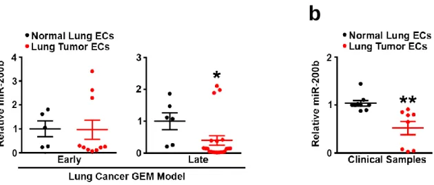
microenvironment (TME) in lung cancer. In lung cancer samples, both miR-200b suppression and QKI overexpression corresponded with tumor ECs relative to normal ECs, and QKI
ACKNOWLEDGEMENTS
There are no words to do justice to the amount of gratitude and love I hold in my heart for the friends and family who have brought me to where I am today. I give thanks first and foremost to my loving parents, Naweed and Marriam Azam, who have showered me with relentless and unconditional love, support, and prayers my entire life and have always
encouraged me to shoot for the stars; to my grandmother (my beloved “Nanee”), who is nothing short of the complete and absolute embodiment of love, compassion, and strength; to my great uncle, Dr. Amjad Umar (my dear “Nanaboo”), who was both the first and the most inspiring and loving PhD scientist I ever met; and to Numan Ahmad, for coming into my life with unconditional happiness and love when I needed him the most. And above all else, I say Alhamdolillah, “all praise belongs to God,” for blessing me with so many wonderful and caring people in my life, for answering all my prayers, and for granting me the opportunity to pursue my passion.
TABLE OF CONTENTS
CHAPTER 1: INTRODUCTION ... 1
Metastasis and the Metastatic Cascade ... 1
Tumor Angiogenesis ... 5
The Angiogenic Switch ... 6
Molecular Basics of Sprouting Angiogenesis ... 8
Role of Basement Membrane in Angiogenesis ...10
Tumor Vessel Morphology ...12
Hypoxia and Tumor Progression ...13
Anti-Angiogenic Therapies and Resistance Mechanisms...14
Overview of microRNAs ...19
The microRNA-200 Family and Metastasis ...20
Overview of Quaking and Molecular Function ...23
Role of QKI in Endothelium and in Cancer ...25
Therapeutic RNAi for Tumor Angiogenesis Targeting ...28
CHAPTER 2: MATERIALS AND METHODS ...31
Cell Lines, Maintenance, Transfection Reagents, and In Vitro Drug Use ...31
Chitosan Nanoparticle (CNP) Preparation ...33
Animals, In Vivo Models and Tissue Processing ...33
Quantitative Real-Time Polymerase Chain Reaction (PCR) ...35
Target Gene Binding Sites, Luciferase Reporter Assays and 3’UTR Site Mutagenesis ...36
Proliferation Assays ...38
Cell Cycle Analysis ...38
RNA Immunoprecipitation ...38
Argonaute-2 Immunoprecipitation ...39
mRNA Stability Assay ...39
In Vivo Angiogenesis Plug Assay ...40
Sprouting Assay ...40
Immunostaining ...51
Adenoviral Cre Recombinase Induction ...52
Flow Cytometry ...52
µCT Imaging ...55
mRNA Microarray ...55
RNA-Sequencing ...55
Tissue Microarray ...56
Affymetrix Microarray ...57
Statistical Analyses...58
CHAPTER 3: RESULTS ...59
miR-200b is Downregulated in Endothelium During Lung Cancer Progression ...59
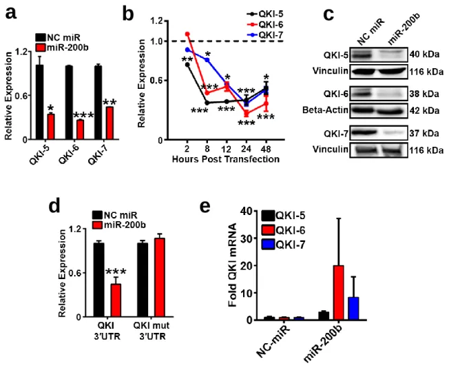
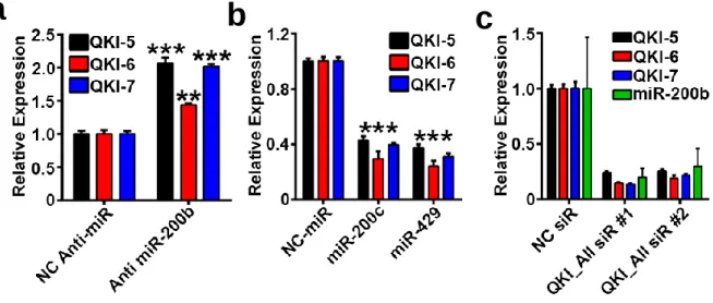
Quaking is a miR-200b Target in Tumor Endothelium ...63
QKI Silencing Recapitulates the Effects of miR-200b on EC Sprouting...70
QKI Expression in Clinical Samples is Associated with Angiogenesis and Poor Survival ...74
QKI Regulates EC Cell Cycle Progression ...79
QKI Promotes Cyclin D1 mRNA Stability to Regulate EC Growth ...82
Palbociclib Recapitulates the Effects of QKI Silencing on EC Function...89
CHAPTER 4: DISCUSSION AND UNANSWERED QUESTIONS ... 101
Working Model ... 101
Beyond the miR-200b/QKI Axis ... 103
QKI Isoform-Specific Effects ... 106
Broadening Understanding of QKI Molecular Mechanisms ... 109
Deconvoluting the Roles of QKI and miR-200b in Cancer and the TME ... 113
Characterizing miR-200b/QKI/CCND1’s Effects on Tumor Vasculature ... 114
New Therapeutic Strategies for Targeting Tumor Angiogenesis ... 120
Developing Novel Angiogenesis Models ... 121
Concluding Remarks ... 124
LIST OF FIGURES
Figure 1. The KRAS;p53;LKB1 mouse can be used to model early and late stage tumor tumor
late stage tumor angiogenesis. ...61
Figure 2 miR-200b is downregulated in tumor endothelium. ...62
Figure 3 QKI is a predicted miR-200b target in cancer and in endothelium. ...65
Figure 4 QKI is a direct miR-200b target in endothelium. ...68
Figure 5 QKI and miR-200 expression. ...69
Figure 6 QKI silencing recapitulates miR-200b mediated inhibition of sprouting. ...71
Figure 7 Pericytes tightly associate with endothelial vessels in the bead sprouting assay. ...72
Figure 8 QKI silencing in either ECs or pericytes recapitulates miR-200b mediated inhibition of mediated inhibition of pericyte-covered vessels. ...73
Figure 9 QKI is expressed in multiple cellular compartments of the tumor microenvironment in microenvironment in clinical lung cancer samples. ...76
Figure 10 Stained TMA sections can be partitioned into endothelial and cancer cell regions and cancer cell regions and scored for QKI staining intensity. ...77
Figure 11 QKI is clinically relevant in tumor angiogenesis and survival. ...78
Figure 12 QKI regulates expression of a large cohort of cell cycle-related genes in ECs. ...80
Figure 13 QKI regulates EC cell cycle progression. ...81
Figure 14 CCND1 is a top cell cycle-related QKI target. ...84
Figure 15 QKI directly regulates CCND1 mRNA. ...85
Figure 16 QKI regulates CCND1 mRNA stability. ...86
Figure 17 CCND1 splicing in ECs is not altered upon QKI knockdown. ...87
Figure 18 The different QKI isoforms have distinct effects on EC function ...88
Figure 19 CCND1 overexpression is sufficient to rescue QKI siR-mediated inhibition of EC inhibition of EC proliferation ...90
Figure 20 The CDK4/6-cyclin D inhibitor palbociclib inhibits EC function. ...91
Figure 21 QKI is highly expressed in tumor endothelium in the 344SQ metastatic lung lung lung lung adenocarcinoma mouse model. ...95
Figure 23 Endothelial QKI and miR-200b targeting alters tumor vasculature and inhibits and and
and inhibits metastasis. ...97
Figure 24 Palbociclib treatment recapitulates the effects of endothelial QKI siR treatment on siR siR treatment on metastasis. ...98
Figure 25 QKI does not regulate primary tumor growth. ...99
Figure 26 Palbociclib treatment recapitulates the effects of endothelial QKI siR treatment on the siR treatment on the tumor microenvironment. ... 100
Figure 27 Working models of the miR-200b/QKI/CCND1 axis of regulation of EC function and of of EC function and angiogenesis. ... 102
Figure 28 Effects of demethylating agent on miR-200b, QKI, and miR biogenesis enzymes biogenesis enzymes expression. ... 105
Figure 29 QKI isoform specific staining in bead sprouting assay. ... 108
Figure 30 QKI expression is cell cycle regulated. ... 112
LIST OF TABLES
LIST OF ABBREVIATIONS
αSMA Alpha smooth muscle actin
µCT micro computed tomography
AES Amino-Terminal Enhancer of Split
AGO Argonaute
ANG2 Angiopoietin-2
ANGPTL4 Angiopoietin-like 4 ARP2/3 Actin-related protein 2/3
ATCC American Type Culture Collection
BLI Bioluminescence
BSA Bovine serum albumin
CA9 Carbonic anhydrous IX
CCND1 Cyclin D1
CCR4 C-C motif chemokine receptor 4 CD31 Cluster of differentiation 31 CDC42 Cell division cycle 42
CDK Cyclin dependent kinase
circRNA Circular RNA
CK Cytokeratin
CLDN5 Claudin-5
CNP Chitosan nanoparticle
COX-2 Cyclooxygenase-2
DCC Deleted in colorectal carcinoma
DCP Decapping mRNA
DDX6 DEAD-box helicase 6
DGCR8 DiGeorge Syndrome Critical Region Gene 8
DLL4 Delta-like 4
DMEM Dulbecco Modified Eagle Medium
DMSO Dimethylsulfoxide
DOPC 1,2-dioleoyl-sn-glycero-3-phosphatidylcholine
EC Endothelial cell
EGF Epidermal growth factor
EGFR Epidermal growth factor receptor EHS Environmental Health and Safety EGM2 Endothelial cell growth medium 2 EMT Epithelial to mesenchymal transition EndoMT Endothelial to mesenchymal transition EPCAM Epithelial cellular adhesion molecule
EPO Erythropoietin
EREG Epiregulin
ETS-1 E26 oncogene homolog 1
EU Ethylene uridine
EV Empty vector
EXP5 Exportin 5
EZH2 Enhancer of zeste 2 polycomb repressive complex 2 subunit FACS Fluorescent activated cell sorting
FDA U.S. Food and Drug Administration
FDR False discovery rate
FGF Fibroblast growth factor FITC Fluorescein isothiocyanate
GAPDH Glyceraldehyde-3-phosphate dehydrogenase GATA Globin transcription factor binding protein 2 GEM Genetically engineered mouse
GFP Green fluorescent protein GLUT-1 Glucose transporter 1
GTP Guanosine triphosphate
GW Glycin tryptophan
HBSS Hanks balanced salt solution
HER2 Human epidermal growth factor receptor 2 HIF Hypoxia inducible factor
HRE Hypoxia response element
HUVEC Human umbilical vein endothelial cells ID1 Inhibitor of cell differentiation 1
IF Immunofluorescence
IGFBP4 Insulin like growth factor binding protein 4
IHC Immunohistochemistry
IL-8 Interleukin-11
IP Intraperitoneal
IPA Ingenuity pathway analysis IRB Institutional Review Board
KH K homology
KLF2 Kruppel like factor 2 L1CAM L1 cell adhesion molecule
LKB1 Liver kinase B1
LNEC Lung normal endothelial cell
LUAD Lung adenocarcinoma
LUSC Lung squamous cell carcinoma
LYVE1 Lymphatic vessel endothelial hyaluronan receptor 1
MBP Myelin basic protein
MEF2C Myocyte enhancer factor 2C
MET Mesenchymal to epithelial transition
miR microRNA
MMP Matrix metalloproteinase
mTOR Mammalian target of rapamycin
MVD Microvessel density
MYOCD Myocardin
NC Negative control
NEO Neogenin
NHLF Normal human lung fibroblast NICD Notch 1 intracellular domain
NKXX2-1 NK2 homeobox 1
NOT Negative regulator of transcription
NP Nanoparticle
NRP1 Neuropillin-1
OA Omphalomesenteric artery
OCLN Occludin
OCT Optimal cutting temperature compound
ORF Open reading frame
PAI-1 Plasminogen activator-1 PARN Poly(A)-specific ribonuclease PBS Phosphate buffered saline PCR Polymerase chain reaction PDGF Platelet-derived growth factor
PDGFRβ Platelet-derived growth factor receptor β PHD2 Prolyl hydroxylase domain protein 2 PIGF Placenta growth factor
PLXDC1 Plexin domain containing 1 pri-miRNA Primary microRNA
PTK Protein tyrosine kinase
QKI Quaking
qkv Quaking viable
QRE Quaking response element
RA Retinoic acid
RBP RNA binding protein
RGD Arginine-glycine-aspartate
RIN RNA integrity number
RISC RNA-induced silencing complex
RNAi RNA interference
ROI Region of interest
S1P Sphingosine-1-phosphate
S1PR Sphingosine-1-phosphate receptors
siR Small interfering RNA
SM22 Smooth muscle protein 22
SRF Serum response factor
STAR Signal transduction and activation of RNA STAT3 Signal transducer and activator of transcription 3 TBS-T Tris-buffered saline-Tween 20
TCGA The Cancer Genome Atlas
TEC Tumor endothelial cell
TGF-β Transforming growth factor beta TIE2 TEK receptor tyrosine kinase
TINAGL1 Tubulointerstitial nephritis antigen like 1
TMA Tissue microarray
TME Tumor microenvironment
TMP Tissue inhibitor of metalloproteinase TNRC6 Trinucleotide repeat containing 6 TPL Tissue Pathology Laboratory
TRBP Trans-activation responsive RNA-binding protein
TSP1 Thrombospondin-1
UTR Untranslated region
UV Ultraviolet
VA Vitelline artery
VE-cadherin Vascular endothelial cadherin VEGF Vascular Endothelial Growth Factor
VEGFR Vascular Endothelial Growth Factor Receptor
VHL von Hippel Lindau
CHAPTER 1: INTRODUCTION
1Metastasis and the Metastatic Cascade
Cancer is the second leading cause of death in the U.S. and among the top 10 killers in the world. Approximately 90% of cancer patients die primarily due to the spread, or metastasis, of disease to distant sites (Gupta and Massague, 2006). Therefore, developing novel approaches for therapeutic targeting of metastasis is critical for improving patient outcomes.
The metastatic cascade refers to the process by which cancer cells shed from a growing primary tumor, invade the endothelial barrier and enter the circulation via intravasation, arrest in capillary beds within distant organs, extravasate, and ultimately re-activate pro-angiogenic and proliferation pathways to support outgrowth from micrometastases into vascularized
macrometastases. The first step of this process consists of cancer cells invading into the tumor-associated stroma and eventually into the healthy tissue parenchyma. One of the greatest barriers to this invasion is the highly-structured basement membrane (Valastyan and Weinberg, 2011). Cancer cells overcome this by activating matrix metalloproteinases (MMPs) which degrade the basement membrane and other components of the extracellular matrix to allow cancer cell invasion and the release of growth factors sequestered within the matrix. The
release of these growth factors helps to further stimulate cancer cell growth (Kessenbrock et al., 2010). Many cancer cells are also believed to activate a biological program known as the
1 The section “Therapeutic RNAi for Tumor Angiogenesis Targeting” was modified and previously
appeared as my authored contribution to the article presented in the journal Frontiers in Pharmacology. The actual citation is as follows: Harrison, E.B., Azam, S.H., and Pecot, C.V. (2018). Targeting
epithelial to mesenchymal transition (EMT) which decreases adherens and tight junctions between epithelial cells, causes a disruption in cellular polarity, and enhances mesenchymal characteristics such as invasiveness to enable metastasis (Thiery et al., 2009). As the cancer cells invade, the surrounding stroma is stimulated to become more reactive as the cancer cells encounter fibroblasts, endothelial cells (ECs), adipocytes, bone marrow-derived cells, and immune infiltrates (Grivennikov et al., 2010; Joyce and Pollard, 2009). These interactions between stromal components and cancer cells help promote tumor progression. For example, tumor associated macrophages have been shown to activate epidermal growth factor receptor (EGFR) signaling in breast carcinoma cells (DeNardo et al., 2009).
Intravasation refers to the process by which cancer cells invade into the lumen of blood or lymphatic vessels and is dependent on molecular alterations that enable cancer cell
penetration of the pericyte-endothelial barrier wall of blood vasculature. For example, the transcriptional modulator amino-terminal enhancer of split (AES) was demonstrated to inhibit colon cancer cell intravasation through regulation of Notch signaling (Sonoshita et al., 2011). Intravasation is believed to be necessary to allow cancer cells to travel throughout the
of metadherin, which has been demonstrated to promote cancer cell arrest in the lungs by enabling binding to pulmonary vasculature (Brown and Ruoslahti, 2004).
Cancer cells shedding from a primary tumor are thought to also be recruited into lymphatic vessels and localized to lymph nodes. It has been clinically observed that the presence of lymphatic metastases predicts a more aggressive stage of cancer and a worse overall patient prognosis in multiple cancer types, but the mechanisms by which the lymphatic system contribute to cancer progression remain poorly characterized. Ultimately, however, lymphatic vessels do not provide a conduit directly to organs, so cancer cells must eventually be returned to the circulation to be able to hone to distant sites (Chambers et al., 2002).
Once cancer cells have arrested at a specific site, they extravasate, or cross the endothelial barrier from the lumen into the extravascular space to begin establishing a micrometastasis. Cancer cells face additional challenges at this step as opposed to the intravasation step, as here the vasculature and tissue microenvironment are much more normalized and impermeable (Valastyan and Weinberg, 2011). One possible mechanism that may help facilitate extravasation is primary tumor secretion of factors that increase vascular permeability at distant sites. For example, secretions of angiopoietin-like 4 (ANGPTL4), epiregulin (EREG), cyclooxygenase-2 (COX-2), MMP-1, and MMP-2 were shown to disrupt pulmonary EC-EC junctions and promote breast cancer cell extravasation (Gupta et al., 2007a; Padua et al., 2008).
The extravasated cancer cells must then establish micrometastases in a new, likely hostile environment that is molecularly and structurally different from the microenvironment of the host primary tumor (Valastyan and Weinberg, 2011). Some evidence suggests that primary tumors may release molecular signals which alter these recipient microenvironments to
undergo molecular changes. For example, it has been shown that breast carcinoma cells activate Src tyrosine kinase signaling to be able to successfully colonize in the bone; however, this cell signaling was not required for initial honing to this site (Zhang et al., 2009).
Once the cancer cells have successfully established at a new site, they may not
immediately outgrow into metastases. They may remain as occult micrometastases for a period of time either due to impaired cancer cell proliferation in the hostile new microenvironment; or due to a balance in cell proliferation and apoptosis and the absence of tumor angiogenesis to tip the scale and promote tumor outgrowth (Chambers et al., 2002). In one study, ANG2 was shown to enable pancreatic carcinoma metastatic colonization by promoting recruited myeloid cell-mediated activation of vascularization of metastases (Mazzieri et al., 2011).
Each cancer type is associated with unique metastasis tropisms, i.e. breast cancer cells preferentially metastasize to brain, bone, and lungs; lung cancer cells preferentially metastasize to liver and thoracic lymph nodes, etc. The unique patterns of tissue infiltration observed in different cancer types suggests that the deposition of cancer cells at distant sites is not due to chance or random distribution alone. It is believed that both the directionality of blood flow as it passes from one organ to another, as well as the unique molecular characteristics of each organ type, allow for the distinct patterns of metastasis observed. The “seed and soil hypothesis” predicts that the molecular nature of different organ/tissue types influences their contribution to metastasis (Paget, 1889). In brief, this suggests that the different chemokines, ligands, etc. expressed by certain tissue types make it more likely for specific cancer cell types to localize to particular organs. For example, breast cancer cells express high levels of C-X-C motif chemokine receptor 4 (CXCR4), which is the receptor for C-X-C motif chemokine ligand 12 (CXCL12) highly expressed in the lungs. Thus, breast cancer cells can find an agonist for their particular expressed receptor more readily at the lungs, making it a more likely site at which the cancer cells will metastasize
sequestered within the bone matrix and ultimately facilitate bone metastasis (Kang et al., 2003; Sethi et al., 2011).
To be able to successfully establish a new metastasis, cancer cells are also believed to require a high self-renewal capacity. Cells harboring such a capacity have been deemed “tumor-initiating cells.” For example, the transcription factors inhibitor of cell differentiation 1 (ID1) and ID3 and the transcription factor NK2 homeobox 1 (NKXX2-1) have all been shown to regulate metastasis in the breast and lung through alterations of the “tumor-initiating” state (Gupta et al., 2007b; Winslow et al., 2011).
In general, the early steps of the metastatic cascade are considered highly effective, whereas the latter steps following cancer cell entry into the circulation are much more prone to failure. It has been estimated that < 0.01% of tumor cells that enter the bloodstream eventually establish metastases, suggesting metastatic colonization of distant organs is a highly inefficient process. Thus, metastasis is believed to be an inefficient process largely due to the inability of micrometastases to activate tumor angiogenesis or continually support their growth in novel sites (Chambers et al., 2002), suggesting angiogenesis has an integral role in regulating both tumorigenesis and cancer spread.
Tumor Angiogenesis
Tumor angiogenesis, which refers to the sprouting of new tumor blood vessels from pre-existing host vasculature into the tumor microenvironment (TME), is a critical cancer hallmark. It is an appealing process to target therapeutically due to its importance in driving multiple
continued growth (Folkman, 1971). However, this newly-established vasculature can also serve as a conduit for cancer cells to metastasize.
The Angiogenic Switch
ECs are some of the longest-living cells in the body apart from those found in the nervous system. Healthy adult vasculature is quiescent due to a controlled balance between pro- and anti-angiogenic factors that regulate EC proliferation and migration. A temporary usurpation of the balance activates angiogenesis to promote fundamental processes such as embryogenesis and wound healing. During embryonic development, blood vessels are first formed via vasculogenesis, or de novo vessel formation, from EC precursors assembling to create a primary capillary plexus. The EC precursors then undergo differentiation, and the plexus is remodeled via sprouting angiogenesis. During physiologic angiogenesis, vessels become mature and stabilized very quickly, but in the pathologic context, tumors hijack the balance between pro- and anti-angiogenic factors and permanently shift the balance to a pathologic, pro-angiogenic state (the “angiogenic switch”) to stimulate constant growth of new vessels (Bergers and Benjamin, 2003; Carmeliet, 2000; Hanahan and Folkman, 1996).
Tumors are believed to start out as avascular lesions that remain less than 1-2 mm in diameter, are dormant, and are at a steady state of proliferation and apoptosis. Once the tumors undergo the “angiogenic switch”, they experience exponential growth. This is believed to be what also happens at a new metastatic site. Many studies in autochthonous, mouse models of cancer have demonstrated that as tumors progress, they undergo distinct stages of growth, and it is after inducing angiogenesis that the tumors reach a more aggressive state (Bergers and Benjamin, 2003).
growth factor (EGF), placenta growth factor (PIGF) (binds VEGFR1), and ANG2 (binds to TEK receptor tyrosine kinase (TIE2)). These angiogenic molecules are all believed to be expressed by tumors. VEGFA expression alone can stimulate angiogenesis from quiescent vasculature and serves as a survival factor for ECs (Ferrara, 2002; Yancopoulos et al., 2000). MMP-9 has been shown to promote angiogenesis by degrading components of the extracellular matrix to release sequestered VEGF (Bergers et al., 2000) which then signals through VEGFR.
Endogenous angiogenesis inhibitors include thrombospondin-1 (TSP1), which modulates EC proliferation and motility by inhibiting the release of VEGF from the extracellular matrix through suppression of MMP activity, directly binding VEGF, and modulating VEGFR signal transduction (Lawler and Lawler, 2012; Rodriguez-Manzaneque et al., 2001). Other endogenous
angiogenesis inhibitors include statins, including angiostatin(a fragment of plasminogen), endostatin, tumstatin, and canstatin(fragments of collagens that bind to integrins). The relative levels of angiogenesis inhibitors and promoters determine whether the vasculature will be actively angiogenic or not (Bergers and Benjamin, 2003).
vessels with less pericyte coverage can have abnormal vessel diameter and more sensitivity to VEGF therapies (Benjamin et al., 1998; Benjamin and Keshet, 1997).
Molecular Basics of Sprouting Angiogenesis
Angiogenesis involves the sprouting, migration, and proliferation of ECs which are regulated by many factors. Tip cells are the migratory and invasive ECs at the leading end of a growing sprouting vessel which extend filopodia that respond to growth factors, the extracellular matrix, and other cues. They are stimulated to become migratory by VEGF. Following the tip cells, ECs known as stalk cells will undergo proliferation to help grow the developing vessel (Viallard and Larrivee, 2017). The Notch pathway has been implicated in modulating the tip/stalk EC phenotype. VEGF activation of VEGFR2 or neuropillin-1 (NRP1) has been shown to
increase expression of the Notch ligand delta-like 4 (DLL4) (Phng and Gerhardt, 2009). DLL4 then binds to NOTCH1 in neighboring ECs, which causes the cleavage and release of the Notch 1 intracellular domain (NICD), which goes on to transcriptionally regulate gene expression to reduce the cell’s response to VEGF (Blanco and Gerhardt, 2013; Bray, 2016). NICD activation has been associated with inhibition of VEGFR2, VEGFR3, NRP1 and upregulation of VEGFR1 (Jakobsson et al., 2010). VEGFR1 strongly associates with VEGF but has weak kinase activity and therefore acts as a competitive “decoy receptor” that reduces VEGF binding to VEGFR2. In this way, the cell with high VEGFR2 signaling and expression of DLL4 is more likely to have a tip cell-like phenotype, while the adjacent cells with higher Notch signaling maintain more of a proliferative/stalk cell phenotype (Ferrara et al., 2003; Jakobsson et al., 2010). In addition, jagged1 (JAG1) is a Notch receptor ligand strongly expressed in stalk cells that antagonizes DLL4-NOTCH1 signaling in the neighboring tip cell to help further maintain the differential phenotype between tip and stalk cells (Benedito et al., 2009).
(GTP)ase cell division cycle 42 (CDC42) promotes filopodia production (Mattila and Lappalainen, 2008). RAC-actin-related protein 2/3 (ARP2/3) complexes regulate actin polymerization as well to help produce the cellular extensions (Ridley, 2015). Axon guidance molecules have also been implicated in endothelial tip cell guidance and vessel patterning: SLIT/roundabout (ROBO), netrin(NTN)/deleted in colorectal carcinoma (DCC)/UNC5 and NRP/plexin/sema families (Larrivee et al., 2009; Viallard and Larrivee, 2017). NRP1 has been shown to inhibit tip cell migration. The receptor UNC5B is activated by NTN1 or via NTN4 binding to neogenin (NEO) to regulate tip cell filopodia repulsion (Larrivee et al., 2007; Viallard and Larrivee, 2017). SLIT activates ROBO4 to induce vessel repulsion and also inhibit VEGFR2 activation (Koch et al., 2011; Viallard and Larrivee, 2017).
Lumen formation of the vessels is needed to initiate blood flow. One mechanism is believed to be that ECs undergo pinocytosis to internalize multiple vesicles of the plasma membrane that will then fuse together to create an intracellular lumen. This process is
considered to be dependent on the signaling of integrins α2β1, αvβ3, and α5β1 (Sacharidou et al., 2012). Another model is that ECs negatively charge the glycoproteins expressed on their apical surface, which causes a repulsion between ECs and opening up of the lumen, followed by redistribution of cell-cell adhesions to the edge of the lumen (Strilic et al., 2009).
receptor sphingosine-1-phosphate receptors (S1PRs) to mediate N-cadherin trafficking and strengthen adhesion between ECs and pericytes (Lucke and Levkau, 2010).
Vascular endothelial cadherin (VE-cadherin) is a transmembrane protein which stabilizes EC-EC interactions and decreases vessel permeability and leakage. The intracellular domain is anchored to the cytoskeleton via β-catenin, whereas the extracellular domain is bound to VE-cadherin on a neighboring EC. VE-VE-cadherin also promotes VEGFR2 dephosphorylation through the recruitment of phosphatases to ultimately enable blood vessel quiescence. Tight junction proteins claudin-5 (CLDN5), occludin (OCLN) and cluster of differentiation 31 (CD31) all also contribute to EC cell-cell adhesion by regulating the passage of solutes and ions and
transducing intracellular signals (Dejana et al., 2009; Viallard and Larrivee, 2017; Wallez and Huber, 2008).
Role of Basement Membrane in Angiogenesis
promote angiogenesis. In addition, disruption of the basement membrane frees sequestered growth factors such as VEGF and FGF, and also allows for pericyte detachment (Bergers et al., 2000; Egeblad and Werb, 2002; Folkman and D'Amore, 1996). When ECs detach, they
eventually come into contact with what is known as the interstitial provisional matrix which includes vitronectin, fibronectin, type I collagen, and thrombin, which are all considered to provide proliferative cues (as opposed to the inhibitory signals given by the structured basement membrane) (Kalluri, 2003). As tumors begin to grow, they start recruiting immune cells,
fibroblasts, and other stromal cells which produce additional VEGF and growth factors; therefore MMP activity is likely more important at the earlier stages of tumor angiogenesis (Kalluri, 2003). As the MMPs degrade the basement membrane even further, what are known as cryptic domains begin to become exposed from partially degraded components of the basement
Tumor Vessel Morphology
Angiogenesis produces chaotic and dysfunctional vasculature. While normal blood vessels consist of a continuous monolayer of tightly adhered ECs, close association of mural cells such as pericytes (which wrap around vessels and provide structural and chemical support), and a continuous basement membrane; tumor vessels have loosely associated ECs with large gaps between them, poor mural cell recruitment, and an irregular and discontinuous basement membrane. This reduced vessel wall integrity promotes leakiness and cancer cell intravasation, thus reducing drug delivery to the primary tumor and promoting distant metastasis (Baluk et al., 2005).
Tumor blood vessels are dilated, tortuous, disorganized, immature, sparsely covered by mural cells, leaky, poorly perfused, and lead to tumor hypoxia. Pericytes and smooth muscle cells help regulate blood flow and make vessels less permeable. In tumor vessels, pericytes have poorer association with the basement membrane and ECs. There is less pericyte coverage and they are abnormal in morphology (Bergers and Song, 2005; Morikawa et al., 2002). Higher EC proliferation and less pericyte coverage of tumor blood vessels contributes to their instability. High VEGF signaling from cancer cells inhibits PDGFRβ signaling in mural cells (Greenberg et al., 2008). Cancer cells release MMPs, elastase or trypsin to cleave VE-cadherin (Dejana et al., 2009). Inflammatory factors such as histamine stimulation can also disrupt vessel wall integrity by cleaving VE-cadherin (Guo et al., 2008). Electron microscopy images of tumor vessel basement membrane imaging type IV collagen, CD31 (marker of ECs) and alpha smooth muscle actin (αSMA, marker of pericytes) are disjointed, suggesting loose association of cells with the basement membrane. Electron microscopy images reveal that the tumor vascular basement membrane is uneven, with irregular thickness and even some perforations (Baluk et al., 2003).
compress or collapse and stem blood flow. This compression also inhibits lymphatics and decreases the ability of the lymphatic system to drain interstitial fluid, further contributing to pressure build up (Jain et al., 2014). There is also increased fluid leakage from the vessels into the interstitial space. All these factors together result in a high interstitial fluid pressure, which disrupts blood pressure and flow within tumors and also makes for more difficult drug delivery. This ultimately causes poor oxygen delivery to the tumor, making it hypoxic. This results in selection of more aggressive cancer cell clones, as well as cells more likely to undergo EMT, which is believed to increase their ability to metastasize (Jain et al., 2014; Reymond et al., 2013).
Hypoxia and Tumor Progression
Due to the “angiogenic switch”, tumors are considered incapable of growing beyond 1--2 mm diameters in size without co-opting a new vascular supply, largely due to the effects of hypoxia. Under normal oxygen conditions, prolyl hydroxylase domain protein 2 (PHD2)
hydrolyzes hypoxia inducible factors (HIFs) transcription factors to target them for ubiquitination by the von Hippel-Lindau (VHL) complex and ultimately proteasomal degradation (Giaccia et al., 2004; Mazzone et al., 2009). In hypoxia, PHD2 is unable to do so, allowing HIFs to bind to DNA sequence hypoxia response elements (HREs) to activate the expression of genes important for angiogenesis, survival, cell proliferation and glucose metabolism which include VEGF,
Anti-Angiogenic Therapies and Resistance Mechanisms
Currently, most anti-angiogenic therapeutic strategies focus on inhibition of the VEGF pathway, which has led to the U.S. Food and Drug Administration (FDA) approval of drugs for several cancer types (Table 1). Unfortunately, however, targeting the VEGF pathway thus far has had limited clinical success (Bergers and Hanahan, 2008). The effects are often temporary, modest, survival is only prolonged on the order of months, and are susceptible to numerous resistance mechanisms that have been shown to sometimes promote EC function or enhance metastasis (Bergers and Hanahan, 2008; Bottsford-Miller et al., 2012). Tumor-activated
resistance mechanisms to VEGF inhibitors include activation of other pro-angiogenic pathways, vessel co-option, cancer cell vascular mimicry, and vasculogenesis (de novo vessel formation) from bone marrow-derived precursor ECs (Bergers and Hanahan, 2008; Ebos and Kerbel, 2011; Welti et al., 2013).
Table 1
FDA Approved Anti-Angiogenic Drugs as of 2016
Drug Type Target/Mechanism Approved Cancer Types
Axitinib TKI VEGFR and PDGFR Renal cell carcinoma
Lenvatinib mesylate
TKI VEGFR2 Thyroid
Pazopanib TKI VEGFR1,2,3, c-kit, PDGFR Renal cell carcinoma, soft tissue carcinoma
Regorafenib TKI VEGFR2,3, Ret, Kit, PDGFR and Raf kinases
Colorectal Sorafenib TKI RAF kinase, VEGFR2,
PDGFRb
Hepatocellular carcinoma, renal cell carcinoma, thyroid Sunitinib TKI VEGFR2, PDGFRb, c-kit,
FLT3
Renal cell carcinoma, pancreatic neuroendocrine
Vandetanib TKI VEGFR2, EGFR Medullary carcinoma of
thyroid Bevacizumab Recombinant
humanized monoclonal antibody
VEGF Colorectal, non-small-cell
lung, renal cell carcinoma, ovarian
Ramucirumab Recombinant, fully human monoclonal antibody
VEGFR2 Gastric, gastro-esophageal
junction, non-small-cell lung, colorectal Ziv-aflibercept Recombinant human soluble decoy receptor
Segments of extracellular domains of VEGFR1 and VEGFR2 fused to the constant region (Fc) of human IgG1; binds VEGF
Colorectal
stromal tissue morphology as opposed to destroying it; preserved expression of normal tissue-specific endothelial markers within tumor endothelium; low expression of
angiogenesis-associated markers; and preserved vascular membrane and vascular integrity of tumor vessels (Kuczynski et al., 2019).
Vessel co-option has been observed both as an intrinsic feature of solid tumors and as an acquired mechanism of resistance in response to anti-angiogenic therapy. For example, it has been demonstrated that hepatocellular carcinomas being treated with the anti-angiogenic tyrosine kinase inhibitor sorafenib undergo EMT to deeply invade the liver parenchyma and co-opt pre-existing liver vessels as a mechanism of resistance in response to angiogenesis inhibition (Kuczynski et al., 2016). In clinical samples of lung metastases, tumors have been demonstrated to vascularize via mechanisms of both angiogenesis or co-option of vessels. Cancer cells were shown to either growth within alveolar air spaces or invade the alveolar walls to co-opt alveolar capillaries, or, alternatively, cancer cells were also shown to grow as
perivascular cuffs along larger lung vessels. In the alveolar growth pattern, cancer cells invaded the alveolar air spaces, facilitating the co-option of alveolar capillaries. In the interstitial growth pattern, cancer cells invaded the alveolar walls to co-opt alveolar capillaries. In the perivascular cuffing growth pattern, cancer cells grew by co-opting larger vessels of the lung (Bridgeman et al., 2017).
Mechanisms of vessel co-option are predicted to be associated with increased cancer cell invasiveness/motility, and increased cancer cell adhesion, both of which enable improved association of cancer cells with a pre-existing source of blood flow. ARP2/3, a protein complex involved in actin-mediated cell motility; zinc finger E-box-binding homeobox 2 (ZEB2), an
Another mechanism of angiogenesis or VEGF inhibition therapy resistance is the compensatory upregulation of alternative pro-angiogenic signaling pathways (Bergers and Hanahan, 2008). In a mouse model of pancreatic neuroendocrine cancer, treatment with the monoclonal antibody DC101 to inhibit VEGFR2 signaling caused an initial transitory inhibition of tumor growth and angiogenesis for 10-14 days, which was followed by a rebound during which tumor growth and vessel density were restored. This was found to be through increased expression of FGF and other growth factors. Treatment with an FGF trap after treatment with the VEGFR2 inhibitor slowed tumor rebound growth and angiogenesis, implicating the role of other signaling pathways in compensating for the loss of VEGF signaling (Casanovas et al., 2005). Similar phenomena have been observed in patients. Glioblastoma patients being treated with the VEGFR inhibitor cediranib had an initial period of responsiveness to the therapy,
followed by a relapse, accompanied by re-initiation of tumor angiogenesis and a loss of vascular normalization as revealed by imaging. Circulating levels of FGF2 were found to be higher in relapsing patients compared to patients still in the response phase, indicating FGF2 may be involved in an angiogenesis resistance mechanism to VEGFR inhibition (Batchelor et al., 2007).
Another mechanism of resistance is the increased recruitment of vascular progenitor cells for incorporation into blood vessels. The hypoxic environment created by potent inhibition of angiogenesis can stimulate increased recruitment of bone marrow-derived cells which can eventually differentiate into ECs or pericytes (Bergers and Hanahan, 2008). For example, one study demonstrated that tumors treated with anti-angiogenic agents underwent acute hypoxia and necrosis, which caused a sudden accumulation of endothelial progenitor cells at the tumor margins and subsequent reactivated vascularization. Tumors not treated with an anti-angiogenic agent did not demonstrate this sudden increase in endothelial progenitors, suggesting their accumulation was an activated resistance mechanism (Shaked et al., 2006).
vasculature by an anti-angiogenic agent, tumors may begin to rely on pericytes to keep a select few vessels alive and functional to continue sustaining the tumor. It is believed that ECs induce pericyte recruitment to help promote their survival due to being deprived of VEGF signaling (Bergers and Hanahan, 2008). It has been shown that tumor vasculature with poor pericyte coverage are more susceptible to VEGF inhibition (Bergers et al., 2003) and that tumor pericytes express VEGF and other factors to support EC survival (Darland et al., 2003). Pericytes also regulate EC proliferation to allow vascular structures to mature and remain stabilized (Hirschi and D'Amore, 1997). Therefore, the remaining pericyte-covered vessels are thought to be less responsive to anti-angiogenic treatment (Bergers and Hanahan, 2008).
Another phenomenon that has been observed in tumors is cancer cell vasculogenic mimicry, in which tumor cells take on the phenotype of ECs. This was first observed as channel-like structures containing red blood cells within tumors (Maniotis et al., 1999). These cancer cells express markers of both tumors and endothelium (including VE-cadherin and CD31) and form perfused, extracellular matrix-rich vasculogenic-like networks (Viallard and Larrivee, 2017). VE-cadherin allows for the cancer cells to undergo tube formation and close cell-cell adhesions to create perfused “vessels” (Paulis et al., 2010). Cancer cells have also been found to integrate into the walls of already formed vessels and create mosaic vessels (Chang et al., 2000).
Channels created by vasculogenic mimicry have been shown to anastomose with endothelial vessels and provide functional blood flow within tumors (Ruf et al., 2003). Because the cancer cells are lining the walls of the vasculature, they are directly exposed to constant blood flow and therefore have the possibility of easily detaching from the vessel wall and traveling via the circulation to distant sites (Viallard and Larrivee, 2017). Patients demonstrating evidence of vasculogenic mimicry have been shown to have worse clinical outcome (Yang et al., 2016).
Overview of microRNAs
microRNAs (miRs) are small, non-coding RNAs 20-23 nucleotides long that are loaded into the RNA-induced silencing complex (RISC) to modulate target mRNA expression by binding to their 3’untranslated regions (UTRs) and promoting degradation or preventing translation (Winter et al., 2009). Because a single miR can regulate the expression of multiple mRNAs simultaneously, they are appealing targets to study for the development of novel cancer therapeutics.
Mammalian miRs are encoded in the genome either as individual genes or clusters that can contain up to a few hundred miRs. Clustered miRs are initially transcribed as polycistronic transcripts which are later processed to separate mature miRNAs. Many miRs are transcribed from intronic regions of the genome (Treiber et al., 2019). Transcription is usually carried out by RNA polymerase II to produced capped and polyadenylated primary miRs (pri-miRNAs) which have a hairpin structure (Ha and Kim, 2014). Pri-miRNAs are then processed by the
microprocessor protein complex to generate single hairpins known as pre-miRNAs (Lee et al., 2003). Microprocessor consists of RNase III enzyme Drosha, the double-stranded RNA binding protein DiGeorge syndrome critical region gene 8 (DGCR8), and other less well-characterized factors (Denli et al., 2004; Gregory et al., 2004; Han et al., 2004a). pre-miRNAs are then
of the miR) usually within 3’UTRs of target mRNAs (Bartel, 2009). Initially miRS are believed to inhibit the translation of their target mRNAs, but at later stages the target mRNAs can be degraded (Bazzini et al., 2012; Bethune et al., 2012; Djuranovic et al., 2012). Once the RISC complex has been brought to a target mRNA, AGO recruits the glycine-tryptophan (GW) repeat protein trinucleotide repeat containing 6 (TNRC6), which has both an AGO-binding domain and a silencing domain (Pfaff et al., 2013; Schirle and MacRae, 2012). The silencing domain allows for interaction with the poly(A) tail shortening deadenylase complexes (poly(A)-specific
ribonuclease (PARN)2-3 or C-C motif chemokine receptor 4-negative regulator of transcription (CCR4-NOT). The shortened or lost poly(A) tail provides a signal for mRNA decapping by the decapping mRNA (DCP)1-DCP2 and DEAD-box helicase 6 (DDX6) containing complex, which is recruited to the 5’ end of the target mRNA to remove the 7-methylguanylate cap. The
unprotected 5’ end is then degraded by a 5’3’ exoribonuclease. DDX6 is important both for inhibiting translation and for promoting mRNA decay (Jonas and Izaurralde, 2015; Treiber et al., 2019).
miRs have been shown to regulate many cancer biology hallmarks, including apoptosis, proliferation, metastasis, and the TME (Pichler and Calin, 2015), including tumor angiogenesis (Collet et al., 2012). In my dissertation, I chose to focus on mechanisms of the miR-200 family modulation of tumor angiogenesis and metastasis.
The microRNA-200 Family and Metastasis
is believed to contribute to cancer metastasis by promoting cancer cell motility and invasiveness by causing cells to undergo changes in morphology and gene expression, including: loss of epithelial cell-to-cell junctions, alteration in cellular polarity, changes in cell cytoskeleton, downregulation of epithelial-related genes (including E-cadherin) and upregulation of
mesenchymal genes (including vimentin), increased cellular protrusions, increased cell motility, and increased ability to degrade extracellular matrix (Lamouille et al., 2014). miR-200 has been shown to potently inhibit EMT-related genes such as the transcription factors ZEB1 and ZEB2 which modulate transcriptional repressors of E-cadherin and other EMT-related genes (Park et al., 2008). miR-200’s effects on EMT-associated gene expression was shown to inhibit lung adenocarcinoma (LUAD) cell line 344SQ invasion, migration, and metastatic gene expression profiles (Gibbons et al., 2009), and was therefore speculated to be a metastasis suppressor. However, miR-200 has also been shown to promote metastatic colonization of 4TO7 breast cancer cells through inhibition of SEC23A, a component of vesicles involved in cellular transport of secreted proteins including metastasis suppressors tubulointerstitial nephritis antigen like 1 (TINAGL1) and insulin like growth factor binding protein 4 (IGFBP4) (Korpal et al., 2011). Thus, miR-200 regulation of cancer cells has been demonstrated to both inhibit and promote metastasis in different models and cancer types. The authors speculate that this may be due in part to which steps of the metastatic cascade are rate-limiting (Korpal et al., 2011). If the ability of cancer cells to undergo EMT and intravasate into vasculature is more critical to metastasis efficiency, then miR-200 may play more of a suppressive role due to its effects on inhibiting ZEB1 and ZEB2 expression and EMT. If the final steps of distant metastatic colonization are more inefficient, then miR-200 mediated inhibition of metastasis suppressors and promotion of the mesenchymal to epithelial transition (MET) and overall promotion of metastasis may play more of a dominant role.
nanoparticles (NPs) potently inhibited primary tumor growth and metastasis with associated decreases in tumor vasculature in orthotopic ovarian and lung cancer models. These effects were due to miR-200 suppression of cancer cell secretion of the pro-angiogenic chemokines IL-8 and CXCL1 (Pecot et al., 2013). In a separate set of experiments, chitosan NPs were used for miR-200 delivery directly to tumor endothelium (as opposed to DOPC NPs which were used for delivery to cancer cells), which again resulted in suppression of primary tumor growth and metastasis and was accompanied by pronounced effects on tumor vasculature, including decreased vessel density, more organized and less tortuous morphology of vessels, increased pericyte coverage, and decreased vessel permeability (Pecot et al., 2013). These effects were speculated to again be due in part to miR-200 inhibition of IL-8 and CXCL1 expression, however the authors of the study hypothesized that other miR-200 targets in tumor endothelium likely also contributed to these effects.
Overview of Quaking and Molecular Function
The qki gene was originally discovered due to the occurrence of a spontaneous mutation quaking viable (qkv) which caused mice to experience epileptic seizures after postnatal day 10. This phenotype was later attributed to poor myelination due to deficient oligodendrocyte function (Sidman et al., 1964). The hypomyelination was revealed to be due to defective mRNA
processing, localization, and translation of myelin components in glial cells and thus implicating QKI as a regulator of RNA metabolism (Hardy, 1998; Vernet and Artzt, 1997). The qkv mutation
was determined to be a 1 mb deletion of QKI promoter and enhancer regions that caused disruption of expression of the two alternatively spliced QKI isoforms (QKI-6 and QKI-7, see below) that are usually elevated during myelination and remain most highly expressed in the adult brain (Ebersole et al., 1996; Hardy et al., 1996; Kondo et al., 1999).
QKI is a highly conserved member of the Signal Transduction and Activation of RNA (STAR) family of RBPs expressed as three major alternatively spliced isoforms that are 5 (QKI-5), 6 (QKI-6), and 7 (QKI-7) kb in length (Ebersole et al., 1996; Vernet and Artzt, 1997). The N terminus (first 311 amino acids) of the coding region is identical across all the isoforms and contains 3 conserved functional domains: K homology (KH) RNA-binding domain and QUA1 and QUA2 domains (Ebersole et al., 1996; Kondo et al., 1999; Zorn et al., 1997). The QUA1 domain is necessary for QKI dimerization, while the KH and QUA2 domains direct specific binding to the bipartite sequence RNA motif ACUAAY and “half-site” (UAAY) separated by at least 1 nucleotide (Beuck et al., 2012; Chen and Richard, 1998; Galarneau and Richard, 2005; Teplova et al., 2013). QKI dimerizes when interacting with its mRNA targets and has been shown to only be stable as a dimer in vivo (Beuck et al., 2012; Teplova et al., 2013). QKI also has a proline-rich domain, which suggests an ability to engage in protein-protein interactions (Vernet and Artzt, 1997; Williamson, 1994).
contains a nuclear localization signal within its C terminus and localizes primarily to the nucleus, but it can shuttle between the nucleus and the cytoplasm (Wu et al., 1999). QKI-6 and QKI-7 are predominately cytoplasmic, and there is evidence to suggest that the C terminus of at least QKI-6 may help promote its localization in the cytoplasm (Fagg et al., 2017; Hardy et al., 199QKI-6; Pilotte et al., 2001). QKI regulates a wide range of functions related to RNA expression and processing, including RNA alternative splicing (de Bruin et al., 2016a; Hall et al., 2013; Wu et al., 2002), stability (Larocque et al., 2005; Li et al., 2000; Lobbardi et al., 2011; Zearfoss et al., 2011), translation (Saccomanno et al., 1999), nuclear export (Larocque et al., 2002), localization (Li et al., 2000), miR processing (Wang et al., 2013), and circular RNA (circRNA) biogenesis (Conn et al., 2015).
The differing subcellular localizations of the QKI isoforms help confer specificity of function, and changes in the ratios of QKI isoforms can impact their mRNA targets. In general, nuclear QKI-5 is considered most important for regulating splicing and nuclear export/retention, while cytoplasmic QKI-6 and QKI-7 are considered to play more roles in regulating RNA
translation and stability. In one study, the authors found that a balance of the different QKI isoforms was required for efficient export of myelin basic protein (MBP) mRNA from the nucleus (Larocque et al., 2002). Overexpression of QKI-5 resulted in increased retention of MBP within the nucleus and caused a decrease in MBP protein expression as well as aberrant MBP protein localization. Overexpression of QKI-6 or QKI-7 in concert with QKI-5 overexpression partially rescued MBP mRNA nuclear export, while simultaneous overexpression of all isoforms completely restored MBP export from the nucleus into the cytoplasm, suggesting the
appropriate ratio of all QKI-isoforms is required for appropriate MBP expression (Larocque et al., 2002). In another study, QKI-7 was found to induce apoptosis when present in the
mechanism of QKI expression (Fagg et al., 2017). They observed that isoform-specific
knockdown of QKI-5 caused an inhibition of expression of all QKI-isoforms, and that there are predicted QKI binding sites within both the QKI-5 and QKI-6 3’UTRs. At low levels of QKI-5 expression, QKI-5 promoted splicing of its own mRNA to generate higher levels of QKI-5. However, at high levels of QKI-5 expression, QKI-5 bound to its own 3’UTR to inhibit its
expression (thus engaging in a negative feedback loop). QKI-6 appeared to negatively regulate QKI-5 protein expression by inhibiting QKI-5 translation while simultaneously stabilizing QKI-5 mRNA transcripts. QKI-6 also promoted its own translation (Fagg et al., 2017).
Role of QKI in Endothelium and in Cancer
Intriguingly, although QKI was initially discovered for its roles in regulating glial cell differentiation, mouse embryos homozygous null or mutant for qkI were determined to die in utero (in the range of E9.5 to E13.5) due to severe vascular defects (Bohnsack et al., 2006; Li et al., 2003; Noveroske et al., 2002).
endodermal signaling to the mesoderm to facilitate vascular progenitor function (Noveroske et al., 2002).
In another study, Li et. al generated a completely null QKI mouse, which was also embryonic lethal. At E9.5 the yolk sacs were lacking in large vitelline vessels and had only a primary vascular plexus, poor vascular remodeling was again observed in the embryo proper, and the connection between the intra-embryonic omphalomesenteric artery (OA) and the extra-embryonic vitelline artery (VA) was disrupted. The authors observed poor smooth muscle cell recruitment to the OA, VA, and dorsal artery, again suggesting poor mural cell recruitment contributed to the defective vascular phenotype. Staining for QKI-5 within wild type embryos revealed expression in ECs and later on in development, smooth muscle cells. Therefore, the authors hypothesized that QKI may play a direct role in smooth muscle cell differentiation to contribute to vascular remodeling. In an aortic explant culture system, the authors observed normal formation of a vascular bed (i.e. early vasculogenesis) suggesting initial differentiation, proliferation, and migration of ECs were not affected. However later stages of vascular network formation were severely impaired and few smooth muscle actin cells developed, suggesting poor vascular progenitor differentiation into mural cells accounted for deficient vascular modeling. In a rescue experiment, CD31/QKI dual positive cells were sorted from wild type E9.05 embryos and added back to the mutant aortic explant system. This resulted in the eventual appearance of smooth muscle cells and restored vascular remodeling. Some of the smooth muscle cells were QKI positive and some were not, suggesting that QKI may elicit intercellular signaling to promote smooth muscle cell differentiation (Li et al., 2003).
RA did not completely account for all of the vascular remodeling defects or the embryonic lethality, suggesting other QKI targets in EC function were equally if not more important in proper vascular development (Bohnsack et al., 2006).
Apart from QKI’s roles in embryologic angiogenesis, QKI has been characterized in a few other endothelial contexts. For example, QKI maintains endothelial barrier function by binding to quaking response elements (QREs) within the 3’UTRs of VE-cadherin and β-catenin and promoting their translation. QKI expression was induced by the exposure of ECs to laminar flow, at least partially due to increased transcription promoted by the flow-induced transcription factor kruppel like factor 2 (KLF2). QKI loss was demonstrated to increase vascular leakage of gut microvascular (de Bruin et al., 2016b). In another study, miR-214 was shown to inhibit endothelial function in vitro by targeting QKI expression, which resulted in decreased expression of pro-angiogenic growth factors including VEGFA, bFGF, and PDGF (van Mil et al., 2012). miR-214 was also shown to de-repress QKI-mediated inhibition of vascular smooth muscle cell differentiation. QKI was shown to inhibit transcription of smooth muscle cell specific genes including smooth muscle protein 22 (SM22), serum response factor (SRF), myocyte enhancer factor 2C (MEF2C) and myocardin (MYOCD) (Wu et al., 2017). Finally, QKI-5 has been shown to be upregulated during induced pluripotent stem cell differentiation into ECs by directly binding to the 3’UTR of STAT3 and stabilizing its mRNA. This stabilization of signal transducer and activator of transcription 3 (STAT3) resulted in increased VEGFR expression. QKI-5
QKI has also been shown to play varying and conflicting roles in cancer progression, including promoting metastasis through activating pro-EMT splicing programs (Pillman et al., 2018), inhibiting lung cancer cell invasion and migration through targeting β-catenin (Zhou et al., 2017), inhibiting lung cancer cell proliferation and Notch signaling activation by regulating
NUMB alternative splicing (Zong et al., 2014), and acting as a p53-regulated tumor suppressor in glioblastoma (Chen et al., 2012).
In my dissertation, I sought characterize the function of QKI in regulating tumor
angiogenesis specifically within tumor endothelium and to assess the therapeutic implications of targeting tumor endothelial QKI expression to inhibit metastasis and cancer progression.
Therapeutic RNAi for Tumor Angiogenesis Targeting
RNA interference (RNAi) has become an increasingly appreciated therapeutic approach in cancer due to its ability to inhibit targets that traditional therapeutics (such as small molecule inhibitors or monoclonal antibodies) cannot. Therapeutic RNAi relies on delivery of usually small interfering RNAs (siRs) or miRs packaged within NP carriers to silence targets that drive cancer progression. As virtually all mRNAs are susceptible to RNAi-mediated inhibition of expression, independent of their protein function or localization, RNAi demonstrates immense promise as a therapeutic strategy. However, there are still many limitations to therapeutic RNAi that need to be overcome, including poor intracellular delivery, exonuclease degradation of siRs (Pecot et al., 2011), and lack of tissue specificity of NP delivery.
with an endothelial stain such as the cell surface marker CD31. Chitosan NPs have been demonstrated to co-localize to both tumor and ECs in vivo and effectively deliver siRs to both cell types (Lu et al., 2010). In an orthotopic model of ovarian carcinoma, treatment with chitosan NPs carrying siRs targeting human enhancer of zeste 2 polycomb repressive complex 2 subunit (EZH2; expressed in the transplanted cancer cells) or murine EZH2 (expressed in the
endogenous murine vasculature) inhibited tumor growth. However, the NPs carrying murine targeting siR had more potent effects on inhibiting disease burden, suggesting chitosan-mediated targeting of tumor vasculature had more potent therapeutic effects than targeting cancer cells directly (Lu et al., 2010). Second-generation NPs rely on incorporation of ligands to target EC-specific surface proteins. For example, ligands to integrin αVβ3, such as the peptide arginine-glycine-aspartate (RGD), can be used to facilitate NP uptake into neo-vasculature. Studies have shown that NPs containing the chemotherapeutic drug doxorubicin can be
directed specifically to tumor vasculature using this ligand, causing loss of tumor blood vessels and decreased metastasis (Murphy et al., 2008). Similarly, delivery of an anti-miR to inhibit the pro-angiogenic miR-132 with these same NPs in an orthotopic xenograft mouse model of human breast cancer yielded therapeutic effects on inhibiting tumor vasculature and decreasing tumor burden (Anand et al., 2010). miRs have also been delivered using RGD-labeled chitosan NPs. Delivery of miR-200 family members using this approach reduced angiogenesis by direct and indirect mechanisms and resulted in reduced disease burden in ovarian cancer models (Pecot et al., 2013). RGD-chitosan mediated delivery of siR targeting plexin domain containing 1 (PLXDC1), a growth-promoting gene, has been shown to effectively silence target gene
receptor that can mediate uptake into the vascular endothelium is CD31, a classical marker of blood vessels. While αVβ3 is thought to be expressed specifically by tumor neovasculature (as well as some cancer cell types), CD31 is expressed on all endothelium (both blood, and to a lesser extent, lymphatic). Using CD31 ligands to deliver siRs resulted in specific decrease of target genes in vascular endothelium. By delivering a siR against CD31 itself, tumor growth and metastasis were inhibited in a prostate cancer model (Santel et al., 2006). An alternative
approach to ligand-based targeting is chemically modified dendrimers that can specifically target the endothelium (Khan et al., 2015). 7C1 NPs are another type of NP that have been reported to localize faithfully and specifically to the endothelium in multiple models of aberrant vascular function, including tumor angiogenesis. These NPs are able to elicit at least 50% knockdown of target endothelial gene expression, and simultaneously deliver siRs targeting multiple genes in the endothelium (Dahlman et al., 2014).
CHAPTER 2: MATERIALS AND METHODS
2Cell Lines, Maintenance, Transfection Reagents, and In Vitro Drug Use
All cell lines were maintained in 5% CO2/95% air at 37°C. 344SQ LUAD cells were kindly provided by Dr. John Kurie (M.D. Anderson Cancer Center; Houston, TX). 344SQ luciferase-expressing cells were generated by stably transducing 344SQ cells with a lentivirus luciferase-expressing a green fluorescent protein (GFP)/firefly luciferase construct kindly provided by Dr. Shawn D Hingtgen (University of North Carolina at Chapel Hill, Chapel Hill, NC). 10T1/2 pericyte-like cells and normal human lung fibroblast (NHLF) cells were obtained from the American Type Culture Collection (ATCC). 10T1/2 ZsGreen cells were generated by stably transducing 10T1/2 cells with a ZsGreen construct. Human umbilical vein endothelial cells (HUVEC) were obtained from Lonza. 344SQ cells were maintained in RPMI 1640, NHLF cells in Dulbecco Modified Eagle Medium (DMEM), and 10T1/2 cells in Basal Medium Eagle, all supplemented with 10% fetal bovine serum (FBS) and 1% penicillin streptomycin. HUVEC were maintained in complete endothelial cell growth medium 2 (EGM2) supplemented with the growth factor bullet kit (Lonza) and 10% FBS. All cell lines were tested to confirm the absence of Mycoplasma, and all in vitro experiments were conducted with 60-80% confluent cultures. All cells were transiently
2 The majority of this chapter was slightly modified and previously appeared as part of an article in the
journal Oncogene. The original citation is as follows: Azam, S.H., Porrello, A., Harrison, E.B., Leslie, P.L., Liu, X., Waugh, T.A., Belanger, A., Mangala, L.S., Lopez-Berestein, G., Wilson, H.L., et al. (2019).
Quaking orchestrates a post-transcriptional regulatory network of endothelial cell cycle progression critical to angiogenesis and metastasis. Oncogene.
The “Sprouting Assay” section was slightly modified and previously appeared as part of an article in the
transfected with RNAiMax reagent (Invitrogen) using mirVana® mature miR mimic (Ambion, miR-200b, miR-200c, miR-249, or scrambled), anti-miRs (Ambion, negative control anti-miR or anti miR-200b), QKI siRs (Sigma or Ambion Silencer Select, QKI siR #1 sense:
5’CAGGCUGCUCCAAGGAUCAdTdT3’, antisense: 5’UGAUCCUUGGAGCAGCCUGdTdT3’; Sigma, QKI siR #2 sense: 5’CUACAGAGAUGCCAACAUUdTdT3’, antisense:
5’AAUGUUGGCAUCUCUGUAGdTdT3’; Ambion Silencer Select, QKI-5 siR sense:
5'CUAUUAACCCACAGCAUUAdTdT3', antisense: 5’UAAUGCUGUGGGUUAAUAGdTdT3’; Ambion Silencer Select QKI-6 siR sense: 5’GUGUAUUAGGUAUGGCUUUdTdT3’, antisense: 5’AAAGCCAUACCUAAUACACdTdT3’; Ambion Silencer Select QKI-7 siR sense:
5’GAGUGGAUUGAAAUGCCAdTdT3’, antisense: 5’UGGCAUUUCAAUCCACUC3dTdT’) or a negative control siR (Sigma or Ambion Silencer Select, sense:
5’UUCUCCGAACGUGUCACGUdTdT3’, antisense: 5’ACGUGACACGUUCGGAGAAdTdT3’) at a final concentration of 20 nM. QKI siRs #1 and #2 targeted sequences that were conserved across all QKI isoforms in both mice and humans; QKI-5, QKI-6, and QKI-7 siRs targeted sequences unique to each mRNA isoform. For rescue experiments, cells were reverse transfected with miR or siR using RNAimax as described above, then the next day cells were forward transfected at a ratio of 2.5 uL of Lipofectamine 2000 (Invitrogen) per 1 µg of plasmid DNA. DNA plasmids used for the cyclin D1 (CCND1) open reading frame (orf) experiments were pCMV-Cyclin D1 (Addgene #19927) and pmCherry-C1 (as empty vector control), DNA plasmids (GeneCopoeia) used for the QKI orf experiments were EX-NEG-Lv224 (as empty vector
control), EX-T4215-Lv224 (QKI-5), EX-H2552-Lv224 (QKI-6), and EX-H2553-Lv225 (QKI-7). Media was changed 4-6 hours following transfections to minimize toxicity. For all in vitro experiments, palbociclib HCl (MedKoo Biosciences, Chapel Hill, NC, # 202173) was
(Griffin, 1976). HUVEC were treated with 5-Azacytidine demethylating agent (Sigma #A2385) at working concentrations of 5 or 10 µM for 24 or 48 hours.
Chitosan Nanoparticle (CNP) Preparation
miRs or siRs for in vivo EC delivery was incorporated into chitosan (molecular weight 50–190 KDa). CNP was prepared based on ionic gelation of anionic tripolyphosphate and oligos. Briefly, predetermined tripolyphosphate (0.25% w/v) and miRs or siRs (1 μg/μl) were added in chitosan solution, and the miR or siR-CNP were spontaneously formed under constant stirring at room temperature. After incubation at 4 °C for 40 min, miR or siR-CNP was collected by centrifugation (Thermo Biofuge, Germany) at 13,000 r.p.m. for 40 min at 4 °C. The pellet was washed by sterile water 3 times to isolate miR or siR-CNP, which was stored at 4 °C until used.
Animals, In Vivo Models and Tissue Processing
Female athymic nude mice were purchased from Jackson Labs and from the UNC Chapel Hill Animal Studies Core. These animals were cared for according to guidelines set forth by the American Association for Accreditation of Laboratory Animal Care and the U.S. Public Health Service policy on Human Care and Use of Laboratory Animals. All mouse studies were approved and supervised by the University of North Carolina at Chapel Hill Institutional Animal Care and Use Committee. All animals used were between 6-10 weeks of age at the time of injection. For all animal experiments, cells were trypsinized, washed, and resuspended in Hanks balanced salt solution (HBSS; Gibco) prior to injection. 344SQ cancer cells were injected either subcutaneously over the posterior flank (1x10⁵ cells in 100 µL HBSS) or by an intra-pulmonary technique (5x10³ cells in 50 µL for therapeutic experiments or 1x10⁵ in 100 µL for the
mg/kg) + acepromazine (1 mg/kg) and placed in the right lateral recumbency. Following sterile skin preparation, an incision parallel to the rib cage between ribs 10-11 was made to visualize the lung through the intact thoracic pleura. A 1 mL tuberculin syringe with a 30-g needle was used to inject the cell suspension directly into the lung parenchyma at the left lateral dorsal axillary line. After injection, the skin incision was closed using surgery clips, and the mice were turned on the left lateral recumbency and observed until fully recovered. Mice were randomized a few days after orthotopic or subcutaneous injection of cancer cells, before initiating treatment. Mice were all combined into one or two cages and then randomly redistributed into treatment groups. Caliper measurements of subcutaneous tumor growth were taken twice weekly and tumor volume was calculated as L X W2 where L is the greatest cross-sectional length across the tumor and W is the length perpendicular to L. Luciferase-labeled tumor progression was monitored once or twice weekly using an IVIS Lumina optical imaging system and D-Luciferin K+ substrate (Perkin Elmer, # 122796) as per the manufacturer’s instructions. Briefly, substrate was reconstituted at a concentration of 15 mg/mL in phosphate buffered saline (PBS) and 200 µL of substrate were administer via intraperitoneal (IP) injection. Mice were imaged after a 10-minute incubation. Palbociclib HCl (MedKoo Biosciences, Chapel Hill, NC, # 202173) was reconstituted in 50 mM sodium lactate buffer (pH 4.0, Sigma, # 71718) at a concentration of 37.5 mg/mL and was administered at a dose of 150 mg/kg/mouse, daily by oral gavage. CNPs packaged with miR or siR were administered at a dose of 150 ug of oligo/kg/mouse and intravenously, twice a week.
In all therapeutic experiments 5-15 mice per group were used. Once mice in any group became moribund, they were all sacrificed, necropsied, and tumors were harvested. Tumor weights and number and location of lymphatic and distant metastases were recorded. For TME assessments, mice were injected intravenously with 100 µL of fluorescein isothiocyanate (FITC)-dextran at 10 mg/mL (2,000,000 molecular weight, Life Technologies, # D7137) 1 hour before sacrifice. All tissues used for immunostaining analysis were fixed in 10% neutral buffered formalin, cryoprotected in 30% sucrose, and then slowly frozen at -20°C in OCT.
Quantitative Real-Time Polymerase Chain Reaction (PCR)
For mRNA quantification, total RNA was extracted from cultured cells using the Quick RNA MiniPrep Zymo Research Kit (Genesee Scientific, # 11-328) or from ex vivo sorted cells using the Quick RNA MicroPrep Zymo Research Kit (Genesee Scientific, # 11-328M). For coding mRNA gene expression analyses, cDNA was synthesized using an iScript cDNA Synthesis Kit (Bio-Rad, # 1708891) as per the manufacturer's instructions. Analysis of mRNA levels was performed on a StepOnePlus Real-Time PCR System (Applied Biosystems). Specific primers for [QKI-5 (forward)-TACCCTATTGAACCCAGTGGTGT,
TCGAACTTTAGTAGCCACCGC; QKI-6 (forward)-ACCCTATTGAACCCAGTGGTGT, TAGCCTTTCGTTGGGAAAGC; QKI-7 (forward)-CCCAGTGGTGTGTTAGAGTGGA, (reverse)-TGAAATATCAGGCATGACTGGC; CCND1 (forward)-GGCGGATTGGAAATGAACTT,
(reverse)-TCCTCTCCAAAATGCCAGAG; CCND1 3’UTR