COGNITIVE CONTROL AND AFFECTIVE PROCESSING DYSREGULATION IN VETERANS WITH COMORBID PTSD AND MTBI: AND FMRI STUDY
Mariko Frances Weber
Dissertation submitted to the faculty at the University of North Carolina at Chapel Hill in partial
fulfillment of the requirements for the degree of Doctor of Philosophy in the Curriculum in Neurobiology in the School of Medicine
Chapel Hill
2016
Approved by: Aysenil Belger Kelly Giovanello Jacqueline Johnson
Darin Knapp
© 2016
Mariko Frances Weber
ABSTRACT
Mariko Frances Weber: Cognitive Control and Affective Processing Dysregulation in Veterans with comorbid PTSD and mTBI: an fMRI Study
(Under the direction of Aysenil Belger)
Background: Deficits in cognitive control and affective processing are important aspects of comorbid
PTSD and mTBI for which there are no effective treatments. Understanding the neural basis of symptoms and domain specific deficits is an important step in developing therapies for treating individuals with PTSD-mTBI. No studies have addressed this question, as most have examined PTSD or mTBI separately. We therefore utilize a large functional magnetic resonance imaging (fMRI) dataset to test the relationship between individual symptom severity, neurocognitive deficits and task-based functional activation. Methods: The relationship between the severity of clinical symptoms and neurocognitive deficits in
patients with PTSD-mTBI and functional activation during an affective 1-back task, affective face matching task, and number Stroop task (n = 100) was assessed using correlation analysis.
Results: Activity in cortico-limbic regions, and regions associated with striatal, default mode, and
salience networks were found to be significantly associated with increased symptom severity and greater impairments in neurocognition.
Discussion: Our findings suggest that researchers and clinicians should examine individuals neural and
ACKNOWLEDGEMENTS
I want to thank my wonderful family, for their unconditional love and support. Without Masumi, Leigh and Christopher as my number one fans, I would not be the person I am today, who strives for her dreams no matter what obstacles stand in the way.
I want to thank my mentor Aysenil Belger. Ayse is not only a one of a kind mentor but she is also very nurturing, and someone who has some of the most profound ideas and truly has a gift for making complex connections between multiple aspects of this universe.
Thank you also to Sarah, Alana, Elizabeth and Joe for being the best lab mates anyone could ask for. These individual’s make up a group of the most kind, smart and encouraging individuals’ who have a good sense of what is important and who truly get me.
Thank you to the rest of my friends who always made me feel impressive for getting my PhD and for encouraging me along the way.
TABLE OF CONTENTS
List of Tables………....viii
List of Figures………...ix
List of Abbreviations………...x
Chapter 1: INTRODUCTION………..……...1
1.1 General Introduction and Dissertation Outline….……….……..1
1.2 Clinical Presentation of PTSD and mTBI……….…...3
1.2.1 What is PTSD?...3
1.2.2 Clinical Dimensions and Diagnostic evaluation of PTSD………...4
1.2.3 mild Traumatic Brain Injury………....5
1.2.3.1 mTBI Diagnosis and Clinical Evaluation………...5
1.2.3.2 Theories regarding mechanisms of mTBI Injury……….…...6
1.3 Cognitive control and affective processing………...7
1.4 Neurobiology of PTSD……… ………..………9
1.5 PTSD functional Magnetic Resonance Imaging (fMRI) Literature Overview………..11
1.5.1 fMRI Overview………..11
1.5.2 fMRI and PTSD………...11
1.5.3 fMRI paradigms in PTSD………..13
1.6 mTBI Imaging and Overview………15
1.7 Rationale and Specific Aims……….…….17
Chapter 2: NEURAL CORRELATES OF AFFECTIVE PROCESSING IN PTSD-MTBI……....23
2.2 Methods……….…..25
2.3 Results……….…29
2.4 Discussion………...35
Chapter 3: NEURAL CORRELATES OF WORKING MEMORY AND AFFECTIVE PROCESSING IN PTSD-MTBI………...39
3.1 Background………...39
3.2 Methods………...42
3.3 Results………...47
3.4 Discussion………...63
Chapter 4: COGNITIVE CONTROL IN PTSD-MTBI………...69
4.1 Background………...69
4.2 Methods………...71
4.3 Results………...76
4.4 Discussion……….…..81
. Chapter 5: MAIN DISCUSSION………..….84
5.1 General summary………..…..84
5.2 Summary of brain-behavior associations in the context of affective face processing………..….86
5.2.1 Affective Face Matching Task: affective processing………..…...86
5.2.2 Affective 1-back Task: affective processing………...87
5.3 Summary of brain-behavior associations in the context of executive functions: working memory and selective attention……….…..88
5.3.1 Affective 1-back Task: working memory……….….88
5.3.2 Number Stroop Task: selective attention………..….89
5.4 Summary of brain-behavior associations in the context of combined executive function and affective face processing……….…..89
LIST OF TABLES
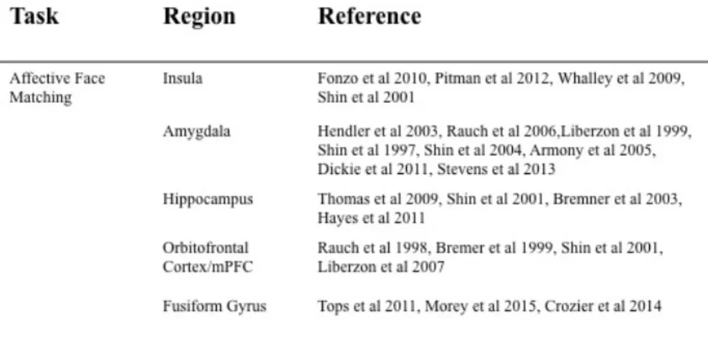
Table 1 – Demographic, clinical symptom and neurocognitive assessment data……… 26 Table 2 – List of brain regions chosen for ROI analyses with references:
Affective Face Matching Task………..29 Table 3 – Behavioral task results: Affective Face Matching Task………...30 Table 4 – Summary of statistically significant correlations between clinical symptom or neurocognitive scores and functional activation: Affective Face Matching Task…………32 Table 5 – Summary of all comparisons between clinical symptom or neurocognitive scores and functional activation: Affective Face Matching Task………...33 Table 6 – Demographic, clinical symptom and neurocognitive assessment data……….43 Table 7 – List of brain regions chosen for ROI analyses with references:
Affective 1-back Working Memory Task……….46 Table 8 – Behavioral task results: Affective 1-back Working Memory Task………..48 Table 9 – Summary of statistically significant correlations between clinical symptom or
neurocognitive scores and functional activation: Affective 1-back Working
Memory Task………52 Table 10 Summary of all comparisons between clinical symptom or neurocognitive scores and functional activation: [Object>Baseline] Affective 1-back Working Memory Task………53 Table 11 Summary of all comparisons between clinical symptom or neurocognitive scores and functional activation: [Face>Object] Affective 1-back Working Memory Task…………..57
Table 12 Summary of all comparisons between clinical symptom or neurocognitive scores and functional activation: [Face>Baseline] Affective 1-back Working Memory Task………..61 Table 13 – Demographic, clinical symptom and neurocognitive assessment data………..72 Table 14 – List of brain regions chosen for ROI analyses with references:
Number Stroop Task………..76 Table 15 – Behavioral task results: Number Stroop Task………...77 Table 16 – Summary of statistically significant correlations between clinical symptom or neurocognitive scores and functional activation: Number Stroop Task………79
LIST OF FIGURES
Figure 1 – Neurocircuitry model of PTSD………..10 Figure 2 – Full brain group activation maps generated from [Face>Object] condition:
Affective Face Matching Task………...31 Figure 3 – Correlation graphs showing significant correlations between clinical symptom or neurocognitive scores and functional activation: Affective Face Matching Task………34 Figure 4 – Full brain group activation maps generated from [Object>Baseline] condition:
Affective 1-back Working Memory Task………..49 Figure 5 – Full brain group activation maps generated from [Face>Object] condition:
Affective 1-back Working Memory Task………..49 Figure 6 – Full brain group activation maps generated from [Face>Baseline] condition:
Affective 1-back Working Memory Task………..50
Figure 7 – Correlation graphs showing significant correlations between clinical symptom or neurocognitive scores and functional activation: [Object>Baseline] condition
Affective Face Matching Task………54
Figure 8 – Correlation graphs showing significant correlations between clinical symptom or neurocognitive scores and functional activation: [Object>Baseline] condition
Affective Face Matching Task………...55
Figure 9 – Correlation graphs showing significant correlations between clinical symptom or neurocognitive scores and functional activation: [Object>Baseline] condition
Affective Face Matching Task……….……..56
Figure 10 – Correlation graphs showing significant correlations between clinical symptom or neurocognitive scores and functional activation: [Face>Object] condition
Affective Face Matching Task……….…………..58
Figure 11 – Correlation graphs showing significant correlations between clinical symptom or neurocognitive scores and functional activation: [Face>Object] condition
Affective Face Matching Task……….………59
Figure 12 – Correlation graphs showing significant correlations between clinical symptom or neurocognitive scores and functional activation: [Face>Baseline] condition
LIST OF ABBREVIATIONS
TBI Traumatic Brain Injury
PTSD Posttraumatic Stress Disorder
mTBI mild Traumatic Brain Injury
MRI Magnetic Resonance Imaging
fMRI functional Magnetic Resonance Imaging
PTSD-mTBI Posttraumatic Stress Disorder-mild Traumatic Brain Injury
CAPS Clinician-Administered PTSD Scale
PSS-I PTSD Symptom Scale-Interview
SI-PTSD Structured Interview for PTSD
PCL PTSD Checklist
ACC anterior cingulate cortex
dACC dorsal anterior cingulate cortex
PCC posterior cingulate cortex
WM white matter
BOLD blood oxygen level dependent
IFG inferior frontal gyrus
PFC prefrontal cortex
mPFC medial prefrontal cortex
vmPFC ventromedial prefrontal cortex
dlPFC dorsolateral prefrontal cortex vlPFC ventrolateral prefrontal cortex
IL infralimbic
PL prelimbic
OFC orbitofrontal cortex
GOS Glascow Outcome Scale
GOSe extended Glascow Outcome Scale
DRS Disability Rating Scale
BC-PSI British Columbia Post-Concussion Symptom Inventory
NSI Neuropsychological Symptom Inventory
DOD Department of Defense
VA Veterans Affairs
GCS Glasgow Coma Scale
LOC Loss of Consciousness
PTA Post-Traumatic Amnesia
ACRM American College of Rehabilitation Medicine
WHO World Health Organization
DAI diffuse axonal injury
DMN Default Mode Network
MFG middle frontal gyrus
OEF/OIF Operation Enduring Freedom/ Operation Iraqi Freedom
AMY amygdala
INS insula
HC hippocampus
APA American Psychological Association
IRB Institutional Review Board
DKEFS Delis Kaplan Executive Function System
BIS-11 Barratt Impulsivity Scale
WMS-III Wechsler Memory Scale-III
DAR Dimensions of Anger
FSL FMRIB Software Library
FMRIB Functional Magnetic Resonance Imaging of the Brain
FILM FMRIB’s Improved Linear Model FFA fusiform face area
DSM Diagnostic and Statistical Manual of Mental Disorders ROI region of interest
MNI Montreal Neurological Institute BET brain extraction tool
BPD Borderline Personality Disorder CEN central executive network
Chapter 1 - Introduction
“It may never be possible to fully distinguish the role of the severity of stress, the capacity for resilience to stress effects, and the presence of mild TBI in PTSD-related distress and disability because these factors are so complex and intimately entwined” [1].
1.1 General Introduction & Dissertation Outline:
While significant behavioral and epidemiological research has been conducted to examine brain mechanisms underlying PTSD and mTBI, leading to an advanced understanding of their clinical
manifestations, less is known about the neural mechanisms underlying these disorders and the association between the clinical manifestations and specific neural circuits or structures, or their function. Because comorbid PTSD-mTBI diagnoses result in very poor adjustment to daily life, elucidating the neural disruptions in PTSD-mTBI associated specifically with core cognitive and affective processing deficits is necessary to help understand the disorder and inform treatment.
To date, scientific research on human cognition and sensory perception has shown that brain function is dependent upon activation among a diverse combination of anatomical regions. This process occurs along a core synaptic hierarchy which includes the primary sensory, upstream unimodal,
downstream unimodal, heteromodal, paralimbic and limbic zones of the cerebral cortex [21].
Additionally, research on psychiatric and neurologic disease and disorders have resulted in speculations that diagnoses such as PTSD and mTBI are related to regional brain changes in proposed brain areas, and functional MRI (fMRI) research has been a useful tool in the development of theoretical models of PTSD that suggest specific regional aberrant functional activation. More recently, studies characterizing aberrant function in multiple brain regions that may be associated with clinical symptoms have opened new and robust avenues for understanding brain functions and their modulation by disease conditions. Considering the diverse regional activation and symptoms of comorbid PTSD and mTBI, we might expect that brain regions may be differentially related to different domains of symptoms.
literature of PTSD and mTBI; describe the methods and results of three neuroimaging experiments; and discuss results, limitations, and future directions-implications.
1.2 Clinical Presentations of PTSD and mTBI
1.2.1 What is PTSD?
Anticipating and adaptively responding to events that emerge in everyday life is a critical life-sustaining function. Evolutionary processes shaped our neural circuitry to consistently be on the side of caution, and thereby foster adaptive and protective behaviors. For example, appropriately recognizing and reacting to threatening stimuli is vital; PTSD has been associated with a potential disruption in this ability. PTSD involves a pattern of dysfunctional responses following exposure to a traumatic event or experience involving threat of death or serious bodily harm, followed by a reaction of intense fear, hopelessness, and/or horror [22]. PTSD develops when the normal fear response persists chronically and is expressed in inappropriate contexts [23]. It is important to highlight, however, the importance of individual differences in terms of the relationship between stressors and stress response, as only a small number of people develop and maintain PTSD after exposure to trauma. While it is unclear why everyone who is exposed to trauma does not develop PTSD, implications have surfaced that suggest that trauma severity is linked to the development of PTSD, although the association between trauma severity and PTSD diagnoses has never been clear due to the difficulty in separating subjective responses from the objective characteristics of traumatic events. Furthermore, trauma severity may provide information about why risk is greater for those who develop PTSD versus those who do not, but does not tell us why PTSD develops nor does it explain varying levels of symptom severity. Therefore, these diverse psychological and biological responses to stress, coupled with varying degrees of trauma severity (type and duration) and potential mediators of outcome such as resilience, all play into the clinical dimensions and
1.2.2 Clinical Dimensions and Diagnostic evaluation of PTSD
PTSD is diagnosed based on criteria defined by the American Psychiatric Association [24], where a diagnosis is confirmed if symptoms in four clusters last more than 30 days. The first symptom cluster is re-experiencing, which manifests as flashbacks, nightmares, and physiological reactivity after being
exposed to reminders of the event. Triggers often include a sound, sight, or smell that causes the experience of re-living the event. The second cluster is the avoidance of reminders of the trauma; for example, trying to avoid situations, people, conversations, or thoughts that trigger memories of the traumatic event. The third symptom cluster is restricted affect or numbing, which includes feeling dead or hollow and sometimes having no feelings. The fourth and last symptom cluster is hyperarousal,
characterized by hypervigilance, exaggerated startle response, and difficulty sleeping or concentrating. Those experiencing hyperarousal may operate on “high alert” at all times and are “on guard” constantly and who may suddenly become angry or irritable.
The Clinician-Administered PTSD Scale (CAPS) is the standard assessment used for evaluating PTSD, and uses a structured interview to provide a categorical diagnosis [25]. Other examples of clinician-rated PTSD measures are the PTSD Symptom Scale-Interview Version (PSS-I) [26] and the Structured Interview for PTSD (SI-PTSD) [27]. Self-rated assessments are also available for PTSD, such as the PTSD Checklist (PCL) [28]. The main clinical symptom clusters are often associated with
an important research focus driven by the exploration of neural network deficits in the military population. Better understanding of how aberrant neural function relates to clinical symptoms and behavioral outcomes will help identify key regions in neurobiological models of specific diagnoses, which will ultimately lead to the identification of predictive biomarkers and guide improved treatment and outcomes.
1.2.3 mild Traumatic Brain Injury (mTBI)
1.2.3.1 mTBI Diagnosis and Clinical Evaluation:
Traumatic brain injury (TBI), more specifically, mild traumatic brain injury (mTBI) is a historically recent epidemic and major health issue. While many mTBIs can be acquired by vehicle crashes, sports injury, and domestic violence, mTBI has been called the “invisible” war wound and is an increasingly common combat-related injury. Veterans often suffer mTBI due to explosive blasts, where the production of heterogeneous brain changes due to various mechanisms of injury is common. Examples of clinician-rated TBI measures are the Glasgow Outcome Scale (GOS), extended Glasgow Outcome Scale (GOSe), and the Disability Rating Scale (DRS) [115]. Self-rated assessments available for TBI include the Davidson Trauma Scale (DTS), the British Columbia Post-Concussion Symptom
Consequences of a false-positive or false-negative mTBI diagnosis have a potentially massive impact on the individual’s work, family, social, and legal status. For these reasons, consensus criteria on the definition and diagnosis of mTBI criteria has been published by several organizations such as the American College of Rehabilitation Medicine (ACRM) in 1993, the World Health Organization (WHO) in 2004, and the VA/DOD in 2010. The diagnosis of TBI is based on clinical interview, collateral information, and review of patient records.
1.2.3.2 Theories regarding mechanisms of mTBI injury and associated clinical symptomatology
illustrates that disturbances in emotion and stress regulation are considerably intertwined with the presence of complaints post-mTBI [128].
In individual patients, persistent post-concussive complaints are often unpredictable, even among those with similar injuries. Because of this, two important questions in mTBI research are to ask which patients are at risk to develop persistent complaints and what about their physiology contributes to this risk. The research is conflicted about whether post-concussive symptoms are caused by direct
neurological injury, emotional and psychological stress, or both [129]. One theory is that patient vulnerability to develop persistent complaints may be due to inter-individual differences
1.3 Cognitive control and affective processing
Despite differences in clinical presentation, both PTSD and mTBI are characterized by deficits in the domains of executive functioning and affective processing. Cognitive control
processes are core aspects of executive functions, and encompass domains such as memory,
planning, problem solving, inhibition, mental flexibility and multi-tasking. Executive functions refer to cognitive operations that include appropriate response selection, inhibition of
task-inappropriate actions and responses, and control needed to overcome urges to produce more favorable customary responses as opposed to those associated with automatic behavior. Executive processes are therefore critical in five types of situations: (i) situations involving planning or
decision-making, (ii) situations that require error-correction or troubleshooting, (iii) situations where the responses are not well learned or contain novel sequences of actions, (iv) situations judged to be dangerous or technically difficult, and (v) situations that require the overcoming of a strong habitual response or the resisting of temptation [4]. Working memory has been associated with dorsolateral prefrontal regions [5], self-monitoring and error detection have been localized to the anterior
cingulate and midfrontal regions [6], planning and sequencing have been linked with anterior frontal regions, and inhibition has been associated with anterior insula and inferior frontal regions [7] . More global attention and attentional orienting processes depend upon the posterior parietal regions, particularly the intraparietal sulcus. Finally, behavioral control of stop-go signals relies on subcortical motor regions, including the basal ganglia. These processes are not only crucial for normal psychological and social development, but impairments in executive functions have been identified as the core feature of neuropsychiatric disorders and clinical diagnoses [8] such as PTSD and mTBI.
that support directed actions. Emotions are critical for determining approach or avoidance behaviors for optimal survival functions [12]. Emotions can be either useful or they can be destructive, depending on prior subjective experience, context, and the regulation of the emotion itself[13]. Emotion processing involves the detection and evaluation of salient stimuli as well as the regulation of one’s emotional (affective) response to these stimuli [14]. Models of emotional processes include a number of
components, such as: (i) attention to and perception of information that could potentially elicit emotional responses, (ii) subsequent emotional arousal to such stimuli, and (iii) the regulation of that arousal via several potential mechanisms (altering attention or perception of stimuli and arousal responses and/or using cognitive reappraisal or other cognitive or behavioral strategies to modulate arousal level. Variability in how emotions are experienced and regulated relies heavily upon individual differences grounded in personality traits, coping styles, emotional intelligence, and past experiences. These processes are not only central to how biological value is assigned to stimuli in one’s environment, but deficits in emotion processing are implicated in nearly every psychiatric illness. Reinhard et al [2009] have shown that the mere experience of intense emotions can acutely affect bodily functions, and that deficits in emotion processing is heavily intertwined in disorders such as PTSD and mTBI [15]. Emotion processing has been associated with the amygdala [16][17], thalamus [18], hippocampus [19] and anterior cingulate gyrus [20].
1.4 Neurobiology of PTSD
Stress response and regulation functions have been consistently associated with PTSD.
Abnormalities of the hypothalamic-pituitary-adrenal axis have been proposed, with findings of decreased cortisol, increased corticotrophin-releasing hormone, and sensitized negative feedback inhibition.
Figure 1. Neurocircuitry model of PTSD.
1.5 PTSD functional Magnetic Resonance Imaging (fMRI) Literature Review
1.5.1 fMRI Overview
the magnetic field caused by blood oxygenation. Specifically, the presence of deoxyhemoglobin in a blood vessel causes a susceptibility difference between the vessel and its surrounding tissue. Such susceptibility differences cause dephasing of the MR proton signal [40], leading to a reduction in the value of T2*. Therefore, the presence of deoxyhemoglobin in the blood vessels causes a darkening of the image in those voxels containing vessels [41]. Since oxyhemoglobin is diamagnetic and does not produce the same dephasing, changes in oxygenation of the blood can be observed as the signal changes in T2* weighted images [41][42][43]. Blood oxygen level dependent (BOLD) fMRI signal has been reported by numerous studies over the past decade [44][45][46][47] to directly reflect neuronal responses elicited by a stimulus, and is therefore the reason why fMRI is currently the most widely used method for brain mapping and studying the neural basis of human cognition [48]. To study brain function using fMRI it is necessary to repeatedly image the brain while a subject is presented with a stimulus or required to carry out a task. The images are then compared during one condition to the images during another to see how the blood flow differs between the two. The success of the experiment is dependent on three aspects; the scanning sequence used, the design of the stimulus paradigm, and data analysis.
1.5.2 fMRI and PTSD
FMRI is an important method for further investigating the neurocircuitry of PTSD. Liberzon and colleagues (2008) hypothesized that impaired contextualization represents a pathophysiological
Another region that modulates amygdala responses is the vmPFC. The vmPFC receives strong hippocampal projections and is densely connected to the amygdala [59][60][61]. Inhibitory control by vmPFC over the amygdala has been revealed in numerous studies [62][63]. In animal studies, the rat PFC is subdivided into infralimbic (IL) and prelimbic (PL) regions. IL facilitates fear inhibition [63], whereas PL facilitates fear expression [64]. The human homologues of IL and PL are thought to be vmPFC and dorsal anterior cingulate cortex (dACC), respectively [65]. Both structures receive modulating input from the hippocampus [60][66][67]. The above-mentioned structures have been shown to be dysfunctional in PTSD subjects across a variety of paradigms such as symptom provocation and active task
paradigms[68]. Rauch et al [2006] provided evidence for a neurocircuitry model of PTSD that implicates the amygdala, medial prefrontal cortex (mPFC), and hippocampus as dysfunctional in PTSD individuals [34] . According to this model, the amygdala is hyper-responsive, which leads to an exaggerated fear response. In contrast, regions of the mPFC including rostral anterior cingulate cortex (rACC) and ventral medial frontal gyrus are hypo-responsive and fail to inhibit the amygdala. This hypo-responsivity may also be related to impaired fear extinction in PTSD. Finally, abnormal hippocampal function may underlie declarative memory impairments and deficits in identifying safe contexts in PTSD. Emerging evidence suggests that the dorsal anterior cingulate cortex (dACC) may be hyper responsive in PTSD and play a role in this disorder [69] . Although this model did not originally include the insula, recent findings suggest that the anterior insula may be functionally abnormal and related to symptoms of numbing (dissociation, lack of feeling) in PTSD [70][71].
1.5.3 fMRI paradigms in PTSD
Common tasks used in neuroimaging studies examining PTSD include both symptom provocation and active task paradigms. Symptom provocation paradigms often use provocative and neutral or
as graphic war images and emotionally neutral images (control condition) such as pictures of animals or nature scenes. While there is some discrepancy in neuroimaging findings, multiple studies have shown exaggerated limbic response coupled with decreased medial prefrontal activation [72][73][74][75], including Shin et al. (2005) showed that patients with PTSD experience heightened amygdala responses in response to traumatic imagery, accompanied by diminished mPFC activity, compared to healthy controls [76]. These findings support the hypothesized neurobiological model of PTSD-related neural circuit dysfunction that is characterized by a hyperactive amygdala and hypoactive mPFC. The
hyperactive amygdala suggests a failure to extinguish conditioned fear responses and hypoactive mPFC in the ineffective modulation of bottom-up information provided by the amygdala. Dysregulation of the insula, which is involved in integrating emotional feelings and body physiology, has also been observed [77][78]. All in all, findings from studies using symptom provocation paradigms have provided a theoretical model of neural circuitry underlying PTSD that includes aberrant activity in the mPFC, amygdala and insula.
Active task paradigms are most often designed to probe the circuitry responsible for affective
findings have been argued to account for deficits in identifying ‘safe’ contexts as well as in explicit memory [92][93]. Additionally, Shin et al. (2007) reported that diminished hippocampal findings in PTSD was due to greater hippocampal blood flow in the baseline condition, as well as across all conditions [94].
The majority if neuroimaging studies have found relative reductions in ACC function. For example reduced ACC activity was observed during an oddball task requiring response to salient non-trauma targets and hypo responsive activity was detected in the dACC during an emotional Stroop task in veterans with PTSD versus veterans with no PTSD, and during the presentation of traumatic narratives [81][95][96][97]. Bremner et al (2005) observed extinction after fear conditioning to be associated with diminished activation in the ACC in PTSD [98]. It is important to note however, that while the majority of studies have reported relatively diminished activation of medial prefrontal cortical regions in PTSD, a few studies have yielded discrepant results, such as both increased and decreased activation in this region [99][100] or increased activation [34][101][102]. Possible explanations for such discrepancies may lie in the imaging techniques used or in the dissociative state of the participants. Patients with PTSD who dissociated during traumatic narratives had greater activation in medial prefrontal cortex than control subjects [103] whereas patients who did not dissociate had relatively less activation in this region than control subjects [95].
Hypo-activation to tasks engaging more frontal regions have been observed in the OFC during an emotional counting Stroop task using aversive pictures and autobiographic narratives in PTSD versus non-PTSD groups [104][105][86][49]. Additionally, in a study of PTSD veterans versus veterans without, exaggerated dlPFC deactivation was observed during cognitive reappraisal versus passive viewing content of aversive stimuli [100] [106][107][108]. In a study of PTSD versus controls using an emotional declarative memory task, the PTSD group showed increased activation in the vlPFC [109][[91].
[34][105][106][110][111]; decreased vmPFC activity was reported during a classic inhibition task [112][89].
In summary, multiple studies indicate aberrant activity in cortico-limbic circuitry, including the prefrontal cortex, amygdala, insula, and hippocampus, making these key regions consistently good candidates for investigation in PTSD-related neuroimaging studies [83], consistent with the hypothesis that PTSD deficits may be, in part, caused by inadequate inhibition of the limbic system [113] by the prefrontal cortex. Neuroimaging studies have also helped illustrate that PTSD symptoms are maintained by hyperactive amygdala centric functional networks [49][34][114] in parallel with the diminished activation of top-down inhibitory control networks.
1.6 mTBI Imaging and Overview
Neuropathological and neuroimaging studies in humans also show that blunt force damages due to TBI cause white matter changes characterized by axonal stretching, disruption, and eventual
separation of nerve fibers [135][117]. TBI neuroimaging studies demonstrate volume loss that correlates with TBI severity in nearly every brain region, with strongest correlations observed in the frontal, temporal, and cingulate regions [136]. Additionally, both diffuse WM damage and damage to distinct WM bundles, such as the superior longitudinal, uncinate, external capsule, internal capsule, corpus callosum, and others have been described [137].
While animal studies indicate that diffuse axonal injury occurs after mTBI [133][134], ,established neuroimaging modalities typically used in TBI research often fail to detect any structural brain alterations in patients with mTBI [138]. The lack of observed structural changes occurs even though these same patients report post-concussive complaints. These negative imaging findings often contribute to the debate
conjunction with post-injury complaints. These studies have demonstrated abnormalities in several brain networks, including the salience and default mode [140] [141][142]. Few studies have investigated the role of networks in emotion processing and post-concussive complaints, even when anxiety and depression are common comorbidities [133].
Several fMRI studies investigating mTBI showed that a group of regional activity consistently increased compared to control groups. Nathan and colleagues, in a study on working memory
performance in patients with mTBI depression, reported increased activity of areas within the DMN and decreased activity of areas associated with executive functioning [143]. Using both auditory and visual N-back tasks, McAllister et al. (2001) found that, compared with controls, those with mTBI showed
increased activation while completing moderately difficult tasks and decreased activation as complexity increased [144]. In a separate study, the group also found that prefrontal activity was greater than controls even when symptoms had resolved [145].
Therefore, based on mTBI neuroimaging literature and common post-concussive complaints, we can assert that mTBI can affect a widely distributed network of structures and that these disruptions lead to deficits in multiple domains. The measureable specifics of these effects, however, are undetermined. It may be that due to widespread injury, neural networks responsible for executive function are over-compensating and that this over-compensation leads to a lack of resources for emotional regulation.
1.7 Rationale of current study: Distinct neural and clinical correlates of
co-morbid PTSD and mTBI diagnoses
Up to half of returning service members who have sustained a mild traumatic brain injury (mTBI) have also experienced life-threatening events that result in (PTSD). A cross-sectional study of Iraq and Afghanistan veterans found that PTSD was the strongest factor associated with post-concussive symptoms, even after removing overlapping symptoms from the PTSD score [153]. Young and
colleagues (2006) found that in combat veterans PTSD is an important mediator between TBI and poor health outcomes [138], and Howlett and colleagues (2014) found that the association between TBI and functional impairment disappeared after controlling for PTSD [154].
The risk of PTSD increases when TBI is sustained, likely because the areas of the brain that are most vulnerable to TBI (i.e., the frontotemporal regions) are the same as those implicated in PTSD [31] [155]. Substantial symptom overlap between PTSD and mTBI includes depressed mood, anxiety,
insomnia, irritability, difficulty concentrating, fatigue, hyperarousal, and avoidance. Emotional numbing, derealization, reduced awareness of one’s surroundings, depersonalization, and amnesia are also related to either PTSD or mTBI [156], and there is substantial evidence supporting cognitive and affective sequelae associated with mTBI are compounded by the presence of PTSD symptoms.
integrated trauma memories and emotional regulation [157][113][155]. In addition, loss of inhibitory control of the limbic system related to TBI may exacerbate PTSD symptoms [155]. Equally, individuals who experience prolonged or posttraumatic stress before sustaining a mTBI may experience greater cognitive impairment after brain injury due to additive effects [158][159]. Beyond the overlap in symptoms, there may also be more complex interactions between the symptomatology of PTSD and mTBI. For example, pain related to a traumatic injury may serve as a trigger for the re-experiencing symptoms of PTSD [160].
Cognitive deficits such as information processing speed and executive functioning have been found to be significantly impaired in veterans with mTBI when symptoms of PTSD were also present [161]. Fronto-striate networks are thought to play an important role in executive processing and can include regions such as the dorsolateral prefrontal cortex (dlPFC), ventrolateral prefrontal cortex (vlPFC), dorsal anterior cingulate cortex (dACC), orbitofrontal cortex (OFC), and medial prefrontal cortex
(mPFC). Neuroimaging findings suggest that activity in these regions are decreased in PTSD and TBI but the biological basis underlying deficits seen in executive functioning in comorbid PTSD-mTBI remains unknown.
Studies of PTSD and mTBI suggest an overlap between PTSD and mTBI in findings of the sensitization of the amygdala and hippocampus and aberrant dlPFC/MFG and OFC activity [164]. There are no known studies to date that examine the role of both executive dysfunction and emotional
dysregulation in a PTSD-mTBI population and how these deficits relate to clinical symptoms. The neuroimaging findings mentioned above suggest that frontal lobe and limbic system dysfunction have a large part in cognitive and emotional sequelae by reducing the capacity to adapt to environmental change and process emotional and non-emotional stimuli. Hoffman and Harrison (2009) focus on these prefrontal regions as being important in the comorbidity of PTSD and mTBI, suggesting that when TBI occurs during a traumatic event, a flood of stress hormones reduces repair and limits recovery [165].
In summary, PTSD and mTBI share neuropsychological and functional neuroanatomical
characteristics [113]. It is hypothesized that prefrontal cortex damage in mTBI can lead to disinhibition of cerebral structures that control fear and anxiety. Reactive systemic inflammatory processes related to mTBI may also impair psychological health, and impaired psychological health may lead to increased psychological distress that impedes brain repair due to release of stress-related hormones.
Neurocognitively, PTSD and mTBI overlap within the domains of attention, working memory, executive functioning and episodic memory [166][155]. Correspondingly, imaging studies implicate abnormalities in cortico-limbic circuitry in both disorders [167]. The potential overlay of mTBI-related neural and neuropsychological compromise onto similar abnormalities is thought to play a role in perpetuating PTSD and may offer clues as to why TBI is associated with increased risk of PTSD.
function associated with specific executive function and affective regulation domain deficits and clinical symptom severity.
Therefore, based on key findings in PTSD and mTBI literature, and the knowledge that in the presence of one another clinical symptom severity and cognitive deficits are exacerbated, we proposed a study seeking to identify aberrant activation in regions within the cortico-limbic and fronto-striate circuit associated with symptom severity and neurocognitive deficits in PTSD- mTBI. Our study has three aims, each implementing independent functional neuroimaging tasks designed to probe both affective and executive function in order to examine the associations between task-based brain activity and symptom severity.
Aim 1: To measure the relationship between individuals’ PTSD-mTBI symptom severity and activity
during an affective face matching task. The relationship between functional BOLD activation (in response
to the face relative to object target) and symptom severity was assessed by measuring the correlation between symptom severities and functional activation. The severity of PTSD symptoms has been associated with increased functional activity in limbic areas, which are known to be interrogated by emotional face matching tasks. We expect that individual symptoms and deficits in neurocognitive function will be positively correlated with patterns of functional activity. This aim will be addressed in Chapter 2.
Aim 2: To measure the relationship between individuals’ PTSD-mTBI symptom severity and activity
during an affective 1-back working memory task. The relationship between BOLD activation in response
response to the working memory component, patterns of increased functional activity during the affective face processing component, and even greater increased and decreased patterns of activation in frontal and limbic regions when examining effects of the combined working memory and affective face processing. This aim will be in Chapter 3.
Aim 3: To measure the relationship between individuals’ PTSD-mTBI symptom severity and activity
during a number Stroop task. The relationship between BOLD activation in response to the
[Incongruent-Congruent] condition and PTSD symptom severity or neurocognitive deficits will be assessed by measuring the correlation between symptom severities and functional activation. The severity of PTSD and mTBI has been associated with decreased functional activity in cognitive control areas, which are known to be interrogated by the Stroop task. We expect that symptoms and neurocognitive deficits will be negatively correlated with patterns of functional activity in cognitive control areas. This aim will be addressed in Chapter 4.
Chapter 2- Neural correlates of affective processing in PTSD-mTBI
2.1 Background
Studies have shown that the affective sequelae associated with mTBI are compounded by the presence of PTSD symptoms among veterans who have served in Operation Enduring Freedom and Operation Iraqi Freedom. Schneiderman et al. (2008) showed that the strongest factor associated with postconcussive symptoms was PTSD, Hoge et al. (2008) found that mTBI among soldiers was
significantly associated with PTSD and physical health problems after returning home war, and two other groups have found strong links between comorbid PTSD-mTBI and anger and violence [168][169][162]. Postconcussive symptoms related to mTBI and symptoms reported in PTSD are characterized by deficits across multiple domains, however overlapping symptoms reported in PTSD-mTBI are typically
associated with affective processing. While there has been extensive research on PTSD and mTBI separately, there remains an outstanding need to evaluate the associations between the neural function underlying deficits in affective processing in patients with PTSD-mTBI and associations with symptoms common to both.
A common element of PTSD may be an abnormally elevated fear response, fear and avoidance of trigger cues are common to many anxiety disorders (APA) and resemble the arousal and avoidance responses shown by normal subjects to conditioned fear cues [170][171]. Additionally, a hallmark symptom of those affected by PTSD is to have aggravated emotional responses to innocuous everyday stimuli. Based on animal models of fear learning [16][172], this hypothesis leads to the prediction that amygdala dysfunction is also common. Indeed, amygdala hyperactivity has been observed during
findings have led to a theoretical model of PTSD that involves hyper-responsivity in the amygdala as well as other regions involved in emotional processing. Neuroimaging studies of PTSD have revealed
hyperactivation in the insula and amygdala [175][176], where activation has been associated with stressresponses. Specifically, activation in the amygdala has been strongly linked to negative affective states in PTSD [177][178][179][78]. In order to examine affective circuits in PTSD, many neuroimaging studies have used reliable face tasks to probe affective circuits to better understand PTSD symptomology [99][88][180]. While face tasks do not use trauma-related stimuli and do not directly provoke
re-experiencing symptoms in PTSD, they do require the evaluation of social emotions. Therefore, they appear to provide a method to measure affective circuitry in a theoretically and clinically meaningful way. Neuroimaging findings have not been as consistent in PTSD as it has been in other anxiety disorders [181] but research has supported the hypothesis that PTSD is in part due to the manifestation of ineffective top-down inhibitory modulation of limbic circuitry by the prefrontal cortex [178][49].
With regards to neuroimaging findings in mTBI, findings support aberrant network activity in several brain networks [182][142][153] . However, these studies mainly focused on the role of brain network function in relation to cognitive problems post-mTBI, whereas few studies have investigated the
role of networks regarding affective processing and postconcussive complaints. Yet, anxiety and
depression are common post-mTBIand are associated with cognitive complaintsand vocational outcome
[126][139]. These findings highlight the need to sufficiently explore functional brain activation related to
affective processing and postconcussive symptoms.
In the current study, we aimed to map neural responses in regions implicated in affective processing and relate them to symptom severity and neurocognitive measures in PTSD/mTBI. We
would show greater limbic circuit activation during response to face targets as symptom severity and neurocognitive deficits increase.
2.2 Methods
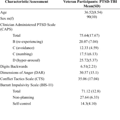
Participants
Table 1. Demographic and assessment data for all participants (N=100) showing the group mean and standard deviation. CAPS scores above 60 are considered PTSD; 6 is the average score for a non-clinical population on the Digit Backwards; the range for DAR is 0-56; CTS is calculated based off a 7-point scale where midpoints are averaged; and the BIS range is 0-120.
Assessments
Dimensions of Anger (DAR) [189].
Stimulus Presentation
Task: An emotional face-matching task was administered as a single run. This particular task has been
previously found to yield reliable activation of the amygdala and fusiform gyrus [190][183]. During this task, which was adapted by Hariri et al (2002), participants performed 1 run consisting of 4 blocks of perceptual face-processing conditions interleaved with 5 blocks of sensorimotor control conditions [183]. Face-processing condition: each trial consisted of a presented image with 3 faces expressing either fear or
anger, oriented such that participants were instructed to match either one of the bottom face stimuli to the top, by pressing button 1 if the left image matches and button 2 if the right image matches. There were no trials in which the same face appeared with different expressions, and both face and expression matched for the matching item. Each face-processing block consisted of 6-trials, balanced for gender and target affect (angry or fearful). Face stimuli were derived from a standard set of pictures of facial affect [191]. Sensorimotor control condition: each trial consisted of images with 3 geometric shapes (circles and
vertical and horizontal ellipses) and each block consisted of 6 different shape trials. For the shape trials, identical to instructions for the face trials, participants indicated with a button press which of the 2 bottom shapes matches the top one. All blocks were preceded by a brief instruction “match faces or “match shapes” that lasted for 2s. In both conditions times, each trial was presented for 5 s without ISI, for a total block length of 30 s. The total task run-time was 5minutes. Previous studies using this paradigm have demonstrated reliable and consistent robust activation of the amygdala and fusiform gyrus during the processing of the emotional faces [183].
fMRI Procedures
Acquisition: A 3T Siemens Magnetom TimTrio syngo MR B17 was used to acquire images. Whole-brain
%, flip angle: 80 degrees). Functional runs were preceded with four discarded RF excitations. High-resolution T1-weighted anatomical images were acquired using an MP-RAGE sequence (sagittal plane, 1mm3 TR: 2300ms, TE: 2.98ms, 256 x 256 matrix, FoV: 256 mm, FoV phase: 93.8%, flip angle: 9 degrees). Signal-to-noise ratio, and displacement of the center-of-mass were assessed for each scan session to ensure image quality. No participant had greater than a 4-mm deviation in the center of mass in any plane [192].
Data Analysis
region of interest was then correlated with pre-selected neurocognitive and clinical symptom severity measures using Pearson’s correlation. Values more than 3 standard deviations from the mean of the other data and outcome data with the value of zero, were all set to missing.
Table 2 Summary of brain regions with references chosen a priori for ROI correlation analyses
2.3 Results
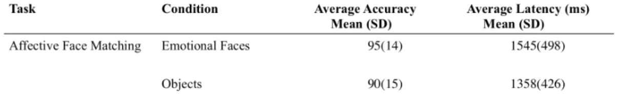
Behavior
Table 3 Behavioral task results. Summary of performance data during the emotional face condition and objects condition. Group average accuracy and latency are reported by mean and standard deviation.
Whole-Brain
Figure 2 Full brain group activation maps depicting brain regions activated by the [Face>Object] condition. A) z= 39 B) z= 27 C) FSL generated image depicting region of interest masks used in correlation analyses as defined by the Harvard Oxford Cortical/Subcortical Atlas z=30. Insula=Green; Amygdala=Blue;
Hippocampus=Orange; Fusiform Gyrus=Red; Orbitofrontal Cortex= Pink
Region of Interest Correlation Analyses: correlations of brain activation and behavioral measures
The [Face-Object] condition was analyzed in order to isolate activation in a priori regions of interest during angry or fearful face processing. Data representing mean percent signal change for each region was then used in correlation analyses to determine significant associations between regional brain activity and cognitive or clinical measures of interest.
A summary of significant correlations as determined by Pearson’s R correlation analyses
All significant correlations suggest that as symptom severity increases activation in limbic regions decreases. A summary of significant findings can be found in Table 4, and plots can be found in Figure 3. A summary of all comparisons including null results can be found in Table 5.
2.4 Discussion
In the present study, when PTSD-mTBI participants viewed fearful and angry faces relative to
objects, activation was observed in the fusiform gyrus, right and left hippocampus, and the right amygdala. The observed activation was significantly negatively correlated with measures of
re-experiencing. Additionally, increased total PTSD symptom severity and measures of anger and violence were found to be related to decreased fusiform activation.
Results from functional neuroimaging research of emotion processing in PTSD has generally supported the hypothesis that the amygdala is hyper-responsive and the mPFC is hypo-responsive. Hippocampus functional abnormalities have also been reported, and while the direction of activation tends to vary, aberrant activation has been linked to deficits in explicit memory and the identification of safe contexts. Therefore, in this study we hypothesized that the proposed regions traditionally associated with affective processing and facial emotion processing would increase in activation as severity increased. However, what we found was the opposite: regional activity associated with emotional face processing decreased as severity increased. In other words, we found blunted response in these regions. It is important to note that besides modulating emotional responses, the amygdala is thought to interact with sensory processing via back-projections and also modulates the fusiform cortex and early sensory processing regions [16][198][199] which may in part explain some of our findings Specifically, that we found the same direction of activation in the fusiform as well as the amygdala and that activation in these regions were found to be significantly related to symptoms of re-experiencing.
possible that in this cohort those with greater symptoms may exhibit higher “at-rest” baseline activity, and may therefore reflect a “ceiling effect.” Such a ceiling effect may explain their reduced activation relative to those who have less severe symptoms. Alternatively, subjects may have increased activation in these regions during object processing, which may lead to smaller relative face-specific activation. We may also conclude that as symptom severity increases and the observed ceiling has been reached, there has been an allocation of additional cognitive resources for emotional processing in regions that we did not investigate. Additionally, the fusiform gyrus has been found to show a strong response to images with trauma-related content [74]. While our study does not use trauma-related images, the task stimuli do possess emotional information, which could be contributing to the significant findings specific to
increased affective dysregulation relating to limbic and fusiform activation . In addition, activation in the fusiform gyrus and amygdala may also support the notion that traumatic and stressful events could modify visual processing by the limbic system (e.g. the amygdala).
While no mTBI studies have used pure emotional face-processing tasks to explore aberrant regional activity, some neuroimaging studies have produced findings that support altered network dynamics in switching between internally and externally focused mental states. These network findings were found to be associated with post-concussive symptoms, although few mTBI studies have
investigated the relationship to symptoms related to affective dysregulation. Neumann and colleages (2015) examined deficits in face recognition in TBI and reported decreased activity in the fusiform gyrus related to facial recognition deficits [201]. Therefore, related to our findings, it is possible that this cohort experienced deficits in face recognition that are related to symptoms, and that these deficits are related to reduced fusiform activity.
In terms of brain-behavior associations involving measures of affective dysregulation, it may be that the reported brain activity associated with increased measures of behavioral affective dysregulation (violence, anger) are due to deficits in the ability to switch attention from internally salient information processing to external salient cues and stimuli. Anger and violence may result from the suppression of emotion, that is related to an imbalance of attention to internal versus external stimuli [202].
Alternatively, these findings may be due to the suppression of other, more appropriate emotions that are replaced by anger. Therefore, it is possible that face stimuli in our study are either perceived as non-threatening, resulting in a lack of attention to social cues and attributed emotional salience, or that the veteran is unable to switch from internal to external processing and regulate information.
Our data are novel in supporting emotional face-processing-related neural activity in PTSD-mTBI and the potential role of these networks in relation to symptom severity and behavioral and cognitive function disturbance. Rather than hyper responsivity in suggested brain regions consistently being
characteristic of this population in a social or affective context, we instead showed decreased activation in response to emotional faces as symptom severity increased. We can infer from these data that, in response to these specific social cues, veterans either have a blunted affect in response to task stimuli or, as
processing. This is turn could result in a smaller window for variance, namely increases, in activation. It is also possible that neural resources are being over-exerted from attention to the cognitive aspect of task completion, resulting in fewer resources available for emotional face processing. However, due to the participants high performance on the task it is likely neural resources are not being over exerted.
Additionally, all significant associations were between increased measures of affective dysregulation, so it appears the reduction in activation is not likely due to blunted affect but rather heightened baseline neural activity.
Specific focus on the fusiform gyrus would be an appropriate region to further investigate for neural overlap in PTSD and mTBI due to its consistent associations with clinical symptoms and measures of affective dysregulation. Additionally, deficits in facial recognition have been reported in patients in TBI but not mTBI, and while the underlying neurobiological mechanisms of this deficit remains unknown, decreased activation in the fusiform gyrus was found to be associated with facial recognition deficits [201]. Therefore, blunted response may be a failure to ID faces and therefore a failure to respond to emotion stimuli, contributing to the underlying blunted amygdala response we found.
Chapter 3 –Neural Correlates of Working Memory and Affective Processing
3.1 Introduction
Comorbid PTSD-mTBI diagnosis results in executive function impairments, particularly in the domain of working memory. Working memory has been defined as the maintenance and manipulation of information in a temporary memory store [203]. Importantly, working memory has a limited capacity, suggesting that a small amount of information is processed at a given time. An implication of this limited capacity is that interference from distracting stimuli can reduce an individual's ability to maintain goal-relevant information. The interference of distracting stimuli, such as intrusive thoughts and trauma memories seems to be a particular difficulty in PTSD and may underlie the hallmark symptom of difficulty with concentration. Working memory deficits in patients with PTSD have been demonstrated using both verbal and visual stimuli. Schweizer and Dalgleish (2011) reported poorer working memory performance in patients vs. trauma-exposed controls on a verbal sentence task, in which participants were instructed to remember words presented following trauma-related or neutral sentences [204]. Consistent with the idea that trauma-related material is particularly disruptive to working memory performance, memory was worse for words presented after trauma vs. neutral sentences.
neutral and trauma-specific distracters were presented during the working memory delay in comparison to a trauma-exposed control group [192]. Furthermore, Morey and colleagues (2009) showed disrupted activity in the dorsal executive function network during the working memory delay in PTSD that could explain the diminished performance, and that performance was disrupted for both trauma-specific and neutral distracters, suggesting evidence of generalized hypervigilance [205].
Since the first published fMRI study of mTBI in 1999 [113] fewer than 20 papers have
investigated cognitive functions such as working memory after adult mTBI using fMRI. Neuroimaging studies that have focused on investigating working memory deficits in patients with mTBI [205][150] have reported hyperactivation in right dlPFC and lateral parietal regions for mTBI patients compared to healthy controls under moderate processing loads (1-back to 2-back conditions), and hypoactivation for lower processing loads (0- to 1-back conditions). Additional studies confirmed that mTBI patients exhibited frontoparietal hyper activation in the moderate load condition, but also found hypo activation at higher processing loads (going from 2- to 3-back). Upon further analysis it was observed that the overall magnitude of change in activation in frontal and parietal cortical areas when comparing the 0-back control
task with the 2-back task was similar between the two groups. However, the interaction of working
memory processing load and degree of increased brain activation differed between the two groups,
particularly in the right dorsolateral frontal and right lateral parietal regions. In those areas, the control
subjects showed increased activation going from the 0-back to the 1-back condition, and a much smaller
increase going from the 1-back to the 2-back condition. By contrast, the mTBI patients showed relatively
little increase going from the 0-back to the 1-back condition, but significantly more activation when going
from the 1-back to the 2-back condition. There are no known studies to date that have tested the effects of
emotion on working memory in mTBI patients.
literature reviewed above, there is a need to examine how functional activation related to working memory relates to symptom severity and deficits specific to patients with PTSD-mTBI, and also how emotion processing interferes with working memory.
Thus, our study aimed to identify brain-behavior relationships using a well-established affective 1-back fMRI. We investigated neural mechanisms underlying working memory, affective face processing and combined working memory and affective face processing in order to identify associations between functional activation and behavioral dyregulation in this clinical cohort. Our group implemented an affective 1-back working memory paradigm using fearful and angry faces. Using this task, we are able to characterize activation during emotional face processing [Face-Object], working memory [Object-Baseline], and the effects of combined emotional stimuli and working memory [Face-Baseline]. Furthermore, we examined how brain behavior during task completion relates to clinical symptom severity and neurocognitive deficits. Based off of the existing predominant neurobiological model of PTSD that hypothesizes a hypoactive dlPFC and hyperactive amygdala, we chose to examine these brain regions as well additional regions implicated in PTSD (insula, orbitofrontal cortex,medial PFC/ACC, hippocampus) and those most vulnerable in mTBI (PCC, IFG, MFG/dlPFC). We posited that as
symptom severity and neurocognitive deficits increase, activity in the IFG, MFG/dlPFC, and mPFC/ACC during emotional face processing and working memory decreases, and that these associations are coupled with an increase in activation in the OFC, amygdala, insula, and hippocampus. Additionally, we
hypothesize that the association between frontal hypo-activation and symptom severity is going to be greater during completion of an executive task in the context of emotional stimuli, suggesting that
individuals with greater symptom severity will show greater executive deficits in the emotional condition.
Within this framework, we can further examine changes in neural mechanisms associated with
executive function by examining working memory with the use of emotional stimuli, and determine how
3.2 Methods
Participants
Table 6 Demographic and assessment data for all participants (N=100) showing the group mean and standard deviation. CAPS scores above 60 are considered PTSD; 6 is the average score for a non-clinical population on the Digit Backwards; the range for DAR is 0-56; CTS is calculated based off a 7-point scale where midpoints are averaged; and the BIS range is 0-120.
Neurocognitive and Clinical Assessments
Selective attention was measured using the Stroop color word interference portion of the Delis-Kaplan
the Dimensions of Anger (DAR) [189]. PTSD symptom severity was measured using the Clinician Administered PTSD scale for DSM-IV (CAPS). Re-experiencing (B), avoidance and numbing (pulled from C), and hyper-arousal (D) were assessed [207].
Affective 1-back Task: Participants completed a total of 6 runs, each lasting 136 seconds, of an affective
1-back working memory task adapted from Kanwisher et al. (1997) while undergoing functional MR imaging [208]. Each run consisted of one block of each of three different types of stimuli including: emotional faces (the affective condition), objects, and scrambled images. Each block lasted 28 seconds, and participants were asked to press a button using an MR-safe 4-button box whenever they saw an image that matched the previous image (target), which occurred 4 times in each block. Blocks were separated by a 17s rest period where participants saw “Rest, Please do not move your head” on the screen. The face images are either angry or fearful, and were derived from a standard set of pictures of facial affect [209]. Each block had a total of 24 stimuli per block. Stimuli were presented for 300 ms with a jittered inter-stimulus interval (ISI) of 700-1100ms. Completion of the task (6 runs total) took 13 minutes.
Performance on the task was measured by calculating accuracy and response latency for the targets of each task condition (faces, objects, scrambled).
Task Imaging Parameters
MRI Data Acquisition: Images were acquired on a 3T Siemens Magnetom TimTriosyngo MR B17.
Image Processing: FMRI data processing was completed using the FMRI Expert Analysis Tool v5.98
(FEAT), which is part of FSL (FMRIB’s Software Library, www.fmrib.ox.ac.uk/fsl). Image pre-processing included motion correction using MCFLIRT [194] which utilizes FLIRT (FMRIB’s Linear Registration Tool), slice-timing correction, skull stripping using FSL Brain Extraction Tool (BET), spatial smoothing using a Gaussian kernel (5mm FWHM), and high-pass temporal filtering (cut-off of
100s)[195]. Image registration of all participants’ functional runs was carried out using the FMRIB Linear Image Registration Tool (FLIRT)[194]. Functional images were co-registered with skull-stripped T1-weighted anatomical images for each participant. Next, each participant’s T1-T1-weighted anatomical image was registered to the Montreal Neurological Institute (MNI) atlas. Finally, each participant’s functional images were transformed to MNI space.
First-level, within-participant analysis started with pre-whitening each voxel’s time series using FMRIB’s Improved Linear Model (FILM) [210]to correct for voxel autocorrelation. Task based regressors were created for each task condition by convolving the time course of stimulus onsets separately for each condition (faces, objects, scrambled) with a double-gamma function that approximates the hemodynamic response function. Only stimulus onsets for trials where participants responded correctly were used. A General Linear Model (GLM) analysis was used in order to measure the parameter estimate between the time course of each voxel in the brain and the task based regressors for each condition during each of the 6 runs. A second-level of analysis was then performed where functional activations during the same condition were combined across runs for each participant and were contrasted with other conditions ([Faces], [Objects], [Scrambled], [Faces > Objects], [Faces < Objects], [Faces>Scrambled],
fMRI Analysis
Region of Interest Analysis
Our primary analysis was a region of interest (ROI) analysis. We defined 15 ROIs a priori using the Harvard-Oxford Cortical and Subcortical Structural Atlases (version 3.0.3) in MNI space. Chosen regions and associated references are shown in Table 6 and were included because they have been previously shown to be activated by working memory or affective face processing tasks. Featquery [211] was used to calculate the mean percent signal change from the parameter estimate maps for each ROI mask. This was done for each participant using data generated from the second-level of analysis for experimental
conditions and contrasts of interest ([Objects>Baseline], [Faces > Objects]).
Statistical Analysis
Brain-Behavior Correlation Analyses:
Before analyses, distributions of all outcomes and predictors were inspected for outliers. Any value more than 3 standard deviations from the mean of the other data was set to missing. Outcome data values of zero, were also set to missing in all analyses. Pearson correlations were computed for data within each condition between mean percent signal change of each specified region and each predictor of interest. P-values describe a hypothesis test of whether each correlation is different from zero. P-P-values are presented without correction for multiple comparisons (p<.05). Backwards eliminations of predictors were
performed for each combination of ROI(15) and condition contrast (2), eliminating predictors starting with the least significant predictor. Predictors with p<0.05 are reported.
3.3 Results
Behavior

![Figure 2 Full brain group activation maps depicting brain regions activated by the [Face>Object] condition](https://thumb-us.123doks.com/thumbv2/123dok_us/8320084.2205121/43.918.104.853.116.372/figure-brain-activation-depicting-regions-activated-object-condition.webp)
![Table 4 Statistically significant (p<.05) results depicting associations between activation from ROI analyses to the [Face>Object] task condition and clinical or neurocognitive measures](https://thumb-us.123doks.com/thumbv2/123dok_us/8320084.2205121/44.918.129.796.376.671/statistically-significant-depicting-associations-activation-condition-neurocognitive-measures.webp)
![Table 5 Summary of all comparisons between a priori ROI activation and clinical and neurocognitive measures specific to the [Face>Object] condition and clinical or neurocognitive measures](https://thumb-us.123doks.com/thumbv2/123dok_us/8320084.2205121/45.918.93.798.146.503/comparisons-activation-clinical-neurocognitive-specific-condition-neurocognitive-measures.webp)
![Figure 3 Correlation graphs showing signification correlations (p<.05) between either cognitive or clinical measures and regional mean percent signal change in response to the [Face> Object]](https://thumb-us.123doks.com/thumbv2/123dok_us/8320084.2205121/46.918.111.785.128.597/correlation-signification-correlations-cognitive-clinical-measures-regional-response.webp)
![Table 9 Statistically significant (p<.05) results depicting associations between activation from ROI analyses to the [Face>Object], [Object>Baseline], and [Face>Baseline] task conditions and clinical or neurocognitive measures](https://thumb-us.123doks.com/thumbv2/123dok_us/8320084.2205121/64.918.121.801.180.679/statistically-significant-depicting-associations-activation-baseline-conditions-neurocognitive.webp)
![Table 10 Summary of all comparisons between a priori ROI activation and clinical and neurocognitive measures specific to the [Object>Baseline] condition](https://thumb-us.123doks.com/thumbv2/123dok_us/8320084.2205121/65.918.110.791.170.796/summary-comparisons-activation-clinical-neurocognitive-measures-baseline-condition.webp)
