ACTIVATION OF ADAPTIVE IMMUNITY
Özgün Erdoğan
A dissertation submitted to the faculty at the University of North Carolina at Chapel Hill in partial fulfillment for the degree of Doctor of Philosophy in the Department of
Biochemistry and Biophysics in the School of Medicine.
Chapel Hill
2016
Özgün Erdoğan: PHF8 regulates the gene-‐specific, transcriptionally active chromatin state to promote TLR4-‐induced acute inflammation and adaptive immunity
(Under the direction of Xian Chen)
I would like to thank my thesis advisor Dr. Xian Chen for giving me the opportunity to work in his lab. I appreciate that he welcomed me to his lab during a desperate time when I needed the help and encouragement the most. I would like to thank Dr. Beverly Errede, Dr. William F. Marzluff, Dr. Leslie Parise, and Dr. Yisong Wan, for their time, insightful comments, informative discussions, and endless support over the years; I am grateful that their doors were always open to me, and they were
extremely kind in providing me guidance. I would like to acknowledge the
Bioinformatics and Computational Biology certificate program as they provided funding for the first year of my graduate studies.
I would like to thank the members of the Chen lab, past and present, especially Dr. Qing Kong, Dr. Cui Liu, Dr. Li Wang, Dr. Xin Wei, and Dr. Ling Xie, for their help, discussion, and guidance; their input has been invaluable for my research. I would also like to thank our collaborator Dr. Bing Wu for his help in T cell experiments.
friends. I am grateful to Dr. Racha Moussa, Dr. Wafa Hassouneh, Dr. Mikaëla M. Adams, Jennifer Park, Ayșe Gündoğdu-‐Șentürk, Esra Erdoğan, Berke Soyuer, Barıș Can, İrem -‐ Onur Dağlıyan, Selcan -‐ Mert Aydın, Amy Stencel, and Jacques P. Le Roux for their enormous love, support, patience, kindness, and encouragement. I am grateful to my Chapel Hill parents Narjes -‐ Khalil Moussa, who made Chapel Hill home for me with their warmth and kindness.
My soul-‐sister Gülfem Türel has been a source of encouragement, support, love, and patience for every step of my life. I can’t imagine having survived all these years without her support and cheers. Her positivity was the only thing I could count on when I was in need. Her cheerful attitude was my guide during the darkest times. I am also grateful to my pseudo-‐wife Dr. Tishan Williams for her amazing companionship! She has become the most stable part of my last eight years. There was no place else to go to than her desk, in times of desperation or joy. She has been an inspiration to level up, excel, and go beyond limits.
LIST OF TABLES ... xiv
LIST OF FIGURES ... xv
CHAPTER 1: INTRODUCTION ... 32
OVERVIEW ... 32
INNATE IMMUNITY AND TOLL-‐LIKE RECEPTORS ... 32
MyD88-‐Dependent Signaling ... 34
NFκB IN INFLAMMATION RESPONSE ... 35
IκB Family Proteins Regulate p65 Activity ... 36
Regulation of Transcriptional Activity of p65 via Phosphorylation ... 37
ENDOTOXIN TOLERANCE ... 38
EPIGENETIC REGULATION OF INFLAMMATION RESPONSE ... 40
Epigenetic Regulation of Transcription Through Methylation PTM ... 41
Role of Lysine Methylation in Regulation of Inflammation Response ... 45
TOLERANCE ... 59
OVERVIEW ... 59
INTRODUCTION ... 60
Protein Phosphatases and PP2A ... 60
PP2A: A Major Regulator of NFκB Signaling ... 61
Phosphoprotein Sequencing Using Quantitative Proteomics ... 63
MATERIALS AND METHODS ... 68
Reagents ... 68
Cell Culture ... 68
Quantitative Phosphoproteomic Analysis using AACT ... 70
Detection of the changes in Histone PTMs via Western Blotting ... 73
RESULTS ... 74
Phosphoproteomic analysis reveals broad range of signaling pathways regulated by PP2Ac in ET-‐macrophages ... 74
PP2Ac dephosphorylates various chromatin modifiers to regulate gene transcription in ET macrophages ... 76
PP2Ac regulates the cross-‐talk between TLR4 and AKT pathways through modulating the phosphoproteome in ET macrophages ... 78
DISCUSSION ... 82
FUTURE WORK ... 84
FIGURES ... 86
CHAPTER 3: PHF8 IS ACTIVATED IN LPS-‐INDUCED MACROPHAGES TO POSITIVELY REGULATE CYTOKINE EXPRESSION AND NFκB ACTIVITY ... 106
OVERVIEW ... 106
INTRODUCTION ... 107
Histone Demethylase PHF8 ... 107
MATERIALS AND METHODS ... 110
Reagents ... 110
Cell Culture ... 111
Immunoblotting of Histone PTMs ... 111
RNA Isolation and qPCR for PHF8 mRNA expression analysis ... 112
Nuclear-‐Cytoplasmic Fractionation for Immunoblotting ... 112
Co-‐immunoprecipitation of PHF8-‐p65 Complexes ... 113
NF-‐κB reporter assay ... 114
RNA Isolation and qPCR for Pro-‐inflammatory mRNA expression ... 115
PHF8 is involved in regulation of p65-‐dependent gene-‐specific transcription
of pro-‐inflammatory cytokines in macrophages upon LPS stimulation ... 118
PHF8 regulates gene-‐specific pro-‐inflammatory mRNA expression via regulating NFκB in LPS-‐stimulated macrophages ... 121
DISCUSSION ... 122
FUTURE WORK ... 123
FIGURES ... 125
CHAPTER 4: PHF8 REGULATES THE SECRETION OF SPECIFIC ‘TOLERIZABLE’ GENES INCLUDING CYTOKINES AND CHEMOKINES IN LPS-‐INDUCED MACROPHAGES FOR SUCCESSFUL ACTIVATION OF ADAPTIVE IMMUNITY ... 133
OVERVIEW ... 133
INTRODUCTION ... 134
The Role of Macrophage Secretome in Inflammation Response ... 134
Secretome Analysis Methods ... 138
MATERIALS AND METHODS ... 140
Reagents ... 140
Cell Culture ... 140
PHF8-‐dependent Secretome Profiling ... 141
P14 CD8+ T cell proliferation assay ... 147
RESULTS ... 147
PHF8 selectively promotes the secretion of a specific group of ‘tolerizable’ proteins including cytokines and chemokines ... 147
PHF8-‐dependent T-‐class secretome is involved in regulation of diverse extracellular processes and pathways upon LPS stimulation ... 150
PHF8 positively regulates the activation and proliferation of T cells via secretion of specific proteins involved in antigen presentation and activation of adaptive immunity ... 158
DISCUSSION ... 160
FUTURE WORK ... 163
FIGURES ... 164
CHAPTER 5: PHF8 AND G9A WORK ANTAGONISTICALLY TO REGULATE THE SECRETION OF ‘TOLERIZABLE’ PROTEINS IN A PHENOTYPE-‐ DEPENDENT MANNER ... 180
OVERVIEW ... 180
INTRODUCTION ... 181
Regulation of K9me-‐mediated Macrophage Inflammation Response ... 181
G9a Histone Methyltransferase ... 182
G9a in regulation of immunity ... 184
RESULTS ... 187
PHF8 and G9a regulate a subset of inflammatory protein secretion in LPS-‐ induced macrophages in a phenotype-‐dependent manner ... 187
PHF8 is a G9a-‐antagonist that regulates the inflammatory phenotype in acute inflammation ... 188
PHF8 and G9a are antagonistic regulators of translation, immune response, and cell adhesion/communication ... 189
DISCUSSION ... 190
FUTURE WORK ... 192
FIGURES ... 193
APPENDIX 1: T-‐CLASS SECRETOME ... 204
APPENDIX 2 : NT-‐CLASS SECRETOME ... 206
APPENDIX 3: COMMON PROTEINS WITH SECRETOME FROM MEISSNER ET AL. ... 207
APPENDIX 4: COMMON PROTEINS WITH SECRETOME FROM LIU ET AL. ... 210
APPENDIX 5: COMMON PROTEINS WITH LPS-‐INDUCIBLE SECRETOME FROM LIU ET AL. ... 216
APPENDIX 6: PHF8-‐G9a COMMON SECRETOME ... 217
Table 1 TLR ligands and cellular localization (Kumar et al, 2011) ... 34
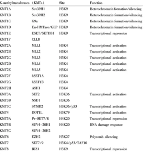
Table 2 Histone lysine methyltransferases ... 42
Table 3 Epigenetic regulators with phosphosites in the phosphoproteome dataset ... 76
Table 4 ET-‐specific PP2Ac-‐targeted phosphosites of chromatin regulators ... 79
Table 5 Primer Sequences for qPCR ... 116
Table 6 Secretory products of mononuclear phagocytes (Manes et al, 2011). ... 135
Table 7 Macrophage products with similar activities (Manes et al, 2011) ... 137
Table 8 Enzymes secreted by macrophages (Takemura & Werb, 1984) ... 137
Table 9 Primer Sequences used in the qPCR of T-‐cell regulatory genes ... 146
Table 10 List of cytokines and chemokines identified in the LPS-‐inducible secretome ... 150
Table 11 T-‐class GOBP enrichment ... 151
Figure 1 TLR structure ... 48
Figure 2 Mammalian TLR signaling pathways ... 48
Figure 3 Domain structure of the NFκB signaling proteins ... 49
Figure 4 NFκB dimers ... 49
Figure 5 NFκB bound to IκB proteins ... 50
Figure 6 Phosphorylated residues of p65 (RelA) ... 50
Figure 7 Model of regulation of NFκB transactivation through phosphorylation ... 51
Figure 8 Chromatin modifications and regulators ... 52
Figure 9 Dynamic epigenetic regulation ... 53
Figure 10 Modifications of the histone histone tails ... 54
Figure 11 Chemical mechanism for FAD-‐dependent demethylation ... 55
Figure 12 Schematic diagrams of LSD1 Domains ... 55
Figure 13 Chemical mechanism for demethylation by JmjC ... 56
Figure 14 Structure of JMJD2A bound to substrate ... 56
Figure 17 Regulatory function of SET7/9 on NFκB and inflammatory gene
expression ... 58
Figure 18 Role of PP2A in various signaling pathways ... 86
Figure 19 Schematic representation of diversity of the PP2A holoenzymes ... 87
Figure 20 Chronic-‐active PP2Ac regulates chromatin modifications ... 88
Figure 21 Schematic representation of phosphoprotein analysis workflow ... 89
Figure 22 Proteomic analysis using two-‐dimensional gel electrophoresis ... 90
Figure 23 Quantitative phosphoproteomics workflow. ... 91
Figure 24 Phosphoproteomic analysis shows 99% phospho-‐enrichment efficiency. ... 91
Figure 25 Identified phosphopeptides mostly cover single phosphorylation ... 92
Figure 26 Distribution of Phospho STY identifications ... 92
Figure 27 Identified mass error ... 93
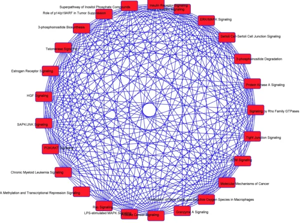
Figure 28 The top signaling pathways targeted by PP2Ac in ET macrophages ... 93
Figure 29 Top signaling pathways that are targeted by PP2Ac ... 98
Figure 33 The MS spectra of KMTs that are targeted by PP2Ac in ET. ... 101
Figure 34 The MS spectra of PP2Ac-‐targeted deacetylases and acetyltransferases ... 102
Figure 35 MS-‐MS Spectra of known PP2Ac targets. ... 103
Figure 36 Growth factors and insulin regulation of mTORC signaling via AKT. ... 104
Figure 37 Histone PTM changes regulated by PP2Ac activity in ET ... 105
Figure 38 The MS/MS (Left) and MS (Right) spectra of PHF8 phosphopeptides. ... 105
Figure 39 Domain structure and repeats of PHF8 ... 125
Figure 40 Mouse and human PHF8 sequence alignment. ... 125
Figure 41 The JmjC domain contains residues required for Fe(II) and αKG binding ... 126
Figure 42 Transcriptional transcriptional co-‐activation by KDM7-‐family proteins ... 127
Figure 43 Knockdown of PHF8 on mouse macrophage RAW cell line ... 127
Figure 44 PHF8 regulates LPS-‐induced demethylation of H3K9me1/me2 ... 128
Figure 45 PHF8 regulates nuclear translocation of p65 in LPS response. ... 129
Figure 48 Nucleus-‐specific CoIP of PHF8 and p65 ... 130
Figure 49 PHF8 positively regulates the transcriptional activity of p65. ... 131
Figure 50 PHF8 regulates the expression of select pro-‐inflammatory cytokines. ... 132
Figure 51 Schematic of ELISA procedure. ... 164
Figure 52 Scheme of the protein profiling process with antibody microarrays. ... 165
Figure 53 Workflow of the LFQ secretome analysis ... 166
Figure 54 Immunoblot of cell lysates for phenotype detection ... 166
Figure 55 Scatter plots showing Pearson correlation of samples. ... 167
Figure 56 The LFQ secretome heatmap ... 168
Figure 57 Comparison of the secretome to previous studies ... 169
Figure 58 T-‐class GOBP (Erdoğan et al, 2016). ... 170
Figure 59 T-‐class PHF8-‐dependent KEGG pathways (Erdoğan et al, 2016). ... 171
Figure 60 Processes associated with activated PHF8. ... 171
Figure 61 T-‐class GOCC (Erdoğan et al, 2016). ... 172
Figure 64 The overall protein-‐protein interaction network of the T-‐class
secretome ... 175
Figure 65 IPA network analysis of T-‐class secretome ... 176
Figure 66 PHF8 regulates the mRNA expression of T cell activating proteins ... 177
Figure 67 T cell activation markers ... 178
Figure 68 T cell proliferation assay ... 179
Figure 69 Histone H3K9 methyltransferases (HMTs) and demethylases (HDMs). ... 193
Figure 70 G9a structure. ... 193
Figure 71 Transcriptional repression and activation by G9a. ... 194
Figure 72 G9a regulates hypoxia response. ... 195
Figure 73 GOBP enrichment of the common secretome ... 196
Figure 74 GOMF enrichment of the common scretome ... 197
Figure 75 GOCC enrichment of the common secretome ... 198
Figure 76 Protein-‐protein interaction network of the common secretome ... 199
Figure 77 Top 10 canonical pathways regulated antagonistically by G9a and PHF8 ... 200
Figure 80 The mechanism of the G9a-‐ vs PHF8-‐ dependent chromatin
plasticity ... 203
2D Two-‐dimensional 53BP1 p53-‐binding protein 1
5mC methylation of the 5 position of the pyrimidine ring of cytosine AACT Amino-‐acid-‐coded mass tagging
ACN Acetonitrile
ADAM17 metalloproteinase domain-‐containing protein 17 ADAM8 metalloproteinase domain-‐containing protein 8 AP1 Activating Protein 1
APL Acute promyelocytic leukemia APP amyloid precursor protein AR androgen receptor
ASH1 Absent, small, or homeotic 1 ASH1L Ash1 Like
ATRA All-‐trans retinoic acid B2m beta-‐2-‐microglobulin BCL-‐3 B-‐cell lymphoma-‐3 Bcl2 B-‐cell Lymphoma 2
BMDM bone marrow-‐derived macrophages BP Biological Processes
CDK Cyclin-‐dependent kinase CDK Cyclin-‐dependent kinase CDS Cytosolic DNA sensors
CEBP CCAAT-‐enhancer binding protein Cfb Complement Factor B
Cfh Complement Factor H
CFSE Carboxyfluorescein diacetate succinimidyl ester ChEP Chromatin enrichment for proteomics
ChIP Chromatin immunoprecipitation ChIP-‐seq ChIP-‐sequencing
CID Collision-‐induced dissociation CKII Casein kinase II
CKII Casein kinase II CL/P cleft lip/palate
CLR C-‐type lectin receptors CoIP co-‐immunoprecipitation
CoREST Corepressor for element-‐1-‐silencing transcription factor CREB cAMP response element-‐binding protein
CSF Macrophage colony stimulating factor CtBP C-‐terminal binding protein
DMEM Dulbecco’s minimal essential media DMP Dimethyl Pimelimidate
DNMT DNA Methyl-‐transferase DNMT3A/B DNA Methyl-‐transferase 3A/B Dot1 Disruptor of telomeric silencing dsDNA Double stranded DNA
DTT Dithiothreitol DTX1 Deltex 1
ECL enhanced chemiluminescence EIF2 Eukaryotic initiation factor 2
ELISA Enzyme-‐linked immunosorbent assay ESCC Esophageal squamous cell carcinoma ET Endotoxin tolerance
EV empty vector
EZH2 Enhancer of zeste 2 polycomb repressive complex 2 subunit FAD flavin adenine dinucleotide
FBS Fetal bovine serum FBXL11 F-‐box LRR protein 11 FDR false-‐discovery-‐rate FOXP3 Forkhead box P3
GOCC GO Cellular Components GOMF GO Molecular Functions
GSK3β Glycogen synthase kinase-‐3 Beta H3PO4 phosphoric acid
HAT Histone acetyl-‐transferase HDAC Histone deacetylase
HES1 hairy and enhancer of split-‐1 HIF1-‐α Hypoxia-‐inducible factor 1-‐alpha
HIPK2 Homeodomain Interacting Protein Kinase 2 HP1 Heterochromatin protein 1
IAA iodoacetamide
Icam1 intercellular adhesion molecule 1 ID Intermediate domain
Ifi30 IFNγ-‐Inducible Protein 30 IFNβ Interfeuron-‐β
IFNγ Interfeuron-‐γ IKK IκB kinase IL-‐1 Interleukin-‐1
IL-‐1R Interleukin-‐1 Receptor IL10 Interleukin 10
IL8 Interleukin 8
ILF3 Interleukin Enhancer Binding Factor 3 IMAC Immobilized Metal Affinity Chromatography IPA Ingenuity Pathway Analysis
IRAK IL-‐1R-‐associated kinase Itga4 Integrin alpha 4
Itgb2 Integrin beta 2
Itmb2 integral membrane protein 2B IκB Inhibitor of kappaB
JARID1A Jumonji/ARID domain-‐containing protein 1A (KDM5a or RBP2) JARID1B Jumonji/ARID domain-‐containing protein 1B or KDM5B
JmjC Jumonji C-‐terminal domain Jmjd2 Jumonji domain-‐containing 2 JmjN Jumonji N-‐terminal domain KDM Lysine demethylase
KDM1A Lysine demethylase 1
KDM5A Lysine-‐specific Demethylase 5A also known as JARID1A or RBP2 KDM5B Lysine demethylase 5B or JARID1B
KEGG Kyoto Encyclopedia of Genes and Genomes Kme Lysine methylation
LFQ Label-‐free Quantification
Lgals3BP Lectin-‐Galactoside-‐Binding Soluble 3-‐Binding Protein LHSCC Laryngeal and hypopharyngeal squamous cell carcinoma Lif Leukemia inhibitory factor
LPS Lipopolysaccharide LRR Leucine-‐rich repeat
LSD1 lysine-‐specific demethylase 1 Ly86 lymphocyte antigen 86 m/z Mass-‐to-‐charge ratio
MCES mRNA cap guanine-‐N7 methyltransferase MCM minichromosome maintenance
MD2 Lymphocyte antigen 96 MeCP2 methyl CpG binding protein 2 MHC Major histocompatibility complex MLL Mixed lineage leukemia
MS Mass spectrometry
MS/MS tandem mass spectrometry
MSK1 Mitogen-‐ and stress-‐activated protein kinase 1 MT Methyltransferases
mTOR Mechanistic target of rapamycin
MyoD Myoblast determination protein NEMO NFκB essential modulator NFκB Nuclear factor kappa B NLR NOD-‐like receptors
NLS Nuclear localization signal
NOTCH3 Neurogenic locus notch homolog protein 3 NSCLC Non-‐small cell lung cancer
NSD1 Nuclear receptor-‐binding SET domain-‐containing protein 1 NSD2 Nuclear SET domain-‐containing protein 2
NSD3 Nuclear SET domain-‐containing protein 3 NT Non-‐tolerizable
OA Okadaic acid
P-‐p65 Phosphorylation of p65 at Ser536 PAMP Pathogen-‐associated molecular patterns PcG Polycomb-‐group
PCR Polymerase chain reaction PHD Plant Homeodomain PHF8 PHD finger 8
PHF8-‐KD PHF8 Knock-‐down
PKAc Cyclic AMP-‐dependent kinase PKCζ Protein kinase C-‐ζ
PP2A Protein phosphatase-‐2A
PP2AA Protein phosphatase-‐2A scaffold subunit PP2AB Protein phosphatase-‐2A regulatory subunit PP2Ac Protein phosphatase 2A catalytic domain PP2Ac-‐KD PP2Ac Knock-‐down
PP4 protein phosphatase 4 PPI Protein-‐protein interaction
PPM1D Protein Phosphatase, Mg2+/Mn2+ Dependent, 1D PPPs Phosphoprotein phosphatases
PRC Polycomb repressive complex PrCA Prostate cancer
PRMT5 Protein arginine methyltransferase 5 PRR Pattern recognition receptors
PSPs protein serine/threonine phosphatases PTM Post-‐translational modification
PVDF Polyvinylidene fluoride Pvrl1 poliovirus receptor-‐related 1 PYPs phosphotyrosine phosphatases qPCR Real-‐time Polymerase chain reaction Rac1 Ras-‐related C3 botulinum toxin substrate 1
RhoA Ras homolog gene family, member A RLR RIG-‐like receptors
RNA-‐PII RNA polymerase II RP Reverse phase rRNA ribosomal RNA
RSK1 Ribosome-‐associated kinase-‐1 RT room temperature
SAM S-‐adenosylmethionine
SATB2 Special At-‐rich sequence-‐binding protein 2 SCX strong cation exchange chromatography Sdc4 syndecan 4
SDS-‐PAGE sodium dodecyl sulfate polyacrylamide gel electrophoresis SDS3 Suppressor of defective silencing 3 protein homolog
SerB phosphoserine phosphatase
SET Su(var)3-‐9, Enhancer-‐of-‐zeste and Trithorax Set7 SET domain protein 7
Set9 SET domain protein 9 shCON Wild-‐type
shPHF8 PHF8 Knock-‐down SIRT1 Sirtuin 1
SRGN serglycin
STAT6 Signal Transducers and Activators of Transcription 6 STRING Search Tool for the Retrieval of Interacting Genes/Proteins Suv39h1 Suppressor Of Variegation 3-‐9 Homolog 1
SWI/SNF Switch/Sucrose Non-‐Fermentable T-‐class “tolerizeable”
TAD Trans-‐activation domain TAK1 TGF-‐β-‐associated kinase 1 TAP Tandem affinity purification TFA Trifluoroacetic acid
TGF-‐β Transforming growth factor-‐β Th T helper cell
TiO2 Titanium dioxide TIR Toll-‐IL-‐1R
TLR Toll-‐like receptors
TLR4/MD2 TLR4/Lymphocyte antigen 96 complexes TNF Tumor necrosis factor
TRIF TIR-‐domain containing adapter-‐inducing interferon β
TRM6 tRNA (adenine-‐N(1)-‐)-‐methyltransferase non-‐catalytic subunit URA Upstream Regulator Analysis
WD Tryptophan-‐Aspartic acid WDR5 WD-‐repeat domain 5
WIP1 wild-‐type p53-‐induced phosphatase 1 WT Wild-‐type
OVERVIEW
The mammalian immune system is a complicated and tightly regulated system that can recognize self and non-‐self cells or molecules, providing protection from a wide variety of pathogens. Recognition of non-‐self molecules leads cells of immunity to activate various signaling pathways, which lead to activation or repression of specific genes to induce inflammation response. At the core of this gene-‐specific regulation of inflammation is the chromatin remodeling. As the need to understand the tight regulation of epigenetics in inflammation response increases, various chromatin modifiers functioning as pro-‐ or anti-‐inflammatory factors have been studied
extensively. Here I provide an overview of innate immunity, Toll-‐like receptors (TLR), and epigenetic regulation of both transcription and inflammation response through methylation.
INNATE IMMUNITY AND TOLL-‐LIKE RECEPTORS
(RLR), cytosolic DNA sensors (CDS), and the C-‐type lectin receptors (CLR) (Kawai & Akira, 2011). The first PRR family to be identified and most widely studied was TLR that recognizes a wide range of PAMPs and activates downstream pathways.
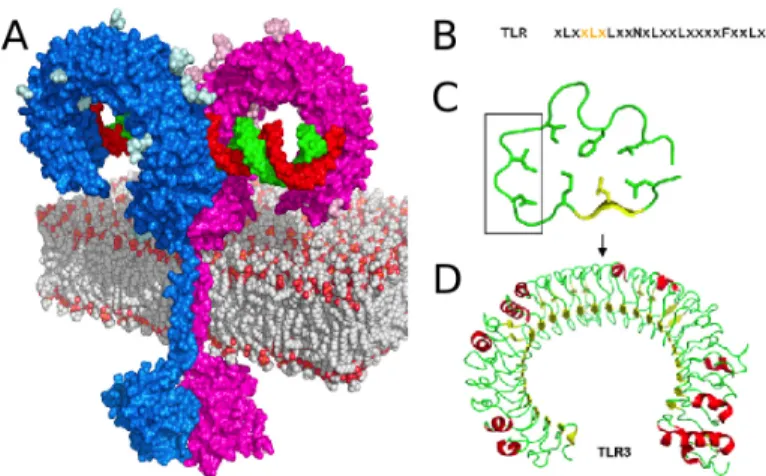
As type I transmembrane proteins, TLRs are composed of an extracellular ectodomain containing Leucine-‐rich-‐repeat (LRR) motif and a cytoplasmic Toll-‐
interleukin-‐1 Receptor (Toll-‐IL-‐1R or TIR) domain responsible for relaying the signal to the downstream adapters (Akira et al, 2006) (Figure1A). LRR domain consists of 19-‐25 tandem copies of the leucine-‐rich motif of 24-‐29 amino acids such as XLXXLXLXX and XΦXXΦXXXXFXXLX where X represents any amino acid and Φ represents a hydrophobic amino acid (Figure 1B) (Akira & Takeda, 2004). These repeats are composed of a β-‐ strand and an α-‐helix connected with loops creating a concave surface responsible for ligand recognition (Figure 1C-‐D). On the other hand, TIR domains have only 20-‐30% sequence homology with three conserved regions required for signaling.
TLRs are divided into subfamilies based on the ligand they recognize: TLR1, TLR2, and TLR6 recognize lipids, TLR7, TLR8, and TLR9 recognize nucleic acids, TLR3 recognizes double stranded DNA (dsDNA) from viruses, TLR4 recognizes
Upon ligand recognition, TLRs induce host defense genes through activation of two major downstream pathways based on the adapter protein involved: Myeloid
differentiation primary response gene 88 (MyD88)-‐dependent and MyD88-‐independent, also known as TIR-‐domain containing adapter-‐inducing interferon-‐β (TRIF)-‐dependent, pathways (Janeway & Medzhitov, 2002). All TLRs except TLR3 are MyD88-‐dependent pathways while TLR4 stimulation activates both MyD88-‐dependent and –independent pathways (Figure 2). Throughout this dissertation, I focused mainly on studying the events downstream MyD88-‐dependent TLR4 signaling.
MyD88-‐Dependent Signaling
a short intermediate domain (ID) (Lin et al, 2010). TIR domain is responsible for the recruitment of MyD88 as a scaffold to the activated TLR to relay the signal to
downstream elements (Ohnishi et al, 2009). On the other hand, DD is responsible for homophilic interaction with the downstream elements such as IL-‐1R-‐associated kinases (IRAKs) IRAK-‐1 and IRAK-‐4 (Akira et al, 2006). Activated IRAKs associate with Tumor necrosis factor (TNF) receptor (TNFR)-‐associated factor 6 (TRAF6), inducing
ubiquitination of the Inhibitor of kappaB (IκB) kinase (IKK)/Nuclear factor kappaB (NFκB) essential modulator (NEMO) complex and itself. In addition, another complex composed of Transforming growth factor-‐β (TGF-‐β)-‐activated kinase 1 (TAK1) and TAK1 binding proteins is recruited to TRAF6. TAK1 recruitment is crucial for
phosphorylation and subsequent activation of IKK-‐β for the degradation of IκB, which in return exposes the nuclear-‐localization signal, thus enables the early-‐phase
activation of NFκB transcription factor for the pro-‐inflammatory cytokine expression (Figure 2).
NFκB IN INFLAMMATION RESPONSE
Downstream TLR pathways, transcription factor NFκB regulates the transcription of many immune response-‐related genes such as cytokines, growth factors, effector enzymes in response to activation of immunity-‐related receptors (Hayden & Ghosh, 2004). Proteins in the NFκB family are divided into two subfamilies called the ‘NFκB’ and the ‘Rel’ proteins, all of which include an amino-‐terminal
activated through removal of these inhibitory domains by proteolysis leading to the exposure of the active forms p52 and p50, respectively (Hoffmann & Baltimore, 2006). These activated forms, as they don’t have trans-‐activation domains (TADs), require heterodimerization with the Rel proteins to initiate transcription (Hayden & Ghosh, 2012). The Rel proteins p65/RelA, RelB, and RelC have a C-‐terminal TADs, which can activate transcription across many species although their sequences are not conserved the same way (Gilmore, 2006). All five members of NFκB family can form homodimers or heterodimers in vivo, except RelB, which exists only in heterodimers in vivo (Figure 4). The variety of different homodimers and heterodimers create a multi-‐layered regulation system as different dimers have different DNA-‐binding site specificities and protein-‐protein interactions at the promoters.
In this thesis, I mainly focus on the transcriptional activity of the p65 subunit downstream MyD88-‐dependent TLR4 pathways, because its nuclear translocation and DNA-‐binding are indicators of transcriptional activation of NFκB target genes.
IκB Family Proteins Regulate p65 Activity
NFκB activity is regulated by interaction with seven IκB family proteins IκBα, IκBβ, IκBγ, IκBε, B-‐cell lymphoma-‐3 (BCL-‐3), IκBζ, and IκBζ as well as the precursor proteins p100 and p105 (Hayden & Ghosh, 2012). IκB family proteins contain five to seven ankyrin repeats that modulate the interaction NFκB proteins (Hayden & Ghosh, 2004). This interaction covers the Nuclear localization signal (NLS) of p65 to
and cytoplasmic localization of NFκB, respectively (Sun & Andersson, 2002).
Regulation of Transcriptional Activity of p65 via Phosphorylation
NFκB activity is also regulated by post-‐translational modifications (PTMs), such as phosphorylation, acetylation, and methylation. Currently, phosphorylation of p65 is the most common and most widely studied NFκB PTM because it is associated with rapid transactivation and transcriptional activity of p65 (Figure 6, Figure 7) (Vermeulen et al, 2006); thus, in this overview I will focus on the p65 regulation through phosphorylation.
Currently, five phosphorylation sites have been discovered to regulate p65 activity: S276, S311, S468, S529, and S536 (Figure 6). S276 resides in the RHD and gets phosphorylated by cyclic AMP-‐dependent kinase (PKAc) (Zhong et al, 1997) and by Mitogen-‐ and stress-‐activated protein kinase 1 (MSK1) upon TNF treatment
(Vermeulen et al, 2003). Phosphorylation of this site is essential for gene activation (Zhong et al, 1998). Another important phosphorylation is the phosphorylation of S311 by Protein kinase C-‐ζ (PKCζ) that is responsible for recruitment of cAMP response element binding protein (CREB)-‐binding Protein (CBP) and RNA polymerase II (RNA-‐ PII) to the IL-‐6 promoter (Duran et al, 2003). Moreover, S529 is phosphorylated by casein kinase II (CKII) (Wang & Baldwin, 1998) and IKK2 enhancing transactivity of p65 (Sakurai et al, 1999). In addition to S529, IKK2 also phosphorylates S536 for
transcription factor activity of p65 upon TNF or IL-‐1 (Buss et al, 2004; Mattioli et al, 2004).
It is important to note that the dynamic regulation of p65 phosphorylation status is sustained by the interplay between kinases and phosphatases. Protein phosphatase-‐ 2A (PP2A), protein phosphatase 4 (PP4), protein phosphatase 1γ (PP1γ), wild-‐type p53-‐induced phosphatase 1 (WIP1), and phosphoserine phosphatase (SerB) are some of the known negative regulators of p65 keeping it in the inactive state (Peng et al, 2015; Takeuchi et al, 2013; Yang et al, 2001; Yeh et al, 2004).
ENDOTOXIN TOLERANCE
Inflammatory response, if persistent, may lead to severe reaction in the form of chronic inflammation since excessive cytokine production is harmful to the host (Beutler et al, 1985; Kanterman et al, 2012). Thus, the immune system is programmed to turn of inflammation to prevent tissue damage by immunosuppression. This
phenomenon is called endotoxin tolerance (ET) (Dobrovolskaia & Vogel, 2002). There are multiple inhibitory mechanisms that regulate endotoxin tolerance. First, the expression and availability of the surface receptors are affected by the second exposure to the endotoxin (Fujihara et al, 2003). In ET, RAW 264.7 cells show
Kohler & Joly, 1997; Medvedev et al, 2000), IRAK-‐1 activation (Jacinto et al, 2002; Li et al, 2000), and ratio of p50/p65 heterodimers to p50/p50 homodimers (Adib-‐Conquy et al, 2000). In addition, increased negative regulators of TLR signaling, such as kinase IRAK-‐M (Kobayashi et al, 2002), suppressor of cytokine signaling 1 (SOCS1) (Kinjyo et al, 2002), and MyD88short (MyD88s) (Burns et al, 2003), a short alternative spliced
form of MyD88 lacking the ID, contributes to the ET. The last inhibitory mechanism is the regulation of secreted proteins/mediators via prolonged LPS stimulation,
downregulating of cytokine expression in ET (Fujihara et al, 2003). For example, prolonged LPS-‐induced Interleukin 10 (IL10) secretion in macrophages disrupts expression of other inflammatory molecules such as cytokine Interfeuron-‐γ (IFNγ) (Varma et al, 2001), Tumor necrosis factor α (TNFα), Interleukin 6 (IL6), and macrophage colony stimulating factor (CSF) in ET (Berlato et al, 2002). Similarly, increased Interfeuron-‐β (IFNβ) leads to decreased TNFα and Interleukin 8 (IL8) expression in ET (Zaric et al, 2011).
ET, or also known as acquired immune tolerance, including that to endotoxin or LPS, if deregulated, is a major molecular feature of the pathogenesis of many chronic diseases including asthma, sepsis, and cancer (Biswas & Tergaonkar, 2007).
Deciphering the mechanisms that regulate LPS-‐induced diverse inflammatory
responses will be beneficial for diagnosis of, prevention of, and therapies for various inflammation-‐associated diseases.
Inflammation response is also regulated at the epigenetic level by the LPS-‐ induced gene-‐specific chromatin modifications, whereby the promoters of a select class of pro-‐inflammatory or “tolerizeable” (T-‐class) genes have differentially programmed chromatin based on the “inflammatory-‐phenotype”, acutely versus chronically inflamed nature, of the stimulated cells (Foster et al, 2007).
As the core components of the chromatin, the properties of histones can be altered by different PTMs to their N-‐ terminal “tails” that leads to a predetermined combination of histone marks known as the “histone code” (Strahl & Allis, 2000). These differentially modified histone marks determined whether the promoter they are in is in an open/active or closed/repressed chromatin state called euchromatin or
heterochromatin, respectively (Figure 8); thus, gene-‐specific transcription is regulated through histone PTMs to achieve specific biological outcomes, such as inflammation response (Adcock et al, 2007). The histone PTMs are added or removed by different enzymes termed “writers” and “erasers”, respectively (Figure 8, Figure 9) (Falkenberg & Johnstone, 2014). Moreover, to signal for recruitment and activity of downstream effectors, histone “readers” are recruited to histone PTM sites (Yun et al, 2011).
Currently there are various PTMs known to modify histone N-‐tails: methylation, acetylation, phosphorylation, citrullination, ubiquitination, SUMOylation, ADP-‐
modifying enzymes such as acetyltransferases, deacetylases, methyltransferases,
demethylases, kinases, phosphatases, ubiquitilases, and proline isomerases (Kouzarides, 2007). Here, I will focus on histone methylation since the scope of this research is more relevant to this PTM and provide information on how this PTM is regulated.
Epigenetic Regulation of Transcription Through Methylation PTM
Histone lysine methylation (Kme) is the most widely studied chromatin PTM with six well characterized lysines including H3K4, -‐9, -‐27, -‐36, -‐79, and H4K20 (Shi & Whetstine, 2007). In eukaryotes, Kme patterns regulate the chromatin architecture by dictating activated or repressed conformation (Barski et al, 2007). Particularly on histone H3, the transcription-‐associated Kme occurs at multiple lysine residues: methylated H3K4, H3K36, and H3K79 are associated with transcriptional activation of specific genes while methylated H3K9 and H3K27 are associated mostly with gene repression (Martin & Zhang, 2005). Site-‐scpecific Kme levels are tightly regulated by the antagonistic activities of lysine methyltransferases (KMTs) and lysine demethylases (KDMs) to maintain cell homeostasis. Specific biological processes that promote gene activation, such as induced inflammatory response, require coordinated changes in the activity of LPS-‐induced KMTs and KDMs increasing the activating methylation marks while decreasing the repressive methylation marks, respectively, at the promoters of those specific genes.
Trithorax (SET) domain. KMTs are relatively specific; they modify the target lysine to a specific degree (mono-‐, di-‐, or tri-‐methyl state); for instance SET domain proteins 7 and 9 (Set7/9) can only mono-‐methylate H3K4. Various targets of known KMTs are listed in
Table 2.
Table 2 Histone lysine methyltransferases
Methyltransferases, their target site, and function (Bannister & Kouzarides, 2011)
Autoregulatory Factor 1 (MYND) domain-‐containing 3 (SMYD3) during gene activation (Gibbons, 2005).
H3K9 methylation is involved in many biological processes such as
transcriptional regulation, X chromosome silencing, heterochromatin formation, and DNA methylation (Wang & Zhu, 2008). One of the most widely studied H3K9 KMT is G9a, which dimethylates H3K9 in euchromatic regions; this G9a-‐mediated H3K9
methylation is responsible for the silencing of individual genes (Martin & Zhang, 2005). In addition to G9a, KMT Suppressor Of Variegation 3-‐9 Homolog 1 (Suv39h1)
methylates H3K9 (Wang & Zhu, 2008). Suv39h1 targets both euchromatin and heterochromatin and regulates heterochromatin formation as well as transcriptional inhibition by tri-‐methylating H3K9. Moreover, Suv39h1-‐dependent H3K9 methylation recruits the chromodomain of Heterochromatin protein 1 (HP1) during meiosis and cell cycle leading to DNA methylation for transcriptional repression (Biel et al, 2005; Martin & Zhang, 2005; Peters et al, 2001).
H3K27 methylation is catalyzed by Enhancer of zeste 2 polycomb repressive complex 2 subunit (EZH2), which forms the Polycomb repressive complex (PRC) with other Polycomb-‐group (PcG) proteins (Wang & Zhu, 2008). As an H3K27
methyltransferase, EZH2 has an inhibitory effect on transcription and is known to regulate cell differentiation, fetal development, cell proliferation, and X chromosome inactivation (Biel et al, 2005; Wang & Zhu, 2008).
Histone demethylation was considered irreversible for decades due to the stability of the C-‐N bond (Mosammaparast & Shi, 2010). The first experimental evidence for an enzyme that catalyzes demethylation was the functional
characterization of lysine-‐specific demethylase 1 (LSD1, also known as KDM1A) as a member of the amine oxidase superfamily (Shi et al, 2004). Demethylation by the amine oxidases occurs through cleavage of the α-‐carbon bond of the substrate leading to an imine intermediate that further gets hydrolyzed to produce an aldehayde and amine (Shi & Whetstine, 2007). This is a flavin adenine dinucleotide (FAD)-‐dependent reaction that requires a protonated nitrogen making it recognize only mono-‐ or di-‐methylated lysine substrates (Figure 11) (Bannister & Kouzarides, 2011; Shi & Whetstine, 2007). Thus, LSD1 is specific for demethylation of H3K4me1/me2 (Varier & Timmers, 2011). However, LSD1 specificity and activity is regulated by protein-‐protein interactions (Shi & Whetstine, 2007). Although in vitro purified LSD1 can demethylate H3K4me1/2, nucleosomal H3K4me1/2 demethylation by LSD1 requires an interaction with the corepressor complex Corepressor for element-‐1-‐silencing transcription factor
(CoREST) through the tower domain leading to increased stability of LSD1 (Figure 12) (Shi & Whetstine, 2007). Another protein-‐protein interaction regulating the LSD1 substrate specificity is the androgen receptor (AR) complex formation, which switches LSD1 from transcriptional repressor to transcriptional activator by changing the substrate specificity from H3K4me1/2 to H3K9me1/2 (Figure 12).
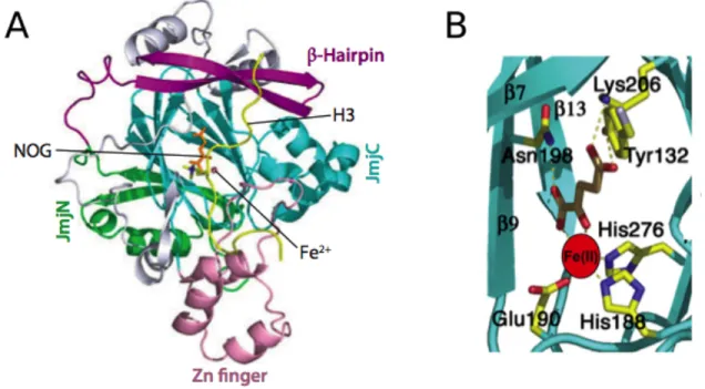
Timmers, 2011). The oxidative decarboxylation of αKG is coupled to hydroxylation of the methyl group that creates an unstable hydroxymethyl ammonium intermediate releasing formaldehyde (Figure 13) (Mosammaparast & Shi, 2010). The first JmjC proteins that were identified as KDMs are the Jumonji domain-‐containing 2 (JMJD2) family proteins and the very first characterized high-‐resolution structure belonged to JMJD2A, an H3K9 and H3K36 KDM, in this family. JMJD2A structure revealed that it consists of a Jumonji N-‐terminal domain (JmjN), a JmjC domain, a C-‐terminal zinc-‐finger motif, and a β-‐hairpin (Figure 14A). The catalytic core consists of the JmjC domain in which two histidine and one glutamate residue are essential for catalytic activity as they chelate the catalytic iron atom as shown in Figure 14B.
Role of Lysine Methylation in Regulation of Inflammation Response
The first histone Kme discovered regulating the inflammatory response is the dynamically regulated H3K9 methylation (Saccani & Natoli, 2002). Gene-‐specific activation and post-‐induction repression of some of the inducible inflammatory genes happens through demethylation and remethylation of H3K9, respectively. Moreover, demethylation and remethylation of H3K9 is correlated with RNA-‐PII recruitment and release, respectively, to the promoters.
meythylation (Figure 15) (El Gazzar et al, 2008). G9a also recruits members of other repressive complexes such as CoREST, C-‐terminal binding protein (CtBP), and
Switch/Sucrose Non-‐Fermentable (SWI/SNF), creating a strong transcriptionally repressive environment for immunosuppression at ET and leading to global changes in the histone PTM landscape, chromatin remodeling, and activities of select transcription factors (Figure 16) (Liu et al, 2014).
H3K27me3, another repressive histone PTM, regulates expression of specific inflammatory genes (De Santa et al, 2007). Regulation of H3K27me3 is achieved by the opposite activities of KDM Jumonji domain-‐containing 3 (JMJD3, also known as KDM6B) and KMT complex PcG in macrophage response (De Santa et al, 2007; Ishii et al, 2009; Köhler & Villar, 2008). JMJD3 is induced with inflammatory stimulation and gets recruited to PcG target genes, decreasing H3K27me3 levels at Signal Transducers and Activators of Transcription 6 (STAT6) promoter and enhancing transcriptional activity. The subsequent activation of STAT6 enhances binding to JMJD3 promoter and
positively regulates the expression of specific inflammatory genes. Interestingly, JMJD3 controls the transcription of the 70% of the LPS-‐inducible genes (De Santa et al, 2009), indicating the essential role of histone PTM changes in macrophage activation.
increased recruitment of CCAAT-‐enhancer binding protein (CEBP) and p65 to the IL6 promoter (Hanzu et al, 2013). With obesity, LSD1 gets decreased leading to expression of pro-‐inflammatory cytokines and obesity-‐associated inflammation. There have been other reports demonstrating evidence for the role LSD1 in cytokine repression of IL1α, IL1β, IL6, and IL8 (Janzer et al, 2012).
Histone KMTs, which target activating methyl-‐marks on H3, may act as co-‐ activators of inducible inflammatory genes; one such example is the H3K4 KMT Set7/9 that regulates select inflammatory gene transcription through regulation of NFκB recruitment to the cytokine-‐induced inflammatory promoters (Figure 17) (Fujimaki et al, 2015; Li et al, 2008). Similarly, a H3K4 KMT MLL1 regulates the TNF-‐induced
activation of NFκB target genes (Wang et al, 2014b). Activated NFκB recruits MLL1, through binding, to the target genes.
Figure 1 TLR structure
(A) Structural model of TLR3-‐dsRNA complex based on the mTLR3-‐dsRNA structure (3CIY) and TLR3 TIR domain homology model on the TLR10 TIR structure structure (2J67) (Botos et al, 2011). (B) LRR consensus sequence for TLR3 where orange highlighted residues represent the β-‐ sheets (Botos et al, 2011). (C) A LRR loop from hTLR3 with the conserved residues forming the hydrophobic core where the box highlights the ligand binding residues. (D) Ribbon diagram of TLR3 ectodomain structure (2A0Z) (Bell et al, 2005).
Figure 2 Mammalian TLR signaling pathways
Figure 3 Domain structure of the NFκB signaling proteins
The general domain structure of Rel and NFκB subfamily transcription factors (left) consists of a conserved DNA-‐binding/dimerization domain called the Rel homology domain (RHD) with nuclear localization and IkB binding sequences. The C-‐terminal halves of the Rel proteins have transcriptional activation domains (TAD). The C-‐terminal halves of the NFκB subfamily proteins have ankyrin repeat-‐containing inhibitory domains (red bars), which can be removed by
proteasome-‐mediated proteolysis. As with the C-‐terminal domains of the NF-‐kB proteins, the independent IκB proteins consist mainly of ankyrin repeats, and several (IκBα, IκBβ, IκBε, IBγ) have two N-‐terminal serine residues (S) that serve as IKK phosphorylation sites, which signal the protein for ubiquitination and degradation. The generalized structures of IKKα and β (kinase domain; HLH, helix-‐loop-‐helix; LZ, leucine zipper; NBD, NEMO binding domain) and of NEMO (CC, coiled coil; LZ, leucine zipper; ZF, zinc finger) are also shown (right) (Gilmore, 2006).
Figure 4 NFκB dimers
Figure 5 NFκB bound to IκB proteins
(A) The structure of IBα to the p50:RelA heterodimer where the NLS of RelA, which is
unstructured in the absence of IκB proteins, folds into a helical structure when bound to IκBα while the NLS of p50 doesn’t interact with IκBα. (B) The structure of IBβ bound to RelA
homodimer. One NLS interacts with IκBβ as that in A while the other has weak contacts (Hoffmann & Baltimore, 2006).
Figure 6 Phosphorylated residues of p65 (RelA)
The known phosphorylated residues are S276, S311, S468, S529, and S536 (Vermeulen et al, 2006).
Figure 7 Model of regulation of NFκB transactivation through phosphorylation
Figure 8 Chromatin modifications and regulators
Figure 9 Dynamic epigenetic regulation
Figure 10 Modifications of the histone histone tails
Figure 11 Chemical mechanism for FAD-‐dependent demethylation
The reaction schematic shows LSD1 removing a methyl group from a dimethylated lysine residue and the reaction can be repeated until lysine is unmethylated (Shi & Whetstine, 2007).
Figure 12 Schematic diagrams of LSD1 Domains
Figure 13 Chemical mechanism for demethylation by JmjC
The reaction schematic shows Fe(II) and the α-‐keto-‐glutarate-‐dependent dioxygenase-‐mediated
demethylation. Red indicates carbons that are demethylated in each reaction (Mosammaparast & Shi, 2010).
Figure 14 Structure of JMJD2A bound to substrate
Figure 15 Proinflammatory gene silencing by G9a during endotoxin tolerance
G9a is responsible for the recruitment of DNA methyltransferases in addition to increased repressive histone H3K9 methylation (El Gazzar et al, 2008).
Figure 16 G9a stabilizes c-‐Myc and promotes cell survival in ET
Figure 17 Regulatory function of SET7/9 on NFκB and inflammatory gene expression
CHROMATIN MODIFIERS TO REGULATE ENDOTOXIN TOLERANCE
OVERVIEW
Protein phosphatase PP2A is a major regulator of ET in macrophages (Xie et al, 2013). PP2Ac not only dephosphorylates NFκB for successful suppression of target genes, but also affects the chromatin modeling in ET; however, it is still not clear how PP2Ac broadly affects the changes on the chromatin state. Here, we proposed to screen ET-‐specific PP2Ac targets that get dephosphorylated in ET using Amino-‐acid-‐coded mass
tagging (AACT)-‐based quantitative phosphoproteomics to clarify the exact pathways and
biological processes modulated by chronically active PP2Ac activity. Our
Protein Phosphatases and PP2A
Protein phosphorylation regulates many cellular processes, including regulation of proteolysis, transcription, metabolism, cell-‐cycle progression, cell differentiation, cytoskeleton organization, cell movement, apoptosis, cell-‐cell communication, neuronal functions, and immune responses (Johnson, 2009). With such diverse functional roles in the cell, phosphorylation mainly targets hydroxyl-‐group containing amino acids serine, threonine, and tyrosine with 86.4%, 11.8% and 1.8% degree of phosphorylation, respectively (Seshacharyulu et al, 2013). The high diversity of proteins regulated by phosphorylation demands high diversity of these enzymes enabling the proper regulation of the phosphorylation-‐dependent events in the cell (Virshup, 2000).
Phosphoprotein phosphatases (PPPs), grouped into protein serine/threonine phosphatases (PSPs) and phosphotyrosine phosphatases (PYPs), are the enzymes that are responsible for the removal of the phosphate group in the presence of a water molecule. There are many subfamilies of PPPs: PP1, PP2A, PP2B, PP4, PP5, PP6, and PP7 (Seshacharyulu et al, 2013).