109
http://www.ijmess.com
Application of Intelligence Computing to
Optimizing Enzymatic Bioprocessing in Cartilage
Hydrolysis
Tzu-Miao Lin1 *Hsi-Chieh Lee3
Wen-Jia Kuo5
Chih-Ching Chien2
Yao-Horng Wang4
Chai-Li Chen6
1
Dept. of Nursing, Hsin Sheng College of Medical Care and Management, Taiwan
2
Graduate School of Biotechnology and Bioengineering, Yuan Ze University, Taiwan
3
Dept. of Computer Science and Information Engineering, Quemoy University, Taiwan
4
Dept. of Nursing, Yuanpei University of Medical Technology, Taiwan
5
Dept. of Information Management, Yuan Ze University, Taiwan
6
Dept. of Information Management, Lunghwa University of Science and Technology, Taiwan
This study uses the Taguchi orthogonal method and artificial neural network to optimize enzymatic bioprocessing of animal waste cartilage (chicken, mini pig and hog). Specifically, the artificial neural network is used in parallel with the Taguchi orthogonal array process for enzymatic hydrolysis of the cartilage tissue to optimize the best quality of bioactive peptides. The experiment was designed using Taguchi orthogonal array optimal level L25 physical parameters and key media components, namely temperature, pH, enzyme/substrate ratio, substrate concentration, and reaction time. The experimental results were used to train the artificial neural network (ANN) to predict the optimizing enzymatic bioprocessing in animal cartilage hydrolysis. The analysis was performed on a personal computer using NeuroSolutions 6.0 software. The experiment of an enzymatic hydrolysate of three animal cartilages followed the Taguchi orthogonal design, and we discovered that 60±1℃ is the most effective temperature to hydrolyze cartilage. These peptides of molecular size smaller than 10kDa (with 95% values between 10.7kDa and 2.5kDa) were capable of stimulating the porcine chondrocytes to produce glycosaminoglycan (GAG) and type II collagen in vitro. NeuroSolutions 6.0 back-propagation analysis achieved a convergence value of R2=0.9762, indicating that the enzymatic bioprocessing has good performance. Therefore, this study suggests that integrating artificial neural network and Taguchi method when constructing an optimal enzymatic bioprocessing model could significantly increase and improve the quality of final bioactive peptide products. It also suggests that integrating artificial neural network and Taguchi method in the construction of an optimal enzymatic bioprocessing in cartilage hydrolysis could be used as nutraceutical component in bone and joint health
Keywords: Intelligence computing, Taguchi orthogonal array, Neural network, Cartilage hydrolysis, Enzymatic bioprocessing
Cartilage is a flexible connective tissue found in various parts of humans and other animals, including the
Manuscript received May 22, 2017; revised June 17, 2017; accepted July 20, 2017. © The Author(s); CC-BY-NC; Licensee IJMESS
International Journal of Management, Economics and Social Sciences
110
joints between bones, the rib cage, the ears, the nose, the bronchial tubes and the intervertebral discs [1]. It
is chemically abundant in collagen, proteoglycans, acidic polysaccharides and water. Conceivably, cartilage
can be hydrolyzed to be a potential bioactive material of collagen extraction. In fact, hydrolyzed collagen has
been applied in the leather and film industries, pharmaceuticals, cosmetics, biomedical materials and food
manufacturing [2]. In particular, some clinical studies report that oral ingestion of hydrolyzed collagen ease
the joint pain of osteoarthritis or rheumatoid arthritis, with those having the most severe symptoms showing
the most benefit [3, 4 and 5]. Normally, hydrolyzed collagen is isolated from animal (chicken, bovine, porcine,
rabbit, duck and antler) cartilages [6, 7 and 8], and fish (skate and shark) cartilages [2, 9 and 10].
A review of methods for extracting hydrolyzed collagen from animal cartilage shows the following. A study
conducted in 2005 found that hydrolyzed collagen absorbed small peptides in blood, indicating that the
process of hydrolysis involves breaking down the molecular bonds between individual collagen strands using
combinations of heat, acids, alkalis, or enzymes, and then reducing collagen proteins of about 300,000 Da
into small peptides with an average molecular weight between 2000 and 5000 Da [11]. Specifically, the
process is characterized by the following steps: (1) obtaining raw materials from fresh animal cartilage,
removing the adipose tissue, cutting adipose tissue into small pieces and soaking a 3% sodium hydroxid for
10-30 hours; (2) using phosphate-buffered saline (abbreviated PBS) to adjust pH value to 3.5-9.5, in
proportion to the concentration of animal cartilage particle in g substrate; (3) Adding a plant extract of papain,
bromelain or alkaline protease enzymatic protecting agent based on the ratio of animal cartilage particle to g
substrate and then adding the composite (enzyme/substrate ratio), incubating with stirring hydrolysis for 4-8
hours; (4) transferring the reaction contents to 45-100℃ water-bath for 8-15 minutes off the enzyme while
awaiting completion of incubation; and (5) after cooling to room-temperature, centrifuging the reaction
mixture at 3500-5000rpm for 30 minutes at 4℃. Finally, the supernatant fluid and precipitate are separated
and freeze-dried [12-17].
Resorting to the above methods [12-17], whether enzymatic hydrolysis, acid hydrolysis or alkaline
hydrolysis is used for extracting animal cartilage collagen, the products need to be assured of safety,
efficiency, and quality testing before being marketed. Thus, to optimize bioprocessing of cartilage hydrolysis
111
necessary for a chondroprotective effect [2]. It is believed that intelligence computing technology can bring
about a significant advancement in product development (as archived pharmaceutical industry) as well as in
future business execution and prediction.
-Specific Aims
The primary aim of this study is to use intelligence computing technologies (such as Taguchi orthogonal
method and artificial neural network) to optimize enzymatic bioprocessing of animal waste cartilage (chicken
sternal cartilage, mini pig laryngeal and tracheal, and hog laryngeal and tracheal) and generate hydrolysis
conditions for preparation of hydrolyzate collagen. Specifically, the artificial neural network is used in parallel
with the Taguchi orthogonal array process for enzymatic hydrolysis of the cartilage tissue to produce the best
yield and quality of bioactive peptides with clinical efficacy for functional ingredients of nutraceuticals.
Ultimately, the goal is to develop a valuable and market-potential nutraceutical product for bone and articular
joint health care.
A systematic review of the scientific evidence put forth between 1979~2010 [18, 19] shows that a new era
in the management of osteoarthritis and nutrition, from nutraceuticals to functional foods, is possible.
According to a report published in 2007, nutraceuticals sales were projected to reach $74.7 billion at an
AAGR (Average Annual Growth Rate) of 9.9%. In fact, the global market for nutraceuticals is growing day by
day and was expected to reach $176.7 billion in 2013 [20]. Particularly, chondroprotective agents including
collagen hydrolysate have been used widely as nutraceuticals in osteoarthritis treatments [21]. As such,
identification and selection of bioactive factors would be important for engineering mimetic biomaterials/
biological biomaterials to provide not only mechanical support but also biological cues in the induction of a
synthesized special cartilage extracellular matrix by chondrocytes. Motivated by such a trend, this study was
conducted to advance both academic and industrial interests.
MATERIALS AND METHODS
-Materials
International Journal of Management, Economics and Social Sciences
112
This study obtained chicken sternal cartilage from an industrial poultry meat processor (Kai Shing Trading
Co., Ltd., Yunlin, Taiwan). The mini pig laryngeal and tracheal and hog laryngeal and tracheal were obtained
from PigModel Animal Technology Co., Ltd. The three animal cartilage tissues were stored and frozen before
they were processed and prepared as cartilage hydrolysates. Fresh porcine knee joints were purchased from
a local meat market and used for isolation of chondrocytes in less than 6hrs. Papain was obtained from
Merck (No.107147 Papain 6000USP-U/mg). DMEM/Ham’s F12 medium, Fetal Bovine Serum (FBS),
Penicillin and Streptomycin were from Invitrogen Corporation, (Carlsbad, CA, USA). Peopidium iodide,
Alcian blue, MTT (3-(4, 5-Dimethylthiazol-2-yl)-2,5-diphenyltetrazolium bromide, a yellow tetrazole),
1,9-dimethyl-methylene blue (DMB), chondroitin sulfate A, calf thymus DNA and Hoechst 33324 and reagent
graded chemicals were obtained from Sigma-Aldrich (St. Louis, MO, USA).
Taguchi Orthogonal Working Model
This study obtained the Qualitek-4 from a local agent in Taiwan.
Artificial Neural Network Software
NeuroSolutions 6.0 software was obtained from the local agent that provided the Qualitek-4.
-Methods
Enzymatic Bioprocessing Materials
Optimal levels L25 of physical parameters and key media components, namely temperature, pH,
enzyme/substrate, substrate concentration, and reaction time were determined.
Getting Sarted with Qualitek-4 [31]
Preparation of Cartilage Hydrolysate (see Table 1)
Papain Hydrolysis of Animal Cartilage
Based on the hydrolyzing procedure of avian cartilage used in the study by Vouland et al. [14, 15], efforts for
the Taguchi orthogonal working model (Qualitek-4 Windows software) were made to reach optimal levels of
physical parameters, namely temperature, pH, enzyme/substrate ratio, substrate concentration and duration
time [22, 31]. From published information [12-17], this study established the papain hydrolysis of animal
cartilage to achieve the following conditions: 60-70±1℃, pH4.5, pH5.5 and pH6.5, enzyme/substrate ratio
113
end of incubation time, the reaction contents were transferred to 100℃ water bath for 10 minutes. After
cooling to room temperature, the reaction mixture was centrifuged at 3500rpm (2200×g) for 30minutes at 4℃.
The supernatant fluid and precipitate were separated and freeze-dried. The dried products were stored at
room temperature in tide container vials.
Table 1. Blending Animal Cartilage to 5mm Particle Size
Isolation, Cultivation and Identification of Porcine Articular Chondrocytes
Full-thickness cartilage slices were harvested from porcine knee joints. Then the slices were cut into 1mm3
pieces and placed in dishes containing 0.2% typeIcollagenase and transferred to a 37℃ incubator for 16
hours. The chondrocytes liberated were filtered through 10μ m filter to remove undigested cartilage
fragments. The isolated cells were washed and re-suspended in completed medium (DMEM/Ham’ s F12
medium containing 10% FBS and 100 U/ml Penicillin and 100 μ g/mL Streptomycin) and their viability was
determined using trypan blue dye exclusion.
Cells were expanded by monolayer culture in completed medium. At 80 to 90% confluences, the cells
were trypsinized and re-suspended in completed medium at a concentration of 1×10⁵ cells/ ml. The 5ml cell
suspension was placed into culture flask and at 37℃ in a humidified 5% CO2 incubator. This was the P1
passage of the primary culture of chondrocytes.
International Journal of Management, Economics and Social Sciences
114
A cell climbing sheet of P3 chondrocytes was washed with PBS, fixed with 4% paraformaldehyde for 15
minutes, stained with 1% alcian blue for 30 minutes and washed with ddH2O. Cell morphology was observed
using a microscope and photography was taken at a magnification of 100× for further examination.
Evaluation of Cell Cytotoxicity and Proliferative Effects
The cartilage hydrolysate on the chondrocyte cultures by MTT assay. The chondrocytes were cultured in
completed medium and cultured overnight. For cytotoxicity assay, chondrocytes were treated with cartilage
hydrolysate in serial dilutions of concentrations 200mg/ ml to 0.0002mg/ml for 24hours. For proliferation
analysis, the cells were treated with various concentrations of cartilage hydrolysate (2mg/ml, 0. 2mg/ml and
0.02mg/ml) for 2, 7 and 14 days. After treatment, 10μ M MTT (5mg/ml in PBS) were added to each culture
well, and the cultures were incubated at 37℃ for 4hours. The purple-blue MTT formazan precipitate was
dissolved in 100μ l DMSO and cells were shaken for 5 minutes. Absorbance was measured at 570 nm using
ELISA reader.
Biochemical Analysis
The P3 chondrocyte culture were treated with cartilage hydrolysate and assessed for Glycosaminoglycans
(GAG) type II collagen and DNA content. Briefly, cells were washed with PBS and digested by incubation for
16 hours at 60℃ in papain digest buffer. The measurement of total sulphated GAG in the chondrocyte
culture was performed using the 1,9-dimethyl-methylene blue (DMB) dye method. Chondrotin sulfate A from
bone cartilage was diluted with distilled water to prepare a standard curve (0 to 80μ g/ml). A 96-well plate
was used and 40 μ l of GAG standards or the papain-digested samples were piped into each well, then 250
μ l of DMB solution was added to each well. Absorbance at 525 nm was determined using a
spectrophotometer (Biorad Plate-Reader, BioRad Laboratories, Hemel Hempstead, UK) at a wavelength of
595 nm.
Determination of DNA Content Using Hoechst 33324 Fluoresce-Metric Assay
DNA from calf thymus was used to prepare a standard dilution curve ranging from 0 to 2.5μ g/ml. 100μ l
aliquots of standards or sample and Hoechst 33324 (1μ g/ml) were added into a 96-well plate. The samples
were read on a fluorimeter with a 365nm excitation filter emission set at 460nm, and the GAGs were
115
Quantification of Type II Collagen
Porcine type II collagen was determined by means of the Collagen type II ELISA kit (MD Bioscience, Inc,
Switzerland). To solubilize the newly synthesized collagen, the cell culture medium was removed and pepsin
solution was added to the cell layer and digested the collagen at 2-8℃overnight. Subsequently, TXB (0.1M
Tris, 0.2M NaCl, 5mM CaCl2, pH 7.8-8.0) was added to stop the reaction. To monomerize the remaining
polymeric collagen, pancreas elastase solution was added to the samples and incubated at 2-8℃ overnight.
After centrifugation (10,000rpm for 5 minutes), the supernatant was assayed for type II collagen by ELISA.
Molecular Size Distribution
The cartilage hydrolysate was characterized by HPLC analysis using an LC system consisting of a Waters
model 600E solvent delivery system (a Waters model 996 Photodiode Array Detector and a Waters 717 Plus
auto-sampler using a 200 μ l sample loop). Empower software was used to control the system and to
perform the data analysis. The size exclusion experiments were performed at room temperature using a 10×
300nm SuperdexTM peptide column (GE Healthcare) eluted at 0.4ml/min with 10mM phosphate buffer, pH7.4
and containing 0.15M NaCl as the mobile phase. Sample volumes of 40μ l were used. The molecular mass
of the peptides was estimated with molecular weight markers of range 2,512-16,949 (Amersham Biosciences,
Code No. 80-1129-83). Above data obtained were analyzed using SPSS software. Results are expressed as
mean ± SD.
Application Supervised ANN for Optimizing Enzymatic Bioprocessing
NeuroSolutions is a neural network development environment by NeuroDimension, Inc. It combines a
modular, icon-based (component-based) network design interface with an implementation of advanced
learning procedures, such as conjugate gradients, Levenberg-Marquardt and backpropagation through time.
The software is used to design, train and deploy neural network (supervised learning and unsupervised
learning) models to perform a wide variety of tasks, such as data mining, classification, function
approximation, multivariate regression and time-series prediction.
care service training, service redesign, and Standard Operating Procedures (SOP). However, these actions
International Journal of Management, Economics and Social Sciences
116
2006; Chen et al., 2004; Chuang, 2007; Keleher et al., 2007).
RESULTS
Biological and Enzymatically Experimental Results
-Hydrolysis of Three Animal Cartilages
This biochemical structure of cartilage has a high water content of 60-97%. Our hydrolyzed rate was in good
agreement with published results. The high water content was also in the range of reported data [12-15].
-Molecular Size Distribution
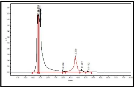
The molecular weight distribution of the cartilage papain hydrolyzed product is shown in Figure 1
(SEC-HPLC chromatograms). Based on the area under cover the molecular weight distribution, more than 97% of
cartilage hydrolysate’ s molecular weight was less than 10.7kDa. We also have observed that all of the
molecular weight of cartilage lysate was less than Ribonuclease A (Mwt. 13700) as analyzed by Superdex
75 (data not shown)]. Additionally, 58% of cartilage hydrolysate has a molecular weight of between 10.7 and
6.2kDa, while 40% of cartilage lysate has a molecular weight of less than 6.2kDa.
Figure 2 shows the heat-hydrolyzed (not enzyme-hydrolyzed) cartilage hydrolysate. More than 83% of
molecules in the heat-hydrolyzed cartilage product appeared to be above the upper separation limit of
SEC-HPLC (before retention time of 20 minutes). The result indicated that the majority of heat-hydrolyzed
cartilage products still retained their high molecular weight structures. As shown in Figure 3, about 14% of
small molecular weight product appeared after a retention time of 42 minutes. Its identity and properties
require further study.
-Cytotoxic and Mitogenic Effects of Cartilage Hydrolysate
Figure 4 shows the cartilage papain-hydrolysate is not cytotoxic to the primary porcine chondrocytes at a
concentration below 20mg/ml and lower. At a concentration of 200mg/ml, it displayed a significantly lowered
viability that could be due to other physical properties or its having very weak toxicity.
Figure 5 shows the cartilage papain-hydrolysate had no mitogenic effects on porcine chondrocyte culture.
117
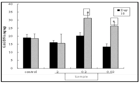
Figure 6 shows the cartilage papain-hydrolysate induced GAG production by the primary porcine
chondrocyte cultures at day 14. Figure 7 shows the treatment of cultured chondrocytes with 0.2 and
0.02mg/ml sample induced a marked increase in type II collagen synthesis. At the end of the culture period
(18 days), type II collagen synthesis was almost 11.9-fold higher in sample stimulated cultures in comparison
with the control cells.
Our cartilage hydrolysate was presented in molecular size smaller than 10kDa (Fig.1, 2 and 3) with the
capability of stimulating chondrocytes to express GAG (Fig. 6) and type II collagen (Fig. 7) , thus fulfilling the
requirement of being dietary peptides. Dietary peptides are known to have biological importance beyond
their nutritive value of intact protein and individual amino acids [12-17, 46]. It is well recognized that apart
from their basic nutritional role many food proteins contain encrypted within their primary structures peptide
sequences capable of modulating specific physiological functions [43-46]. In this study, the cartilage
hydrolysate was prepared by papain hydrolysis and resulted in various sizes of a fragment including peptides
and saccharide-aggregates, with stimulating/enhancing of the anabolic activity of chondrocyte. This bioactive
property was similar to reports of bioactive peptides having been found in enzymatic protein hydrolysates
[12-17]. A U 0.00 0.02 0.04 0.06 0.08 0.10 0.12 0.14 0.16 0.18 0.20 0.22 0.24 0.26 Minutes
5.00 10.00 15.00 20.00 25.00 30.00 35.00 40.00 45.00 50.00 55.00 60.00 65.00 70.00
19 .0 93 19 .3 93 35 .6
88 38.1
28 43 .5 68 47 .8 90 51 .8 31
International Journal of Management, Economics and Social Sciences
118 Figure 2. HPLC Chromatogram of Amersham Standards (80-1129-83)
Figure 3. HPLC Chromatogram of Heat-Hydrolyzed Cartilage Products
119 Figure 5. Effects of the Sample on Chondrocytes Proliferation Chondrocytes were treated with various concentrations of the sample for 2, 7 and 14 days. Chondrocytes viability was determined by MTT assay
Figure 6. Effects of the Sample on GAG Content of Chondrocytes. Chondrocytes were treated with various concentrations of the sample for 10 and 14 days. GAG and DNA content were determined by DMMA assay and
International Journal of Management, Economics and Social Sciences
120 Figure 7. Effects of the Sample on Type Ⅱ Collagen Expression of Chondrocytes. Chondrocytes were treated with three concentrations of cartilage hydrolysate product for indicated durations. *p< 0.05 compared with untreated
controls
Neural Network Assessment and Statistical Analysis
NeuroSolutions 6.0 neural network software (Back-propagation Network) was into the production process to
parallel the orthogonal array in the construction of the prediction model for the 89 experiments. The
biological enzymatically results (target value: supernatant recovery rate) demonstrated good consistency in
the sample duplications and experimental groups, and the artificial intelligence computing convergence value
(R2=0.9762) indicated this enzymatic bioprocessing had good performance (Figure 8-24).
121 Figure 9. Training Report: Papain Hydrolysis of Chicken Sternal Cartilage
Figure10. Testing Trend: Papain Hydrolysis of Chicken Sternal Cartilage
International Journal of Management, Economics and Social Sciences
122 Figure 12. Testing Report: Papain Hydrolysis of Chicken Sternal Cartilage
Figure 13. Training MSE: Papain Hydrolysis of Mini Pig Laryngeal and Tracheal Cartilage
123 Figure 15. Testing Trend: Papain Hydrolysis of Mini Pig Laryngeal and Tracheal Cartilage
Figure 16. Testing Report: Papain Hydrolysis of Mini Pig Laryngeal and Tracheal Cartilage
International Journal of Management, Economics and Social Sciences
124 Figure 18. Training Report: Papain Hydrolysis of Hog Laryngeal and Tracheal Cartilage
Figure 19. Testing Trend: Papain Hydrolysis of Hog Laryngeal and Tracheal Cartilage
125 Figure 21. Training MSE: Papain Hydrolysis of Mixture of Three Animal Cartilages
Figure 22. Training Report: Papain Hydrolysis of Mixture of Three Animal Cartilages
International Journal of Management, Economics and Social Sciences
126 Figure 24. Testing Report: Papain Hydrolysis of Mixture of Three Animal Cartilages
DISCUSSION
Neural Network Performance
NeuroSolutions 6.0 neural network software (Back-propagation Network) was incorporated into the
production process to parallel the Taguchi orthogonal array in the construction of the prediction model for the
89 experiments. The biological enzymatically results were 92.09-93.03% (target value: supernatant recovery
rate) appeared to have good consistency in the sample duplications and experimental groups, and the
artificial intelligence computing convergence value was R2=0.9762, indicating this enzymatic bioprocessing
had good performance. However, the analysis indicated the papain enzyme/substrate ratio as the major
impact factor in this enzymatic hydrolysis to produce high yields of final bioactive peptide products (Figure
8-24).
Optimizing Enzymatic Bioprocessing
In 1999, collagen hydrolysate (prepared from gelatin) was shown to be absorbed in its high-molecular weight
by containing peptides of 2.5kDa to 15kDa [13-18]. Recently, a report showed hydrolyzed chicken sternal
cartilage extract of novel low molecular weight (from 0.05kDa to 10kDa with average 5.5 kDa), indicating an
improved osteoarthritis-related system in a Randomized, Double-Blind, Placebo-Controlled Trail [13-16]. The
molecular size of our cartilage hydrolysate was smaller than 10.7 kDa, which is comparable to the published
127
Significantly, this study provides a simple, easy and economical way of preparing 10 to 100 grams of
cartilage hydrolysate required for further investigations.
Hydrolyzed chicken cartilage powder is a natural source of BSE free type II collagen and chondroitin
sulfate. Extensive research has shown the benefit of these two substances for both healthy and unhealthy
joints. Furthermore, ongoing research and studies continue to uncover their mechanisms of action and to
strengthen the evidence of their efficiency [14-17].
-Hydrolyzed collagen type II having an average molecular weight
Animal cartilage-derived material comprising hydrolyzed collagen type II, said hydrolyzed collagen type II
having an average molecular weight of between about 1,500 and 2,500 Daltons [14-17].
-Comprising hydrolyzed collagen type II having an average molecular weight
A method for treating an individual with a connective tissue disorder, comprising orally administering to said
individual an effective daily amount of chicken sternal cartilage-derived material comprising hydrolyzed
collagen type II having an average molecular weight of between 1,500 and 2,500 Daltons [14-17].
-Intelligence computing technology can optimize enzymatic bioprocessing in animal waste cartilage
It is believed that intelligence computing technology can bring about significant advancement in product
development (as archived pharmaceutical industrial) as well as in future business execution and prediction
(such as Taguchi orthogonal method and artificial neural network) by optimizing enzymatic bioprocessing in
animal waste cartilage (chicken sternal cartilage, mini pig laryngeal and tracheal, and hog laryngeal and
tracheal) and generating hydrolysis conditions for preparation of hydrolyzate collagen. Specifically, the
artificial neural network is used in parallel with the Taguchi orthogonal array process for enzymatic hydrolysis
of the cartilage tissue to produce the best yield and quality of bioactive peptides with clinical efficacy for
functional ingredients of nutraceuticals.
CONCLUSION
This study comprises two parts: one is the biological experiment of papain hydrolysis of animal (chicken, mini
pig and hog pig) cartilage and the other is the neural network computing and analysis in an attempt to locate
a predicting model for the formulation of a better enzymatical production procedure for bioactive-functional
International Journal of Management, Economics and Social Sciences
128
The enzymatic hydrolysate of three animal cartilages was processed following the Taguchi orthogonal
design. It was discovered that a hydrolyzing temperature of 60±1℃ is the most effective to hydrolyze the
cartilage. The chicken sternal cartilage could be hydrolyzed by papain to produce a molecular size smaller
than 10kDa (with 95% in 10.7kDa and 2.5kDa). These peptides were demonstrated to be capable of
stimulating the porcine chondrocytes to produce glycosaminoglycan (GAG) and type Ⅱ collagen in vitro.
Thus, it is concluded that animal cartilage papain-hydrolysate contain bioactive peptides or factors and could
be a good agent for chondrogenesis and regeneration of cartilage tissue. The product of this research can
be applied in nutraceuticals since it has been granted “ GRAS” status by the US FDA.
In the second part of this study, through NeuroSolutions 6.0 analysis, it is concluded that the good
conversion value (R2=0.9762) is an indication that based on this study experiments can perform neural
network analysis. The training MSE-papain hydrolysis of a mixture of three animal cartilages also indicated
the best final MSE =0.00779.
However, the testing input/output data: papain hydrolysis of a mixture of three animal cartilages indicate
the papain enzyme/substrate ratio to be the major impact factor in this enzymatic hydrolysis to produce high
yields of the final bioactive peptide products.
Indeed, the chicken sternal cartilage can be efficiently hydrolyzed by papain to produce a molecular
weight smaller than 10kDa with 95% in 10.7kDa, 6kDa and 2.5kDa fragments or peptide with biological
activity capable of stimulating the primary porcine chondrocyte to express GAG and type II collagen in
culture. The cartilage hydrolysate could be used as ingredients in food supplements and nutraceuticals. Also
importantly, it should be considered as safe as collagen hydrolysate which has been granted GRAS status
by the US FDA in 2003. Finally, the cartilage hydrolysate could also be a biomimetic biomaterial for bone
and connective tissue regeneration studies.
FUTURE DIRECTIONS
This study results suggest that intelligence computing manipulation of bioprocessing engineering could
improve the goods of final bioactive peptide products. Nevertheless, waste animal cartilages can indeed
become the green resources for biotechnology to convert low valued by-products of the meat industry for
129
Bioprocess engineering is a conglomerate of mathematics, biology and industrial design. It also consists
of various biotechnological processes used in industries for large-scale production of biological products for
optimization of yield and quality [48]. It is believed that the intelligence computing technology can bring about
significant advancements in product development (as archived pharmaceutical industrial) as well as in future
business execution and prediction.
REFERENCES
[1]Asanbaeva, A., Masuda, K., Thonar, E. J., Klisch, S. M. and Sah, R. L.. 2008. Osteoarthritis and Cartilage. Cartilage growth and remodeling. Modulation of balance between proteoglycan and collagen network in vitro with β -aminopropionitrile1, 16 (1), 1– 11. [2] Phanat Kittiphattanabawon, Soottawat Benjakul, Wonnop Visessanguan and Fereidoon Shahidi, 2010. LWT - Food Science and
Technology. Isolation and characterization of collagen from the cartilages of brown banded bamboo shark (Chiloscyllium punctatum) and blacktip shark (Carcharhinus limbatus), 43, 792– 800.
[3] Moskowitz, R.. 2000. Seminars in arthritis and rheumatism. Role of collagen hydrolysate in bone and joint disease, 30 (2), 87– 99. [4] Ruiz-Benito, P.; Camacho-Zambrano, M.M., Carrillo-Arcentales, J.N., Mestanza-Peralta, M.A., Vallejo-Flores, C.A., Vargas-Lopez,
S.V., Villacis-Tamayo, R.A. and Zurita-Gavilanes, L.A.. 2009. International journal of food science and nutrition. A randomized controlled trial on the efficacy and safety of a food ingredient, collagen hydrolysate, for improving joint comfort", 12, 1– 15.
[5] Sarah Brien, Phil Prescott, and George Lewith. 2011. Evidence-Based Complementary and Alternative Medicine. Meta-Analysis of the Related Nutritional Supplements Dimethyl Sulfoxide and Methylsulfonylmethane in the Treatment of Osteoarthritis of the Knee, Volume 2011, Article ID 528403, 12 pages.
[6] Alexander G. Schauss, Jerome Stenehjem, Joosang Park, John R. Endres and Amy Clewell. 2012. J. Agric. Food Chem. Effect of the Novel Low Molecular Weight Hydrolyzed Chicken Sternal Cartilage Extract, BioCell Collagen, on Improving Osteoarthritis-Related Symptoms: A Randomized, Double-Blind, Placebo-Controlled Trial, 60 (16), 4096– 4101.
[7] Vittayanont, M. and Jaroenviriyapap, T.. 2014. International Food Research Journal. Production of crude chondroitin sulfate from duck trachea, 21(2), 791-797.
[8] Chong-Tai Kim, Naiyana Gujral, Advaita Ganguly, Joo-Won Suh and Hoon H. Sunwoo. 2014. Biotechnology Reports. Chondroitin sulphate extracted from antler cartilage using high hydrostatic pressure and enzymatic hydrolysis, 4, 14– 20.
[9] Jin-Ho Jo, Duck-Chun Park, Jeong-Ryong Do, Young-Myoung Kim, Dong-Soo Kim, Yong-Kon Park, Taek-Kyun Lee1 and Seung-Mock Cho. 2004. Food Sci. Biotechnol. Optimization of Skate (Raja flavirostris) Cartilage Hydrolysis for the Preparation of Chondroitin Sulfate, 13(5), 622 ~ 626.
[10] Jin-Ho Jo, Jeong-Ryong Do, Young-Moung Kim, Dong-Soo Kim, Taek-Kyun Lee, Seon-Bong Kim, Seung-Mock Cho, Suk-Nam Kang and Douck-Choun Park. 2005. Food science and biotechnology. Optimization of Shark (Squatina oculata) Cartilage Hydrolysis for the Preparation of Chondroitin Sulfate, 14(5), 651-655.
[11] Iwai, K., Hasegawa, T., Taguchi, Y., Morimatsu, F., Sato, K., Nakamura, Y., Higashi, A., Kido, Y., Nakabo, Y. and Ohtsuki, K.. 2005. Journal of Agricultural and Food Chemistry. Identification of food-derived collagen peptides in human blood after oral ingestion of gelatine hydrolysates, 53 (16), 6531– 6536.
[12] Lignot B1, Lahogue V, and Bourseau P.. 2003. J Biotechnol. Enzymatic extraction of chondroitin sulfate from skate cartilage and concentration-desalting by ultrafiltration, Aug 15, 103(3), 281-4.
[13] Anna Gunilla Oberg Sturrock and Dale P. DeVore. 1998. US Patent 5840848 A. Facilitating removal of perichondrial membrane from vertebrate cartilage tissue using acidic solution containing acid protease
[14] Eric V. 2010. US Patent 7671041. Hydrolysate of Avian Cartilage, Process of Preparation and Uses Thereof. [15] Eric V. 2010. European Patent. Hydrolysate of Avian Cartilage, Process of Preparation and Uses Thereof.
[16] Lin TM. 2012. 3 rd International Conference Strategies in Tissue Engineering. Cartilage Hydrolysate-Derived Biomaterials for Chondrogenesis, Wurzburg, Germany, May 23 to 25, 2012.
International Journal of Management, Economics and Social Sciences
130 [18] Ameye LG and Chee W SS. 2006. Arthritis Research & Therapy. Osteoarthritis and nutrition. From nutraceuticals to functional
foods: a systematic review of the scientific evidence, 8:R127, p1-22, DOI: 10.1186/ar
[19] Henrotin Y.2011. Osteoarthritis Cartilage. Nutraceuticals: do they represent a new era in the management of osteoarthritis? - a narrative review from the lessons taken with five products, 19(1), 1-21.
[20] Baby Chauhan, Gopal Kumar, Nazia Kalam, and Shahid H. Ansari. 2013. J Adv Pharm Technol Res.. Current concepts and prospects of herbal nutraceutical: A review, Jan-Mar; 4(1): 4– 8. DOI: 10.4103/2231-4040.107494
[21] Jennifer M. Grossman1, Rebecca Gordon, Veena K. Ranganath, Chad Deal, Liron Caplan, Weiling Chen, Jeffrey R. Curtis, Daniel E. Furst, Maureen McMahon, Nivedita M. Patkar, Elizabeth Volkmann and Kenneth G. Saag. 2009. Rheumatic Disease Clinics of North America. Nutraceuticals as Therapeutic Agents in Osteoarthritis, 25(2), 379-395.
[22] Roy R.K.. 2001. Design of Experiments Using the Taguchi Approach, 16 Steps to Product and Process Improvement.
[23] Lars Enochson, Mats Brittberg and Anders Lindahl. 2012. Biores Open Access. Optimization of a Chondrogenic Medium Through the Use of Factorial Design of Experiments, Dec 2012; 1(6): p.306– 313. DOI: 0.1089/biores.2012.0277
[24] Bourseaua P., Vandanjona, L., Jaouena P., Chaplain-Derouiniota M., Masséa A., Guérardc F., Chabeaudb, A., Fouchereau-Pérond M., Le Gald Y., Ravallec-Plée R., Bergéf J.-P., Picotg L., Piotg J.-M., Batistah I., Thorkelssoni G., Delannoyj C., Jakobsenk G. and Johanssonk I.. 2009. Desalination. Fractionation of fish protein hydrolysates by ultrafiltration and nanofiltration: impact on peptidic populations, August, 244(1-3), 303-320.
[25] Anthony Ratcliffe. 2014. US 8808730 B2. Composite material for tissue repair.
[26] The University of York Department of Mathematics. 2004. Orthogonal Arrays (Taguchi Designs).
[27] Bin Wang, Hong-yu Luo and You-le Qu. 2011. IEEE Xplore, Bioinformatics and Biomedical Engineering, (iCBBE) 2011 5th International Conference. Microwave-Assisted Extraction and Antioxidant Activity of Chondroitin Sulfate from the Cartilage of Dasyatis akajei, 10-12 May, 1-4.
[28] Lyu SR, Wu WT, Hou CC and Hsieh WH. 2010. Cryobiology. Study of cryopreservation of articular chondrocytes using the Taguchi method, Apr, 60(2), p.165-76.DOI: 10.1016/j.cryobiol.2009.10.008. Epub 2009 Oct 24
[29] Nagarjun P A, Rao RS, Rajesham S, Rao LV.. 2005. The Journal of Microbiology. Optimization of Lactic Acid Production in SSF by Lactobacillus amylovorus NRRL B-4542 Using Taguchi Methodology, 38-43.
[30] Prakasham R. S., Rao CS, Rao RS, Rajesham S. 2005. Applied Biochemistry and Biotechnology. Optimization of alkaline protease production by Bacillus sp. using taguchi methodology, 120(2), 133-144, DOI: 10.1385/ABAB:120:2:133.
[31] Pitopech CO., LTD. The qualitek-4 windows software [32] Wikipedia. 2015. "Neural Networks"
[33] Jure Zupan. 1994. Acta Chimica Slovenica. Introduction to Artificial Neural Network (ANN) Methods: What They Are and How to Use Them*, 41(30), 327-352.
[34] Dominique A. Heger. 2011. DH Technologies. An Introduction to Artificial Neural Networks (ANN) - Methods, Abstraction, and Usage.
[35] Lee H. C.. 1994. University of Missouri—Rolla. Skin Cancer Diagnosis Using Hierarchical Neural Networks, 1-103.
[36] Fikret Ercal, Senior Member, IEEE Anurag Chawla, William V. Stoecker, Hsi-Chieh Lee, and Randy H. Moss, Senior Member, IEEE. 1994. IEEE Transactions on Biomedical Engineering. Neural Network Diagnosis of Malignant Melanoma From Color Images, 41(9), 837-845.
[37] http://en.wikipedia.org/wiki/Multilayer_perceptron
[38] Witten IH., Eibe Frank and Mark A. Hall. 2011. Data Mining. Practical machine learning tools and techniques, 3rd Edition, Morgan Kaufmann, San Francisco. http://www.cs.waikato.ac.nz/~ml/weka/book.html.
[39] http://whiteboard.ping.se/uploads/MachineLearning/layers700.png
[40] Dana L. Nettles, Mansoor A. Haider, Ashutosh Chilkoti and Lori A. Setton. 2010. Tissue Engineering Part A. Neural Network Analysis Identifies Scaffold Properties Necessary for In Vitro Chondrogenesis in Elastin-like Polypeptide Biopolymer Scaffolds, 16(1), 11-20. DOI:10.1089/ten.tea.2009.0134.
[41] Nikzad M, Movagharnejad L K, Talebnia F.2012. International Journal of Computer Applications. Comparative Study between Neural Network Model and Mathematical Models for Prediction of Glucose Concentration during Enzymatic Hydrolysis, October 2012, 56(1), (0975 – 8887).
131 [43] Suvarna Mahavir Patil and Mudholkar R.R.. 2012. International Journal of Advances in Engineering & Technology. An osteoarthritis
classifier using backpropagation neural network, Sept 2012, ISSN: 2231-1963.
[44] From Wikipedia, the free encyclopedia. 2006. Damaged cartilage in gross pathological specimen from sows. Bouchard's nodes and Heberden's nodes may form in osteoarthritis. http://en.wikipedia.org/wiki/Osteoarthritis
[45] Beth Israel Deaconess Medical Center (A Teaching Hospital of Harvard Medical School). 2012. Functional Imaging of Cartilage Research.
[46] Sorana D. Bolboacă and Jäntschi L. 2007. Bulletin. Amino acids sequence analysis on collagen, usamv-cn, 63 - 64/2007. [47] Shular, Michael A., kargi, Fikret. 2005. Prentice Bioprocess engineering- Basic concepts, Prentice Hall of India. [48] http://www.neurosolutions.com/resources/whatsnew60.html
[49] Pitopech CO., LTD. NeuroSolutions software 6.0
[50] Aynsley M., Hofland A., Morris A. J., Montague G. A. and Massimo C. Di. 2005. Bioprocess Design and Control Advances in Biochemical Engineering/Biotechnology. Artificial intelligence and the supervision of bioprocesses (real-time knowledge-based systems and neural networks) 48, .1-27.
ACKNOWLEDGMENT
The author wishes to convey her sincere thanks to Dr. Ling-Hui Hsu and the Industrial Technology Research
Institute, Biomedical Technology and Device Research Laboratories. Special thanks too to Dr. Chao-Ying