R E S E A R C H
Open Access
Using low-risk factors to generate
non-integrated human induced pluripotent
stem cells from urine-derived cells
Linli Wang
1*†, Yuehua Chen
1†, Chunyan Guan
1†, Zhiju Zhao
1, Qiang Li
1, Jianguo Yang
1, Jian Mo
1, Bin Wang
1,
Wei Wu
2, Xiaohui Yang
1, Libing Song
3and Jun Li
1,4*Abstract
Background:Because the lack of an induced pluripotent stem cell (iPSC) induction system with optimal safety and efficiency limits the application of these cells, development of such a system is important.
Methods:To create such an induction system, we screened a variety of reprogrammed plasmid combinations and multiple compounds and then verified the system’s feasibility using urine cells from different individuals. We also compared large-scale iPSC chromosomal variations and expression of genes associated with genomic stability between this system and the traditional episomal system using karyotype and quantitative reverse transcription polymerase chain reaction analyses.
Results:We developed a high-efficiency episomal system, the 6F/BM1-4C system, lacking tumorigenic factors for human urine-derived cell (hUC) reprogramming. This system includes six low-risk factors (6F),Oct4,Glis1,Klf4,
Sox2,L-Myc, and the miR-302 cluster. Transfected hUCs were treated with four compounds (4C), inhibitor of
lysine-demethylase1, methyl ethyl ketone, glycogen synthase kinase 3 beta, and histone deacetylase, within a short time period. Comparative analysis revealed significantly decreased chromosomal variation in iPSCs and significantly increasedSirt1expression compared with iPSCs induced using the traditional episomal system. Conclusion:The 6F/BM1-4C system effectively induces reprogramming of urine cells in samples obtained from different individuals. iPSCs induced using the 6F/BM1-4C system are more stable at the cytogenetic level and have potential value for clinical application.
Keywords:Induced pluripotent stem cells, Human urinary cells, 6F/BM1-4C system, iPSC safety
Background
Advancements in induced pluripotent stem cell (iPSC) technology have provided great opportunities for regenera-tive medicine and tumor immunotherapy [1–3]. However, human iPSCs (hiPSCs) are primarily induced using retro-viral or lentiretro-viral vectors carrying reprogramming factors [4, 5], and exogenous DNA fragments can randomly insert into genomic DNA and induce cell transformation, thereby preventing the clinical application of iPSCs. To date, many non-integrating methods have been generated, including
mRNA and protein transfection [6, 7], Sendai virus (SeV) [8], piggyback (PB) transposons [9], and episomal vectors [10]. Nonetheless, mRNA and protein transfections are associated with high preparation costs and low induction efficiency, and a risk of transformation is associated with the retention of SeV RNA in the first passages of iPSC lines [8, 11] and PB transposons. Although episomal induction systems can avoid these problems and are widely used for reprogramming, most of these systems utilize at least one tumorigenic factor such as c-Myc, SV40-LT, and p53 inhibitors, includingp53 RNA interference (RNAi) or small molecule inhibitors [10, 12–17]. As induction efficiency varies when the same method is used to reprogram different types of somatic cells or when different methods are applied to reprogram the same type of somatic
* Correspondence:[email protected];[email protected] †Equal contributors
1Guangzhou Biocare Institute of Cancer, Building D, Guangzhou International
Business Incubator, No. 3, Juquan Road, Guangzhou Science Park, Guangzhou 510663, Guangdong, People’s Republic of China Full list of author information is available at the end of the article
cells [12], it is crucial to optimize the induction method for each type of cell. Human urine-derived cells (hUCs) are ideal donors for iPSC generation: their isolation is simple and nontraumatic. In addition, these cells are easy to ex-pand in vitro and can be used as the main source for iPSCs; thus, their use is cost-effective and universal [18–20]. In the present study, an episomal vector was used for hUC repro-gramming [14, 17].
The nontransformativeMYCfamily proteinL-Myccan be replaced with c-Myc to induce iPSCs [15, 21].Glis1, which is enriched in oocytes, can also replace c-Myc in the classical OSKM (Oct4,Sox2,Klf4, andc-Myc) system; chimeric mice generated from iPSCs induced using OSK andGlis1 have longer survival times than those generated from iPSCs induced by OSKM [22]. Moreover, there is a positive correlation between chimeric mouse mortality and mouse tumor mortality [21], suggesting that Glis1 is a safety factor for iPSC generation. The miR-302 family, which is specifically expressed in embryonic stem cells (ESCs), can partially or completely replace reprogram-ming factors and increase reprogramreprogram-ming efficiency [14, 23, 24]. Furthermore, the miR-302 family activates
Ink4a andArfto suppress the tumorigenesis of human pluripotency stem cells by targeting the oncogeneBmi1 [25], and Arf/p53 pathway activation suppresses somatic cell reprogramming [15, 16]. Therefore, miR-302 s are typ-ically important factors that promote somatic cell repro-gramming, but targeted factors that inhibit reprogramming exist in some signal pathways. Several studies to date have suggested that long noncoding RNAs (lncRNAs) regulate development and tumorigenesis; for example, long intergenic noncoding RNA, regulator of reprogram-ming (lincRNA-ROR) regulates the self-renewal and pluri-potency of human ESCs (hESCs) and the reprogramming of hiPSCs [26, 27]. In this study, we applied hUCs as donor cells to induce iPSCs using low-risk factors, and then we screened a combination of low-risk reprogram-ming factors, includingOct4,Glis1,Klf4,Sox2,L-Myc, and the miR-302 cluster.
To improve non-integrated reprogramming efficiency, we optimized our culture system and observed iPSC induc-tion with high efficiency when four compounds (Parnate, PD0325901, CHIR99021, and sodium butyrate) were added to the medium for no more than 4 days. To analyze iPSC safety, a karyotype analysis was performed, and the results showed that significantly lower iPSC chromo-somal variation was induced when using this system than when using episomal systems containingSV40-LT andc-Myc.
Methods Cell culture
hUCs were collected according to methods reported previ-ously [14, 18]. Briefly, 100–1000 ml of urine was collected
from donors, centrifuged at 1010 ×gfor 5 minutes, and washed with phosphate-buffered saline (PBS). The cells were maintained in 24-well plates coated with 0.1% gel-atin (ES-006-B; Millipore, Germany) in RM1 medium (50% Renal Epithelial Cell Growth Medium (REGM) (CC-3190; Lonza, USA) and 44% Dulbecco's Modified Eagle Medium (DMEM) (SH30022; HyClone, USA) supplemented with 5% fetal bovine serum (FBS) (P30-3302; PAN Biotech, Germany), 0.5% nonessential amino acids (NEAA) (11140050; Gibco, USA), 0.5% GlutaMax (35050-061; Gibco, USA)) and 1 × Primocin (ant-pm-2; InvivoGen, USA); 0.25% trypsin-EDTA (25200072; Gibco, USA) was used for dissociation of primary hUCs. RM1 or RM2 (82% DMEM (SH30022; HyClone, USA) supple-mented with 5% FBS, 1% human keratinocyte growth sup-plement (HKGS) (S-001-5; Gibco, USA), 1% NEAA, and 1% GlutaMax) was used for hUC culture.
The HN4 hESC line was obtained from the Chinese Academy of Sciences, and both HN4 and hiPSCs were maintained in the hESC medium BioCISO (BC-PM0001; BIOCARE Biotech, China) in plates coated with Matrigel (354277; Corning, USA).
Plasmids
pCEP4 (V04450; Invitrogen, USA) was digested using the restriction enzymes NruI and SalI and ligated with synthesized multiple cloning site (MCS) oligonucleotides to obtain the plasmid pE2.1. The EF1αpromoter (NruI,
iPSC generation
For hUC16 reprogramming, 1.5 × 10⁶hUC16 cells were transfected with plasmids (Additional file 3: Table S2) using the T-020 program of a Lonza Nucleofector 2b Device and a Basic Epithelial Cells Nucleofector Kit (VPI-1005; Lonza, USA). Transfected hUC16 cells were seeded into six-well plates coated with 0.1% gelatin and cultured using RM2 medium. After 24 h, the cells were dissociated using 0.25% trypsin-EDTA (25200-072; Gibco, USA), and 2 × 10⁴ cells were seeded into 12-well plates coated with Matrigel. To induce iPSCs, the medium was changed to BioCISO-BM1 medium (BC-BM001; BIOCARE Biotech, China) containing 4i (A83-01 (0.5μM, BC-SMC-A01-10; BIOCARE Biotech, China), Thiazovivin (0.5 μM, BC-SMC-T01-10; BIOCARE Biotech, China), CHIR99021 (3 μM, BC-SMC-C01-10; BIOCARE Biotech, China), and PD03254901 (0.5 μM, BC-SMC-P01-10; BIOCARE Biotech, China)) after 24 h. The medium was then changed to BioCISO on day 15. Alkaline phosphatase (AP) staining was performed on day 18, and the induction efficiency was calculated according to the formula:
Induction efficiency ¼ AP‐positive colony number
=total seeded cell number 100%:
For reprogramming using the 6F/BM1-4C system, ap-proximately 2.8 × 10⁶–3.5 × 10⁶ hUCs were transfected with 4.0 μg pE3.1-OG- -KS and 2.8 μg pE3.1LMyc -hmiR-302 cluster using the same nucleofector method. The transfected hUCs were placed in plates coated with Matrigel and cultured with RM1 medium. On day 3 after nucleofector addition, the medium was changed to BioCISO-BM1 medium containing 2 μM Parnate (also known as tranylcypromine hydrochloride, 1986-47-6; Curegenix, China). The medium was then changed to BioCISO-BM1 medium containing 2μM Parnate, 0.25 mM sodium butyrate (NaB) (303410-100G; Sigma, USA), 3μM CHIR99021, and 0.5 μM PD03254901 on day 5, to BioCISO-BM1 on day 7, and to BioCISO on day 17. iPSC colonies were collected or stained with AP on day 19. The induction efficiency was calculated according to the formula:
Induction efficiency ¼ AP‐positive colony number
=ðnucleofector cell number
–death cell numberÞ 100%:
Compounds used in the present study also included dimethyloxaloylglycine (DMOG) (0.1 μM, D1070; Frontier Scientific, USA), PS48 (5 μM, 1180676-32-7; Curegenix, China), SC-79 (0.5μM, 4635; Tocris, USA), forskolin (5μM, 66575-29-9; Curegenix, China), and 3-deazaneplanocin A (DZNEP) (0.05μM, 4703; Tocris, USA).
For reprogramming using the 4F2L-6C system, 3.0 × 10⁶ hUCs were transfected with 4.0μg pEP4-E02S-ET2K and 2.8μg pCEP4-M2L using the same nucleofector method.
The transfected hUCs were placed in plates coated with Matrigel and cultured with RM1 medium. The medium was changed to BioCISO-BM1 medium containing 2μM Parnate on day 3 after nucleofector addition, and then changed to BioCISO-BM1 medium containing 2 μM Parnate, 0.25 mM NaB, 3 μM CHIR99021, 0.5 μM PD03254901, 0.5μM A83-01, and 0.5μM Thiazovivin on day 5. The medium was changed to BioCISO-BM1 on day 7 and to BioCISO on day 17. iPSC colonies were collected on day 34.
iPSC characterization
they were removed and fixed with 4% paraformaldehyde. The tissues were embedded with paraffin, sectioned, stained with hematoxylin and eosin, and analyzed under a microscope. The procedures were performed according to IACUC (Institutional Animal Care and Use Committee; YS-YFStudy060-20160315).
Karyotype analysis
Sample preparation for karyotyping was conducted as described previously [28]. Cells were treated with 50 ng/ml colchicine (Xy008; Xiangya Gene Technology, China) for 16 h, and the Ikaros karyotyping system was used to analyze karyotypes. The aneuploid evaluation is shown in Additional file 4: Table S3.
Quantitative reverse transcription polymerase chain reaction
Total RNA was isolated using RNAiso Plus (TaKaRa), and M-MLV Reverse Transcriptase (TaKaRa) was used to synthetize cDNA. Specific stem-loop primers and ran-dom primers were used for reverse transcription of micro-RNAs and mmicro-RNAs into cDNA, respectively. mRNA and miRNA expression levels were determined using SYBR Premix Ex Taq™(TaKaRa). Reactions were performed in triplicate using a LightCycler 480II/96 system (Roche, Switzerland). mRNA expression was normalized to GAPDH, and microRNA (miRNA) expression was normalized to U6 small nuclear RNA (snRNA). The primers are presented in Additional file 2: Table S1.
Western blot analysis
Radioimmunoprecipitation assay (RIPA) buffer (CW2333; Cwbiotech, China) supplemented with a protease inhibitor cocktail (PI003; BOCAI Technology, China) and phenyl-methylsulfonyl fluoride (PMSF; Dingguo Changshen Biotech, China) was used to isolate cellular proteins. Equivalent amounts of protein were separated by sodium dodecyl sulfate polyacrylamide gel electrophoresis (SDS-PAGE) and transferred to polyvinylidene difluoride (PVDF) membranes. The membranes were incubated with specific primary antibodies against Oct4 (2840; Cell Signaling Technology, USA), Glis1 (SAB2700289; Sigma, USA), Klf4 (ab72543; Abcam, UK), Sox2 (3579; Cell Signaling Technol-ogy, USA), L-Myc (sc-790; Santa Cruz BiotechnolTechnol-ogy, USA), and GAPDH (KC-5G4; KangChen Biotech, China), followed by horseradish peroxidase-conjugated secondary antibodies: goat Rabbit IgG (ZB-2301; ZsBio, China) and anti-mouse IgG-HRP (IH-0031; Dingguo Changshen Biotech, China). Bands were visualized using enhanced chemilu-minescence (ECL) (34087; Thermo, USA).
Microarray analysis
GeneChip Human Transcriptome Array 2.0 (Affymetrix HTA 2.0, USA) was utilized to determine the gene ex-pression profiles of human ESCs, iPSCs, and hUCs. The
experiments were conducted according to the manufac-turer’s instructions.
Statistical analysis
SPSS 18.0 was used to perform statistical analysis. The results are presented as the mean ± standard deviation (SD) of at least three repeated individual experiments for each group. Statistical differences were examined using Student’s
ttest. For analysis of the chromosome abnormality rate, a four-table chi-square test was applied.P< 0.05 was consid-ered statistically significant.
Accession numbers
Microarray data for human ESs, iPSCs, and hUCs have been submitted to Gene Expression Omnibus (http:// www.ncbi.nlm.nih.gov/geo/) under accession number GSE85885.
Results
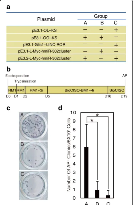
Screening low-risk reprogramming factors using hUCs
Since Yamanaka used OSKM to induce reprogramming, many genes and non-RNAs that improve reprogramming efficiency have been reported. To induce reprogramming in this study, we employed low-risk factors, includingL-Myc,
Glis1, lincRNA-ROR, and the miR-302 cluster, and ran-domly combined them with Oct4, Sox2, and Klf4 in the Epstein–Barr virus-encoded nuclear antigen-1 (EBNA)-oriP episomal vector (Additional file 1: Figure S1a, S1b, S1c). The miR-302 family can increase reprogramming efficiency by replacing reprogramming factors [14, 23, 24]. Further-more, miR-302 family members target the oncogeneBmi1 and suppress tumorigenesis, which can inhibit somatic cell reprogramming [15, 16, 25]. Therefore, miR-302 s have a positive impact on somatic cell reprogramming, but in some pathways these family members can inhibit reprogramming by indirectly activating targets that inhibit reprogramming. Because the expression levels of the miR-302 cluster must be precisely regulated, we used different promoters to exogenously express the miR-302 cluster and screened for optimal expression (Additional file 1: Figure S1a, S1b).
hUCs to ensure reliability. To induce reprogramming, we transfected the three plasmid combinations into hUCs and cultured in hESC basal medium (BioCISO-BM1) with the addition of four small inhibitors that have been widely used for reprogramming [12, 29]: 4i, the TGF-β/Activin/Nodal receptor inhibitor A83-01 (0.5 μM); the MEK inhibitor PD0325901 (0.5 μM); the GSK3β inhibitor CHIR99021 (3 μM); and the ROCK inhibitor Thiazovivin (0.5 μM) (Fig. 1a, b). As revealed by AP staining, the combination termed 6F, which includes Oct4, Glis1, Klf4, Sox2, L-Myc, and the miR-302 cluster initially expressed from the CMV promoter (Fig. 1c, d), showed the highest reprogramming efficiency at 19 days post nucleofection. Thus, we selected 6F, which does not contain high-risk tumorigenic factors such asc-Myc,SV40-LT, and p53 inhibitors, as the repro-gramming induction combination for hUCs and found that it successfully induced hUCs into iPSCs.
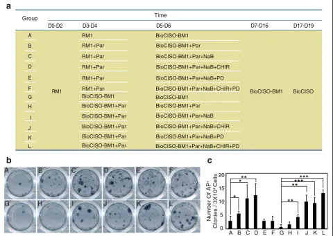
Optimization of compounds in the 6F combination system
Different cell lineages exhibit different gene expression profiles, and ideal reprogramming factor combinations and induction conditions depend on the cell type [12, 30, 31]. To determine whether 4i is the best induction condition for the 6F combination, we first examined the effects of com-pounds that are reported to regulate reprogramming in our system, including many compounds involved in signaling pathways. Some inhibitors or activators were observed to be unsuitable for our system; for example, the TGF-β /Acti-vin/Nodal receptor inhibitor A83-01 and the ROCK inhibi-tor Thiazovivin did not promote programming (Additional file 6: Figure S3a–S3f). In addition, other compounds [31, 32], such as the PDK1 activator PS48, adenylyl cy-clase activator forskolin, histone methyltransferase in-hibitor DZNEP, HIF-αprolyl hydroxylase inhibitor DMOG, and Akt activator SC-79, did not promote or inhibit pro-gramming (Additional file 6: Figure S3g–S3l). Next, we examined the effects of combinations of compounds that promote reprogramming on the reprogramming efficiency. These compounds included the MEK inhibitor PD0325901 (PD, 0.5 μM), the GSK3β inhibitor CHIR99021 (CHIR, 3 μM), the histone deacetylase inhibitor NaB (0.25 mM), and the lysine-specific demethylase 1 inhibitor Parnate (Par, 2μM). We observed that when Par was used in 6F combin-ation reprogramming for an extended time interval, many cells in the cell layer shrank and were floating; however, the reprogramming efficiency increased when Par was added for a short time period (Additional file 6: Figure S3m–S3q). Thus, compound combinations, including Par, were used for no longer than 4 days. AP staining showed the high-est reprogramming efficiency and the bhigh-est iPSC quality when Par was added to the BioCISO-BM1 medium on days 3–4 and when PD, CHIR, NaB, and Par, termed 4C, were added on days 5–6 (Fig. 2a–c). Therefore, we selected BM1-4C as the optimum induction culture condition for the 6F combination induction system and named it 6F/BM1-4C.
Reprogramming hUCs from different human sources using the 6F/BM1-4C system
When hUCs were isolated from different individuals or from the same individual at different time points, the cells exhibited different morphologies when cultured, suggesting that these cells were of different types, which is consistent with previous reports [17, 33–35]. To confirm that the 6F/BM1-4C system is suitable for reprogramming different types of hUCs, we reprogrammed seven groups of hUCs isolated from different individuals (Fig. 3a, b, Table 1). At approximately 19 days post nucleofection, we col-lected iPSCs for further purification and expansion culture (Fig. 3c). The remaining iPSCs were subjected to AP stain-ing, which demonstrated that the 6F/BM1-4C system re-sulted in effective reprogramming of all hUCs into iPSCs. 0
1 2 3 4 5 6 7 8 9 10
A B C
Number Of AP
+ Clonies/5X10 4 Cells
*
*
C B A
Trypsinization
D0 D1 D2 D5 D16 D19
RM1RM1 RM1+3i BioCISO-BM1+4i BioCISO
Electroporation AP
_
+
+
+
_
+
_
+
_
_
+
_
_
+
_
pE3.1-OG--KSPlasmid Group
pE3.2-L-Myc-hmiR-302cluster pE3.1-OL--KS
B C
A
pE3.1-L-Myc-hmiR-302cluster pE3.1-Glis1--LINC-ROR
a
b
c
d
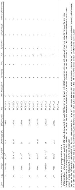
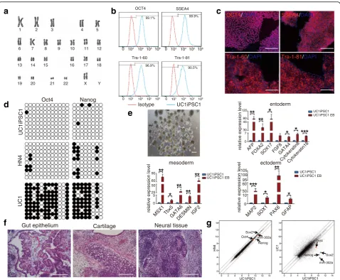
AP staining also revealed an AP-positive rate that varied between 0.00021 and 0.0741% for iPSCs reprogrammed from different types of hUCs (Fig. 3d, Table 1). Further-more, PCR analysis demonstrated a lack of genomic inte-gration of the exogenous gene sequence in 13 of 14 iPSC colonies (Fig. 3e, Additional file 7: Figure S4a). Moreover, karyotype analysis revealed that 13 iPSC colonies had normal chromosome numbers and G-band distributions (Fig. 4a, Additional file 7: Figure S4b). Flow cytometry expression and immunofluorescence analyses revealed that iPSCs induced using the 6F/BM1-4C system express the hESC-specific markers Oct4, SSEA4, 60, and Tra-1-81, suggesting that 6F/BM1-4C-iPSCs possess the molecu-lar characteristics of hESCs (Fig. 4b, c, Additional file 8: Figure S5a, S5b). In addition, bisulfite sequencing PCR ana-lysis indicated that the endogenous pluripotency genes
Oct4 and Nanog were activated and that their promoters were demethylated in 6F/BM1-4C-iPSCs, similar to what occurs in hESCs (Fig. 4d, Additional file 8: Figure S5c).
The generated iPSCs were also able to differentiate into de-rivatives of all three germ layers, as determined using an in-vitro EB differentiation assay and an in-vivo tera-toma formation assay (Fig. 4e, f, Additional file 8: Figure S5d, S5e). The gene expression profile of iPSCs was simi-lar to that of hESCs and differed from that of hUCs, which was determined using Affymetrix gene microarray HTA 2.0. (Fig. 4g, Additional file 8: Figure S5f). The induction efficiencies of the different sources of hUCs as well as the iPSC characteristics are presented in Table 1. Together, these results indicate that the 6F/BM1-4C system has high reliability and versatility for reprogramming hUCs into iPSCs.
The 6F/BM1-4C system is safer than traditional episomal induction systems
We observed that before day 5 following nucleofection, the observed cell masses had high nuclear–cytoplasmic ratios, a phenotype that was similar to the lentiviral
a
b
A B C D E F
L K
J I
H G
c
AB
C
D
E
F G
H
I
J
K L
RM1
D0-D2 D3-D4 D5-D6 D7-D16 D17-D19
RM1
RM1+Par RM1+Par
RM1+Par RM1+Par
RM1+Par
BioCISO-BM1+Par BioCISO-BM1+Par BioCISO-BM1+Par BioCISO-BM1+Par BioCISO-BM1+Par BioCISO-BM1
BioCISO-BM1 BioCISO-BM1+Par
BioCISO-BM1+Par+NaB BioCISO-BM1+Par+NaB+CHIR
BioCISO-BM1+Par+NaB+PD BioCISO-BM1+Par+NaB+CHIR+PD
BioCISO-BM1+Par BioCISO-BM1+Par+NaB BioCISO-BM1+Par+NaB+CHIR BioCISO-BM1+Par+NaB+PD BioCISO-BM1+Par+NaB+CHIR+PD BioCISO-BM1
BioCISO-BM1 BioCISO
Group Time
0 5 10 15 20
A B C D E F G H I J K L
Number Of
AP
+
Clonies / 3X10
4 Cells
*
*
**
**
**
***
***
preprogramming process. From days 9 to 11, the colony mass underwent massive death, and the surviving cells grew slowly to maturity. However, because early induction using the traditional episomal induction system (termed 4F2L-6C), which includes Oct4, Sox2, Klf4, c-Myc,
SV40-LT, and Lin28 [12], is a gradual process, the cells aggre-gated slowly and displayed little massive death (Fig. 5a, Additional file 9: Figure S6a, S6b); these results suggest that the two induction systems differ regarding iPSC gen-eration. Therefore, we examined the safety of iPSCs gener-ated using the 6F/BM1-4C system.
Evaluation of the application of iPSCs is based on safety. Schlaeger et al. [36] analyzed chromosomal variation among
iPSCs induced using retrovirus, mRNA transfection, SeV, episomal, and lentivirus systems and observed large differences using these various methods. For example, the aneuploidy rate of iPSCs induced using the mRNA transfection method was only 2.3%, whereas that of iPSCs induced episomally was as high as 11.5%. How-ever, these methods employ different reprogramming genes, and it is difficult to determine whether differ-ences in chromosomal variation reflect the genes or the methods used for induction. In our study, we uti-lized an episomal vector to reprogram the same batch of hUCs isolated from the same donor using the 6F/ BM1-4C and 4F2L-6C systems, and at least 60 iPSC
UC1 UC2 UC3 UC4 UC5 UC6 UC7
UC1 UC2
UC3 UC4
UC5 UC7
d
e
1 2 3 4 5 6 7 8 9 101112 131415161718
OriP
Oct4 endo L-Myc
Sox2 Klf4 Glis1 EBNA1
(1)
(2)
(3)
Oct4
302cluster
(1)
(2)
(3)
(1)
(2)
(1)
(2)
(1)
(2)
(3)
b
a
D0 D3 D5 D7 D17
RM1 BioCISO-BM1+Par BioCISO-BM1+4C BioCISO-BM1 BioCISO
AP
D19
c
UC2iPSC1 UC3PSC1 UC4iPSC1 UC5iPSC1 UC6iPSC2 UC7iPSC1
UC1iPSC1
Electroporation
Fig. 3Induction of iPSCs from multiple hUCs using the 6F/BM1-4C system.aMorphology of hUCs isolated from seven different donors.bTime schedule of the 6F/BM1-4C reprogramming system.cMorphology of iPSCs induced from seven groups of hUCs using the 6F/BM1-4C system.d AP staining for iPSC generation from multiple hUCs using the 6F/BM1-4C system.eNon-integrating analysis of episomal DNA in iPSCs. Representative lanes: 1, H2O; 2, pE3.1-OG- -KS and pE3.2-L-Myc- -hmiR-302 cluster; 3, UC1; 4, UC1, pE3.1-OG- -KS, and pE3.2-L-Myc- -hmiR-302 cluster; 5, UC1iPSC1; 6,
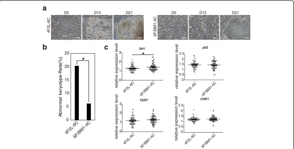
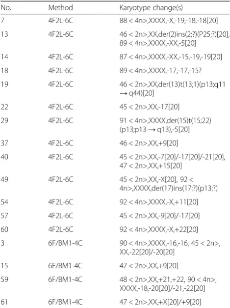
colonies (65 for 6F/BM1-4C and 64 for 4F2L-6C) were se-lected for karyotype analysis (the specific criteria are sum-marized in Methods). We observed a significantly lower chromosomal abnormality rate for 6F/BM1-4C-iPSCs than for 4F2L-6C-iPSCs (P= 0.017,χ2test; Fig. 5b, Table 2). We also determined the expression profiles of genes associated with genomic stability, such as Sirt1, p53, and CHK1, and found significantly high Sirt1expression in iPSCs induced using the 6F/BM1-4C system (Fig. 5c). These data show that iPSCs induced using the 6F/BM1-4C sys-tem are safer than those induced using the traditional episomal induction method, which would be beneficial for clinical applications.
Discussion
Reflecting their high efficiency and controllable cost, episomal plasmid-carried reprogramming factors are the most widely used approach for obtaining non-integrated iPSCs. Most episomal induction methods employ at least one oncogene or tumorigenic molecule, such as c-Myc,
SV40-LT, p53 short hairpin RNA (shRNA), and the p53 small molecule inhibitor cyclin pifithrin-α[10, 12–17], and iPSCs induced using previous methods cannot be used in clinical applications. Furthermore, induction methods that do not include tumorigenic factors are essential. In the present study, we constructed a low-risk 6F/BM1-4C reprogramming system, in which we eliminated the
a
1 2 3 4 5
6 7 8 9 10 11 12
13 14 15 16 17 18
19 20 21 22 X Y
Oct4 Nanog
UC1iPSC1
UC1
HN4
d
Gut epithelium Cartilage Neural tissue
f
c
OCT4/DAPI SSEA4/DAPITra-1-60/DAPI Tra-1-81/DAPI
Oct4 Sox2
miR-302a Nanog
Sox2 Nanog
Oct4
miR-302a
g
b
103 104
0 105106 0 103 104 105 106
103 104
0 105 106 0 103 104 105 106
99.1% 99.9%
96.9% 90.5%
OCT4 SSEA4
Tra-1-60 Tra-1-81
UC1iPSC1 Isotype
e
UC1iPSC1 UC1iPSC1 EB
mesoderm
relative expression level
MSX1 Tbx5GATA6DESMINIGF2 0
9 25 50 60 90
**
**
**
**
*
ectoderm
relative expression level
UC1iPSC1 UC1iPSC1 EB
MAP2 SOX1 PAX6 GFAP
0 10 50 60 95 100
105
**
*
*
entoderm
relative expression level
UC1iPSC1 UC1iPSC1 EB
AFP
FOXA2SOX17FGF8GATA4 Cytokeratin8Cytokeratin18 0
7 30 45 80 120
**
**
*
*
*
* *
*
*
* *
tumorigenic factors used in traditional episomal repro-gramming systems, such as c-Myc, SV40-LT, and p53 in-hibitor, and included Oct4, Glis1, Klf4, Sox2, L-Myc, the miR-302 cluster and four compounds, and then treated cells for no longer than 48 h to efficiently generate iPSCs from hUCs. This system also successfully converted hUCs from different sources into iPSCs and showed good repro-ducibility. Analyzing a large number of iPSCs by karyotype analysis, the 6F/BM1-4C-hiPSCs we generated exhibited fewer chromosome abnormalities compared with trad-itional 4F2L-6C-hiPSCs. In addition, expression of Sirt1, the NAD-dependent deacetylase necessary for maintain-ing iPSC genomic stability [37], in 6F/BM1-4C-iPSCs was high compared with iPSCs induced using the 4F2L-6C system, suggesting that 6F/BM1-4C-iPSC chromosome inheritance is more stable. Moreover, the presented method has a low cost, and the use of episomal plas-mids makes this system suitable for clinical non-integrated iPSC preparation.
To obtain large-scale amounts of clinical-grade iPSCs, a reprogramming method with good reproducibility, non-tumorigenic reprogramming factors, and cost-effectiveness is needed; xeno-free components and a medium for pri-mary somatic cell isolation to iPSC generation are also ne-cessary. Besides, although the 6F/BM1-4C reprogramming system has relatively high reprogramming reproducibility, due to the heterogeneity of hUCs [18, 33, 34], it is difficult to accurately and separately perform multiplication culture
in vitro to obtain a variety of different types of cells that meet the required number of experiments; hUCs from different donors and different batches of cells also show a wide range of induction efficiencies in the 6F/ BM1-4C reprogramming system (Table 1). Therefore, except for a xeno-free induction reprogramming sys-tem, in the future the best reprogramming system should be screened for different types of hUCs or gen-eral suitability for a variety hUCs; in addition, the par-ticular type of hUCs that is more easily reprogrammed or the particular type of hUCs that is more suitable for the 6F/BM1-4C system should be screened to find a more specific cultivation method for a particular type of hUCs, so that we can obtain a high-efficiency repro-gramming system for screening high-quality clinical-grade iPSCs from a large number of iPSCs. Furthermore, when we used the xeno-free hESC E8 medium [38] to induce hUC reprogramming based on episomal vectors, we found it to be unsuitable after the addition of certain compounds, with deformed cells all dying (data not shown). Xeno-free extracellular matrices such as Vitronec-tin exhibit poor maintenance of iPSC self-renewal capacity [38]. Conversely, Laminin521 maintains iPSC self-renewal capacity, but it is extremely expensive [39] and thus is not suitable for large-scale production of clinical-grade iPSCs. Accordingly, the selection of appropriate cell culture materials remains essential for further clinical applications.
c
TERT
0 1 2 3
relative expression level
6F/BM1-4C 4F2L-6C
CHK1
0 1 2 2.5
relative expression level
6F/BM1-4C 4F2L-6C
1.5
0.5
p53
0 1 2 2.5
relative expression level
6F/BM1-4C 4F2L-6C
1.5
0.5 Sirt1
0 1 2 3
relative expression level
6F/BM1-4C 4F2L-6C
*
b
a
D5 D13 D21 D13
6F/BM1-4C
4F2L-6C
D5 D21
0 5 10 15 20 25
Ab
n
o
rm
a
l
k
a
ry
o
ty
p
e
R
a
te(
%
)
6F/BM1-4C 4F2L-6C
*
Conclusion
We developed a safe method based on an episomal vector for inducing iPSCs from hUCs. This method does not in-volve the use of tumorigenic factors, such asc-Myc,
SV40-LT, and p53 inhibitor. Karyotype analysis revealed that the chromosomal variation that occurred during iPSC gener-ation in the present study was significantly low compared with the traditional method. Such low variability is critical for clinical applications of iPSCs.
Additional files
Additional file 1: Figure S1.showing expression of factors from episomal vectors.apE3.1 plasmid construction process chart (upper). Schematic representation of seven constructed episomal vectors. pEF1α EF1αpromoter, pCMV CMV promoter (below).bQuantitative real-time PCR assay forOct4,Glis1,Klf4,Sox2,L-Myc, linc-RoR, miR-367, miR-302a, miR-302b, miR-302c, and miR-302d.cWestern blot assay for Oct4, Glis1, Klf4, Sox2, and L-Myc carried on episomal vectors. GAPDH was used as the loading control. (PDF 171 kb)
Additional file 2: Table S1.Presenting information for primers or functional fragments used in the present study. (XLSX 17 kb)
Additional file 3: Table S2.presenting plasmid combinations used for screening low-risk factors. (XLSX 9 kb)
Additional file 4: Table S3.presenting the method of karyotype analysis. (XLSX 10 kb)
Additional file 5: Figure S2.showing use of hUC16 cells to screen for low-risk factors for iPSC generation.ahUC16 morphology.bStrategy to screen low-risk factor combinations using hUC16 cells.cAP staining for iPSC generation using different factor combinations.dNumbers of AP-positive colonies. (PDF 137 kb)
Additional file 6: Figure S3.showing the effects of multiple compounds in the 6F combination system.aStrategy to optimize six-factor combinations using A83-01.bAP staining for iPSCs induced using A83-01.cNumber of AP-positive colonies induced using A83-01.P(B) = 0.002.dStrategy to optimize six-factor combinations using Thiazovivin (thi).eAP staining of iPSCs induced using thi.fNumber of AP-positive colonies induced using thi.P(D) = 0.000.g Strategy to optimize six-factor combinations using forskolin, PS48, and sc-79.h AP staining for iPSCs induced using forskolin, PS48, and sc-79.iNumber of AP-positive colonies induced using forskolin, PS48, and sc-79.jStrategy to optimize six-factor combinations using DMOG and DZNEP.kAP staining for iPSCs induced using DMOG and DZNEP.lNumber of AP-positive colonies induced using DMOG and DZNEP.P(C) = 0.041.mStrategy to optimize six-factor combination using Parnate in the early induction stage.nAP staining for iPSCs induced using Parnate in the early induction stage. Arrow indicates cell edge hemming.oStrategy to optimize six-factor combination treated with Parnate for a short time.pAP staining for iPSCs induced using Parnate for a short time.qNumber of AP-positive colonies.P(C) = 0.04. Error bars indicate mean ± SD. *P< 0.05, **P< 0.01, ***P< 0.001. Scale bars, 100μm. (PDF 227 kb)
Additional file 7: Figure S4.showing non-integrating analysis and karyotype assays of iPSCs induced with the 6F/BM1-4C system.a Non-integrating analysis of genomic DNA in iPSCs. Representative lanes: 1, H2O; 2, pE3.1-OG- -KS and pE3.2-L-Myc- -hmiR-302 cluster; 3,
UC5; 4, UC5, pE3.1-OG- -KS, and pE3.2-L-Myc- -hmiR-302 cluster; 5, UC5iPSC1; 6, UC5iPSC2; 7, UC6; 8, UC6, pE3.1-OG- -KS, and pE3.2-L-Myc- -hmiR-302 cluster; 9, UC6iPSC1; 10, UC6iPSC2; 11, UC7; 12, UC7, pE3.1-OG- -KS, and pE3.2-L-Myc- -hmiR-302 cluster; 13, UC7iPSC1; 14, UC7iPSC2. OriP in lane 9 exhibited integration.bKaryotype analysis of iPSCs induced from several hUCs. (PDF 340 kb)
Additional file 8: Figure S5.showing pluripotent characterization of iPSCs induced using the 6F/BM1-4C system.aFlow cytometry for expression profiles of the hESC markers OCT4, SSEA4, Tra-1-60, and Tra-1-81.bBisulfite sequencing assay for the methylation status of theOct4andNanogpromoters in iPSCs. Color codes indicate the proportion of methylation.yaxis shows individual CpGs analyzed.xaxis shows different cells.cImmunofluorescence assay for expression profiles of hESC markers.dQuantitative real-time PCR assay for expression profiles of marker genes of the three germ layers.e Hematoxylin and eosin staining of sections of iPSC-generated teratomas.f Scatter plots comparing global gene expression patterns between HN4 hESCs and UC1 iPSCs and between UC2 cells and UC2 iPSCs. Highlighted are the pluripotency factorsOct4,Sox2,Nanog, and miR-302a. Error bars indicate mean ± SD. *P< 0.05, **P< 0.01, ***P< 0.001. Scale bars, 100μm. (PDF 375 kb)
Additional file 9: Figure S6.showing morphology changes during iPSC generation using the 6F/BM1-4C system.aMorphology altered during iPSC generation using the 6F/BM1-4C system.bSchematic of episomal vectors used in the 4F2L-6C system. pEF1αEF1αpromoter, pCMV CMV promoter. Scale bars, 100μm. (PDF 158 kb)
Abbreviations
iPSC:Induced pluripotent stem cell; hUC: Human urine-derived cell; hESC: Human embryonic stem cell; MCS: Multiple cloning site; AP: Alkaline phosphatase; EB: Embryoid body; PD: PD0325901; CHIR: CHIR99021; NaB: Sodium butyrate; Par: Parnate; thi: Thiazovivin; 4C: PD, CHIR, NaB, and Par; 4F2L-6C: episomal-induced system containing six reprogramming factors
(Oct4,Sox2,Klf4,c-Myc,SV40-LT, andLin28) and six compounds (PD, CHIR,
NaB, Par, thi, and A83-01); 6C: PD, CHIR, NaB, Par, thi, and A83-01; 6F/BM1-4C: episomal-induced system containing six reprogramming factors (Oct4,
Glis1,Klf4,Sox2,L-Myc, and miR-302 cluster) and four compounds (PD, CHIR,
Table 2Specific changes in cell karyotypes using the 4F2L-6C and 6F/BM1-4C systems
No. Method Karyotype change(s)
7 4F2L-6C 88 < 4n>,XXXX,-X,-19,-18,-18[20]
13 4F2L-6C 46 < 2n>,XX,der(2)ins(2;?)(P25;?)[20], 89 < 4n>,XXXX,-XX,-5[20]
14 4F2L-6C 87 < 4n>,XXXX,-XX,-15,-19,-19[20]
18 4F2L-6C 89 < 4n>,XXXX,-17,-17,-15?
19 4F2L-6C 46 < 2n>,XX,der(13)t(13;1)(p13;q11 →q44)[20]
22 4F2L-6C 45 < 2n>,XX,-17[20]
29 4F2L-6C 91 < 4n>,XXXX,der(15)t(15;22) (p13;p13→q13),-5[20]
37 4F2L-6C 46 < 2n>,XX,+9[20]
40 4F2L-6C 45 < 2n>,XX,-7[20]/-17[20]/-21[20], 47 < 2n>,XX,+15[20]
49 4F2L-6C 45 < 2n>,XX,-X[20], 92 < 4n>,XXXX,der(17)ins(17;?)(p13;?)
54 4F2L-6C 92 < 4n>,XXXX,-X,+11[20]
57 4F2L-6C 45 < 2n>,XX,-9[20]/-17[20]
60 4F2L-6C 92 < 4n>,XXXX,-X,+22[20]
3 6F/BM1-4C 90 < 4n>,XXXX,-16,-16, 45 < 2n>, XX,-22[20]/-20[20]
15 6F/BM1-4C 47 < 2n>,XX,+9[20]
59 6F/BM1-4C 48 < 2n>,XX,+21,+22, 90 < 4n>, XXXX,-18,-20[20]/-21,-22[20]
61 6F/BM1-4C 47 < 2n>,XX,+X[20]/+9[20]
4F2L-6Cepisomal-induced system containing six reprogramming factors (Oct4,
Sox2,Klf4,c-Myc,SV40-LT, andLin28) and six compounds (PD, CHIR, NaB,
Par, thi, and A83-01),6F/BM1-4Cepisomal-induced system containing six reprogramming factors (Oct4,Glis1,Klf4,Sox2,L-Myc, and miR-302cluster) and four compounds (PD, CHIR, NaB, and Par), [20] 20 metaphases,<2n >abnormal frequency≥3 metaphases,<4n >abnormal frequency≥15%
NaB, and Par); OSKM:Oct4,Sox2,Klf4, andc-Myc; 6F:Oct4,Glis1,Klf4,Sox2, L-Myc, and the miR-302 cluster; 4F2L:Oct4,Sox2,Klf4,c-Myc,SV40-LT, andLin28
Acknowledgements
The authors thank American Journal Experts for English editing.
Funding Not applicable.
Availability of data and materials
Supporting data are available from the corresponding author upon reasonable request.
Authors' contributions
LLW, LBS, and JL conceived and designed the experiments. LLW, YHC, CYG, ZJZ, QL, JGY, JM, BW, WW, and XHY performed the experiments. LLW, YHC, CYG, QL, and JGY analyzed the data. BW, WW, and XHY contributed reagents/ materials/analysis tools. LLW, ZJZ, and JL drafted the paper. All authors read and approved the final manuscript.
Ethics approval and consent to participate
hUCs were obtained from subjects who provided written informed consent for the use of their cells for iPSC generation, and the experiments involving human subjects and animal research were reviewed and approved by the Institutional Review Board at Guangzhou Biocare Institute of Cancer (BioCareIRB201304a#).
Consent for publication Not applicable.
Competing interests
The authors declare that they have no competing interests.
Publisher's Note
Springer Nature remains neutral with regard to jurisdictional claims in published maps and institutional affiliations.
Author details
1Guangzhou Biocare Institute of Cancer, Building D, Guangzhou International
Business Incubator, No. 3, Juquan Road, Guangzhou Science Park, Guangzhou 510663, Guangdong, People’s Republic of China.2The Guangdong Key Lab for Shock and Microcirculation Research, Departments of Pathophysiology, Southern Medical University, Guangzhou 510515, People’s Republic of China.3State Key Laboratory of Oncology in Southern China and Department of Experimental Research, Sun Yat-sen University Cancer Centre, Guangzhou 510060, People’s Republic of China.4Department of Biochemistry, Zhongshan School of Medicine, Sun Yat-sen University, 74 Zhongshan Road II, Yuexiu District, Guangzhou, Guangdong 510080, China.
Received: 22 March 2017 Revised: 21 September 2017 Accepted: 16 October 2017
References
1. Ando M, Nishimura T, Yamazaki S, Yamaguchi T, Kawana-Tachikawa A, Hayama T, Nakauchi Y, Ando J, Ota Y, Takahashi S, Nishimura K, Ohtaka M, Nakanishi M, Miles JJ, Burrows SR, Brenner MK, Nakauchi H. A safeguard system for induced pluripotent stem cell-derived rejuvenated T cell therapy. Stem Cell Rep. 2015;5:597–608.
2. Vizcardo R, Masuda K, Yamada D, Ikawa T, Shimizu K, Fujii S, Koseki H, Kawamoto H. Regeneration of human tumor antigen-specific T cells from iPSCs derived from mature CD8(+) T cells. Cell Stem Cell. 2013;12:31–6. 3. Srivastava D, DeWitt N. In vivo cellular reprogramming: the next generation.
Cell. 2016;166:1386–96.
4. Yu J, Vodyanik MA, Smuga-Otto K, Antosiewicz-Bourget J, Frane JL, Tian S, Nie J, Jonsdottir GA, Ruotti V, Stewart R, Slukvin II, Thomson JA. Induced pluripotent stem cell lines derived from human somatic cells. Science. 2007;318:1917–20.
5. Takahashi K, Tanabe K, Ohnuki M, Narita M, Ichisaka T, Tomoda K, Yamanaka S. Induction of pluripotent stem cells from adult human fibroblasts by defined factors. Cell. 2007;131:861–72.
6. Warren L, Manos PD, Ahfeldt T, Loh YH, Li H, Lau F, Ebina W, Mandal PK, Smith ZD, Meissner A, Daley GQ, Brack AS, Collins JJ, Cowan C, Schlaeger TM, Rossi DJ. Highly efficient reprogramming to pluripotency and directed differentiation of human cells with synthetic modified mRNA. Cell Stem Cell. 2010;7:618–30.
7. Zhou H, Wu S, Joo JY, Zhu S, Han DW, Lin T, Trauger S, Bien G, Yao S, Zhu Y, Siuzdak G, Scholer HR, Duan L, Ding S. Generation of induced pluripotent stem cells using recombinant proteins. Cell Stem Cell. 2009;4:381–4. 8. Fusaki N, Ban H, Nishiyama A, Saeki K, Hasegawa M. Efficient induction of
transgene-free human pluripotent stem cells using a vector based on Sendai virus, an RNA virus that does not integrate into the host genome. Proc Jpn Acad Ser B Phys Biol Sci. 2009;85:348–62.
9. Woltjen K, Michael IP, Mohseni P, Desai R, Mileikovsky M, Hamalainen R, Cowling R, Wang W, Liu P, Gertsenstein M, Kaji K, Sung HK, Nagy A. piggyBac transposition reprograms fibroblasts to induced pluripotent stem cells. Nature. 2009;458:766–70.
10. Yu J, Hu K, Smuga-Otto K, et al. Human induced pluripotent stem cells free of vector and transgene sequences. Science. 2009;324:797-801.
11. Okita K, Ichisaka T, Yamanaka S. Generation of germline-competent induced pluripotent stem cells. Nature. 2007;448:313–7.
12. Yu J, Chau KF, Vodyanik MA, Jiang J, Jiang Y. Efficient feeder-free episomal reprogramming with small molecules. PLoS One. 2011;6:e17557. 13. Valamehr B, Robinson M, Abujarour R, Rezner B, Vranceanu F, Le T, Medcalf
A, Lee TT, Fitch M, Robbins D, Flynn P. Platform for induction and maintenance of transgene-free hiPSCs resembling ground state pluripotent stem cells. Stem Cell Reports. 2014;2:366–81.
14. Xue Y, Cai X, Wang L, Liao B, Zhang H, Shan Y, Chen Q, Zhou T, Li X, Hou J, Chen S, Luo R, Qin D, Pei D, Pan G. Generating a non-integrating human induced pluripotent stem cell bank from urine-derived cells. PLoS One. 2013;8:e70573. 15. Okita K, Matsumura Y, Sato Y, Okada A, Morizane A, Okamoto S, Hong H,
Nakagawa M, Tanabe K, Tezuka K, Shibata T, Kunisada T, Takahashi M, Takahashi J, Saji H, Yamanaka S. A more efficient method to generate integration-free human iPS cells. Nat Methods. 2011;8:409–12.
16. Okita K, Yamakawa T, Matsumura Y, Sato Y, Amano N, Watanabe A, Goshima N, Yamanaka S. An efficient nonviral method to generate integration-free human-induced pluripotent stem cells from cord blood and peripheral blood cells. Stem Cells. 2013;31:458–66.
17. Li D, Wang L, Hou J, Shen Q, Chen Q, Wang X, Du J, Cai X, Shan Y, Zhang T, Zhou T, Shi X, Li Y, Zhang H, Pan G. Optimized approaches for generation of integration-free iPSCs from human urine-derived cells with small molecules and autologous feeder. Stem Cell Rep. 2016;6:717–28.
18. Zhou T, Benda C, Duzinger S, Huang Y, Li X, Li Y, Guo X, Cao G, Chen S, Hao L, Chan YC, Ng KM, Ho JC, Wieser M, Wu J, Redl H, Tse HF, Grillari J, Grillari-Voglauer R, Pei D, Esteban MA. Generation of induced pluripotent stem cells from urine. Clin J Am Soc Nephrol. 2011;22:1221–8.
19. Wang L, Wang L, Huang W, Su H, Xue Y, Su Z, Liao B, Wang H, Bao X, Qin D, He J, Wu W, So KF, Pan G, Pei D. Generation of integration-free neural progenitor cells from cells in human urine. Nat Methods. 2013;10:84–9. 20. Cheng L, Hu W, Qiu B, Zhao J, Yu Y, Guan W, Wang M, Yang W, Pei G.
Generation of neural progenitor cells by chemical cocktails and hypoxia. Cell Res. 2014;24:665–79.
21. Nakagawa M, Takizawa N, Narita M, Ichisaka T, Yamanaka S. Promotion of direct reprogramming by transformation-deficient Myc. Proc Natl Acad Sci U S A. 2010;107:14152–7.
22. Maekawa M, Yamaguchi K, Nakamura T, Shibukawa R, Kodanaka I, Ichisaka T, Kawamura Y, Mochizuki H, Goshima N, Yamanaka S. Direct reprogramming of somatic cells is promoted by maternal transcription factor Glis1. Nature. 2011;474:225–9.
23. Lin SL, Chang DC, Chang-Lin S, Lin CH, Wu DT, Chen DT, Ying SY. Mir-302 reprograms human skin cancer cells into a pluripotent ES-cell-like state. RNA. 2008;14:2115–24.
24. Lin SL, Chang DC, Lin CH, Ying SY, Leu D, Wu DT. Regulation of somatic cell reprogramming through inducible mir-302 expression. Nucleic Acids Res. 2011;39:1054–65.
25. Lin SL, Chang DC, Ying SY, Leu D, Wu DT. MicroRNA miR-302 inhibits the tumorigenecity of human pluripotent stem cells by coordinate suppression of the CDK2 and CDK4/6 cell cycle pathways. Cancer Res. 2010;70:9473–82.
27. Loewer S, Cabili MN, Guttman M, Loh YH, Thomas K, Park IH, Garber M, Curran M, Onder T, Agarwal S, Manos PD, Datta S, Lander ES, Schlaeger TM, Daley GQ, Rinn JL. Large intergenic non-coding RNA-RoR modulates reprogramming of human induced pluripotent stem cells. Nat Genet. 2010;42:1113–7.
28. Meisner LF, Johnson JA. Protocols for cytogenetic studies of human embryonic stem cells. Methods. 2008;45:133–41.
29. Xu Y, Zhu X, Hahm HS, Wei W, Hao E, Hayek A, Ding S. Revealing a core signaling regulatory mechanism for pluripotent stem cell survival and self-renewal by small molecules. Proc Natl Acad Sci U S A. 2010;107:8129–34. 30. Li W, Zhou H, Abujarour R, Zhu S, Young Joo J, Lin T, Hao E, Scholer HR,
Hayek A, Ding S. Generation of human-induced pluripotent stem cells in the absence of exogenous Sox2. Stem Cells. 2009;27:2992–3000. 31. Zhu S, Li W, Zhou H, Wei W, Ambasudhan R, Lin T, Kim J, Zhang K, Ding S.
Reprogramming of human primary somatic cells by OCT4 and chemical compounds. Cell Stem Cell. 2010;7:651–5.
32. Hou P, Li Y, Zhang X, Liu C, Guan J, Li H, Zhao T, Ye J, Yang W, Liu K, Ge J, Xu J, Zhang Q, Zhao Y, Deng H. Pluripotent stem cells induced from mouse somatic cells by small-molecule compounds. Science. 2013;341:651–4. 33. Rahmoune H, Thompson PW, Ward JM, Smith CD, Hong G, Brown J.
Glucose transporters in human renal proximal tubular cells isolated from the urine of patients with non-insulin-dependent diabetes. Diabetes. 2005; 54:3427–34.
34. Dorrenhaus A, Muller JI, Golka K, Jedrusik P, Schulze H, Follmann W. Cultures of exfoliated epithelial cells from different locations of the human urinary tract and the renal tubular system. Arch Toxicol. 2000;74:618–26. 35. Zhou T, Benda C, Dunzinger S, Huang Y, Ho JC, Yang J, Wang Y, Zhang Y,
Zhuang Q, Li Y, Bao X, Tse HF, Grillari J, Grillari-Voglauer R, Pei D, Esteban MA. Generation of human induced pluripotent stem cells from urine samples. Nat Protoc. 2012;7:2080–9.
36. Schlaeger TM, Daheron L, Brickler TR, Entwisle S, Chan K, Cianci A, DeVine A, Ettenger A, Fitzgerald K, Godfrey M, Gupta D, McPherson J, Malwadkar P, Gupta M, Bell B, Doi A, Jung N, Li X, Lynes MS, Brookes E, Cherry AB, Demirbas D, Tsankov AM, Zon LI, Rubin LL, Feinberg AP, Meissner A, Cowan CA, Daley GQ. A comparison of non-integrating reprogramming methods. Nat Biotechnol. 2015;33:58–63.
37. De Bonis ML, Ortega S, Blasco MA. SIRT1 is necessary for proficient telomere elongation and genomic stability of induced pluripotent stem cells. Stem Cell Rep. 2014;2:690–706.
38. Beers J, Gulbranson DR, George N, Siniscalchi LI, Jones J, Thomson JA, Chen G. Passaging and colony expansion of human pluripotent stem cells by enzyme-free dissociation in chemically defined culture conditions. Nat Protoc. 2012;7:2029–40.
39. Rodin S, Antonsson L, Hovatta O, Tryggvason K. Monolayer culturing and cloning of human pluripotent stem cells on laminin-521-based matrices under xeno-free and chemically defined conditions. Nat Protoc. 2014; 9:2354–68.
• We accept pre-submission inquiries
• Our selector tool helps you to find the most relevant journal • We provide round the clock customer support
• Convenient online submission • Thorough peer review
• Inclusion in PubMed and all major indexing services • Maximum visibility for your research
Submit your manuscript at www.biomedcentral.com/submit