*Corresponding author: Md Mesbah Uddin Talukder, Tel: +88 01746637487, Email: [email protected]
©2018 The Authors. This is an Open Access article distributed under the terms of the Creative Commons Attribution (CC BY), which permits unrestricted use, distribution, and reproduction in any medium, as long as the original authors and source are cited. No permission is required from the authors or the publishers.
Adv Pharm Bull, 2018, 8(2), 169-179 doi: 10.15171/apb.2018.021 http://apb.tbzmed.ac.ir
Advanced
Pharmaceutical
Bulletin
Chemical Enhancer: A Simplistic Way to Modulate Barrier Function
of the Stratum Corneum
Tasnuva Haque1, Md Mesbah Uddin Talukder2*
1 Department of Pharmacy, East West University, A/2, Jahurul Islam City Gate, Aftab Nagar Main Rd, Dhaka-1212, Bangladesh. 2 Department of Pharmacy, BRAC University, 66 Bir Uttam AK Khandakar Road, Dhaka 1212, Bangladesh.
Introduction
Since skin is the largest organ of the body, it could be a potential route to deliver drugs into the body. However, barrier property of the outer layer of the skin (stratum corneum, SC) limits the delivery of all types of drug in skin. Topical and transdermal formulations are delivered through the skin, targeting different layers of the skin
and systemic circulation, respectively. Topical
formulation delivers therapeutically effective
concentration of a compound in the specific layer of the skin, to impart a local effect. As for example, sunscreen targets the outer layer of the skin,1 topical analgesic aims
dermal-epidermal layer to reach cutaneous nociceptors,2
topical antifungals to viable epidermis,3 etc. In order to
reach the specific layer of the skin and systemic circulation, a drug molecule must cross the SC and this only possible if barrier property of the skin is overcome. Chemical enhancers (CEs) are chemical agents which modify the SC barrier function and thereby allow molecules to penetrate into the skin. However, the penetration abilities of CEs changes as different CE interact with drug or skin differently. This article aims to summarize the recent findings on some commonly used CEs so that their incorporation into the formulation can develop more effective topical or transdermal or cosmetic products.
Anatomy of skin
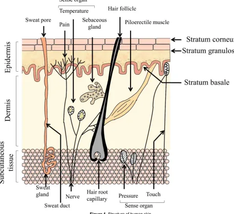
Skin has primarily three layers – epidermis (outer layer), dermis (middle layer) and subcutaneous tissue (bottom layer) (Figure 1).4 Epidermis contains five different cell
strata. From outside to inside, these are stratum corneum (SC), stratum lucidum, stratum granulosum, stratum spinosum and stratum basale. The dermis consists of collagen fibrils and elastic connective tissues.5 This layer
also contains mast cells, macrophages, lymphocytes and
melanocytes.6 Immune and inflammatory responses are
provided by the mast cells.7 Blood vessels, nerves and skin
appendages (sweat and sebaceous glands) are also present in this layer. Because of the structural composition, this layer does not offer the same resistance to drugs as the SC. However, reduced permeation of lipophilic drugs may be observed in this layer.7 In the dermis, there are some
sensory receptors such as thermoreceptors which sense temperature, nociceptors which sense pain and some mechanoreceptors which sense touch and pressure. The mechanoreceptors consist of Messiner’s corpuscles and Pacinian corpuscles which recognize light touch and
pressure, respectively6 (Figure 1). The subcutaneous
tissue is the inner layer containing fat cells interconnected by collagen and elastin fibres. This layer produces and stores large quantities of fat. It also protects the body from mechanical shock and stores large quantities of calories.5,7 Article info
Article History:
Received: 19 February 2018 Revised: 26 May 2018 Accepted: 29 May 2018 ePublished: 19 June 2018
Keywords:
Barrier function Chemical enhancer Drug delivery Modification of skin Stratum corneum
Abstract
Human skin could be a prime target to deliver drugs into the human body as it is the largest organ of human body. However, the main challenge of delivering drug into the skin is the stratum corneum (SC), the outer layer of epidermis, which performs the main barrier function of the skin. Scientists have developed several techniques to overcome the barrier properties of the skin, which include other physical and chemical techniques. The most common and convenient technique is to use special formulation additives (chemical enhancers, CEs) which either drags the drug molecule along with it or make changes in the SC structure, thereby allowing the drug molecule to penetrate in to the SC. The main focus is to deliver drugs in the certain layers of the skin (for topical delivery) or ensuring proper percutaneous absorption (for transdermal delivery). However, skin drug delivery is still very challenging as different CEs act in different ways on the skin and they have different types of interaction with different drugs. Therefore, proper understanding on the mechanism of action of CE is mandatory. In this article, the effect of several CEs on skin has been reviewed based on the published articles. The main aim is to compile the recent knowledge on skin-CE interaction in order to design a topical and transdermal formulation efficiently. A properly designed formulation would help the drug either to deposit into the target layer or to cross the barrier membrane to reach the systemic circulation.
Haque et al.
There are several appendages present in the dermis and epidermis of human skin, such as eccrine and apocrine
sweat glands, sebaceous glands, hair follicles and nails (Figure 1).
Figure 1. Structure of human skin
Stratum corneum (SC), the main barrier of the skin
The SC is the outermost layer of epidermis having a heterogeneous structure and composed of 70 to 80% protein (keratin) and lipid.7 It provides the main barrier
function of the skin. The SC is composed of 10 to 15 layers of compressed corneocytes present in the SC.5,8
Between the SC corneocytes different types of lipids are present.9 If the SC is picturized as a brick wall, the
corneocytes are the ‘bricks’ present in a ‘mortar’ (or
intercellular lipid matrix).10 Desmosomes are the
connectors between the corneocytes. The corneocyte is surrounded by a protein-lipid polymeric envelope.7 The
corneocytes are rigid because of the envelope.11 The
intercellular space between corneocytes is filled with multiple lipid lamellae. The lamellae consist of ceramides, cholesterol, cholesterol esters, cholesterol sulphate and free fatty acids.7,12 The lipid lamellae are arranged
horizontally to the surface of the corneocytes.13,14
Intercellular lipids act as shields to prevent water loss from the body.15 If the lipid from the SC is extracted, the
lipid from the SC enhanced the water loss faster compared
with non-extracted skin.16 Thus, intercellular lipid
lamellae are very important for the barrier function of the
SC and also help in cohesion between corneocytes.17 The
SC also contains approximately 15 to 20% water mainly associated with keratin7 and a small amount in the polar
head group of the intercellular space.18 The lower water
loss and higher barrier function of the SC is because of the unique composition, especially due to the intercellular
lipids and corneocyte envelope.18,19 The epidermis
| 171 Chemical enhancer to modulate the stratum corneum
Advanced Pharmaceutical Bulletin, 2018, 8(2), 169-179 Routes of permeation in the SC
Diffusion is the principle mechanism by which the
permeation of a permeant across human skin takes place.20
A solute can diffuse through the skin by three main routes: the transappendageal route, the intracellular and intercellular route (Figure 2). Permeation through the transappendageal route is known as the permeation via the hair follicles, sebaceous and sweat glands. Appendageal transport provides an easier path of diffusion in parallel to the transepidermal route (intra- and intercellular routes). However, the skin appendages have very low surface area (only 0.1% of the total skin surface area).21 In addition,
permeation of drugs is not direct in the sweat and sebaceous glands. Sweat moves in the reverse direction of the permeant in sweat gland. Moreover, permeation of only hydrophilic molecules is not possible in sebaceous
glands as it has lipid-rich sebum.22 However, the
transappendageal route can be vital for ions and large polar molecules which do not freely cross the SC.23-25
Figure 2. Routes of penetration of a molecule across the SC
Transepidermal pathway is the route which includes intra- and intercellular permeation. If the SC structure is considered as ‘brick and mortar’, the intracellular is the shortest route through the layers of corneocytes and its surrounding intercellular lipid matrix. When such penetration takes place in a tortuous way via the intercellular lipid matrix, it is called the intercellular route. At first it was believed that hydrophilic drugs preferentially diffuse through the intracellular region and hydrophobic drugs through the intercellular matrix21. In
both cases, the compound has to penetrate the intercellular lipid. However, later it was found that the intercellular route was the predominant pathway for permeation of most drugs through the human SC.26-29 Diffusion of a
penetrant through the intracellular route requires undergoing via a series of partitioning and diffusion stages in and out of the relatively hydrophilic corneocytes, lipid
envelope surrounding the corneocytes and the
intercellular matrix. Whereas, the penetrant needs to take a tortuous route consisting of alternating structures of bilayers (containing both aqueous and lipid domains) in the intercellular route. In this route of permeation, a
penetrant passes through a 50-times longer path length compared with the total thickness of the SC. A penetrant also has to undergo sequential partition and diffusion through the aqueous and lipid domains of the intercellular matrix.22
Permeation of drug molecule across the skin
The percutaneous absorption of a solute involves a series of transport processes which is mainly determined by the solubility and diffusivity of the solute. The solubility of a solute in a solvent is determined by the solvent-solute interaction. Firstly, the solute requires to be solubilised into the outermost lipid layer of SC and then diffuses through it. These processes are affected by the CE-skin and solute-skin interactions. During this process the solute may also permeate into the corneocytes. In the next stage, again a series of partition followed by diffusion takes place in the viable epidermis and in the papillary dermis. Absorption of solute by the capillary plexus followed by distribution into the systemic circulation occurs in the papillary dermis. Being the prominent pathway, hydrophilic molecules permeate through the polar head groups and hydrophobic molecules permeate via the lipid chains of the bilayer regions of the intercellular route.30
Drug-CE-skin interactions
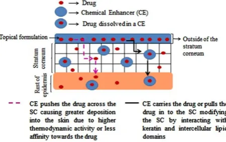
After applying a topical formulation on the surface of the
skin, drug - CE, CE -skin and drug-skin interactions30 may
occur. Drug-CE interactions have effects on the rate and extent of release of drug from the solvent. CE-skin interactions either increase or decrease penetration of a
drug across the skin.31 Drug-CE interaction may be
explained by the solubility parameter. Higher drug-solvent interaction (or higher solubility of the drug in the solvent) will be evident if the solubility parameter difference between these two is low.30 However, if the
drug molecule has a higher affinity for the CE it may remain preferentially in the CE and low permeation of the drug will be observed.20 Solvent-skin interaction will be
discussed in section 3 of this article. Drug-skin interaction is mainly affected by the physicochemical properties (molecular weight, log P, melting point and solubility parameter) of the drug. Drug-solvent-skin interactions may be explained by the ‘push-pull’ effect (Figure 3).32
Haque et al.
‘Push’ effects are of two types. If the solubility parameter difference between drug and CE is high, attraction of drug will be lower towards the CE and the drug will be easily escaped into the skin from the CE.30,32 Drugs having
higher affinity for the CE, it will be held firmly by the CE and will not allow to penetrate through the
SC.20Additionally, by increasing the thermodynamic
activity, the drug will be pushed into the SC by the CE.20,32,33 The ‘pull’ effect explains that CEs change the
SC by structural transformation and therefore, increase the solubility of the drug into the SC or drag the drug molecule while diffusing through the skin.32,33
Modification of the SC to enhance drug penetration
There are physical and chemical methods to enhance the penetration of a drug in the skin. Physical enhancers involve iontophoresis, sonophoresis, phonophoresis,
magnetophoresis, electroporation, thermophoresis,
radiofrequency, needleless injection, microneedle etc. Both techniques involve alter the SC in such a way so that drug can penetrate the SC and reach the target site. Here, the effect of different classes of CE on skin will be discussed elaborately.
Chemical enhancers (CEs)
‘CEs are pharmacologically inactive compounds which partition and diffuse into the skin and interact with SC
components’. They are generally regarded as safe.34 CEs
increase the permeation of drugs by interacting with the intracellular route, interacting in the intercellular route and by modifying the solubility or partition of the SC. In the intercellular route, the solute can interact with the polar head group,in the aqueous regions of intercellular bilayers and interacting in the lipid regions of intercellular bilayers. The skin penetration abilities of selected CEs are discussed below:
Water
Water is the most common and safe penetration enhancer which is used for transporting both drug and cosmetic materials into the skin. Hydrating the skin or using moisturisers can be easiest way to deliver hydrophilic molecules effectively. The water content of the SC is usually 5 to 10%, which can be increased up to 50% under
occlusive condition.35 In 1987, Barry reported that water
molecule acts in both inter and intra-cellular pathways to enhance the permeation of both hydrophilic and lipophilic drugs.36 In case of intracellular region, in dry condition,
the SC provides significant barrier to drug molecules because of the presence of several hydrogen bonding group. Since the SC becomes hydrated, the proteinaceous region takes up water. The arrangement of protein of that region becomes disordered and water starts competing for the hydrogen binding sites on the protein, and therefore, reduces the interaction between them. In this way, permeation of molecule through the intracellular pathway increases.36,37 Barry also stated that water molecule binds
with the polar head group and forms a small hydration shell in the lipid bilayer region via hydrogen bonding.
This leads to loosening of lipid packing and extending hydrophilic domain.36 However, later studies found that
water does not cause a massive lipid disorder,38 it may
cause slight disordering of a small population of the SC lipid.39 Water also found not to swell lipid bilayer but can
be present in very small quantities in the polar head group
of the lipid bilayer region.18 The excess amount of water
the SC absorbs may be present in the corneocytes (intracellular region) or may be present as a separate phase in the intercellular region.18,40
Alcohols
In short chain alcohols, ethanol is the most widely used and studied CE for skin drug delivery in topical and transdermal formulations. Ethanol is also used in such formulations to aid solubility of poorly water soluble drugs or as a cosolvent.41 At low concentration, ethanol
displaces the bound water in the polar head group and disrupts the lipid-polar head/membrane interfacial region. This leads to increase in the interfacial area. At high concentration, ethanol extract lipid and proteins from the SC and thus forms pores in the SC0.18,42 Ethanol helps
penetrating the drug by increasing the solubility in the formulation and by altering the solubility parameter of the SC towards the drug. The residence time of ethanol on skin is short due to its volatile nature.43 Therefore, higher
thermodynamic activity of the drug dissolved in ethanol pushes the drug molecule into or across the SC. In addition, ethanol rapidly penetrates the SC by the mechanisms stated above, which also pulls the drug
molecule along with it through the SC.42 Recently,
Moghadam et al. reported no change in short and long lamellar spacing of the SC structure by ethanol. The authors suggested that the penetration enhancing property of ethanol might be due to the solvent’s ability to
solubilise drug molecule into the SC.44
Fatty or long chain alcohol showed a parabolic relationship with the permeation of melatonin with the carbon chain length of saturated fatty alcohol. Melatonin permeation was found to increase up to chain length of 10 carbons (decanol) and then decreases. Decanol caused
highest permeation enhancement for melatonin.45 Lipid
extraction was the mechanism of enhancing the permeation of drug for D-hexanol and D-octanol. However, D-decanol was not found to disrupt the lipid content.46
Amides Azone
Azone (Laurocapram) is the first compound which was specially developed as a penetration enhancer.35 Azone
contains a seven membered polar head group attached with its 12 carbon chain.43 It mainly reduces diffusional
resistance of a drug into the SC.43 Because of this
structure, it is inserted into the lipid bilayer region with the seven-membered polar group in the polar plane and the dodecyl chain in the lipid region. In this way, it disrupts the highly ordered lipid packing of the lipid
| 173 Chemical enhancer to modulate the stratum corneum
Advanced Pharmaceutical Bulletin, 2018, 8(2), 169-179
of hydrophilic, hydrophobic and some peptides. This CE is effective at low concentration (1 to 5%).35 Azone
imparts its penetration enhancing property more efficiently in conjunction with propylene glycol (PG) rather than alone. Azone only modifies the intercellular region; however, PG acts in the intracellular pathway. Therefore, combination of these two CEs efficiently
delivered a number of drug molecule.36 Although Azone
has been investigated as a CE for over 25 years,35 still it is
not used in any commercial formulation.43
Esters
Alkyl and benzoic acid esters
Ethyl acetate has been found to increase permeation of levonorgestrel. However, its exact mechanism of action is
not confirmed yet.43 Another ester compound, octyl
salicylate (OS) is used as a sunscreen at a concentration up to 5%. OSAL is also found to enhance the penetration of fentanyl and testosterone.43,47,48 Being a lipophilic
solvent, OS previously found to alter the highly ordered lipid bilayer region of the SC converting the gel phase of the lipid lamellae to a liquid phase. However, recent studies did not reveal any lipid distortion result in the SC.49,50 It has been postulated that OS remains in the lipid
gel phase as a ‘pool’ rather than interacting with the lipids
and therefore enhance the diffusivity of the compound.43
Fatty acid esters
This group includes isopropyl myristate (IPM),
propylene glycol monocaprylate (PGMC),
Propyleneglycolmonolaurate (PGML).
IPM
IPM is the most widely investigated fatty acid ester. IPM found to impart fluidisation and hampers the order of lipid lamellae. However, later it was reported that IPM inserted into lipophilic region anchoring its isopropyl group in the polar region and hence interact with the lipid region.51 In
a recent study it has been reported that because of its branched structure and highly mobile terminal isopropyl group, IPM did not mix with other SC lipid. This is why IPM perturbed and disordered the assembly of lipid
lamellae.52 IPM was also found to cause phase segregation
and lipid extraction from the SC.52 In a very recent study,
neat IPM was found to be present in higher quantities in the skin and therefore, aided higher retention of
anthramycin in the skin rather than permeation.53
PGMC is the fatty acid ester which showed enhanced drug
permeation alone54-57sometimes and mostly in
combination with diethyleneglycolmonoethyl ether
(Transcutol®, TC).58,59 However, it was found to deposit
inside the SC, retaining higher quantities of drug.60
Recently, Haque et al. sowed that similar to IPM, PGMC
retains in the skin in higher quantities which also helps to retain higher quantities of drug dissolve in it.53 Takahashi
et al. found that PGMC reduces the resistance to drug
diffusion across the skin by interfering the SC lipid packing. Whereas, no extraction of lipid was evident.60
Moghimipur et al. found contradicting results on
mechanism of action of PGMC. Fourier transform infrared spectroscopy (FTIR) studies showed SC lipid extraction or fluidisation by PGMC. On the other hand, Differential Scanning Calorimetry (DSC) indicated bilayer cohesion by PGMC. Because of the opposite effects, authors concluded that penetration enhancement
effect of PGMC was low.55 Moghimipur et al. also
reported that PGMC interacted mostly with the SC keratins and modifies the skin lipid.55
PGML
PGML is recently used to delivery drug percutaneously in transdermal formulation. Like PGMC, PGML found to increase drug penetration.59,61 Haque et al. showed that
when anthramycin was applied on human skin in pure PGML, retention of small amount of PGML in the skin enhanced the drug permeation significantly.53 However,
much enhanced permeation was observed along with hydrophilic enhancers, such as propylene glycol (PG) and TC.62-65 Parisi et al. showed that combining PGML with
PG in 50:50 ratio increased the skin retention of hexamidinediisethionate in significant quantities.66 The
mechanism of action of the PGML is not clearly stated in the literature. However, it may work in the similar way as PGMC.
Ether alcohol Transcutol® (TC)
TC is a hydrophilic CE with the similar solubility parameter as the skin [10.62 (cal/cm3)1/2].67 TC is used as
a penetration enhancer in both topical and transdermal formulations. TC has been found to increase the flux and retention of drugs.33,68-71 Recently, Haque et al. showed
that TC as a solvent penetrated and retained in the human skin in highest quantities compared with other selected hydrophilic solvents. Therefore, a moderate amount of drug was permeated and accumulated in the skin with the help of TC. TC clearly showed ‘pull’ effect aiding higher absorption of drug molecule.53 The main mechanism of
this solvent to enhance permeation is to increase the partition parameter of the drug into the skin. This may be because of the close solubility parameter of TC with skin. TC has been reported to present inside the SC as intraceutaneous depot. TC being a hydrophilic molecule, is inserted into the aqueous region between the polar head group and induce swelling of the bilayer region without altering the bilayer structure. Therefore, the swollen lipids hold the drugs soluble in the SC. In this way, TC aids to accumulate drugs in the SC (Pull effect).68,72,73 Due to its
hydrophilicity and hydrophobicity, it was suggested to interact with the intracellular lipids of the other layers of epidermis and dermis. The barrier function of the SC was
not altered by TC.72 However, recently Moghadam et al.
suggested that slight disorder in the lamellar structure of
the SC caused by TC, which leads to membrane fluidity.44
Caon et al. showed that skin permeation of ioniaside in TC
Haque et al.
experiments with rat skin after the permeation study. DSC analysis showed that skin lipid disruption was not caused by TC but slight membrane fluidisation. FTR analysis also confirmed that TC increased the order of both lipid and
protein domains of the skin.74
Fatty acids Oleic acid (OA)
OA is an unsaturated lipophilic C fatty acid that is commonly associated with enhanced penetration of polar to fairly polar molecules.75 OA has been used in both
topical and transdermal formulations due to its desirable properties. OA causes temporary and reversible disruption of the SC lipids, increasing fluidisation and diffusivity of the skin. This theory was reinforced in a study using spectrometric and calorimetric measurements which showed that OA increased lipid fluidity and permeant flux in porcine skin.76 Due to the bent structure of OA, it
disrupts the ordered orientation of lipid region and increase the fluidity. The mechanism of action of OA is similar to Azone. However, OA did not found to disturb the structure very drastically.36 More specifically, the
kinked nature of OA (bent cis configuration) has mainly
been associated with the separation of SC lipid regions, which reduces barrier function of the SC. However, despite the advantages of OA, dermal side effects of
unsaturated fatty acids have been reported.77 These can be
overcome by modification of the carboxylic terminal, which reduces the acidic nature of the fatty acid allowing safe use.78
The existence of OA as a separate phase within SC lipids was revealed in a study conducted on porcine SC using Fourier Transform Infrared spectroscopy (FT-IR). Results showed that OA interacted with the SC lipids by reducing the lipid transition temperature (Tm), and by increasing the
“conformational freedom of lipid alkyl chains” higher
than their Tm. OA may cause some sort of permeable
defect within the SC that enhances diffusion of permeants.
This leads to increased permeability75 and diffusion
coefficient in Fick’s law. More recently, a study was conducted using urea, caffeine and diclofenac sodium, in the presence of OA as a CE. In that study OA showed a significant effect on the SC, especially in the model membrane with the higher ratio of phytosphingosine-based ceramide.79 Recently, Atef et al. showed a time
dependent enhancement of OA permeation in rat skin in
terms of spectral change in Raman Spectroscopy.80
Glycols
Propylene glycols (PG)
PG has been used as a cosolvent in topical and transdermal products since long. It is a well-established topical CE. It acts as a CE not only by on its own but also in combination with a number of other CEs. PG mainly increases drug permeation by improving the partition properties of drugs in to the SC. It solvates the α-keratin and therefore reduce drug-tissue binding.36 Bouwstra et al. conducted
Small-angle X-ray scattering (SAXS) and differential thermal analysis (DTA) of the SC after pre-treating with PG. DTA
results showed that PG interacted with the SC lipid. On the other hand, SAXS showed that SC lipids were unaffected by PG. Due to these two contradicting
findings, Bouwstra et al. concluded that PG being a
hydrophilic molecule, incorporated into the polar head group of the lipid bilayer. Therefore, it increased mean interfacial area per lipid molecule. PG also induced lateral swelling (side by side). In order to compensate the lateral swelling (to maintain the density of the alkyl chain region), the chain length of the lipids were decreased. Therefore, no change in the SAXS was observed after
pre-treatment with PG.81 Recently, Moghadam et al. and
Furuishi et al. conducted several experiments on PG and
found no significant skin lipid alteration by PG.44,82
Therefore, Moghadam et al. suggested improvement of
skin partitioning is the main mechanism of PG to enhance
permeation.44 In addition, Mohammed et al. showed that
PG increased transepidermal water loss (TWEL) and KLK 7 protease activity in the skin, therefore reduce the barrier
property of the SC.83 PG was shown to be less penetrating
molecule to the human skin compared with TC. In case of hydrophilic molecule, it imparts its skin penetration effects by both ‘push’ and ‘pull’ effects.53 Atef et al.
showed that in Raman Spectrum, after applying PG on rat skin, the intensity of PG peak (at 840 cm-1) decreased with time in comparison with the skin peaks. The authors concluded that the decrease in PG peak intensity is the indication of increased diffusion of PG in the skin.80
Pyrrolidones
N-methyl-2-pyrrolidone (NMP) and 2-pyrrolidone (2P)
NMP and 2P are the pyrrolidones which have been investigated mostly for years.42 Both of the CEs are
dissolved in water in all proportions. The CEs found to enhance the permeation of both hydrophilic and lipophilic
compounds.42 DSC studies conducted by Barry showed
that these two CEs partition in the corneocyte region (intracellular region) at low concentration and affect the intercellular region at high concentration. These molecules produces a solvation shell around the polar head group, loosens the tight packing of the lipid bilayer and induce lipid fluidity.36 However, Trommer et al.
suggested that comparatively hydrophilic pyrrolidones work by acting on the polar region and hydrophobic pyrrolidones (such as NMP) work on the lipophilic
region.35 However NMP was found to cause erythema,
swelling, skin irritation, thickness of skin etc.84 In
addition, the clinical use of these molecules was restricted due to its skin cytotoxic properties.43
Sulphoxides
Dimethyl sulphoxides (DMSO)
| 175 Chemical enhancer to modulate the stratum corneum
Advanced Pharmaceutical Bulletin, 2018, 8(2), 169-179
interfering with hydrogen bonding and hydrophobic interactions). Therefore, drug/compounds get sufficient loose or flexible areas to penetrate through the SC. However, skin’s impermeable characteristics restores immediately after removing DMSO as the solvent passes out the skin very quickly and gradual removal of protein-DMSO by competitive bonding with cellular water.36 In
the intercellular region, at higher concentration (above 60%), DMSO produces a large solvation shell around the lipid polar head group by displacing water from the polar group. Hence, it loosens the lipid packing more severely compared with water. This leads to increase the aqueous region in the intercellular pathway and helps to promote the permeation of hydrophilic compounds.36 In a recent
study, DMSO was found to change the highly ordered gel phase of Ceremide 2 into loosely packed liquid crystalline phase at greater than 0.4 mole fraction concentration. It has been also shown that DMSO replaced the water from the interface at higher concentration. It also induced lateral swelling by increasing area per lipid in the lipid bilayer region.85
Surfactants
Topical and transdermal formulation contains surfactants in order to increase the solubility of a compound in the formulation. Sodium lauryl or dodecyl sulphate (SLS) (anionic surfactant). Anionic surfactants, such as SLS acts on the skin by affecting both intra- and intercellular pathways.36 For this reason, irritation and skin damage are
very common with this type of surfactant.35,43 SLS swells
the SC and the swollen keratin can absorb more water and help to penetrate drug molecule. Additionally, SLS also unfolds and extends the alpha keratin and opens up the
polar pathway for permeating the drug molecule.36 A
recent study revealed that SLS interacts with lipids and is incorporated there to create a lamellar structure.44
The cationic surfactants include amines,
alkylimidazolines, alkoxylated amines, and quaternary ammonium compounds (or Quats). Cationic surfactants disorders SC lipid organisation more drastically than anionic and non-ionic surfactant. It mainly affects the
lateral packing of the SC lipid.44 Therefore, molecules of
this group are more effective as chemical enhancers than anionic and non-ionic ones.35 However, as this group
cause skin irritation, further use in dermal formulation is
not encouraged.43
The primary non-ionic surfactants used for cosmetics include alcohols (cetyl or stearyl alcohol), alkanolamides, esters, and amine oxides. Non-ionic surfactants disordered the SC less radically compared with ionic surfactants.44
This is why, these surfactants are less irritating to the skin and comparatively less effective as penetration enhancer than the other types and therefore, regarded as safe.42
Terpenes
Terpenes are found in essential oils.42 These are lipophilic
compounds which mainly act on the lipid pathway of the SC. Since it produces less skin irritation, it is regarded as
safe.35 Smaller terpenes are found to enhance drug
permeation more effectively than larger terpenes. Furthermore, polar terpenes (menthol, 1,8-cineole, etc.) improve the penetration of hydrophilic and non-polar terpenes (D-limolene) improve the penetration of hydrophobic molecules in the SC. D-limolene, 1,8 cineole hampers the arrangement of the SC lipid. On the other hand, nerolidol, a sesquiterpene strengthens the lipid
bilayer may be by incorporating into the bilayer.42
However, Moghadam et al. investigated different terpenes
and terpenoids and found that nerol disrupted the lipid lamellae more drastically than other types of molecules. The hydroxyl group and the alkene of the molecule may donate hydrogen bond and disrupt the interaction between the existing hydrogen bond between the ceramide groups of the lipid bilayer.
Phospholipids
The use of phospholipids as penetrations enhancer has been widely studied as a vesicle or liposomes. However, there are fewer studies of this molecule as non-vesicular
chemical enhancers. Because of its lipophilic
characteristics, it occludes the SC and increase the hydration of the skin. Therefore, penetration of a drug molecule is enhanced. As a vesicle it is incorporated or fused into the SC lipid and liberates the molecule in the solvent in which it was poorly soluble. Thus thermodynamic activity as well as drug permeation were increased.42
Cyclodextrines
Cyclodextrins are not typical enhancers as they cannot penetrate the SC. They form inclusion complexes of hydrophobic drugs and improve their aqueous solubility.
However, several studies showed penetration
enhancement of certain drugs when cyclodextrin was used in combination with lipophilic enhancers (fatty acids, azone etc.).35
Discussion
The various sites of action of different types of CE are as follows:
The intracellular route is the polar route where keratin fibrils are present. Aprotic solvents [for example, dimethyl sulfoxide (DMSO)], pyrrolidone, surfactants interact with the keratin and may disrupt the ordered arrangement in corneocytes. Openings may be formed because of the extensive interactions. Therefore, higher
permeability coefficients and fluxes can be observed.24
In the intercellular region, CEs may act in three ways. By interaction with the polar head groups, they modify the hydrogen-bonding and ionic forces. As a result, the packing order of the polar head group in the aqueous region may be distressed. This disturbance fluidises the lipid region which also allows polar CEs to diffuse into the aqueous region and increase the volume of water between the lipid layers.34
Haque et al.
(TC), ethanol (EtOH), pyrrolidones] increase the solubilising property (or change the solubility parameter) of this region so that drug can partition readily into the SC. CEs also act within the alkyl chain (lipid region) of the lipid bilayer by disturbing the lipid packing and enhancing
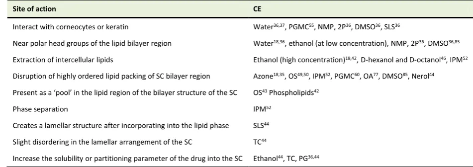
the fluidity of the lipid chains. There are some CEs (DMSO, alcohols, etc.) which may also cause lipid extraction.24,34 Table 1 summarises the site of action of
various types of CE in skin.
Table 1. A summary of reported mechanism of action of commonly used topical and transdermal CEs
Site of action CE
Interact with corneocytes or keratin Water36,37, PGMC55, NMP, 2P36, DMSO36, SLS36
Near polar head groups of the lipid bilayer region Water18,36, ethanol (at low concentration), NMP, 2P36, DMSO36,85
Extraction of intercellular lipids Ethanol (high concentration)18,42, D-hexanol and D-octanol46, IPM52
Disruption of highly ordered lipid packing of SC bilayer region Azone18,35, OS49,50, IPM52, PGMC60, OA77, DMSO85, Nerol44
Present as a ‘pool’ in the lipid region of the bilayer structure of the SC OS43 Phospholipids42
Phase separation IPM52
Creates a lamellar structure after incorporating into the lipid phase SLS44
Slight disordering in the lamellar arrangement of the SC TC44
Increase the solubility or partitioning parameter of the drug into the SC Ethanol44, TC, PG36,44
Conclusion
Several approaches have been taken to enhance penetration of drug molecules across the SC. However, chemical enhancers have been found to be the most efficient and simplest ones. In addition, the chemical enhancers improve the permeation of the molecules in the skin in a most cheap and effective way. Except for some established CEs, the in depth mechanism of action of most of the CEs are poorly understood until now. In this article, we have summarized the studies conducted till now on different CEs. CEs induce structural transformations and enhance drug permeation across the SC by interacting with the major permeation pathway (intercellular region). Some CEs or combination of CEs show dual action, that is, by altering the partition parameter of the skin and by interacting with either the intra-or intercellular regions. ‘Pull- Effect’ is the result of penetration enhancing properties of either a single of combined CE(s). The mechanism of action of some CEs in molecular levels has been included in this article as well. Though, in case of some CEs contradicting mechanism of actions were suggested, the information would still be a basis to do further studies to confirm the exact mechanism of action. Still we need further studies to have a concrete understanding on the CEs. However, the summarized information on CEs would be useful for the formulation scientists to develop simple topical and transdermal formulations with improved permeation or penetration of a compound.
Ethical Issues
Not applicable
Conflict of Interest
There is no conflict of interest.
References
1. Haque T, Crowther JM, Lane ME, Moore DJ. Chemical ultraviolet absorbers topically applied in a skin barrier mimetic formulation remain in the outer
stratum corneum of porcine skin. Int J Pharm
2016;510:250-4. doi:
10.1016/j.ijpharm.2016.06.041.
2. D’Arcy Y. Targeted topical analgesics for acute pain.
PainMedicine News; 2015 [cited 2016 25
November]; Available from:
http://www.painmedicinenews.com/Review- Articles/Article/12-14/Targeted-Topical-Analgesics-For-Acute-Pain/28992/ses=ogst.
3. Güngör S, Erdal MS, Aksu B. New formulation strategies in topical antifungal therapy. J Cosmet
Dermatol Sci Appl 2013;3:56-65. doi:
10.4236/jcdsa.2013.31A009
4. McGrath JA, Eady RAJ, Pope FM. Anatomy and organization of human skin. In: Burns T, Breathnach S, Cox N, Griffiths C, editors. Rook's textbook of dermatology. 7th ed. Blackwell Publishing, Inc.; 2008. p. 45-128.
5. Katz M, Poulsen BJ. Absorption of drugs through the skin. In: Brodie BB, Gillete J, editors. Handbook of experimental pharmacology. Berlin, Heidelberg, New York: Springer- Verlag; 1971. p. 103-74. 6. Wood EJ, Bladon PT. The human skin. Great Britain,
Australia, USA: Camelot Press; 1985.
7. Benson HAE. Skin structure, function, and permeation. In: Benson HAE, Watkinson AC, editors. Topical and transdermal drug delvery: Principles and practice. New Jersey: Jhon Wiley & Sons, Inc.; 2012. p. 3-22. 8. Christophers E. Cellular architecture of the stratum
corneum. J Invest Dermatol 1971;56(3):165–9.
| 177 Chemical enhancer to modulate the stratum corneum
Advanced Pharmaceutical Bulletin, 2018, 8(2), 169-179
structure and lipid composition. J Invest Dermatol
1981;76(4):297-301.
10. Michaels AS, Chandrasekaran SK, Shaw JE. Drug permeation through human skin: Theory and in vitro
experimental measurement. Aiche J
1975;21(5):985-96. doi: 10.1002/aic.690210522
11. Elias PM. Structure and function of the stratum
corneum permeability barrier. Drug Dev Res
1988;13(2-3):97-105. doi: 10.1002/ddr.430130203 12. Wertz PW, Downing DT. Stratum corneum:
Biological and biochemical considerations. In: Hadgraft J, Guy RH, editors. Transdermal drug
delivery: Developmental issue and research
initiatives. New York: Marcel Dekker, Inc.; 1989. p. 1-22.
13. Bouwstra J, Pilgram G, Gooris G, Koerten H, Ponec
M. New aspects of the skin barrier organization. Skin
Pharmacol Appl Skin Physiol 2001;14 Suppl 1:52-62.
doi: 10.1159/000056391
14. Pilgram GSK, Pelt AME-v, Bouwstra JA, Koerten HK. Electron diffraction provides new information on human stratum corneum lipid organization studied
in relation to depth and temperature. J Invest
Dermatol 1999;113(3):403-9. doi:
10.1046/j.1523-1747.1999.00706.x
15. Golden GM, Guzek DB, Kennedy AE, McKie JE, Potts RO. Stratum corneum lipid phase transitions
and water barrier properties. Biochem
1987;26(8):2382-8.
16. Blank IH. Factors which influence the water content
of the stratum corneum. J Invest Dermatol
1952;18(6):433-40.
17. Swartzendruber DC, Wertz PW, Kitko DJ, Madison KC, Downing DT. Molecular models of the intercellular lipid lamellae in mammalian stratum
corneum. J Invest Dermatol 1989;92(2):251-7.
18. Suhonen TM, Bouwstra JA, Urtti A. Chemical enhancement of percutaneous absorption in relation to stratum corneum structural alterations. J Control
Release 1999;59(2):149-61.
19. Potts RO, Francoeur ML. The influence of stratum
corneum morphology on water permeability. J Invest
Dermatol 1991;96(4):495-9.
20. Higuchi T. Physical chemical analysis of percutaneous
absorption process from creams and ointments. J Soc
Cosmet Chem 1960;11(11):85-97.
21. Scheuplein RJ, Blank IH. Permeability of the skin.
Physiol Rev 1971;51(4):702-47. doi:
10.1152/physrev.1971.51.4.702
22. Morrow DIJ, McCarron PA, Woolfson AD, Donnelly RF. Innovative strategies for enhancing topical and
transdermal drug delivery. Open Drug Deliver J
2007;1:36-59. doi: 10.2174/1874126600701010036 23. Scheuplein HJ, Blank IH, Brauner GJ, MacFarlane DJ.
Percutaneous absorption of steroids. J Invest
Dermatol 1969;52(1):63-70. doi: 10.1038/jid.1969.9
24. Benson HAE. Transdermal drug delivery: Penetration
enhancement techniques. Curr Drug Deliv
2005;2:23-33.
25. Hueber F, Wepierre J, Schaefer H. Role of
transepidermal and transfollicular routes in
percutaneous absorption of hydrocortisone and testosterone: In vivo study in the hairless rat. Skin
Pharmacol 1992;5(2):99–107.
26. Albery WJ, Hadgraft J. Percutaneous absorption: In
vivo experiments. J Pharm Pharmacol
1979;31(1):140-7.
27. Potts RO, Guy RH. Predicting skin permeability.
Pharm Res 1992;9(5):663-9.
28. Nemanic MK, Elias PM. In situ precipitation: A novel
cytochemical technique for visualization of
permeability pathways in mammalian stratum
corneum. J Histochem Cytoche 1980;28(6):573-8.
doi: 10.1177/28.6.7190175
29. Boddé HE, van den Brink I, Koerten HK, de Haan FHN. Visualization of in vitro percutaneous penetration of mercuric chloride; transport through intercellular space versus cellular uptake through
desmosomes. J Control Release 1991;15(3):227-36.
doi: 10.1016/0168-3659(91)90114-S
30. Roberts MS, Cross SE, Pellett MA. Skin transport. In: Walters KA, editor. Dermatological and transdermal formulation. New York, Basel: Marcel Dekker, Inc.; 2002. p. 89-183.
31. Benson HA, Sarveiya V, Risk S, Roberts MS. Influence of anatomical site and topical formulation on skin penetration of sunscreens. Ther Clin Risk
Manag 2005;1(3):209-18.
32. Kadir R, Stempler D, Liron Z, Cohen S. Delivery of theophylline into excised human skin from alkanoic
acid solutions: A “push-pull” mechanism. J Pharm
Sci 1987;76(10):774-9.
33. Mura P, Faucci MT, Bramanti G, Corti P. Evaluation of transcutol as a clonazepam transdermal permeation
enhancer from hydrophilic gel formulations. Eur J
Pharm Sci 2000;9:365–72. doi:
10.1016/S0928-0987(99)00075-5
34. Lane ME, Santos P, Watkinson AC, Hadgraft J. Passive skin permeation enhancement. In: Benson HAE, Watkinson AC, editors. Topical and transdermal drug delivery principle and practice. New Jersey: Wiley-Blackwell; 2012. p. 24-42. 35. Trommer H, Neube RHH. Overcoming the stratum
corneum: The modulation of skin penetration. Skin
Pharmacol Physiol 2006;19:106–21. doi:
10.1159/000091978
36. Barry BW. Mode of action of penetration enhancers in
human skin. J Control Release 1987;6:85-97. doi:
10.1016/0378-5173(95)04108-7
37. Gwak HS, Oh IS, Chun IK. Transdermal delivery of ondansetron hydrochloride: Effects of vehicles and
penetration enhancers. Drug Dev Ind Pharm
2004;30(2):187–94. doi: 10.1081/DDC-120028714 38. Mak VW, Potts R, Guy R. Does hydration affect
intercellular lipid organization in the stratum
corneum? Pharm Res 1991;8(8):1064-5.
Haque et al.
degrees c) lipid transitions in human stratum
corneum. J Invest Dermatol 1994;103(2):233-9.
40. Van Hal DA, Jeremiasse E, Junginger HE, Spies F, Bouwstra JA. Structure of fully hydrated human
stratum corneum: A freeze-fracture electron
microscopy study. J Invest Dermatol
1996;106(1):89-95.
41. Trommer H, Neubert RH. Overcoming the stratum corneum: The modulation of skin penetration. A
review. Skin Pharmacol Physiol 2006;19(2):106-21.
doi: 10.1159/000091978
42. Williams AC, Barry BW. Penetration enhancers. Adv
Drug Deliv Rev 2004;56(5):603-18. doi:
10.1016/j.addr.2003.10.025
43. Lane ME. Skin penetration enhancers. Int J Pharm
2013;447(1-2):12-21. doi:
10.1016/j.ijpharm.2013.02.040
44. Moghadam SH, Saliaj E, Wettig SD, Dong C, Ivanova MV, Huzil JT, et al. Effect of chemical permeation enhancers on stratum corneum barrier lipid organizational structure and interferon alpha
permeability. Mol Pharm 2013;10(6):2248-60. doi:
10.1021/mp300441c
45. Andega S, Kanikkannan N, Singh M. Comparison of the effect of fatty alcohols on the permeation of
melatonin between porcine and human skin. J
Control Release 2001;77(1-2):17-25.
46. Dias M, Naik A, Guy RH, Hadgraft J, Lane ME. In vivo infrared spectroscopy studies of alkanol effects
on human skin. Eur J Pharm Biopharm
2008;69(3):1171-5. doi: 10.1016/j.ejpb.2008.02.006 47. Santos P, Watkinson AC, Hadgraft J, Lane ME.
Formulation issues associated with transdermal
fentanyl delivery. Int J Pharm 2011;416(1):155-9.
doi: 10.1016/j.ijpharm.2011.06.024
48. Santos P, Watkinson AC, Hadgraft J, Lane ME. Influence of penetration enhancer on drug permeation
from volatile formulations. Int J Pharm
2012;439(1-2):260-8. doi: 10.1016/j.ijpharm.2012.09.031 49. Casal HL, Mantsch HH. Polymorphic phase behaviour
of phospholipid membranes studied by infrared
spectroscopy. Biochim Biophys Acta
1984;779(4):381-401.
50. El Maghraby GM, Campbell M, Finnin BC. Mechanisms of action of novel skin penetration enhancers: Phospholipid versus skin lipid liposomes.
Int J Pharm 2005;305(1-2):90-104. doi:
10.1016/j.ijpharm.2005.08.016
51. Brinkmann I, Muller-Goymann CC. An attempt to clarify the influence of glycerol, propylene glycol, isopropyl myristate and a combination of propylene glycol and isopropyl myristate on human stratum
corneum. Pharmazie 2005;60(3):215-20.
52. Engelbrecht TN, Deme B, Dobner B, Neubert RH. Study of the influence of the penetration enhancer isopropyl myristate on the nanostructure of stratum corneum lipid model membranes using neutron
diffraction and deuterium labelling. Skin Pharmacol
Physiol 2012;25(4):200-7. doi: 10.1159/000338538
53. Haque T, Rahman KM, Thurston DE, Hadgraft J, Lane ME. Topical delivery of anthramycin i.
Influence of neat solvents. Eur J Pharm Sci
2017;104:188-95. doi: 10.1016/j.ejps.2017.03.043 54. Takahashi K, Komai M, Kinoshita N, Nakamura E,
Hou XL, Takatani-Nakase T, et al. Application of hydrotropy to transdermal formulations: Hydrotropic solubilization of polyol fatty acid monoesters in water
and enhancement effect on skin permeation of 5-fu. J
Pharm Pharmacol 2011;63(8):1008-14. doi:
10.1111/j.2042-7158.2011.01308.x
55. Moghimipour E, Salimi A, Zadeh BSM. Effect of the various solvents on the in vitro permeability of
vitamin b12 through excised rat skin. Trop J Pharm
Res 2013;12(5):671-7.
56. Gwak HS, Kim SU, Chun IK. Effect of vehicles and enhancers on the in vitro permeation of melatonin
through hairless mouse skin. Arch Pharm Res
2002;25(3):392-6.
57. Lee J, Chun I. Effects of various vehicles and fatty
acids on the skin permeation of lornoxicam. J Pharm
Invest 2012;42(5):235-41. doi:
10.1007/s40005-012-0035-2
58. Gwak HS, Chun IK. Effect of vehicles and penetration enhancers on the in vitro percutaneous absorption of
tenoxicam through hairless mouse skin. Int J Pharm
2002;236(1-2):57-64.
59. Cho YA, Gwak HS. Transdermal delivery of ketorolac tromethamine: Effects of vehicles and penetration
enhancers. Drug Dev Ind Pharm 2004;30(6):557-64.
doi: 10.1081/ddc-120037486
60. Takahashi K, Sakano H, Yoshida M, Numata N, Mizuno N. Characterization of the influence of polyol fatty acid esters on the permeation of diclofenac
through rat skin. J Control Release
2001;73(2-3):351-8.
61. Lee KE, Choi YJ, Oh BR, Chun IK, Gwak HS. Formulation and in vitro/in vivo evaluation of
levodopa transdermal delivery systems. Int J Pharm
2013;456(2):432-6. doi:
10.1016/j.ijpharm.2013.08.044
62. Kim KH, Gwak HS. Effects of vehicles on the percutaneous absorption of donepezil hydrochloride
across the excised hairless mouse skin. Drug Dev Ind
Pharm 2011;37(9):1125-30. doi:
10.3109/03639045.2011.561352
63. Jung SY, Kang EY, Choi YJ, Chun IK, Lee BK, Gwak HS. Formulation and evaluation of ubidecarenone
transdermal delivery system. Drug Dev Ind Pharm
2009;35(9):1029–34. doi:
10.1080/03639040902755205
64. Choi JS, Cho YA, Chun IK, Jung SY, Gwak HS. Formulation and evaluation of ketorolac transdermal
systems. Drug Deliv 2007;14(2):69-74. doi:
10.1080/10717540600640336
| 179 Chemical enhancer to modulate the stratum corneum
Advanced Pharmaceutical Bulletin, 2018, 8(2), 169-179
66. Parisi N, Paz-Alvarez M, Matts PJ, Lever R, Hadgraft J, Lane ME. Topical delivery of hexamidine. Int J
Pharm 2016;506(1-2):332-9. doi:
10.1016/j.ijpharm.2016.04.069
67. Liron Z, Cohen S. Percutaneous absorption of alkanoic acids ii: Application of regular solution theory. J
Pharm Sci 1984;73(4):538-42.
68. Chadha G, Sathigari S, Parsons DL, Jayachandra Babu R. In vitro percutaneous absorption of genistein from
topical gels through human skin. Drug Dev Ind
Pharm 2011;37(5):498-505. doi:
10.3109/03639045.2010.525238
69. Puglia C, Bonina F, Trapani G, Franco M, Ricci M. Evaluation of in vitro percutaneous absorption of lorazepam and clonazepam from hydro-alcoholic gel
formulations. Int J Pharm 2001;228(1-2):79-87.
70. Prasanthi D, Lakshmi PK. Effect of chemical enhancers in transdermal permeation of alfuzosin
hydrochloride. ISRN Pharm 2012;2012:965280. doi:
10.5402/2012/965280
71. Shah PP, Desai PR, Patlolla R, Klevans L, Singh M. Effect of combination of hydrophilic and lipophilic permeation enhancers on the skin permeation of
kahalalide f. J Pharm Pharmacol 2014;66(6):760-8.
doi: 10.1111/jphp.12206
72. Panchagnula R, Ritschel WA. Development and evaluation of an intracutaneous depot formulation of corticosteroids using transcutol as a cosolvent:
In-vitro, ex-vivo and in-vivo rat studies. J Pharm
Pharmacol 1991;43(9):609-14.
73. Ritschel WA, Panchagnula R, Stemmer K, Ashraf M. Development of an intracutaneous depot for drugs. Binding, drug accumulation and retention studies,
and mechanism of depot. Skin Pharmacol
1991;4(4):235-45.
74. Caon T, Campos CE, Simoes CM, Silva MA. Novel
perspectives in the tuberculosis treatment:
Administration of isoniazid through the skin. Int J
Pharm 2015;494(1):463-70. doi:
10.1016/j.ijpharm.2015.08.067
75. Ongpipattanakul B, Burnette RR, Potts RO, Francoeur ML. Evidence that oleic acid exists in a separate
phase within stratum corneum lipids. Pharm Res
1991;8(3):350-4.
76. Golden GM, McKie JE, Potts RO. Role of stratum corneum lipid fluidity in transdermal drug flux. J
Pharm Sci 1987;76(1):25-8.
77. Sintov A, Ze'evi A, Uzan R, Nyska A. Influence of
pharmaceutical gel vehicles containing oleic
acid/sodium oleate combinations on hairless mouse
skin, a histological evaluation. Eur J Pharm
Biopharm 1999;47(3):299-303.
78. Ben-Shabat S, Baruch N, Sintov AC. Conjugates of unsaturated fatty acids with propylene glycol as potentially less-irritant skin penetration enhancers.
Drug Dev Ind Pharm 2007;33(11):1169-75. doi:
10.1080/03639040701199258
79. Ochalek M, Podhaisky H, Ruettinger HH, Neubert RH, Wohlrab J. Sc lipid model membranes designed for studying impact of ceramide species on drug diffusion and permeation, part iii: Influence of penetration enhancer on diffusion and permeation of
model drugs. Int J Pharm 2012;436(1-2):206-13. doi:
10.1016/j.ijpharm.2012.06.044
80. Atef E, Altuwaijri N. Using raman spectroscopy in studying the effect of propylene glycol, oleic acid,
and their combination on the rat skin. AAPS
PharmSciTech 2018;19(1):114-22. doi:
10.1208/s12249-017-0800-7
81. Bouwstra JA, de Vries MA, Gooris GS, Bras W, Brussee J, Ponec M. Thermodynamic and structural aspects of the skin barrier. J Control Release
1991;15(3):209-20. doi:
10.1016/0168-3659(91)90112-Q
82. Furuishi T, Kato Y, Fukami T, Suzuki T, Endo T, Nagase H, et al. Effect of terpenes on the skin
permeation of lomerizine dihydrochloride. J Pharm
Pharm Sci 2013;16(4): 551-63
83. Mohammed D, Hirata K, Hadgraft J, Lane ME. Influence of skin penetration enhancers on skin barrier function and skin protease activity. Eur J
Pharm Sci 2014;51:118-22. doi:
10.1016/j.ejps.2013.09.009
84. Åkesson B. Concise international chemical assessment document 35: N-methyl-2-pyrrolidone. Geneva: World Health Organization, 2001.
85. Notman R, den Otter WK, Noro MG, Briels WJ, Anwar J. The permeability enhancing mechanism of dmso in ceramide bilayers simulated by molecular
dynamics. Biophys J 2007;93(6):2056-68. doi: