EFFECT OF MODIFIED METHOD OF AUTONOMIC NERVOUS SYSTEM ACTIVITY
ASSESSMENT ON RESULTS OF HEART RATE VARIABILITY ANALYSIS
Michal Botek*, Jakub Krejčí, Filip Neuls, Jiří Novotný
Faculty of Physical Culture, Palacký University, Olomouc, Czech Republic
Submitted in August, 2013
BACKGROUND: Quickness and simplicity of examining procedures belong to the key criteria of decision-making process whether the spectral analysis of heart rate variability (SA HRV) would be implemented into the sports train-ing routine as a diagnostic tool.
OBJECTIVE: The main aim of this study was to find out how results of the SA HRV are influenced when reducing duration of the first position of standardized orthoclinostatic maneuver (supine–standing–supine) from 5 minutes to 60 seconds.
METHODS: The sample consisted of 28 healthy persons divided into two groups (G1 – n = 14, 7 males and 7 females aged 19 to 23; G2 – n = 14, 9 males and 5 females aged 17 to 22). The G1 underwent the autonomic nervous system (ANS) activity assessment firstly by the modified procedure and consecutively by the standard procedure. This order was reversed in the G2. The ANS activity was represented by both, individual HRV parameters and complex indices of the SA HRV.
RESULTS: When comparing the standard and modified procedure of the ANS assessment, no significant differ-ences in any of the assessed parameters of the HRV were found. The correlation analysis that the results of the SA HRV indices between both measures of the ANS are highly related was found. This dependence was also present in a majority of the individual HRV parameters.
CONCLUSIONS: According to the results of this research, the time-modified method of the ANS assessment pro-vides results of the SA HRV which are comparable to the traditionally applied orthoclinostatic maneuver.
Keywords: Orthoclinostatic maneuver, vagal activity, sympathovagal balance, cardiovascular system.
branches of the autonomic nervous system (ANS), (Task Force, 1996). During short-term five-minute analysis (i.e. “short-term spectral analysis”) of the elec-trocardiographic (ECG) record, power spectrum is divided into three basic frequency bands, namely very low frequency power (PVLF) 0.02−0.05 Hz, low frequen-cy power (PLF) 0.05−0.15 Hz, and high frequency pow-er (PHF) 0.15−0.50 Hz (Salinger et al., 1998). Origins of fluctuations within the area of VLF remain unclear. However, very often they have been associated with thermoregulation sympathetic activity of vessels (Hyn-dman, Kitney, & Sayers, 1971) or with oscillations of renin-angiotensin system (Akselrod et al., 1981). In the case of spectral power modulators within the area of PLF, complete agreement does not exist, either. While some authors consider spectral power in the area of PLF to be an indicator of sympathetic activity (Kamath & Fallen, 1991; Montano et al., 1994), others suppose that this area is under common influence of both, sym-pathetic and parasymsym-pathetic activity (Appel, Berger, Saul, Smith, & Cohen, 1989; Berger, Saul, & Cohen, 1989) or it reflects activity of baroreceptors affected by the sympathetic activity (Casadei, Cochrane, John-INTRODUCTION
Heart rate variability (HRV) is a term widely used in literature to denote oscillation of consequential R-R intervals which is generally considered to be a non-in-vasive index of autonomous cardiac regulation (Aksel-rod et al., 1981; Arai et al., 1989; Javorka, 2008; Malik & Camm, 1995). For its high sensitivity to changes of internal environment, the HRV evaluation became ap-plicable not only in medical area (diabetology, cardiol-ogy or neurolcardiol-ogy) but also in a field of sports, e.g. as an instrument for detection of fatigue and/or overload (Achten, & Jeukendrup, 2003; Aubert, Seps, & Beck-ers, 2003, Botek, McKune, Krejčí, Stejskal, & Gába, 2013; Chen et al., 2011; Pichot et al., 2002).
Spectral analysis (SA) of the HRV belongs to non-invasive methods enabling quantification of both
of studies, the band of the PHF is under dominant influ-ence of respiratory related vagal activity (Grossman, Wilhelm, & Spoerle, 2004; Hayano et al., 1991; Malik & Camm, 1995; Malliani, Pagani, Lombardi, & Cerut-ti, 1991; Opavský, 2002; Warren, Jaffe, Wraa, & Steb-bins, 1997).
The high number of individual HRV parameters (30+) often leading to interpretive collisions and effort to increase the sensitivity of whole method to iden-tify even small changes in the ANS activity were the main motives when creating the complex indices of the SA HRV (Stejskal, Šlachta, Elfmark, Salinger, & Gaul-Aláčová, 2002) which would combine all age-de-pendent spectral parameters obtained during the stan-dardized orthoclinostatic maneuver (supine–standing– supine position).
From practical experience with monitoring applica-tion of ANS activity when using the complex indices of the SA HRV within sports training process (Botek, Stejskal, & Svozil, 2010; Cipryan et al., 2007) it is evi-dent that, for coaches and athletes, quickness and sim-plicity of the examining procedure becomes one of the key criteria of the decision-making process if the meth-ods of SA HRV should or should not be implemented into the training routine as a diagnostic tool. In fact, it is less known that only the standing position and the second supine position are necessary for a calculation of the complex indices of the SA HRV while the first supine position serves only for standardization of con-ditions before the examination itself.
Hence, the main aim of this study was to find out how results of the SA HRV would be affected when reducing duration of the first position of standardized orthoclinostatic maneuver (supine–standing–supine) from 5 minutes to 60 seconds.
METHODS The sample
This research was performed in two groups of volun-teers with good health status who were not (except females using hormonal contraception) under any in-fluence of pharmaceuticals significantly affecting the ANS activity. The first group (G1) consisted of 14 per-sons (7 males and 7 females) aged 19 to 23 years. Also the second group (G2), that included different volun-taries compared to G1, included 14 persons (9 males and 5 females) aged 17 to 22. All the subjects involved in the research were familiarized in detail with the en-tire course of the examination and signed the informed agreement. The study was approved by the Ethical Committee of the Faculty of Physical Culture (FTK) , Palacký University, Olomouc.
ing morning hours (8:00 to 10:00) in standardized con-ditions of a gym inside a building which belongs to the FTK facilities. The examined persons were instructed to avoid vigorous physical activity one day before the examination. They were also instructed not to drink coffee or alcohol or use other substances influencing regulations of the HR at least two hours before the ex-amination.
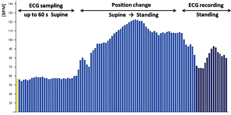
The research was implemented in two stages. The G1 was tested during the first stage. The examined per-sons underwent the ANS activity examination primar-ily by the modified testing procedure when the first position of the standardized orthoclinostatic maneu-ver accompanied by launching of the ECG scanning lasted approximately 60 seconds (Figure 1). Before the ECG scanning in the first position (supine) is started, it is necessary to get each person identified with the diagnostics system which takes about 1 minute. During this period, the examined persons were already in the lying position. After the modified procedure was com-pleted, tested persons had a 10 minute break consisting of light conversation in a seated position. Then they underwent the examination of the ANS by the standard procedure, i.e. 5 minute supine position, consecutive 5 minute standing position and again, 5 minute supine position (Salinger et al., 1993). To exclude possible influences of a factor of the order of partial examina-tions on the SA HRV results, the whole procedure was repeated after 4 weeks from the first testing in the G2 whilst the series of the examinations followed each oth-er revoth-ersely than the G1.
The ANS activity assessment
The ECG data were continually sampled during the standardized orthoclinostatic maneuver (supine– standing–supine position) by DiANS PF8 (Dimea Group, Czech Republic) system which requires both, 300 R-R intervals and 300 seconds per position for the short-term SA HRV (Salinger & Gwozdziewicz, 2008). Modified software version of the diagnostic DiANS PF8 device enables to perform the reduced 60 seconds supine position followed by the standardized stand-ing and supine positions (i.e. 300 R-R intervals and 300 seconds per position).
intervals to define a relevant position of examined per-son while records with a number of scanned intervals lower than three were declared to be out of evaluation. In the modified version of the software, only re-cords with none or one scanned interval are not evalu-ated while default intervals for the definition of the position of examined person are the first and the sec-ond interval (i.e. the supine and the standing position) within the record.
To maintain compatibility of the evaluation also for records acquired by the standard procedure, it was nec-essary to adjust also “a detection” mode of the record-ing; meaning to distinguish between the standard test and its shortened modification. When the software de-tects three or more measured intervals, it is assumed to be related to the record made by the standard method (the supine–standing–supine position). When only two measured intervals are detected, it is associated with the record of the R-R intervals based on the modified method. The major advantage of this adjustment is that the same program can be used for testing and following evaluation in both, standard and shortened version of the test. That is why it depends only on the user, which version will be chosen in a given moment.
Frequency domain analyses were performed ac-cording to the methods described by Salinger et al. (1998). Amplitude density of the collected signal was estimated using the fast Fourier transform method with a partly modified Coarse-Graining Spectral Analyze algorithm (Yamamoto & Hughson, 1991). Power of mean spectral components were calculated by integrat-ing area under the power spectral density curve in the frequency ranges according to Salinger et al. (1998) – power very low frequency (PVLF) 0.02–0.05 Hz; power low frequency (PLF) 0.05–0.15 Hz, power high
frequen-cy (PHF) 0.15–0.5 Hz, and total spectral power (PT) 0.02–0.5 Hz, respectively. Resting heart rate (HRrest) was computed as a mean heart rate within the second 5 minute long lying position. The autonomic cardiac activity was also expressed by the complex indices of the SA HRV (Stejskal et al., 2002) – the complex in-dex of the vagal activity (VA), the complex inin-dex of the sympathovagal balance (SVB), the total spectral power (PT), and the complex index of the total score (TS). The reference values of the SA HRV complex in-dices range from –5.0 to +5.0 points. The physiological values have been established for both, VA and SVB in range from –2.0 to +2.0 points; for PT from –2.5 to +2.5 points, and for TS from –1.5 to +1.5 points, re-spectively.
Statistical analyses
All statistical analyses were processed using STA-TISTICA 12.0 (StatSoft, Tulsa, OK, USA). Normal distribution of the analyzed data was verified by the Kolmogorov-Smirnov test. The R-R had normal distri-bution. All spectral variables (PVLF, PLF, PHF, and PT) and theirs ratios were not of normal distribution. To correct skewed distributions of spectral parameters, natural logarithm transformation (Ln) was applied. After logarithmic transformation, all spectral powers and theirs ratios had normal distribution except for Ln PLF/PHF in group G2 (p = .041). The indices VA, SVB and TS had normal distribution. The index PT in group G1 was not of normal distribution (p = .048).
Table 1
Statistical analysis of the HRV results measured in the first group (G1)
Parameter (units)
Standing (M ± SD) 2nd supine (M ± SD)
Modified Standard p Modified Standard p
Ln PVLF (ms2) 5.40 ± 1.25 5.38 ± 0.99 .945 5.72 ±0.74 5.57 ± 0.94 .632
Ln PLF (ms2) 6.43 ± 1.04 6.48 ± 1.03 .876 6.05 ± 1.15 6.07 ± 1.24 .934
Ln PHF (ms2) 5.63 ± 1.19 5.50 ± 1.20 .593 8.04 ± 0.97 8.00 ± 1.11 .762
Ln PVLF/PHF –0.22 ± 1.25 –0.12 ± 1.11 .811 –2.31 ± 1.24 –2.43 ± 1.23 .718
Ln PLF/PHF 0.81 ± 0.94 0.98 ± 0.99 .372 –1.99 ± 1.38 –1.93 ± 1.21 .818
R-R (ms) 0.70 ± 0.10 0.69 ± 0.10 .403 1.03 ± 0.17 1.07 ± 0.18 .053
Ln PT (ms2) 7.17 ± 0.95 7.14 ± 0.95 .898 8.39 ± 0.79 8.33 ± 0.97 .697
Note. Modified = modified orthoclinostatic maneuver, Standard = standard orthoclinostatic maneuver. Ln PVLF = natural logarithm of very low frequency power, Ln PLF = natural logarithm of low frequency power, Ln PHF = natural logarithm of high frequency power, Ln PT = natural logarithm of total power, R-R = length between subsequent heart beats.
p = significance of paired t-test, * p < .05.
Table 2
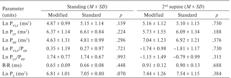
Statistical analysis of the HRV results measured in the second group (G2)
Parameter (units)
Standing (M ± SD) 2nd supine (M ± SD)
Modified Standard p Modified Standard p
Ln PVLF (ms2) 4.87 ± 0.99 5.15 ± 1.14 .159 5.16 ± 1.12 5.10 ± 1.15 .750
Ln PLF (ms2) 6.37 ± 1.14 6.61 ± 0.84 .224 5.73 ± 1.55 6.09 ± 1.34 .188
Ln PHF (ms2) 4.63 ± 1.31 4.81 ± 0.99 .296 7.04 ± 1.23 6.92 ± 1.21 .376
Ln PVLF/PHF 0.35 ± 1.19 0.27 ± 0.97 .721 –1.74 ± 0.98 –1.81 ± 1.17 .730
Ln PLF/PHF 1.74 ± 0.77 1.74 ± 0.67 .993 –1.15 ± 1.49 –0.79 ± 0.99 .315
R-R (ms) 0.65 ± 0.09 0.66 ± 0.08 .448 0.91 ± 0.12 0.90 ± 0.13 .688
Ln PT (ms2) 6.81 ± 1.01 7.05 ± 0.80 .070 7.44 ± 1.26 7.54 ± 1.15 .384
Note. Modified = modified orthoclinostatic maneuver, Standard = standard orthoclinostatic maneuver. Ln PVLF = natural logarithm of very low frequency power, Ln PLF = natural logarithm of low frequency power, Ln PHF = natural logarithm of high frequency power, Ln PT = natural logarithm of total power, R-R = length between subsequent heart beats.
p = significance of paired t-test, * p < .05.
RESULTS
Tables 1 and 2 show that no significant differences were found in any of the individual HRV variables be-tween two related measurements within the examined groups (G1 and G2). In standing position, a correla-tion between related parameters of PVLF, PHF, R-R, PT, and PLF/PHF ratio collected during the standard and modified HRV assessment was found in G1 (Table 3). The relation between the supine PLF, PHF, R-R, PT and
shows that, in the both position in G2, exists the re-lationship between all assessed variables except the PLF/PHF ratio in standing.
DISCUSSION
The main findings of this study are following – 1) there are no significant differences, either in results of the indi-vidual HRV variables or in results of the complex indices of SA HRV, between the standard orthoclinostatic ma-neuver (5 min. supine – 5 min. standing – 5 min. supine)
and its modified version (up to 60 s supine – 5 min. standing – 5 min. supine); 2) a strong intra-class correla-tion between all indices of the SA HRV assessed in both, G1 and G2 was found. Moreover, a positive relationship between majority of the individual HRV parameters ob-tained during the standard and the modified version of the orthoclinostatic maneuver in G1 and G2 was found. Table 3
Correlation analysis of assessed variables during different HRV measurements
Parameter
Modified → Standard (G1), r Standard → Modified (G2), r
Standing 2nd supine Standing 2nd supine
Ln PVLF .611* –.052 .808* .854*
Ln PLF .400 .706* .807* .809*
Ln PHF .724* .925* .908* .924*
Ln PVLF/PHF .161 .490 .759* .761*
Ln PLF/PHF .729* .748* .530 .568*
R-R .897* .939* .898* .950*
Ln PT .575* .862* .905* .955*
Note. Modified = modified orthoclinostatic maneuver, Standard = standard or-thoclinostatic maneuver. Ln PVLF = natural logarithm of very low frequency power, Ln PLF = natural logarithm of low frequency power, Ln PHF = natural logarithm of high frequency power, Ln PT = natural logarithm of total power, R-R = length between subsequent heart beats.
r = Pearson correlation coefficient, * p < .05.
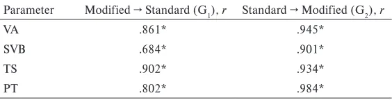
Table 5
Correlation analysis of the SA HRV indices during different HRV measurements
Parameter Modified → Standard (G1), r Standard → Modified (G2), r
VA .861* .945*
SVB .684* .901*
TS .902* .934*
PT .802* .984*
Note. G1 = the first group, G2 = the second group. VA = index of vagal activity, SVB = index of sympathovagal balance, TS = index of total score, PT = age-dependent parameter of total power.
r = Pearson correlation coefficient, * p < .05.
Table 4
Results of statistical analysis in the complex indices of the SA HRV
Parameter (units)
Modified → Standard (G1), M ± SD Standard → Modified (G2), M ± SD
Modified Standard p Standard Modified p
VA (points) 1.25 ± 2.09 1.25 ± 1.95 .996 –0.94 ± 2.30 –0.69 ± 2.23 .250
SVB (points) 0.58 ± 1.77 0.72 ± 1.78 .698 –1.43 ± 1.34 –1.49 ± 1.45 .756
TS (points) 1.02 ± 1.90 1.07 ± 1.81 .820 –1.11 ± 1.83 –0.97 ± 1.79 .441
PT (points) 1.77 ± 2.53 1.76 ± 2.77 .977 –1.54 ± 3.59 –1.12 ± 3.60 .059
Note. G1 = the first group, G2 = the second group. VA = index of vagal activity, SVB = index of sympathovagal balance, TS = index of total score, PT = age-dependent parameter of total power.
assessment when “the short-term spectral analysis” is performed (Malik & Camm, 1995; Task Force, 1996). The standardization process may include a resting pe-riod that the examined persons usually spend in sit-ting or supine position. For example, Plews, Laursen, Stanley, Kilding, and Buchheit (2013) assessed HRV from the last 5 minutes of 6 minutes supine rest pe-riod. In another study, Ewing, Borsey, Bellavere, and Clarke (1981) put 3 minute rest before the R-R interval sampling in diabetic patients. The first position of the orthoclinostatic challenge (supine–standing–supine) that has been established as a fundamental procedure of the ANS activity assessment at the FTK in Olomouc (Salinger et al., 1993) serves as 5 minute rest before the HRV assessment is started. However, in other stud-ies, the R-R intervals were quantified in given position without preceding resting period (Gamelin, Baquet, Berthoin, & Bosquet, 2008; Kiviniemi, Hautala, Kin-nunen, & Tulppo, 2007; Sartor, Vailati, Valsecchi, Vailati, & La Torre, 2013).
Our results show that the shortening of the first supine position down to 60 seconds did not evoke any significant changes in all the individual HRV pa-rameters and HR compared to the standard orthocli-nostatic challenge in both positions. With exception of these findings, the only difference was found in the supine R-R interval where the mean R-R interval in the supine position during the standard maneuver compared to the modified maneuver reached almost a significant level (p = .053) in the G2. This difference in the mean R-R interval values represents approximately HR = 2 beats per minute. Our results further show that the age-dependent PT parameter was higher (p = .059) in the modified maneuver compared to the standard test in the G1. It seems that increase in the ANS activity was associated with the discrete an increase in vagal ac-tivity during the second orthoclinostatic maneuver. An ascendant tendency in vagal activity during multiple orthoclinostatic maneuvers was previously described by Kalina, Stejskal, and Jakubec (2001) who assigned this behavior to the summation effect of clinostasis.
We suggest that the 60 second rest in the supine position preceding the orthoclinostatic maneuver (standing–supine) may represent an adequate time for cardiovascular adjustment. Our opinion leans on the previous study (Bellavere & Ewing, 1982) showing that the initial HR response to the lying down consisted of an immediate shortening of the R-R interval reaching a maximum around the third or fourth beat after ly-ing down, followed by a lengthenly-ing beyond the rest-ing value to reach a steady level around beats 25−30. In addition, Toska and Walløe (2002) found that the cardiovascular adjustment took up to 10 seconds in
su-ANS measurement may be considered as useable only if the rest supine is not performed immediately after high stress stimuli, e.g. intensive physical and mental activity, respectively.
Based on our results, we suggest that the ANS activ-ity assessment performed by the modified orthoclino-static maneuver seems to be useful especially from the aspect of time. From this point of view, shorter time demands in the modified maneuver could potentially increase the compliance to the longitudinal ANS mea-surements primarily in elite athletes. The abovemen-tioned advantages of the modified HRV assessment are primarily recommended to athletes in both, individual and team sports when the ANS activity is quantified by the complex indices of the SA HRV. Main limit of this study is related with the fact that the two different groups were compared. Therefore, our results should be clarified by further study where it is needed to use randomized cross-over design in one group of tested persons.
CONCLUSIONS
To conclude, we can state that the ANS activity could be assessed by using both, standard orthoclinostatic maneuver and its time-modified version where both challenges provide reciprocally comparable results in healthy young persons. It is important to point out that further research is needed because this study was per-formed in young healthy subjects only.
ACKNOWLEDGMENT
The study has been supported by the research grant from the Ministry of Education, Youth and Sports of the Czech Republic (No. MSM 6198959221) “Physical Activity and Inactivity of the Inhabitants of the Czech Republic in the Context of Behavioral Changes”.
REFERENCES
Achten, J., & Jeukendrup, A. E. (2003). Heart rate monitoring: Applications and limitations. Sports Medicine, 33(7), 517−538.
Akselrod, S., Gordon, D., Ubel, F. A., Shannon, D. C., Berger, A. C., & Cohen, R. J. (1981). Power spec-trum analysis of heart rate fluctuation: A quanti-tative probe of beat to beat cardiovascular control. Science, 213, 220−222.
Arai, Y., Saul, J. P., Albrecht, P., Hartley, L. H., Lilly, L. S., Cohen, R. J., & Colucci, W. S. (1989). Modula-tion of cardiac autonomic activity during and im-mediately after exercise. American Journal of Physi-ology, 256, 132−141.
Aubert, A. E., Seps, B., & Beckers, F. (2003). Heart rate variability in athletes. Sports Medicine, 33, 889−919.
Bellavere, F., & Ewing, D. J. (1982). Autonomic con-trol of the immediate heart rate response to lying down. Clinical Science, 62(1), 57−64.
Berger, R. D., Saul, J. P., & Cohen, R. J. (1989). Trans-fer function analysis of autonomic regulation I. Ca-nine atrial rate response. American Journal of Physi-ology, 256, 142–152.
Botek, M., McKune, A., Krejčí, J., Stejskal, P., & Gába, A. (2013). Change in performance in response to training load adjustment based on autonomic activ-ity. International Journal of Sports Medicine, 34, 1–7. Botek, M., Stejskal, P., & Svozil, Z. (2010). Autonomic
nervous system activity during acclimatization after rapid air travel across time zones: A case study. Acta Universitatis Palackianae Olomucensis. Gymnica, 39(2), 13−21.
Casadei, B., Cochrane, S., Johnston, J., Conway, J., & Sleight, P. (1995). Pitfalls in the interpretation of spectral analysis of the heart rate variability during exercise in humans. Acta Physiologica Scandinavica, 153(2), 125−131.
Chen, J. L., Yeh, D. P., Lee, J. P., Chen, C. Y., Huang, C. Y., Lee, S. D., Chen, C. C., Kuo, T. B., Kao, C. L., & Kuo, C. H. (2011). Parasympathetic nervous activity mirrors recovery status in weightlifting performance after training. Journal of Strength and Conditional Research, 25, 1546−1552.
Cipryan, L., Stejskal, P., Bartáková, O., Botek, M., Cipryanová, H., Jakubec, A., Petr, M., & Řehová, I. (2007). Autonomic nervous system observation through the use of spectral analysis of heart rate variability in ice hockey players. Acta Universitatis Palackianae Olomucensis.Gymnica,37(4), 17−21. Ewing, D. J., & Borsey, D. Q., Bellavere, F., & Clarke,
B. F. (1981). Cardiac autonomic neuropathy in diabetes: Comparison of measures of RR interval variation. Diabetologia, 21, 18−24.
Gamelin, F. X., Baquet, G., Berthoin, S., & Bosquet, L. (2008). Validity of the polar S810 to measure R-R intervals in children. International Journal of Sports Medicine, 29(2), 134−138.
Hayano, J., Sakakibara, Y., Yamada, A., Yamada, M., Mukai, S., Fujinami, T., Yokoyama, K., Watanabe, Y., & Takata, K. (1991). Accuracy of assessment of cardiac vagal tone by heart rate variability in nor-mal subjects. American Journal of Cardiology, 67(2), 199−204.
Hyndman, B. W., Kitney, R. I., & Sayers, B. M. (1971). Spontaneous rhythms in physiological control sys-tems. Nature, 233, 339−341.
Javorka, K. (2008). Využití hodnocení variability srdeční frekvence ve sportovní medicíně [Heart rate variability application in sports medicine]. In K. Javorka (Ed.), Variabilita frekvencie srdca: Mech-anismy, hodnotenie, klinické využitie [Heart rate vari-ability: Mechanism, assessment, clinical application] (pp. 16−21). Martin: OSVETA.
Kalina, M., Stejskal, P., & Jakubec, A. (2001). Al-goritmus a standardizace vyšetření autonomního nervového systému metodikou spektrální analýzy variability srdeční frekvence [Algorithm and stan-dardization of investigation of autonomic nervous system through spectral analysis of heart rate vari-ability]. In K. Martiník, B. Komeštík, & J. Ryba (Eds.), Sborník referátů z interdisciplinární kon-ference Optimální působení tělesné zátěže a výživy [CD-ROM] (pp. 128−133). Hradec Králové: Uni-verzita Hradec Králové.
Kamath, M. V., & Fallen, E. L. (1993). Power spec-tral analysis of heart rate variability: A noninvasive signature of cardiac autonomic function. Critical Reviews in Biomedical Engineering, 21(3), 245−311. Kiviniemi, A. M., Hautala, A. J., Kinnunen, H., &
Tulppo, M. P. (2007). Endurance training guided individually by daily heart rate variability measure-ments. European Journal of Applied Physiology, 101, 743−751.
Malik, M., & Camm, J. (1995). Geometrical methods for heart rate variability assessment. Heart rate vari-ability. New York, NY: Futura.
Malliani, A., Pagani, M., Lombardi, F., & Cerutti, S. (1991). Cardiovascular neural regulation ex-plored in the frequency domain. Circulation, 84(2), 482−492.
Montano, N., Ruscone, T. G., Porta, A., Lombardi, F., Pagani, M., & Malliani, A. (1994). Power spectrum analysis of heart rate variability to assess the chang-es in sympathovagal balance during graded ortho-static tilt. Circulation, 90(4),1826−1831.
Pichot, V., Busso, T., Roche, F., Garet, M., Costes, F., Duverney, D., Lacour, J. R., & Barthelemy, J. C. (2002). Autonomic adaptations to intensive and overload training periods: A laboratory study. Medicine and Science in Sports and Exercise, 34, 1660−1666.
Plews, D. J., Laursen, P. B., Stanley, J., Kilding, A. E., & Buchheit, M. (2013). Training adaptation and heart rate variability in elite endurance athletes: Opening the door to effective monitoring. Sports Medicine, 43(9), 773−781.
ka (Ed.), Variabilita frekvencie srdca: Mechanismy, hodnotenie, klinické využitie [Heart rate variabil-ity: Mechanism, assessment, clinical application] (pp. 57−60). Martin: OSVETA.
Salinger, J., Opavský, J., Bůla, J., Vychodil, R., Novot-ný, J., & Vaverka, F. (1993). Programové vybavení měřícího systému, typ TF-2, určené pro spektrální analýzu variací R-R intervalů v kardiologii [Soft-ware components of diagnostic system TF-2 for spectral analysis of RR intervals variation in cardi-ology]. Lékař a technika, 24, 133−138.
Salinger, J., Opavský, J., Stejskal, P., Vychodil, R., Olšák, S., & Janura, M. (1998). The evaluation of heart rate variability in physical exercise by using the telemetric variapulse TF3 system. Acta Universi-tatis Palackianae Olomucensis. Gymnica, 28, 13−23. Sartor, F., Vailati, E., Valsecchi, V., Vailati, F., & La
Torre, A. (2013). Heart rate variability reflects training load and psychophysiological status in young elite gymnasts. Journal of Strength and Condi-tioning Research, 22, 2782−2790.
Stejskal, P., Šlachta, R., Elfmark, M., Salinger, J., & Gaul-Aláčová, P. (2002). Spectral analysis of heart rate variability: New evaluation method. Acta Uni-versitatis Palackianae Olomucensis. Gymnica, 32, 13–18.
Stejskal, P., & Salinger, J. (1996). Spektrální analýza variability srdeční frekvence – základy metodiky a literární přehled o jejím klinickém využití. Medicina Sportiva Bohemica et Slovaca, 5(2),33−42.
Task Force of the European Society of Cardiology and the North American Society of Pacing and Electrophysiology. (1996). Heart rate variability. Standards of measurement, physiological interpre-tation, and clinical use. Special report. Circulation, 93, 1043−1065.
Toska, K., & Walløe, L. (2002). Dynamic time course of hemodynamic responses after passive head-up tilt and tilt back to supine position. Journal of Ap-plied Physiology, 92(4), 1671−1676.
Warren, J. H., Jaffe, R. S., Wraa, C. E., & Stebbins, C. L. (1997). Effect of autonomic blockade on power
Yamamoto, Y., & Hughson, R. L. (1991). Coarse-graining spectral analysis: New method for studying heart rate variability. Journal of Applied Physiology, 71(3), 1143−1150.
VLIV MODIFIKOVANÉHO ZPŮSOBU VYŠETŘENÍ AKTIVITY AUTONOMNÍHO
NERVOVÉHO SYSTÉMU NA VÝSLEDKY ANALÝZY VARIABILITY SRDEČNÍ FREKVENCE
(Souhrn anglického textu)
VÝCHODISKA: Rychlost a jednoduchost vyšetřovací procedury patří k základním kritériím rozhodování, zda zařadit spektrální analýzu variability srdeční frek-vence (SA HRV) jakožto diagnostický nástroj v rámci každodenní tréninkové rutiny.
CÍL: Hlavním cílem práce bylo zjistit, jak ovlivní zkrácení doby první polohy standardizovaného orto-klinostatickém manévru (leh–stoj–leh) z 5 minut na 60 sekund výsledky SA HRV.
METODIKA: Testovaný soubor tvořilo 28 zdravých osob rozdělených do dvou skupin (S1 a S2). S1 tvořilo 14 osob (7 mužů, 7 žen) ve věku 19−23 let, v S2 bylo 14 osob (9 mužů, 5 žen) ve věku 17−22 let. S1 podstou-pila vyšetření ANS nejprve modifikovaným a následně standardním způsobem. U S2 bylo toto pořadí otoče-no. Aktivita ANS byla reprezentována individuálními parametry HRV a komplexními indexy SA HRV. VÝSLEDKY: Při vzájemném porovnání standardní-ho a modifikovanéstandardní-ho způsobu vyšetření ANS nebyly u žádného ze zkoumaných parametrů HRV zjištěny sig-nifikantní rozdíly. Korelační analýzou bylo zjištěno, že výsledky indexů SA HRV mezi oběma měřeními ANS těsně korelují. Závislost byla prokázána také u většiny individuálních parametrů HRV.
ZÁVĚR: Bylo zjištěno, že časově modifikovaný způsob vyšetření ANS poskytuje výsledky SA HRV srovnatel-né s tradičně užívaným ortoklinostatickým masrovnatel-névrem.